Báo cáo y học: "Apoptosis is not the major death mechanism induced by celecoxib on rheumatoid arthritis synovial fibroblasts" pps
Bạn đang xem bản rút gọn của tài liệu. Xem và tải ngay bản đầy đủ của tài liệu tại đây (1.74 MB, 11 trang )
Open Access
Available online />Page 1 of 11
(page number not for citation purposes)
Vol 9 No 6
Research article
Apoptosis is not the major death mechanism induced by celecoxib
on rheumatoid arthritis synovial fibroblasts
Rachel Audo
1
, Véronique Deschamps
2
, Michael Hahne
1
, Bernard Combe
1,2
and Jacques Morel
1,2
1
Institut de Génétique Moléculaire de Montpellier, 1919 route de Mende, CNRS UMR5535, Montpellier, France
2
Service d'immuno-rhumatologie et Université Montpellier 1, 371 Ae du doyen Gaston Giraud, Montpellier, France
Corresponding author: Jacques Morel,
Received: 11 May 2007 Revisions requested: 19 Jun 2007 Revisions received: 15 Oct 2007 Accepted: 12 Dec 2007 Published: 12 Dec 2007
Arthritis Research & Therapy 2007, 9:R128 (doi:10.1186/ar2342)
This article is online at: />© 2007 Audo et al.; licensee BioMed Central Ltd.
This is an open access article distributed under the terms of the Creative Commons Attribution License ( />),
which permits unrestricted use, distribution, and reproduction in any medium, provided the original work is properly cited.
Abstract
Synovial hyperplasia in rheumatoid arthritis (RA) has been
associated with apoptosis deficiency of RA fibroblast-like
synoviocytes (FLSs). Celecoxib is a non-steroidal anti-
inflammatory drug that has been demonstrated to induce
apoptosis in some cellular systems. We have therefore
examined the dose- and time-dependent effects of celecoxib on
RA FLS viability. Treatment of RA FLSs with celecoxib for 24
hours reduced their viability in a dose-dependent manner.
Analysis of celecoxib-treated RA FLSs for their content of
apoptotic and necrotic cells by Annexin V staining and TO-PRO-
3 uptake displayed only few apoptotic cells. Caspase 3, a key
mediator of apoptosis, was not activated in celecoxib-treated
RA FLSs, and the presence of specific caspase 3 or pan-
caspase inhibitors did not affect celecoxib-induced cell death.
Moreover, we could not detect other signs of apoptosis, such as
cleavage of poly(ADP-ribose) polymerase, caspase 8 or 9, or
DNA fragmentation. We therefore conclude that apoptosis is
not the major death pathway in celecoxib-treated RA FLSs.
Introduction
Cyclooxygenases (COXs) are key enzymes in the conversion
of arachidonic acid to prostanoids, which mediate mitogene-
sis, apoptosis, angiogenesis, blood flow, secondary injury
(lipid peroxidation and oxidative stress), and inflammation [1].
The COX-1 isoform is constitutively expressed under physio-
logical conditions, whereas expression of the COX-2 isoform
is inducible under pathophysiological, mainly inflammatory,
conditions [2]. Consequently, the current pharmacological
strategy is to selectively inhibit COX-2 and thereby avoid unfa-
vorable effects of combined COX-1 and COX-2 blocking
[1,2].
Rheumatoid arthritis (RA) is an autoimmune disease charac-
terized by chronic inflammation of joints, leading to a progres-
sive and irreversible joint destruction [3,4]. The aggressive
front of synovial tissue, called pannus, invades and destroys
the local articular structure [3,4]. The pannus is characterized
by a synovial hyperplasia that is mainly composed of fibroblast-
like synoviocytes (FLSs) combined with a massive infiltration
of lymphocytes and macrophages [3,4]. Increased prolifera-
tion and insufficient apoptosis might contribute to the expan-
sion of RA FLSs, and several reports suggest inducing
apoptosis of RA FLSs as a therapeutic approach [3,4].
Celecoxib (4-[5-(4-methylphenyl)-3-(trifluoromethyl)-1H-pyra-
zol-1-yl] benzenesulfonamide) is an anti-inflammatory drug that
specifically inhibits the COX-2. Celecoxib has been described
as a pro-apoptotic factor in several human carcinoma cells [5-
7]. In addition, it has been reported that high doses of
celecoxib have a pro-apoptotic effect on RA FLSs [8]. Here,
we report that the cell death induced by high doses of
celecoxib on RA FLSs is rapid without displaying characteris-
tics of apoptosis.
Materials and methods
Reagents
Celecoxib and valdecoxib were generously provided by Pfizer
Inc (New York, NY, USA) and dissolved in dimethyl sulfoxide
(DMSO) at 100 mM. Indomethacin (Sigma-Aldrich, St Quentin
Fallavier, France) was dissolved in ethanol at a final
COX = cyclooxygenase; DMSO = dimethyl sulfoxide; FCS = fetal calf serum; FLS = fibroblast-like synoviocyte; PARP = poly(ADP-ribose) polymer-
ase; PBS = phosphate-buffered saline; RA = rheumatoid arthritis; TRAIL = tumor necrosis factor-related apoptosis-inducing ligand; z-DEVD-fmk =
benzyloxycarbonyl-Asp(OMe)-Glu(OMe)-Val-Asp(OMe)-FMK inhibitor; z-VAD-fmk = benzyloxycarbonyl-Val-Ala-Asp (OMe) fluoromethylketone.
Arthritis Research & Therapy Vol 9 No 6 Audo et al.
Page 2 of 11
(page number not for citation purposes)
concentration of 100 mM. Pan-caspase inhibitor (z-VAD-fmk
[benzyloxycarbonyl-Val-Ala-Asp (OMe) fluoromethylketone]),
caspase 3 inhibitor (z-DEVD-fmk [benzyloxycarbonyl-
Asp(OMe)-Glu(OMe)-Val-Asp(OMe)-FMK inhibitor]), and the
caspase control inhibitor z-FA-fmk (benzyloxycarbonyl-phenyl-
alanyl-fluoromethylketone) specific for cathepsins B and L
(R&D Systems, Lille, France) were dissolved at 20 mM in
DMSO. Annexin V was purchased from Roche Diagnostic
(Meylan, France) and TO-PRO-3 from Invitrogen Corporation
(Cergy Pontoise, France). Anti-caspase antibodies were
obtained from Cell Signaling Technology (St Quentin Yveline,
France), poly(ADP-ribose) polymerase (PARP) antibody from
BD Pharmingen (BD Biosciences, Le-Pont-de-Claix, France),
and peroxidase-conjugated secondary antibodies were pur-
chased from Sigma-Aldrich.
Preparation of fibroblast-like synoviocytes of patients
with rheumatoid arthritis
Fibroblasts were isolated from synovium obtained from
patients who met the American College of Rheumatology cri-
teria for RA (revised 1987) and who had undergone surgery
for synovectomy or total joint replacement surgery [9]. Fresh
synovial tissues were minced and digested in a solution of dis-
pase (Gibco, now part of Invitrogen Corporation) and colla-
genase (Sigma-Aldrich) and DNase (Calbiochem, now part of
EMD Biosciences, Inc., San Diego, CA, USA). Synovial fibrob-
lasts were cultured in RPMI 1640 supplemented with 10%
fetal calf serum (FCS). Cells were used at passages 4 to 10,
when they constitute a homogeneous population of fibrob-
lasts, free of detectable T cells or macrophages. Upon reach-
ing confluence, the cells were passaged by brief trypsinization.
For experimentation, the content of FCS in the media was pro-
gressively decreased from 10% to 1% with final starvation for
12 hours in RPMI 1640 media containing 1% FCS, as
described previously [10].
Analysis of cell viability and apoptosis
Cell viability was measured by taking metabolic activity as a
readout using the Celltiter 96 AQueous cell proliferation
(MTS) assay (Promega Corporation, Charbonnières, France)
after 24 hours of cell culturing according to the manufacturer's
instructions. Apoptotic RA FLSs were identified by resuspend-
ing 1 × 10
5
cells in 100 μL of Annexin V Binding buffer con-
taining 5 μL of Annexin V-fluorescein isothiocyanate (10 μg/
mL; R&D Systems) for 15 minutes at room temperature. Upon
addition of TO-PRO-3 (1:2,000), cells were analyzed by flow
cytometry (FACSCalibur; BD Biosciences) [11].
Cell proliferation assay
Proliferation was evaluated measuring DNA synthesis by
incorporation of tritiated [
3
H]thymidine. FLSs were seeded in
96-well flat-bottom culture plates at a density of 1 × 10
4
cells
per well. Cells were cultured in RPMI 1640 with decreasing
concentrations of FCS (10% and 5%) and then synchronized
for 24 hours with RPMI 1640 and 1% FCS. FLSs were stimu-
lated for 72 hours. Every condition was tested in quadrupli-
cate. [
3
H]thymidine (1 μCi/well) was added 24 hours before
the end of the assay. FLSs were lysed using a round of freeze-
thaw cycle and then transferred onto a membrane filter using
Harvester 96 (Tomtec, Hamdem, CT, USA). [
3
H]thymidine
incorporated into DNA was quantified using a scintillation
counter 1450 MicroBeta Trilux (Wallac, now part of Perk-
inElmer Life and Analytical Sciences, Inc., Waltham, MA,
USA').
Western blotting analysis
Synovial cells were seeded in six-well plates at 2 × 10
5
cells
per well or in 6-cm dishes at 4 × 10
5
cells. After serum starva-
tion, RA FLSs were treated as indicated. Both detached and
adherents cells were collected, washed twice with cold phos-
phate-buffered saline (PBS), and treated with lysis buffer as
described previously [10]. Total cell extracts were resolved by
SDS-PAGE and proceeded for immunoblot analysis as
described previously [12]. Primary antibodies were diluted
according to the manufacturer's instructions (1:1,000 dilu-
tions for anti-caspase and anti-PARP antibodies and 1:5,000
dilution for anti-β-actin antibody), and nitrocellulose mem-
branes were incubated overnight at 4°C with the primary anti-
bodies (or for 1 hour at room temperature for β-actin). Equal
loading was confirmed by β-actin expression.
Ac-DEVD-AMC (caspase 3/7 fluorogenic substrate)
protease assay
Synovial fibroblasts were seeded in six-well plates at 2 × 10
5
cells per well. After serum starvation, RA FLSs were treated
with either celcoxib or tumor necrosis factor-related apopto-
sis-inducing ligand (TRAIL), and caspase 3 activation was
measured using the Ac-DEVD-AMC protease assay according
to the manufacturer's instructions (BD Biosciences).
DNA fragmentation
FLSs were seeded in 96-well flat-bottom culture plates at 1 ×
10
4
cells per well, cultured with decreasing concentrations of
FCS (10%, 5%, and 1%) as described above, and incubated
for 12 hours with either celecoxib or TRAIL. Cells were col-
lected, washed with PBS, and processed for quantification of
DNA fragments using an enzyme-linked immunosorbent assay
according to the manufacturer's instructions (Cell Death
Detection ELISA
PLUS
; Roche Diagnostic).
Results
Celecoxib decreases cell activity and proliferation of
rheumatoid arthritis fibroblast-like synoviocytes
We first analyzed the effect of celecoxib on metabolic activity
of RA FLSs. For this, RA FLSs were treated for 24 hours with
different concentrations of celecoxib and subsequently ana-
lyzed for cell activity using MTS assay. Cell activity of RA FLSs
was clearly reduced by the addition of 40 μM celecoxib and
completely abrogated in the presence of 60 μM (Figure 1a).
RA FLS viability was only modestly affected by lower
Available online />Page 3 of 11
(page number not for citation purposes)
concentrations of celecoxib. Indomethacin, an inhibitor for
both COX-1 and COX-2, and valdecoxib, another specific
COX-2 inhibitor, had no effect on RA FLS activity, demonstrat-
ing that the observed effect is specific for celecoxib.
The decreased viability of celecoxib-treated RA FLSs was mir-
rored by their decreased proliferation capacity as measured by
thymidine incorporation. Celecoxib inhibited RA FLS prolifera-
tion in a dose-dependent manner at all of the concentrations
tested (20, 40, 50, and 60 μM), and only marginal thymidine
incorporation was detectable in the presence of 60 μM
celecoxib (Figure 1b).
Celecoxib-treated rheumatoid arthritis fibroblast-like
synoviocytes bypass the state of early apoptosis
We next analyzed celecoxib-treated RA FLSs for their content
of apoptotic and necrotic cells by Annexin V staining and TO-
PRO-3 uptake. This technique allows investigators to distin-
Figure 1
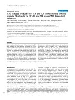
Celecoxib reduces viability and proliferation of synovial fibroblasts extracted from patients with rheumatoid arthritis (RA)Celecoxib reduces viability and proliferation of synovial fibroblasts extracted from patients with rheumatoid arthritis (RA). (a) Fibroblast-like synovio-
cytes (FLSs) of patients with RA were cultured for 24 hours in the presence of the cyclooxygenase inhibitors celecoxib, valdecoxib, or indomethacin
at the indicated concentrations. Metabolic activity was determined by means of the MTS assay. Untreated cells (Co) and cells treated only with sol-
vent served as controls. The graph presents relative cell viability toward cells treated only with solvent as the mean ± standard error of the mean
(SEM) of three individual experiments. (b) Celecoxib strongly inhibits RA FLS proliferation in a dose-dependent manner. RA FLSs were stimulated
for 24 hours with the indicated concentrations of celecoxib, and proliferation was assessed using [
3
H]thymidine incorporation. The graph presents
relative cell viability toward cells treated only with solvent as the mean ± SEM of three individual experiments.
Arthritis Research & Therapy Vol 9 No 6 Audo et al.
Page 4 of 11
(page number not for citation purposes)
guish early apoptotic cells (Annexin
+
/TO-PRO-3
-
) from late
apoptotic/necrotic cells (Annexin
+
/TO-PRO-3
+
) [11] (Figure
2).
RA FLSs were treated with different concentrations (10 to
100 μM) of celecoxib, and cell death was observed only at
celecoxib concentrations of at least 60 μM (Figure 2). Val-
decoxib, used at the same concentration, induced no cell
death (data not shown). Twenty-four hours of treatment of RA
FLSs with 60 μM celecoxib induced death in nearly all cells,
which displayed the characteristics of late apoptotic/necrosis
(that is, were Annexin
+
and TO-PRO-3
+
). A similar pattern was
observed after 4 hours of treatment with celecoxib, when no
early apoptotic cells (Annexin V
+
and TO-PRO-3
-
) were
detectable (Figure 2). Moreover, when RA FLSs were treated
for shorter time points (30 minutes and 1 and 2 hours) with 60
μM celecoxib, no early apoptotic cells were detectable,
whereas cells were already detectable in the Annexin
+
and
Figure 2
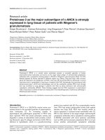
Celecoxib induces cell death in rheumatoid arthritis fibroblast-like synoviocytesCelecoxib induces cell death in rheumatoid arthritis fibroblast-like synoviocytes. Cells were treated at indicated concentrations and times with either
celecoxib or staurosporine. Apoptosis was evaluated by fluorescence-activated cell sorting analysis using Annexin V binding and TO-PRO-3 uptake.
Values are expressed as the percentage of total cell death (upper panel) or apoptosis (lower panel) and are the mean ± standard error of the mean
of three individual experiments.
Available online />Page 5 of 11
(page number not for citation purposes)
TO-PRO-3
+
gate (Figure 3a). Two hours of treatment of RA
FLSs with 60 μM celecoxib was sufficient to induce cell death
in more than 90% of the cells, whereas only 11% dead cells
were detectable upon treatment with 40 μM celecoxib. Inter-
mediate concentrations between 40 and 60 μM demonstrated
that celecoxib induces cell death in a dose-dependent manner
and this cell death is also characterized by an immediate shift
into the necrosis/late apoptosis gate (Figure 3a). Although the
percentage of dead cells detectable on celecoxib-treated cells
varied between the FLSs of different patients, it never reached
those differences detected by measuring cell viability (Figure
1). This is most likely due to the fact that the MTS assay used
for measuring viability quantifies the metabolic activity of cells
and that metabolically inactive cells are not necessarily dead
cells.
To validate our approach for the detection of apoptotic cells,
we treated RA FLSs with staurosporine and TRAIL (also called
APO-2L). Staurosporin is a non-selective protein kinase inhib-
itor that is known to induce apoptosis in several cell types [13],
whereas TRAIL is a member of the tumor necrosis factor family
which induces apoptosis in a wide variety of tumor cells as
well as RA FLSs [10]. Indeed, early apoptotic cells were
observed in cells that were treated for 4 hours with either stau-
rosporine or TRAIL (Figure 3b). TRAIL-treated RA FLSs dis-
played morphological changes characteristic for apoptosis,
including cell shrinkage and membrane blebbing, that were
not detectable on celecoxib-treated RA FLSs. Celecoxib ini-
tially induced a compression of the cells which was followed
by cellular swelling associated with the formation of dendritic-
like structures (Figure 4).
No caspase activity is detectable in celecoxib-treated
rheumatoid arthritis fibroblast-like synoviocytes
One mechanism that is consistently implicated in apoptosis is
the activation of a cascade of cytosolic proteases called cas-
pases. Caspases are synthesized as inactive proenzymes that
are processed by proteolytic cleavage to form an active
enzyme. A member of this family, caspase 3 (CPP32, apopain,
and YAMA), plays a central role in the execution of apoptosis
in mammalian cells, and activation of caspase 3 is therefore a
hallmark of apoptotic cells [14].
To assess the contribution of caspases in celecoxib-mediated
cytotoxicity, RA FLSs were treated with the pan-caspase inhib-
itor z-VAD-fmk or with z-DEVD-fmk, a specific inhibitor of cas-
pase 3, and subsequently with celecoxib (60 μM). A 5-μM
concentration of neither a pan-caspase inhibitor nor the spe-
cific caspase 3 inhibitor could protect RA FLSs against
celecoxib-induced cell death, whereas cell death induced by
TRAIL was significantly reduced by a 5-μM concentration of
either pan-caspase inhibitor z-VAD-fmk or caspase 3 inhibitor
(Figure 5). Also, higher concentrations (50 μM) of the pan-cas-
pase inhibitor z-VAD-fmk had no inhibitory effect on celecoxib-
induced cell death (Figure 6a). Caspase inhibitors also did not
affect cell death of RA FLSs treated with lower concentrations
(40 μM) of celecoxib (Figure 6b).
Cleavage of caspases is an indicator for their activation. We
therefore analyzed whether the cleaved forms of caspases 3,
8, and 9 were detectable in celecoxib-treated RA FLSs. Cell
death happened faster in RA FLSs treated with celecoxib than
in those treated with TRAIL. For this reason, we compared RA
FLSs treated for 2 hours with celecoxib and cells treated for at
least 3 hours with TRAIL. The cleaved forms of caspase 3, 8,
and 9 were not detectable in cell lysates of RA FLSs treated
with celecoxib but were detectable in those treated with TRAIL
(Figure 7a,c). Finally, we analyzed the caspase 3 activity using
an Ac-DEVD-AMC protease assay in celecoxib- and TRAIL-
treated cells but detected caspase 3 activity only in TRAIL-
treated cells (Figure 7b).
Active caspase 3 proteolytically cleaves and activates, among
other targets, PARP involved in DNA repair and DFF40/CAD
DNase, the executor of nuclear DNA fragmentation. Apoptotic
cells are characterized by cleavage of the native 116-kDa form
of PARP into 85-kDa and 25-kDa forms. Concurring with the
observed absence of the active form of caspase 3, PARP
cleavage was not observed in celecoxib-treated RA FLSs but
was observed in those treated with TRAIL (Figure 7c). Moreo-
ver, we could not detect DNA fragmentation in RA FLSs
treated for either 12 or 24 hours with celecoxib concentrations
of 40, 50, or 60 μM, but we could in TRAIL-treated cells (Fig-
ure 7d). Taken together, our results suggest that the cell death
pathway induced by celecoxib on RA FLSs occurs in a cas-
pase-independent manner.
Discussion
Apoptosis is a form of cell death in which a programmed
sequence of events leads to the elimination of cells without
releasing harmful substances into the surrounding area. Apop-
tosis plays a crucial role in controlling cell numbers by
eliminating old cells, unnecessary cells, and unhealthy cells.
Deregulation of apoptosis thus can lead to the survival and
hyperproliferation of unwanted cells such as FLSs in RA.
Therefore, one strategy for treatment is the design of drugs
that can restore the normal apoptotic pathways in hyperprolif-
erative cells.
The anti-inflammatory drug celecoxib, an inhibitor of COX-2,
was reported by Kusunoki and colleagues [8] to be pro-apop-
totic on RA FLSs. In that study, the viability of synovial cells
was reduced by celecoxib in a dose-dependent manner similar
to our observation. Kusunoki and colleagues observed that
celecoxib strongly reduced cell viability of RA FLSs when used
at concentrations of at least 30 μM. The authors concluded
that celecoxib induces apoptosis in RA FLSs as they observed
a strong DNA fragmentation in RA FLSs treated with 30 μM
celecoxib.
Arthritis Research & Therapy Vol 9 No 6 Audo et al.
Page 6 of 11
(page number not for citation purposes)
Figure 3
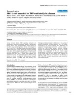
Characterization of celecoxib-induced cell death in rheumatoid arthritis fibroblast-like synoviocytesCharacterization of celecoxib-induced cell death in rheumatoid arthritis fibroblast-like synoviocytes. Cells were treated at indicated concentrations
and times with (a) celecoxib or (b) staurosporine or tumor necrosis factor-related apoptosis-inducing ligand. Apoptosis was evaluated by fluores-
cence-activated cell sorting analysis using Annexin V binding and TO-PRO-3 uptake. Representative data of three different experiments are shown.
DMSO, dimethyl sulfoxide.
Available online />Page 7 of 11
(page number not for citation purposes)
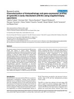
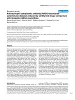
Figure 4
Comparison of morphological changes of rheumatoid arthritis fibroblast-like synoviocytes (RA FLSs) treated with either celecoxib or tumor necrosis factor-related apoptosis-inducing ligand (TRAIL) by light microscopy (magnification × 300)Comparison of morphological changes of rheumatoid arthritis fibroblast-like synoviocytes (RA FLSs) treated with either celecoxib or tumor necrosis
factor-related apoptosis-inducing ligand (TRAIL) by light microscopy (magnification × 300). Untreated RA FLSs (upper panel) and cells treated for
either 1 hour with 60 μm celecoxib (middle panel) or 8 hours with 1 nM TRAIL (lower panel) are shown. Apoptotic cells are indicated by arrows.
Arthritis Research & Therapy Vol 9 No 6 Audo et al.
Page 8 of 11
(page number not for citation purposes)
We compared the characteristics of celecoxib-induced cell
death in RA FLSs with those induced by the established pro-
apoptotic factor TRAIL by Annexin V staining/TO-PRO-3
uptake. We have previously reported that 4 hours of co-cultur-
ing with TRAIL resulted in apoptosis of approximately 30% of
RA FLSs as determined by Annexin V staining/TO-PRO-3
uptake. This technique allows investigators to distinguish early
apoptotic cells (Annexin
+
/TO-PRO-3
-
) from late apoptotic/
necrotic cells (Annexin
+
/TO-PRO-3
+
) [11]. Annexin V is a
Ca
2+
-dependent phospholipid-binding protein with high affin-
ity for phospatidylserine and can be used as a sensitive probe
for the early phase of apoptosis that is characterized by phos-
patidylserine exposure on the cell membrane. Because of
increased permeability, Annexin V binding can also occur dur-
ing cell necrosis, and uptake of DNA stain TO-PRO-3 is taken
as a parameter to distinguish necrotic, and thus permeable,
cells [11].
Whereas we observed pre-apoptotic cell death in RA FLSs
upon 4 hours of TRAIL treatment, pre-apoptotic cells were
hardly detectable in celecoxib-treated cells. We exposed RA
FLSs to different concentrations of celecoxib for various incu-
bation times, but under none of the tested conditions were
pre-apoptotic (that is, Annexin V
+
and TO-PRO-3
-
) cells
detectable. Cell death was observed only at concentrations
above 40 μM celecoxib (Figure 2), although we detected a
decrease in the metabolic activity of FLSs treated with lower
concentrations of celecoxib, which is in agreement with Kusu-
noki and colleagues [8]. We also confirm that celecoxib
strongly inhibited RA FLS proliferation, as shown in their study
[8].
It has been suggested that cell death should be classified as
apoptosis only if execution of cell death is dependent on
caspase activity [14]. We therefore tested whether caspase 3
is activated in celecoxib-treated cells as caspase 3 is a key
executor of apoptosis. Whereas the cleaved (thus active) form
of caspase 3 was detectable in cell lysates of TRAIL-treated
RA FLSs, only the proform of caspase 3 was visible in
celecoxib-treated cells. Concurring with this observation, nei-
ther a pan-caspase inhibitor nor a specific caspase 3 inhibitor
could protect RA FLSs against celecoxib-induced cell death.
Similar observations were made with caspase 8 and caspase
9 inhibitors (data not shown). We therefore conclude that
celecoxib-induced cell death in RA FLSs is independent of
caspases.
These results are in contrast to those of Kusunoki and col-
leagues [8], who observed DNA fragmentation in RA FLSs
treated for 24 hours with celecoxib and an inhibition of this DNA
fragmentation by the addition of caspase 3 inhibitors. A possible
explanation for these differences might be the different cell sys-
Figure 5
Celecoxib-induced cell death in rheumatoid arthritis fibroblast-like synoviocytes is caspase-independentCelecoxib-induced cell death in rheumatoid arthritis fibroblast-like synoviocytes is caspase-independent. Effect of caspase inhibition on celecoxib-
induced cell death. Cells were pre-treated with caspase inhibitors (pancasp-In: pan-caspase inhibitor z-VAD-fmk; casp3-In: caspase 3 inhibitor z-
DEVD-fmk) or control inhibitor z-FA-fmk (co-In: control inhibitor) for 1 hour and subsequently cultured in the presence of either 60 μM celecoxib or
0.5 nM tumor necrosis factor-related apoptosis-inducing ligand (TRAIL) for an additional 24 hours. Cells pre-treated only with solvent (dimethyl sul-
foxide [DMSO]) served as controls. Cell death was determined using Annexin V binding and TO-PRO-3 uptake and expressed as relative cell death.
(For this, cell death induced by TRAIL or celecoxib plus inhibitor was first subtracted by cell death of cells treated with inhibitor alone and then
expressed as percentage versus cell death induced by TRAIL or celecoxib alone.) Data from three patients were averaged and are shown as the
mean ± standard error of the mean.
Available online />Page 9 of 11
(page number not for citation purposes)
Figure 6
Celecoxib-induced cell death in rheumatoid arthritis fibroblast-like synoviocytes is caspase-independentCelecoxib-induced cell death in rheumatoid arthritis fibroblast-like synoviocytes is caspase-independent. Effect of caspase inhibition on celecoxib-
induced cell death using a higher concentration of caspase inhibitor (a) and a lower concentration of celecoxib (40 μM) (b). In these conditions, inhi-
bition of cell death could not be observed. Representative data of three different experiments are shown. NS, unstimulated cells; TRAIL, tumor
necrosis factor-related apoptosis-inducing ligand.
Arthritis Research & Therapy Vol 9 No 6 Audo et al.
Page 10 of 11
(page number not for citation purposes)
Figure 7
Celecoxib does not induce caspase activation in rheumatoid arthritis fibroblast-like synoviocytes (RA FLSs)Celecoxib does not induce caspase activation in rheumatoid arthritis fibroblast-like synoviocytes (RA FLSs). (a) FLSs were stimulated for 2 hours
with celecoxib at indicated concentrations or for 4 hours with tumor necrosis factor-related apoptosis-inducing ligand (TRAIL) (0.5 nM) as positive
control. Cell lysates were analyzed by immunoblot for caspase 3 expression. The same blot was stripped and reprobed with a mouse anti-human β-
actin antibody to confirm equal loading. One representative immunoblot is shown. (b) RA FLSs were stimulated for indicated time points with 60 μM
celecoxib or with TRAIL (0.5 nM) as positive control, and caspase 3 activity was measured using Ac-DEVD-AMC protease assay. Caspase 3 activity
is expressed as fold increase to unstimulated cells (NS) and is represented as the mean ± standard error of the mean (SEM) of different experiments
using RA FLSs from three different patients. (c) RA FLSs were stimulated for indicated time points with celecoxib at indicated concentrations or with
TRAIL (0.5 nM) as positive control. Cell lysates were analyzed by immunoblot for poly(ADP-ribose) polymerase (PARP) and caspase 8 and 9 expres-
sion. One representative immunoblot is shown. (d) RA FLSs were stimulated for 12 hours with celecoxib at indicated concentrations or with TRAIL
(0.5 nM) as positive control, and DNA fragmentation was measured using the Cell Death Detection ELISA
PLUS
kit. The enrichment of mono- and oli-
gonucleosomes released into the cytoplasm is calculated as the ratio of the absorbance of the sample cells to the absorbance of control cells and is
shown as the mean ± SEM from three experiments performed in duplicate.
Available online />Page 11 of 11
(page number not for citation purposes)
tems used. Kusunoki and colleagues used the first two pas-
sages of fibroblasts prepared from synovial tissue for their
experimentation. We, however, employed RA FLSs between
passages 4 and 10 in order to exclude a contamination of other
cell types because approximately 30% of the synovium is com-
posed of macrophage-like synoviocytes.
Moreover, evidence is accumulating that taking DNA fragmen-
tation as the readout of caspase inhibition is not sufficient to
conclude the contribution of caspases to cell death [15]. It is
therefore possible that the caspase dependency of DNA
fragmentation described by Kusunoki and colleagues is a sec-
ondary event during celecoxib-induced cell death, which could
also explain their observed dose-independent effect of the
caspase inhibitors. To conclude an apoptotic cell death, other
parameters of cell death such as mitochondrial dysfunction,
phosphatidylserine exposure, and plasma membrane permea-
bilization have to be considered [15]. Nevertheless, DNA frag-
mentation can also be observed in non-apoptotic cell death
pathways, as for example during necrosis [16].
Conclusion
We conclude from our results that apoptosis is not the major
death pathway in RA FLSs induced by high concentrations of
celecoxib. This observation is in line with that of a previous
report, in which celecoxib was described as being able to
induce non-apoptotic cell death in cardiac myocytes [17].
Administration of antioxidants, such as dithiothreitol, N-acteyl-
cysteine, or calcium-blocking reagents, did not alter celecoxib-
induced cell death, excluding an implication of oxidative stress
and intracellular calcium signals (data not shown). The mor-
phological changes together with the rapid membrane perme-
abilization of RA FLSs during celecoxib treatment suggest
rather a necrotic-like cell death. Necrosis, however, leads to
the release of inflammatory cellular contents which is
unfavorable in the treatment of RA. The proposed local admin-
istration of higher celecoxib concentrations in the synovial
space of the joint of arthritis patients [8] must therefore be
reconsidered.
Competing interests
The authors declare that they have no competing interests.
Authors' contributions
RA performed the experimental work and the analysis of the
data and participated in the writing of the manuscript. VD per-
formed several experiments. MH participated in the analysis of
the study and in the writing of the manuscript. BC and JM
participated in the design of the study and in the writing of the
manuscript. All authors read and approved the manuscript.
Acknowledgements
This work was supported by Société Française de Rhumatologie and
Pfizer Laboratory (New York, NY, USA). We thank Lourdes Planelles for
help with the fluorescence-activated cell sorting analysis, Solange Des-
agher and Nelly Noraz for critical reading of the manuscript, and Michel
Chammas and Bertrand Coulet for providing synovial tissues.
References
1. Williams C, Mann M, DuBois R: The role of cyclooxygenases in
inflammation, cancer, and development. Oncogene 1999,
18:7908-7916.
2. Crofford L: COX-1 and COX-2 tissue expression: implications
and predictions. J Rheumatol Suppl 1997, 49:15-19.
3. Pope RM: Apoptosis as a therapeutic tool in rheumatoid
arthritis. Nat Rev Immunol 2002, 2:527-535.
4. Firestein GS: Evolving concepts of rheumatoid arthritis. Nature
2003, 423:356-361.
5. Maier TJ, Schilling K, Schmidt R, Geisslinger G, Grosch S:
Cyclooxygenase-2 (COX-2)-dependent and -independent
anticarcinogenic effects of celecoxib in human colon carci-
noma cells. Biochem Pharmacol 2004, 67:1469-1478.
6. Liu X, Yue P, Zhou Z, Khuri FR, Sun SY: Death receptor regula-
tion and celecoxib-induced apoptosis in human lung cancer
cells. J Natl Cancer Inst 2004, 96:1769-1780.
7. Jendrossek V, Handrick R, Belka C: Celecoxib activates a novel
mitochondrial apoptosis signaling pathway. FASEB J 2003,
17:1547-1549.
8. Kusunoki N, Yamazaki R, Kawai S: Induction of apoptosis in
rheumatoid synovial fibroblasts by celecoxib, but not by other
selective cyclooxygenase 2 inhibitors. Arthritis Rheum 2002,
46:3159-3167.
9. Arnett FC, Edworthy SM, Bloch DA, McShane DJ, Fries JF, Cooper
NS, Healey LA, Kaplan SR, Liang MH, Luthra HS, et al.: The Amer-
ican Rheumatism Association 1987 revised criteria for the
classification of rheumatoid arthritis. Arthritis Rheum 1988,
31:315-324.
10. Morel J, Audo R, Hahne M, Combe B: Tumor necrosis factor-
related apoptosis-inducing ligand (TRAIL) induces rheuma-
toid arthritis synovial fibroblast proliferation through mitogen-
activated protein kinases and phosphatidylinositol 3-kinase/
Akt. J Biol Chem 2005, 280:15709-15718.
11. Vermes I, Haanen C, Steffens-Nakken H, Reutelingsperger C: A
novel assay for apoptosis. Flow cytometric detection of phos-
phatidylserine expression on early apoptotic cells using fluo-
rescein labelled Annexin V. J Immunol Methods 1995,
184:39-51.
12. Morel JC, Park CC, Zhu K, Kumar P, Ruth JH, Koch AE: Signal
transduction pathways involved in rheumatoid arthritis syno-
vial fibroblast interleukin-18-induced vascular cell adhesion
molecule-1 expression. J Biol Chem 2002, 277:34679-34691.
13. Posmantur R, McGinnis K, Nadimpalli R, Gilbertsen RB, Wang KK:
Characterization of CPP32-like protease activity following
apoptotic challenge in SH-SY5Y neuroblastoma cells. J
Neurochem 1997, 68:2328-2337.
14. Leist M, Jaattela M: Four deaths and a funeral: from caspases
to alternative mechanisms. Nat Rev Mol Cell Biol 2001,
2:589-598.
15. Kroemer G, Martin SJ: Caspase-independent cell death. Nat
Med 2005, 11:725-730.
16. Dong Z, Saikumar P, Weinberg JM, Venkatachalam MA: Internu-
cleosomal DNA cleavage triggered by plasma membrane
damage during necrotic cell death. Involvement of serine but
not cysteine proteases. Am J Pathol 1997, 151:1205-1213.
17. Hasinoff BB, Patel D, Wu X: The cytotoxicity of celecoxib
towards cardiac myocytes is cyclooxygenase-2 independent.
Cardiovasc Toxicol 2007, 7:19-27.