Báo cáo y học: "Indoleamine 2,3-dioxygenase-expressing dendritic cells are involved in the generation of CD4+CD25+ regulatory T cells in Peyer''''s patches in an orally tolerized, collagen-induced arthritis mouse model" ppt
Bạn đang xem bản rút gọn của tài liệu. Xem và tải ngay bản đầy đủ của tài liệu tại đây (1.37 MB, 10 trang )
Open Access
Available online />Page 1 of 10
(page number not for citation purposes)
Vol 10 No 1
Research article
Indoleamine 2,3-dioxygenase-expressing dendritic cells are
involved in the generation of CD4
+
CD25
+
regulatory T cells in
Peyer's patches in an orally tolerized, collagen-induced arthritis
mouse model
Min-Jung Park
1
*, So-Youn Min
1
*, Kyung-Su Park
1,2
, Young-Gyu Cho
1
, Mi-La Cho
1
, Young-
Ok Jung
1
, Hyun-Sil Park
1
, Soog-Hee Chang
1
, Seok Goo Cho
3
, Jun-Ki Min
1,2
, Sung-Hwan Park
1,2
and Ho-Youn Kim
1,2
1
The Rheumatism Research Center, Catholic Research Institute of Medical Science, The Catholic University of Korea, Banpo-dong, Seocho-gu, Seoul
137-701, South Korea
2
Center for Rheumatic Disease, Division of Rheumatology, Department of Internal Medicine, Kangnam St Mary's Hospital, The Catholic University of
Korea, Banpo-dong, Seocho-gu, Seoul 137-701, South Korea
3
Department of Hematology, Catholic Hematopoietic Stem Cell Transplantation, Youido St Mary's Hospital. The Catholic University of Korea, Youido-
dong, Youngdungpo-Gu, Seoul 150-713, South Korea
* Contributed equally
Corresponding author: Ho-Youn Kim,
Received: 12 Apr 2007 Revisions requested: 16 May 2007 Revisions received: 17 Jan 2008 Accepted: 25 Jan 2008 Published: 25 Jan 2008
Arthritis Research & Therapy 2008, 10:R11 (doi:10.1186/ar2361)
This article is online at: />© 2008 Park et al.; licensee BioMed Central Ltd.
This is an open access article distributed under the terms of the Creative Commons Attribution License ( />),
which permits unrestricted use, distribution, and reproduction in any medium, provided the original work is properly cited.
Abstract
Introduction The present study was devised to understand the
role of systemic indoleamine 2,3-dioxygenase (IDO) in the
tolerance induction for orally tolerized mice in collagen-induced
arthritis (CIA). We examined whether IDO-expressing dendritic
cells (DCs) are involved in the generation of CD4
+
CD25
+
regulatory T cells during the induction of oral tolerance in a
murine CIA model.
Methods Type II collagen was fed six times to DBA/1 mice
beginning 2 weeks before immunization, and the effect on
arthritis was assessed. To examine the IDO expression, the DCs
of messenger RNA and protein were analyzed by RT-PCR and
Flow cytometry. In addition, a proliferative response assay was
also carried out to determine the suppressive effects of DCs
through IDO. The ability of DCs expressing IDO to induce
CD4
+
CD25
+
T regulatory cells was examined.
Results CD11c
+
DCs in Peyer's patches from orally tolerized
mice expressed a higher level of IDO than DCs from
nontolerized CIA mice. IDO-expressing CD11c
+
DCs were
involved in the suppression of type II collagen-specific T-cell
proliferation and in the downregulation of proinflammatory T
helper 1 cytokine production. The suppressive effect of IDO-
expressing CD11c
+
DCs was mediated by Foxp3
+
CD4
+
CD25
+
regulatory T cells.
Conclusion Our data suggest that tolerogenic CD11c
+
DCs
are closely linked with the induction of oral tolerance through an
IDO-dependent mechanism and that this pathway may provide
a new therapeutic modality to treat autoimmune arthritis.
Introduction
Repeated oral administration of autoantigen can suppress
autoimmune responses in collagen-induced arthritis (CIA) and
experimental autoimmune encephalomyelitis, and can sup-
press diabetes in nonobese diabetic mice [1-10]. Although the
mechanisms responsible for the induction of oral tolerance
have not been elucidated fully, repeated oral administration of
a high dose of antigen can induce oral tolerance by anergy or
APC = antigen-presenting cell; bp = base pair; CIA = collagen-induced arthritis; CII = type II collagen; cpm = counts per minute; DC = dendritic cell;
ELISA = enzyme-linked immunosorbent assay; IDO = indoleamine 2,3-dioxygenase; IFN = interferon; IL = interleukin; 1-MT = 1-methyl tryptophan;
mAb = monoclonal antibody; MHC = major histocompatibility complex; PBS = phosphate-buffered saline; PCR = polymerase chain reaction; RT =
reverse transcriptase; TGFβ = transforming growth factor beta.
Arthritis Research & Therapy Vol 10 No 1 Park et al.
Page 2 of 10
(page number not for citation purposes)
deletion of antigen-specific T cells. In contrast, repeated feed-
ing of a low dose of antigen favors the induction of active
immune regulation involving regulatory T cells, including trans-
forming growth factor beta (TGFβ)-producing T helper 3 cells,
IL-10-producing T regulatory 1 cells, and CD4
+
CD25
+
T cells
[1,10,11]. Previous studies have demonstrated that, after
repeated oral administration of type II collagen (CII) and sub-
sequent induction of CIA, the mean arthritis index is lower in
tolerized mice than in CIA mice [12] and the proportion of IL-
10-producing CD4
+
CD25
+
T cells increases in Peyer's
patches and spleens of tolerized mice [13]. Among the various
immune cells involved in the induction of oral tolerance, den-
dritic cells (DCs) may play a major role in linking orally admin-
istered antigen to antigen-specific tolerance.
DCs are professional antigen-presenting cells (APCs) that
play a decisive role in determining immunity or immune toler-
ance; this determination is based on the maturation or activa-
tion state and the subset of DCs, and cytokine profiles in the
microenvironment at the time of antigen uptake [1,14-16]. A
previous study demonstrated that CD11c
+
CD11b
+
DCs,
which increase in number in Peyer's patches during the induc-
tion of tolerance to CII, suppress T-cell proliferation and
induce CD4
+
CD25
+
regulatory T cells. CD11c
+
CD8α
+
DCs,
however, promote T-cell proliferation [12]. The mechanisms
underlying the suppression by DCs of the expansion and dif-
ferentiation of effector T cells and promotion of T-cell tolerance
remain elusive.
One regulatory mechanism of DCs is the suppression of pro-
liferation by producing the enzyme indoleamine 2,3-dioxygen-
ase (IDO), which degrades the essential amino acid
tryptophan. Murine macrophages and DCs expressing IDO
inhibit T-cell proliferation or induce T-cell apoptosis in vitro
and in vivo [17]. Munn and colleagues reported that plasma-
cytoid DCs in tumor-draining lymph nodes express IDO con-
stitutively, which causes local immunosuppression and T-cell
anergy in vivo [18,19]. Mellor and colleagues reported that
systemic administration of CpG oligodeoxynucleotides
induces IDO expression in splenic CD19
+
DCs, which acquire
regulatory functions in an IDO-dependent manner [20]. Upon
exposure to allergen within the mucosa, Langerhans-like DCs
expressing high-affinity IgE receptors produce IL-10 and
TGFβ, upregulate IDO expression, and suppress the allergic
response in humans [16,21,22]. Whether the tolerogenic
activity of DCs from Peyer's patches in orally tolerized mice is
IDO dependent, however, is unknown.
To elucidate the expression of IDO and its role in the induction
of oral tolerance, we prepared DCs from Peyer's patches of
DBA/1 mice after induction of oral tolerance by repeated oral
administration of CII and subsequent induction of CIA. We
examined whether IDO-expressing DCs have tolerogenic
characteristics and whether they can induce CD4
+
CD25
+
regulatory T cells. Our results demonstrate that IDO-express-
ing DCs in Peyer's patches play an essential role in the induc-
tion of oral tolerance in this model of autoimmune disease.
Materials and methods
Animals
Six-week-old to 8-week-old male DBA/1J mice (SLC, Inc., Shi-
zuoka, Japan) were maintained in groups of two to four animals
in polycarbonate cages in a specifically pathogen-free environ-
ment and were fed standard mouse chow (Ralston Purina, St
Louis, MO, USA) and water ad libitum. All experimental proce-
dures were examined and approved by the Animal Research
Ethics Committee at the Catholic University of Korea.
Preparation of type II collagen
Bovine CII was kindly provided by Professor Andrew Kang of
the University of Tennessee. CII was extracted in its native
form from the fetal calf articular cartilage and was purified as
described previously [23].
Induction of oral tolerance in DBA/1 mice
DBA/1 mice were sacrificed either with 100 μg bovine CII dis-
solved in 0.05 N acetic acid and 4 mg/ml solution of 25 μl CII
solution + 175 μl PBS for the tolerance group or with an equal
volume of an acetic acid–PBS mixture (25 μl of 0.05 N acetic
acid + 175 μl PBS) for the CIA group. Administration was per-
formed using an oral Zonde needle (Natsume, Tokyo, Japan)
every 2 days over 2 weeks, beginning 2 weeks before
immunization.
Induction and evaluation of arthritis
Bovine CII was dissolved in 0.05 N acetic acid to 4 mg/ml con-
centration and was emulsified (1:1 ratio) with complete Fre-
und's adjuvant. As a primary immunization, 0.1 ml emulsion
containing 100 μg CII was injected into the tail. Two weeks
later, a booster injection of 100 μg CII dissolved similarly and
emulsified 1:1 with incomplete Freund's adjuvant was admin-
istered to the hind leg.
Starting 18 days after the primary immunization, three inde-
pendent observers examined the severity of arthritis three
times a week for up to 11 weeks. The severity of arthritis was
recorded as the mean arthritic index on a 0 to 4 scale accord-
ing to the following criteria [24]: 0 = no edema or swelling, 1
= slight edema and erythema limited to the foot or ankle, 2 =
slight edema and erythema from the ankle to the tarsal bone,
3 = moderate edema and erythema from the ankle to the tarsal
bone, and 4 = edema and erythema from the ankle to the entire
leg. The sum of the values from three legs, excluding the hind
leg into which CII–incomplete Freund's adjuvant was injected,
was determined and divided by three to obtain an average. The
final value represents the average recorded by three inde-
pendent observers.
Available online />Page 3 of 10
(page number not for citation purposes)
Isolation of dendritic cells
Mononuclear cells from Peyer's patches were incubated with
anti-mouse CD11c-coated magnetic beads (Miltenyi Biotec,
Auburn, CA, USA) and then subjected to positive selection
through magnetic-activated cell sorting. Separated cells rou-
tinely showed >98% viable DCs.
Determination of the type II collagen-specific T-cell
proliferative response
Mice were killed 5 weeks after primary immunization. The
Peyer's patches were removed, treated for 90 minutes at 37°C
with media containing dithiothreitol and ethylenediamine
tetraacetic acid to remove epithelial cells, and washed exten-
sively with Hanks' balanced salt solution. The Peyer's patches
were then digested with collagenase D and DNase, and were
incubated in the presence of 5 mM ethylenediamine tetraace-
tic acid for 5 minutes at 37°C. Prepared mononuclear cells
were then plated in 96-well microtiter plates at a concentration
of 2 × 10
5
cells/well and cultured for 3 days with 40 μg/well
CII in 0.3 ml Click's medium supplemented with 0.5% mouse
serum. CD11c
+
DCs (1 × 10
4
cells) isolated from Peyer's
patch mononuclear cells of tolerized or CIA mice were cul-
tured for 3 days with CII-reactive CD4
+
T cells (1 c 10
5
cells)
and irradiated APCs (1 × 10
5
cells) obtained from Peyer's
patch cells of CIA mice. Cells were pretreated with the IDO-
specific inhibitor 1-methyl tryptophan (1-MT) (200 μM) for 2
hours before CII stimulation. Eighteen hours before the termi-
nation of culture, 0.5 μCi [
3
H]thymidine (New England
Nuclear, Boston, MA, USA) was added to each well. Cells
were harvested onto glass fiber filters and were counted in a
Matrix-96 direct ionization counter (Packard Instrument Co.,
Downers Grove, IL, USA). Data are presented as the mean
counts per minute (cpm) of triplicate cultures.
Various numbers of CD4
+
CD25
+
T cells that had been
expanded by exposure to CD11c
+
DCs from Peyer's patches
of tolerized mice in the presence of CII stimulation were cul-
tured for 3 days in the presence of CII (40 μg/well) with CII-
reactive CD4
+
T cells (1 × 10
5
cells) and irradiated APCs (1 ×
10
5
cells) obtained from CIA mice. Proliferative responses
were measured as the amount of [
3
H]thymidine incorporated
during the last 18 hours of incubation.
Reverse transcription–polymerase chain reaction
analysis of indoleamine 2,3-dioxygenase and Foxp3
expression
Total RNA (2 μg) was reverse transcribed into cDNA using a
transcription kit (TaqMan Reverse Transcription Reagents;
Applied Biosystems, Darmstadt, Germany). The resulting
cDNA was amplified by PCR using IDO sense (5'-CACTG-
TACCAGTGCAGTAG-3') and antisense (5'-ACCAT-
TCACACACT CGTTAT-3') primers, and using Foxp3 sense
(5'-CAGCTGCCTACAGTGCCCCTAG-3') and antisense (5'-
CATTTGCCACGAGTGGGTAG-3') primers. PCR products
were separated on a 1.5% agarose gel and stained with ethid-
ium bromide. Fragments of 472 bp for IDO and 390 bp for
Foxp3 were obtained.
Detection of cytokine production by enzyme-linked
immunosorbent assay
CD4
+
T cells isolated from Peyer's patches of CIA mice were
cocultured with CD11c
+
DCs from tolerized mice or CIA mice
in the absence or presence of CII (40 μg/well). Cells were pre-
treated for 2 hours with 1-MT (200 μM). After 2 days, the cul-
ture medium was harvested from each well and stored at -
70°C. The concentrations of IL-17, IL-10, and TGFβ in the cul-
ture supernatant were measured by sandwich ELISA.
Flow cytometric analysis of intracellular indoleamine
2,3-dioxygenase and regulatory T cells
Single mononuclear cells were prepared from Peyer's patches
of tolerized and CIA mice, stained with Fluorescein isothiocy-
anate-labeled anti-CD11c mAb, permeabilized, and fixed with
CytoPerm/CytoFix (Pharmingen, BD, San Diego, CA, USA) as
instructed by the manufacturer. Cells were stained further with
rabbit anti-IDO polyclonal antibody (Transgenic Inc, Kobe,
Japan), followed by Phycoerythrin-conjugated goat anti-rabbit
immunoglobulin, and then subjected to flow cytometric analy-
sis (FACSCalibur; Becton Dickinson, San Jose, CA, USA).
Rabbit IgG was used as the corresponding isotype antibody
control.
To isolate CD4
+
CD25
-
T cells, mononuclear cells from Peyer's
patches from tolerized mice were stained with a mixture of anti-
CD4 PerCP and CD25 allophycocyanin mAbs (Pharmingen,
BD) and were sorted using the Vantage FACSorter (BD Bio-
science, San Diego, CA). The purity of the sorted cells was
95% to 99% as evaluated by flow cytometry. CD11c
+
DCs (1
× 10
4
cells) isolated from Peyer's patch mononuclear cells of
tolerized or CIA mice were cultured for 3 days with
CD4
+
CD25
-
T cells (1 × 10
5
cells) obtained from Peyer's
patch cells of tolerized mice in the presence or absence of CII.
To measure the amount of intracellular Foxp3 in CD4
+
CD25
+
T cells, CD4
+
T cells and DCs were cocultured and surface-
stained with PerCP-labeled anti-mouse CD4 and allophycocy-
anin-labeled anti-mouse CD25, and then with 0.5 μg Phyco-
erythrin-conjugated anti-mouse Foxp3 or Phycoerythrin-
conjugated rat IgG2a isotype control using the regulatory T
Cell Staining Kit (eBioscience, San Diego, CA, USA).
Confocal microscopy
Intracellular immunofluorescence staining was performed
using the intracellular flow cytometric method described
above. The cells were allowed to adhere to the glass slide
using a Shandon CytoSpin III cytocentrifuge (GMI, Ramsey,
MN, USA). The slides were then mounted using fluorescent
mounting medium (Dako, Trappes, France). Confocal analysis
was performed with a confocal laser scanning microscope
(LSM 510 meta; Carl Zeiss, Heidelberg, Germany) equipped
with a krypton–argon mixed-gas laser as the light source.
Arthritis Research & Therapy Vol 10 No 1 Park et al.
Page 4 of 10
(page number not for citation purposes)
Statistical analysis
The arthritis scores at the different times were compared
between the two groups using the nonparametric Mann–Whit-
ney U test. All data are expressed as the mean ± standard
deviation. Statistical analysis was performed using SPSS 10.0
for Windows (SPSS, Chicago, IL, USA). The differences
between groups were analyzed using an unpaired Student's t
test, assuming equal variances. P < 0.05 was considered
significant.
Results
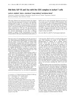
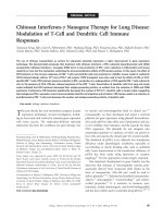
Repeated oral administration of type II collagen induces
immune tolerance and inhibits arthritis development
The arthritis index remained low in both the tolerance and CIA
groups until 4 weeks after primary immunization with CII–com-
plete Freund's adjuvant. In the CIA group, the arthritis index
began to increase after week 5, reached a peak between
weeks 6 and 9 after primary immunization, and then decreased
by week 11. In the tolerance group, the arthritis index peaked
between weeks 6 and 9, but the index was significantly lower
than that of the CIA group throughout the examination period
(Figure 1).
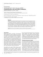
Induction of oral tolerance increases the proportion of
indoleamine 2,3-dioxygenase-expressing CD11c
+
dendritic cells in Peyer's patches of tolerized mice
DCs are potent stimulators of naïve T cells and are key induc-
ers of immune tolerance [25]. One molecular mechanism by
which DCs regulate T cells is through the expression of IDO,
which degrades the essential amino acid tryptophan [26]. To
determine whether IDO expression increases in tolerized mice,
we examined the relative proportion of IDO
+
DCs among
CD11c
+
DCs in Peyer's patches after repeated oral adminis-
tration of CII and subsequent CIA induction.
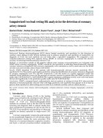
IDO expression was significantly higher in DCs from tolerized
mice than in CIA mice (mean fluorescence index, 87.4 ± 11.2
versus 36.7 ± 10.2, P < 0.05). Normal DBA/1 mice had the
lowest mean fluorescence index of IDO
+
DCs (10.2 ± 5.2, P
< 0.05; data not shown) (Figure 2a). These data were con-
firmed by measuring the mRNA level of IDO in CD11c
+
DCs
obtained from Peyer's patches of tolerized and CIA mice. We
used semiquantitative RT-PCR to measure IDO mRNA expres-
sion in CD11c
+
DCs isolated from Peyer's patches. The IDO
transcripts were upregulated markedly in tolerized mice com-
pared with CIA mice (Figure 2b).
Using confocal microscopy, we identified the expression of
IDO in situ at the single-cell level. Single cells were stained
simultaneously with a pandendritic cell marker, anti-CD11c
(green), and an IDO-specific antibody (red). About one-third of
the CD11c
+
DCs from tolerized mice expressed IDO (yellow).
In contrast, the DCs from CIA mice rarely expressed IDO (Fig-
ure 2c).
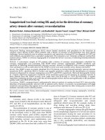
Indoleamine 2,3-dioxygenase-expressing CD11c
+
dendritic cells have an immature phenotype
Human IDO
+
DCs show a mature phenotype characterized by
CD14
-
, CD83
+
, CD80
+
, CD86
high
, and HLA-DR
high
[27,28]. In
contrast, DCs expressing IDO in mice show an immature phe-
notype and suppress T-cell proliferation both in vitro and in
vivo. The maturational status of IDO-expressing DCs in
Peyer's patches of tolerized mice, however, has never been
considered.
We examined the expression of major histocompatibility com-
plex II, CD80, and CD86 by CD11c
+
IDO
+
DCs from tolerized
mice and CIA mice. CD11c
+
IDO
+
DCs from tolerized mice
expressed low levels of MHC II and CD86, suggesting an
immature phenotype. In contrast, the expression of these sur-
face molecules was significantly higher on DCs from CIA mice,
which is characteristic of mature DCs (Figure 3). The expres-
sion of CD80, however, did not differ significantly between the
two groups.
CD11c
+
dendritic cells of tolerized mice inhibit type II
collagen-specific T-cell proliferation in an indoleamine
2,3-dioxygenase-dependent manner
APC-induced T-cell activation requires contact-dependent
bidirectional signaling between the APCs and the T cells [29].
This signaling may upregulate IDO expression in DCs, and this
may control autoreactive T cells by depleting tryptophan
[25,30]. We explored whether IDO affects the ability of DCs
isolated from Peyer's patches of tolerized mice to regulate T-
Figure 1
Inhibition of arthritis development in tolerized miceInhibition of arthritis development in tolerized mice. Mice in the toler-
ance group were fed 100 μg type II collagen (CII) six times for 2 weeks
before immunization. For collagen-induced arthritis (CIA) induction, CII
emulsified with complete Freund's adjuvant was injected into the tail of
mice in the tolerance group and in the CIA group as a primary immuni-
zation. Two weeks later, CII emulsified with incomplete Freund's adju-
vant was injected into a hind leg as a booster injection. The mean
arthritis index was significantly lower in the tolerance group than in the
CIA group throughout the examination period. Values are presented as
the mean ± standard deviation of three independent experiments involv-
ing 20 tolerized mice and 20 CIA mice per group. *P < 0.05, **P <
0.005.
Available online />Page 5 of 10
(page number not for citation purposes)
cell responses. Mixed lymphocyte cultures were performed in
the presence or absence of the IDO-specific inhibitor 1-MT.
CD11c
+
DCs from Peyer's patches of tolerized mice or CIA
mice were cocultured for 3 days with CII-reactive CD4
+
T cells
and irradiated APCs obtained from CIA mice in the absence or
presence of CII.
Without CII stimulation, the proliferation of CII-reactive CD4
+
T cells was inhibited more by CD11c
+
DCs from Peyer's
patches of tolerized mice than by CD11c
+
DCs from Peyer's
patches of CIA mice (687 ± 159 cpm versus 1,257 ± 103
cpm, P < 0.05) (Figure 4a). With CII-specific stimulation, the
suppressive effect was more prominent in CD11c
+
DCs from
tolerized mice. As shown in Figure 4a, proliferation of CD4
+
T
cells induced by CD11c
+
DCs of tolerized mice was sup-
pressed to be about one-third of that in CD4
+
T cells cultured
with CD11c
+
DCs of CIA mice (1,507 ± 817 cpm versus
4,204 ± 95 cpm, P < 0.05). Addition of the IDO inhibitor 1-
MT, however, abolished the suppressive effect of CD11c
+
DCs from tolerized mice on CII-specific T-cell proliferation
(1,507 × 10
3
± 817 cpm versus 3,128 × 10
3
± 101 cpm, P <
0.05). Without CII stimulation, 1-MT had no significant effect
on T-cell proliferation in either group.
To investigate the effect of IDO on the suppression of antigen-
specific T cells exerted by CD11c
+
DCs from tolerized mice,
cytokine concentrations were measured in the coculture
supernatants. The IL-17 concentration was significantly lower
Figure 2
Oral tolerance induction in indoleamine 2,3-dioxygenase-expressing CD11c
+
dendritic cells of tolerized miceOral tolerance induction in indoleamine 2,3-dioxygenase-expressing CD11c
+
dendritic cells of tolerized mice. The induction of oral tolerance
increases the proportion of indoleamine 2,3-dioxygenase (IDO)-expressing CD11c
+
dendritic cells (DCs) in Peyer's patches of tolerized mice. (a)
Flow cytometric analysis of IDO in CD11c
+
DCs isolated from Peyer's patches. Mononuclear cells obtained from Peyer's patches of tolerized mice
and of CIA mice were probed with Fluorescein isothiocyanate-labeled anti-CD11c mAb and were fixed with CytoPerm/CytoFix for 20 minutes. Cells
were probed for intracellular IDO using anti-mouse IDO antibody and were analyzed by flow cytometry. The histograms were gated on CD11c
+
DCs.
Dotted histogram lines represent cells stained with isotype-matched control monoclonal antibodies. Results are the mean ± standard deviation of
replicate samples from seven independent experiments. SSC. (b) Analysis of IDO transcription in tolerized mice and CIA mice. CD11c
+
DCs were
isolated from Peyer's patch mononuclear cells using the magnetic-activated cell sorting system. The expression of IDO mRNA was analyzed using
RT-PCR. β
2
-Actin was used as an internal control. Each value is the mean ± standard deviation of replicate determinations in one of four experi-
ments. *P < 0.05. (c) Immunofluorescent confocal microscopic examination of IDO expression by CD11c
+
DCs. Mononuclear cells obtained from
Peyer's patches of tolerized and CIA mice were stained with Fluorescein isothiocyanate-labeled anti-CD11c (green) and anti-IDO (red), fixed, and
were examined using confocal microscopy. Isotype-matched control antibody staining was negative (data not shown). Data are representative of
three independent experiments.
Arthritis Research & Therapy Vol 10 No 1 Park et al.
Page 6 of 10
(page number not for citation purposes)
in the culture supernatants of CD11c
+
DCs of tolerized mice
than in the supernatants of CD11c
+
DCs of CIA mice cultured
in the presence of CII (82 ± 17 pg/ml versus 158 ± 9 pg/ml,
P < 0.05) (Figure 4b). Addition of 1-MT into the coculture in
the presence of CII, however, markedly increased the produc-
tion of IL-17 by tolerized CD11c
+
DCs (from 82 ± 17 pg/ml to
141 ± 15 pg/ml, P < 0.01). The concentrations of anti-inflam-
matory cytokines such as IL-10 and TGFβ were higher in the
culture supernatants of CD11c
+
DCs of tolerized mice than in
the supernatants of CD11c
+
DCs of CIA mice (IL-10, 131 ±
14 pg/ml versus 79 ± 13 pg/ml (P < 0.05); and TGFβ, 260 ±
11 pg/ml versus 195 ± 16 pg/ml (P < 0.05)). The addition of
1-MT, however, significantly decreased the production of IL-
10 and TGFβ in the tolerance group (IL-10, from 131 ± 14 pg/
ml to 92 ± 16 pg/ml (P < 0.05); and TGFβ, from 260 ± 11 pg/
ml to 126 ± 9 pg/ml (P < 0.05)), suggesting that IDO expres-
sion on CD11c
+
DCs plays an important role in immune
suppression.
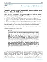
Indoleamine 2,3-dioxygenase-dependent induction of
antigen-specific CD4
+
CD25
+
T cells by CD11c
+
dendritic
cells in tolerized mice
Regulatory APCs may form a bridge between regulatory T
cells and responder T cells, and this has been proposed as a
mechanism contributing to the phenomenon of linked sup-
pression and dominant tolerance [26]. Experimental evidence
indicates that murine regulatory T cells can induce the expres-
sion of IDO [31]. We hypothesized that CD11c
+
DCs in toler-
ized mice are likely to be the IDO-dependent biologically
relevant trigger for the generation or conversion of
CD4
+
CD25
+
regulatory T cells. In a previous study,
CD11c
+
CD11b
+
DCs isolated from Peyer's patches of toler-
ized mice seemed necessary for the expansion and differenti-
ation of CD4
+
CD25
+
T cells, which suppress CII-specific T-
cell proliferation [12]. Highly purified CD4
+
CD25
-
T cells iso-
lated from tolerized mice (purity > 99%) were cocultured with
CD11c
+
DCs from tolerized or CIA mice for 3 days, without
CII. The proportion of CD4
+
CD25
+
T cells expanded by
CD11c
+
DCs from tolerized mice was similar to that obtained
by CD11c
+
DCs from CIA mice (4.65% ± 0.66% versus
3.68% ± 0.46%). 1-MT had no significant effect on the pro-
portion of CD4
+
CD25
+
T cells in these systems (4.74% ±
0.91% versus 3.56% ± 0.33%).
We next examined whether IDO would induce naïve
CD4
+
CD25
-
T cells to differentiate into CD4
+
CD25
+
Foxp3
+
T
cells in an antigen-specific manner when cocultured with
CD11c
+
DCs. Figure 5a,b shows that the percentage of
CD4
+
CD25
+
T cells was higher in CD4
+
CD25
-
T cells cocul-
tured with CD11c
+
DCs from tolerized mice in the presence of
CII than in CD4
+
CD25
-
T cells cocultured with CD11c
+
DCs
from CIA mice (13.4% ± 1.89% versus 5.56% ± 0.22%, P <
0.05). 1-MT abrogated the increase in the proportion of
CD4
+
CD25
+
T cells induced by CD11c
+
DCs from tolerized
mice (5.67% ± 0.72%) but not that induced by CD11c
+
DCs
from CIA mice (5.41% ± 0.20%). We used flow cytometry to
investigate the possible conversion of CD4
+
CD25
-
T cells into
CD4
+
CD25
+
T cells in concomitance with Foxp3 appearance.
CD4
+
CD25
+
T cells obtained by coculture with CD11c
+
DCs
from tolerized mice expressed significantly higher levels of
Foxp3 expression than those cells obtained by coculture with
CD11c
+
DCs from CIA mice; this effect was diminished by the
addition of 1-MT (CII stimulation, 72.1%; CII + 1-MT, 32.1%)
(Figure 5c). Using RT-PCR, we also examined Foxp3 tran-
scripts involved in the conversion of CD4
+
CD25
-
T cells into
CD4
+
CD25
+
T cells (Figure 5d). Our findings show that highly
purified CD4
+
CD25
-
T cells can be converted into
CD4
+
CD25
+
Foxp3
+
T cells by DCs from tolerized mice by
CII-specific stimulation through an IDO-dependent
mechanism.
To verify that the increased population of CD4
+
CD25
+
Foxp3
+
T cells retained their suppressive function, varying numbers of
CD4
+
CD25
+
T cells induced by exposure to CD11c
+
DCs
from tolerized mice were cocultured with CII-reactive CD4
+
T
cells and irradiated APCs from CIA mice for 3 days in the pres-
ence of CII. The CD4
+
CD25
+
T cells induced by CD11c
+
DCs
inhibited the proliferative response of CII-reactive CD4
+
T cells
in a concentration-dependent manner (Figure 5e).
Figure 3
Indoleamine 2,3-dioxygenase-expressing CD11c
+
dendritic cells dis-play a phenotype consistent with the immature stateIndoleamine 2,3-dioxygenase-expressing CD11c
+
dendritic cells dis-
play a phenotype consistent with the immature state. Indoleamine 2,3-
dioxygenase (IDO)
+
CD11c
+
dendritic cells (DCs) were stained for
markers of DC maturity and were analyzed using three-color flow
cytometry. Mononuclear cells from Peyer's patches were stained for
CD11c, IDO, and Major Histocompatibility Complex (MHC) II, or cos-
timulatory molecule markers. All plots were first gated on IDO
+
CD11c
+
cells. Dotted line shows the isotype-matched controls. Data are the
mean ± standard deviation of three experiments. MFI, mean fluores-
cence index.
Available online />Page 7 of 10
(page number not for citation purposes)
Discussion
Oral tolerance is initiated in the gut-associated lymphoid tis-
sues, a well-developed immune network in the alimentary tract
that comprises the mucosal epithelium, lamina propria, Peyer's
patches, and mesenteric lymph nodes [1,2]. Peyer's patches
are essential sites for the induction of mucosal immune
responses and oral tolerance to soluble antigen, and Peyer's
patch DCs are thought to play an important role in mucosal
immunity and tolerance [1-4]. The selection between immunity
and tolerance depends on several factors, including the occur-
rence of specialized DC subsets and the maturation state of
the DCs [1,12]. One mechanism, exploited by tolerogenic
DCs, involves IDO [17-21]. IDO-competent DCs exert regula-
tory effects on T cells that are mediated by tryptophan deple-
tion and by the production of metabolic byproducts
collectively known as kynurenines [16,21,22].
Demonstrating the concomitant induction of IDO enzymatic
activity by DCs, our data support IDO-dependent mechanisms
that have been associated with induction of T-cell tolerance
and immune inhibition in the induction of oral tolerance in the
murine CIA model. We examined the change in the expression
of IDO in CD11c
+
DCs of Peyer's patches after repeated oral
administration of CII and subsequent induction of CIA. In
freshly isolated Peyer's patches, the proportion of CD11c
+
IDO
+
DCs was higher in tolerized mice than that in CIA mice.
We found that IDO expression was induced most by oral CII
feeding plus CII immunization, and that the expression of IDO
did not correlate with disease severity. Next, we investigated
whether the expression of IDO by DCs takes place during the
initial phase of oral CII feeding or after the immunization with
CII for CIA induction. In these studies, Peyer's patches were
isolated from mice that had been fed with CII or PBS but not
Figure 4
Tolerized mice CD11c
+
dendritic cells induce indoleamine 2,3-dioxygenase-dependent inhibition of type II collagen-specific T-cell proliferationTolerized mice CD11c
+
dendritic cells induce indoleamine 2,3-dioxygenase-dependent inhibition of type II collagen-specific T-cell proliferation. (a)
Type II collagen (CII)-reactive CD4
+
T cells (1 × 10
5
/well) and irradiated antigen-presenting cells (APCs) (1 × 10
5
/well) isolated from Peyer's
patches of collagen-induced arthritis (CIA) mice were cultured with CD11c
+
dendritic cells (DCs) (1 × 10
4
/well) from tolerized mice or CIA mice in
the presence or absence of CII (40 μg/ml) in 96-well, U-bottomed plates for 3 days. Some DCs were pretreated with 1-methyl tryptophan (1-MT)
(200 μM) for 2 hours before coculture. Data presented as the mean counts per minute (cpm) of triplicate cultures. Values are the mean ± standard
deviation from three independent experiments. *P < 0.05. (b) Cytokine concentrations in the coculture supernatants. Concentrations of IL-17, IL-10,
and transforming growth factor beta (TGFβ) in the culture supernatants were measured by ELISA. Values are the mean ± standard deviation from
three independent experiments. *P < 0.05, **P < 0.001.
Arthritis Research & Therapy Vol 10 No 1 Park et al.
Page 8 of 10
(page number not for citation purposes)
immunized with CII. We examined the expression of IDO in
situ. The expression of IDO by DCs increased in Peyer's
patches of CII-fed mice, even though these mice had not been
immunized. In contrast, IDO
+
DCs were rare in the Peyer's
patches of PBS-fed mice (data not shown). Our results show
that IDO is induced by oral CII feeding and enhances the
induction of immune tolerance after oral administration of
antigens.
The immunosuppressive mechanism of IDO is shared by sev-
eral different cell types in the immune system [30,32-36].
Splenic CD11c
+
CD19
+
DCs found in the mice administered
CpG oligodeoxynucleotides, human monocyte-derived macro-
phages, and in vitro-derived DCs induce IDO expression and
inhibit T-cell proliferation. In this regard, it will be interesting to
investigate IDO expression in different subsets of DCs from
tolerized mice and to characterize their role in the induction
and maintenance of immune tolerance.
Figure 5
Indoleamine 2,3-dioxygenase-expressing CD11c
+
dendritic cells essential for antigen-specific CD4
+
CD25
+
regulatory T-cell induction in tolerized miceIndoleamine 2,3-dioxygenase-expressing CD11c
+
dendritic cells essential for antigen-specific CD4
+
CD25
+
regulatory T-cell induction in tolerized
mice. (a) Increased CD4
+
CD25
+
T-cell proportion through an indoleamine 2,3-dioxygenase (IDO)-dependent mechanism. For regulatory T-cell
induction, isolated CD4
+
CD25
-
T cells (1 × 10
5
/well) were cultured with CD11c
+
dendritic cells (DCs) (2 × 10
4
/well) from tolerized mice or colla-
gen-induced arthritis (CIA) mice in the absence or presence of type II collagen (CII) (40 μg/ml) for 3 days. 1-Methyl tryptophan (1-MT) was added to
selected cultures. The proportion of CD4
+
CD25
+
T cells was determined using flow cytometry. Numbers represent the percentage of double-posi-
tive cells. (b) Summary of the percentages of CD4
+
CD25
+
T cells from the coculture experiments in (a). Values are the mean from four independent
experiments; individual symbols are the mean in individual animals, and bars show the group means. *P < 0.02. (c) Analysis of Foxp3 expression by
converted CD4
+
CD25
+
T cells. Plots were gated on CD4
+
CD25
+
DCs. Dotted histogram lines represent cells stained with isotype-matched control
monoclonal antibodies. Data represent the mean ± standard deviation and are representative of four independent experiments. (d) Foxp3 mRNA
expression in the same conditions as (a). β -Actin was used as an internal control. Results are representative of four independent experiments. (e)
Regulatory function of the CII-induced CD4
+
CD25
+
T cells. CD4
+
CD25
+
T cells were expanded by exposure to CD11c
+
DCs from Peyer's patches
from tolerized mice in the presence of CII antigen stimulation. Varying numbers of CD4
+
CD25
+
T cells were cultured for 3 days with CII-reactive
CD4
+
T cells (1 × 10
5
) and irradiated antigen-presenting cells (1 × 10
5
) from mice with CIA in the presence of CII (40 μg/ml). Values are the mean
± standard deviation from three independent experiments. *P < 0.05, **P < 0.001. cpm, counts per minute.
Available online />Page 9 of 10
(page number not for citation purposes)
Tolerogenic DCs are immature, maturation-resistant, or alter-
natively activated DCs that express surface MHC molecules
and have a low ratio of costimulatory to inhibitory signals, such
as IL-10, programmed death-ligand 1, CTLA4/CD28, and IDO
[37,38]. IDO expression is detected constitutively in human
regulatory plasmacytoid DCs and can be induced by classical
DC maturation stimuli, namely IFNγ and lipopolysaccharide or
prostaglandin E
2
, which contribute to their immunoregulatory
capacity [37-40]. In our study, the phenotype of DCs from
tolerized mice showed an immature tolerogenic state and low
levels of surface MHC II and CD86 molecules, and expressed
high levels of IDO compared with DCs from CIA mice. Our
study suggests that IDO is expressed constitutively in imma-
ture DCs upon repeated oral administration of CII in an animal
model without artificial administration of CTLA-4 immunoglob-
ulin or other DC-modifying agents.
Because IDO expression on DCs plays a crucial role in the
induction of regulatory T cells and inhibition of the antigen-
specific T-cell response, we performed mixed lymphocyte cul-
tures to determine whether DCs isolated from Peyer's patches
of tolerized mice and CIA mice have an IDO-dependent effect
on CII-specific T cells. DCs from the tolerized mice inhibited
the proliferation of CII-specific T cells and inflammatory
cytokine production compared with DCs from CIA mice, and
these suppressive effects were more evident after CII stimula-
tion and were abrogated by addition of 1-MT, an IDO inhibitor.
These findings suggest that the functional activities of IDO
from tolerized mice could affect the T-cell response.
In the resting state, autoreactive T cells residing in the periph-
ery are suppressed effectively by regulatory T cells, which are
thought to prevent the development of autoimmune diseases
[39]. Our group previously demonstrated that the proportion
of IL-10-producing CD4
+
CD25
+
T cells increases more in
Peyer's patches and spleens of tolerized mice than in those of
CIA mice [12]. Bozza and colleagues reported that
CD4
+
CD25
+
regulatory T cells in candidiasis are strictly
dependent on the expression of B7 costimulatory molecules
by IL-10-producing DCs, and are involved in the IFNγ/IDO-
dependent pathway that controls the local inflammatory
pathology [39]. Saito and colleagues also found that CTLA-4
on CD4
+
CD25
+
regulatory T cells induces the expression of
IDO on DCs [40]. Considering these findings, we next sought
to determine whether IDO
+
DCs increase the proportion of
antigen-specific CD4
+
CD25
+
regulatory T cells in tolerized
mice. Figure 5 shows that the percentage of CD4
+
CD25
+
cells increased significantly after coculture of CD4
+
CD25
-
T
cells with CD11c
+
DCs from tolerized mice in the presence of
CII through an IDO-dependent mechanism. In the absence of
CII, however, the proportion of CD4
+
CD25
+
cells cocultured
with CD11c
+
DCs of tolerized mice was similar to that in cells
cocultured with CD11c
+
DCs from CIA mice. This result sug-
gests that the conversion into CD4
+
CD25
+
T cells without CII
stimulation result from the IDO-independent expansion or
selected survival of residual CD4
+
CD25
+
T cells. Foxp3 is a
transcription factor specific for CD4
+
CD25
+
regulatory T
cells. Foxp3 may be necessary for the induction of
CD4
+
CD25
+
regulatory T cells by conversion of CD4
+
CD25
-
T cells after antigen stimulation [41]. We found that
CD4
+
CD25
+
T cells obtained after coculture of CD4
+
CD25
-
T
cells and CD11c
+
DCs of tolerized mice with CII expressed a
large amount of Foxp3 transcript and Foxp3 protein in vitro in
an IDO-dependent manner. In addition, the conversion of
Foxp3
+
CD4
+
CD25
+
T cells, which are thought to be regula-
tory T cells, inhibited the proliferation of CII-reactive CD4
+
T
cells in a dose-dependent manner.
CD4
+
CD25
+
regulatory T cells converted from peripheral
CD4
+
CD25
-
naïve T cells by TGFβ induction of Foxp3 were
reported to be unresponsive to T-cell receptor stimulation, to
produce neither T helper 1 nor T helper 2 cytokines, to express
TGFβ, and to inhibit normal T-cell proliferation in vitro [42]. Dif-
ferent to this regulatory T-cell population, we found that the
Foxp3
+
CD4
+
CD25
+
T cells in our model system may result
partly from the expansion or selected survival of residual
CD4
+
CD25
+
regulatory T cells and partly from the conversion
of CD4
+
CD25
-
T cells into Foxp3
+
CD4
+
CD25
+
regulatory T
cells through an IDO-dependent mechanism.
Conclusion
IDO expression by DCs is crucial for the induction of
Foxp3
+
CD4
+
CD25
+
T cells and for the suppression of CII-
reactive T-cell function in induction of oral tolerance to CII.
These results demonstrate that the induction of IDO
+
DCs in
Peyer's patches plays an essential role in the induction of oral
tolerance and may provide a new modality of immune-based
treatments for autoimmune diseases.
Competing interests
The authors declare that they have no competing interests.
Authors' contributions
M-JP and S-YM performed the experimental work and pre-
pared the manuscript. Y-GC, M-LC, K-SP, and S-GC advised
on the study. H-SP, Y-OJ, and S-HC performed the experimen-
tal work H-YK is the head of the laboratory, supervised the
experimental work, and advised on the study. S-HP, and J-KM
are the senior researchers, and they supervised the
experimental work and advised on the study. All authors read
and approved the final manuscript.
Acknowledgements
The authors are grateful to Professor Andrew H. Kang for providing the
CII. This work was supported by a grant (R11-2002-098-01001-0) from
the Korea Science and Engineering Foundation through the Rheuma-
tism Research Center and by the Korean Research Foundation Grant
(KRF-2005-217-E00003) funded by the Korean Government (MOE-
HRD, Basic Research Promotion Fund).
Arthritis Research & Therapy Vol 10 No 1 Park et al.
Page 10 of 10
(page number not for citation purposes)
References
1. Mayer L, Shao L: Therapeutic potential of oral tolerance. Nat Rev
Immunol 2004, 4:407-419.
2. Mowat AM: Anatomical basis of tolerance and immunity to intes-
tinal antigens. Nat Rev Immunol 2003, 3:331-341.
3. Nagler-Anderson C, Bober LA, Robinson ME, Siskind GW, Thor-
becke GJ: Suppression of type II collagen-induced arthritis by
intragastric administration of soluble type II collagen. Proc Natl
Acad Sci USA 1986, 83:7443-7446.
4. Whitacre CC, Song F, Wardrop RM 3rd, Campbell K, McClain M,
Benson J, Guan Z, Gienapp I: Regulation of autoreactive T cell
function by oral tolerance to self-antigens. Ann N Y Acad Sci
2004, 1029:172-179.
5. Song F, Guan Z, Gienapp IE, Shawler T, Benson J, Whitacre CC:
The thymus plays a role in oral tolerance in experimental
autoimmune encephalomyelitis. J Immunol 2006,
177:1500-1509.
6. Dubois B, Goubier A, Joubert G, Kaiserlian D: Oral tolerance and
regulation of mucosal immunity. Cell Mol Life Sci 2005,
62:1322-1332.
7. Mowat AM, Parker LA, Beacock-Sharp H, Millington OR, Chirdo F:
Oral tolerance: overview and historical perspectives. Ann N Y
Acad Sci 2004, 1029:1-8.
8. Derry CJ, Harper N, Davies DH, Murphy JJ, Staines NA: Importance
of dose of type II collagen in suppression of collagen-induced
arthritis by nasal tolerance. Arthritis Rheum 2001,
44:1917-1927.
9. Garcia G, Komagata Y, Slavin AJ, Maron R, Weiner HL: Suppres-
sion of collagen-induced arthritis by oral or nasal administration
of type II collagen. J Autoimmun 1999, 13:315-324.
10. Weiner HL: Oral tolerance: immune mechanisms and treatment
of autoimmune diseases. Immunol Today 1997, 18:335-343.
11. Friedman A, Weiner HL: Induction of anergy or active suppres-
sion following oral tolerance is determined by antigen dosage.
Proc Natl Acad Sci USA 1994, 91:6688-6692.
12. Min SY, Park KS, Cho ML, Kang JW, Cho YG, Hwang SY, Park MJ,
Yoon CH, Min JK, Lee SH, Park SH, Kim HY: Antigen-induced,
tolerogenic CD11c
+
, CD11b
+
dendritic cells are abundant in
Peyer's patches during the induction of oral tolerance to type II
collagen and suppress experimental collagen-induced arthritis.
Arthritis Rheum 2006, 54:887-898.
13. Min SY, Hwang SY, Park KS, Lee JS, Lee KE, Kim KW, Jung YO,
Koh HJ, Do JH, Kim H, Kim HY: Induction of IL-10-producing
CD4
+
CD25
+
T cells in animal model of collagen-induced arthri-
tis by oral administration of type II collagen. Arthritis Res Ther
2004, 6:R213-R219.
14. Kelsall BL, Leon F: Involvement of intestinal dendritic cells in oral
tolerance, immunity to pathogens, and inflammatory bowel
disease. Immunol Rev 2005, 206:132-148.
15. Mowat AM: Dendritic cells and immune responses to orally
administered antigens. Vaccine 2005, 23:1797-1799.
16. Wakkach A, Fournier N, Brun V, Breittmayer JP, Cottrez F, Groux H:
Characterization of dendritic cells that induce tolerance and T
regulatory 1 cell differentiation in vivo. Immunity 2003,
18:605-617.
17. Fallarino F, Vacca C, Orabona C, Belladonna ML, Bianchi R, Mar-
shall B, Keskin DB, Mellor AL, Fioretti MC, Grohmann U, Puccetti P:
Functional expression of indoleamine 2,3-dioxygenase by
murine CD8 alpha(+) dendritic cells. Int Immunol 2002,
14:65-68.
18. Munn DH, Sharma MD, Hou D, Baban B, Lee JR, Antonia SJ,
Messina JL, Chandler P, Koni PA, Mellor AL: Expression of
indoleamine 2,3-dioxygenase by plasmacytoid dendritic cells in
tumor-draining lymph nodes. J Clin Invest 2004, 114:280-290.
19. Munn DH, Sharma MD, Baban B, Harding HP, Zhang Y, Ron D, Mel-
lor AL: GCN2 kinase in T cells mediates proliferative arrest and
anergy induction in response to indoleamine 2,3-dioxygenase.
Immunity 2005, 22:633-642.
20. Mellor AL, Baban B, Chandler PR, Manlapat A, Kahler DJ, Munn DH:
Cutting edge: CpG oligonucleotides induce splenic CD19
+
den-
dritic cells to acquire potent indoleamine 2,3-dioxygenase-
dependent T cell regulatory functions via IFN type 1 signaling. J
Immunol 2005, 175:5601-5605.
21. Bilsborough J, George TC, Norment A, Viney JL: Mucosal CD8α
+
DC, with a plasmacytoid phenotype, induce differentiation and
support function of T cells with regulatory properties. Immunol-
ogy 2003, 108:481-492.
22. Moingeon P, Batard T, Fadel R, Frati F, Sieber J, Overtvelt L:
Immune mechanisms of allergen-specific sublingual
immunotherapy. Allergy 2006, 61:151-165.
23. Stuart JM, Cremer MA, Kang AH, Townes AS: Collagen-induced
arthritis in rats. Evaluation of early immunologic events. Arthritis
Rheum 1979, 22:1344-1351.
24. Barnett ML, Kremer JM, St Clair EW, Clegg DO, Furst D, Weisman
M, Fletcher MJ, Chasan-Taber S, Finger E, Morales A, Le CH,
Trentham DE: Treatment of rheumatoid arthritis with oral type II
collagen. Results of a multicenter, double-blind, placebo-con-
trolled trial. Arthritis Rheum 1998, 41:290-297.
25. Belz GT, Heath WR, Carbone FR: The role of dendritic cell sub-
sets in selection between tolerance and immunity. Immunol Cell
Biol 2002, 80:463-468.
26. Munn DH, Sharma MD, Mellor AL: Ligation of B7-1/B7-2 by
human CD4
+
T cells triggers indoleamine 2,3-dioxygenase
activity in dendritic cells. J Immunol 2004, 172:4100-4110.
27. Munn DH, Sharma MD, Lee JR, Jhaver KG, Johnson TS, Keskin DB,
Marshall B, Chandler P, Antonia SJ, Burgess R, Slingluff CL Jr, Mel-
lor AL: Potential regulatory function of human dendritic cells
expressing indoleamine 2,3-dioxygenase. Science 2002,
297:1867-1870.
28. Hwang SL, Chung NP, Chan JK, Lin CL: Indoleamine 2, 3-dioxy-
genase (IDO) is essential for dendritic cell activation and chem-
otactic responsiveness to chemokines. Cell Res 2005,
15:167-175.
29. Swanson KA, Zheng Y, Heidler KM, Mizobuchi T, Wilkes DS: CDllc
+
cells modulate pulmonary immune responses by production of
indoleamine 2,3-dioxygenase. Am J Respir Cell Mol Biol 2004,
30:311-318.
30. Uyttenhove C, Pilotte L, Theate I, Stroobant V, Colau D, Parmentier
N, Boon T, Van den Eynde BJ: Evidence for a tumoral immune
resistance mechanism based on tryptophan degradation by
indoleamine 2,3-dioxygenase. Nat Med 2003, 9:1269-1274.
31. Fallarino F, Grohmann U, Hwang KW, Orabona C, Vacca C, Bianchi
R, Belladonna ML, Fioretti MC, Alegre ML, Puccetti P: Modulation
of tryptophan catabolism by regulatory T cells. Nat Immunol
2003, 4:1206-1212.
32. Yoshida R, Nukiwa T, Watanabe Y, Fujiwara M, Hirata F, Hayaishi O:
Regulation of indoleamine 2,3-dioxygenase activity in the small
intestine and the epididymis of mice. Arch Biochem Biophys
1980, 203:343-351.
33. Malina HZ, Martin XD: Indoleamine 2,3-dioxygenase: antioxidant
enzyme in the human eye. Graefes Arch Clin Exp Ophthalmol
1996, 234:457-462.
34. Moffett JR, Espey MG, Namboodiri MA: Antibodies to quinolinic
acid and the determination of its cellular distribution within the
rat immune system. Cell Tissue Res 1994, 278:461-469.
35. Lee JR, Dalton RR, Messina JL, Sharma MD, Smith DM, Burgess RE,
Mazzella F, Antonia SJ, Mellor AL, Munn DH: Pattern of recruitment
of immunoregulatory antigen-presenting cells in malignant
melanoma. Lab Invest 2003, 83:1457-1466.
36. Mellor AL, Munn DH: Tryptophan catabolism and T-cell toler-
ance: immunosuppression by starvation? Immunol Today 1999,
20:469-473.
37. Hubert P, Jacobs N, Caberg JH, Boniver J, Delvenne P: The cross-
talk between dendritic and regulatory T cells: good or evil? J
Leukoc Biol 2007, 82:781-794.
38. Morelli AE, Thomson AW: Tolerogenic dendritic cells and the
quest for transplant tolerance. Nat Rev Immunol 2007,
7:610-621.
39. Bozza S, Fallarino F, Pitzurra L, Zelante T, Montagnoli C, Bellocchio
S, Mosci P, Vacca C, Puccetti P, Romani L: A crucial role for
tryptophan catabolism at the host/Candida albicans interface. J
Immunol
2005, 174:2910-2918.
40. Saito S, Sasaki Y, Sakai M: CD4(+)CD25
high
regulatory T cells in
human pregnancy. J Reprod Immunol 2005, 65:111-120.
41. Kretschmer K, Apostolou I, Hawiger D, Khazaie K, Nussenzweig
MC, von Boehmer H: Inducing and expanding regulatory T cell
populations by foreign antigen. Nat Immunol 2005,
6:1219-1227.
42. Chen W, Jin W, Hardegen N, Lei KJ, Li L, Marinos N, McGrady G,
Wahl SM: Conversion of peripheral CD4
+
CD25
-
naive T cells to
CD4
+
CD25+ regulatory T cells by TGF-beta induction of tran-
scription factor Foxp3. J Exp Med 2003, 198:1875-1886.