Báo cáo y học: "Cartilage preservation by inhibition of Janus kinase 3 in two rodent models of rheumatoid arthritis" doc
Bạn đang xem bản rút gọn của tài liệu. Xem và tải ngay bản đầy đủ của tài liệu tại đây (559.36 KB, 9 trang )
Open Access
Available online />Page 1 of 9
(page number not for citation purposes)
Vol 10 No 1
Research article
Cartilage preservation by inhibition of Janus kinase 3 in two
rodent models of rheumatoid arthritis
Anthony J Milici
1
*, Elizabeth M Kudlacz
2
*, Laurent Audoly
3
, Samuel Zwillich
2
and Paul Changelian
4
1
Pfizer Global Research and Development, MS#8220-2235, Groton, CT 06340, USA
2
Pfizer Global Research and Development, 50 Pequot Ave, New London, CT 06320, USA
3
Merck Research Laboratories, West Point, PA 19486, USA
4
1009 Glenhill Drive, Northville, MI 48167, USA
* Contributed equally
Corresponding author: Anthony J Milici,
Received: 11 Sep 2007 Revisions requested: 12 Oct 2007 Revisions received: 24 Jan 2008 Accepted: 30 Jan 2008 Published: 30 Jan 2008
Arthritis Research & Therapy 2008, 10:R14 (doi:10.1186/ar2365)
This article is online at: />© 2008 Milici et al.; licensee BioMed Central Ltd.
This is an open access article distributed under the terms of the Creative Commons Attribution License ( />),
which permits unrestricted use, distribution, and reproduction in any medium, provided the original work is properly cited.
Abstract
Introduction CP-690550 is a small molecule inhibitor of Janus
kinase 3 (JAK3), a critical enzyme in the signaling pathway of
multiple cytokines (interleukin (IL)-2, -7, -15 and -21) that are
important in various T cell functions including development,
activation and homeostasis. The purpose of this study was to
evaluate CP-690550 in murine collagen-induced (CIA) and rat
adjuvant-induced (AA) models of rheumatoid arthritis (RA).
Methods CIA and AA were induced using standard protocols
and animals received the JAK3 inhibitor via osmotic mini-pump
infusion at doses ranging from 1.5–15 mg/kg/day following
disease induction. Arthritis was assessed by clinical scores in
the CIA models and paw swelling monitored using a
plethysmometer in the AA model until study conclusion, at which
time animals were killed and evaluated histologically.
Results CP-690550 dose-dependently decreased endpoints of
disease in both RA models with greater than 90% reduction
observed at the highest administered dose. An approximate
ED
50
of approximately 1.5 mg/kg/day was determined for the
compound based upon disease endpoints in both RA models
examined and corresponds to CP-690550 serum levels of 5.8
ng/ml in mice (day 28) and 24 ng/ml in rats (day 24). The
compound also reduced inflammatory cell influx and joint
damage as measured histologically. Animals receiving a CP-
690550 dose of 15 mg/k/d showed no histological evidence of
disease.
Conclusion The efficacy observed with CP-690550 in CIA and
AA suggests JAK3 inhibition may represent a novel therapeutic
target for the treatment of RA.
Introduction
Rheumatoid arthritis (RA) is a chronic, systemic disease char-
acterized by persistent inflammatory synovitis that typically
involves peripheral joints in a symmetric distribution [1]. The
synovial inflammation can cause cartilage destruction and
bone erosions that are irreversible. To minimize the radio-
graphic damage, it has been recognized that initiation of ther-
apy with disease-modifying antirheumatic drugs (DMARDs)
within 3 months after disease diagnosis is critical [2]. The folic
acid antagonist methotrexate (MTX) is the DMARD most com-
monly selected for initial therapy [2] and whose mechanism of
action has been attributed, at least in part, to its ability to func-
tion as an antimetabolite. As such, the compound inhibits cell
proliferation in the inflamed synovium but can affect other pro-
liferating tissues, including gut and bone marrow, producing
associated side effects. The use of biological response modi-
fiers, such as tumor necrosis factor (TNF) antagonists, has
grown due to efficacy observed in many patients and reason-
able safety profile [3]. However, the incomplete efficacy and/
or toxicities observed with agents such as these create a need
for additional therapies with novel mechanisms of action.
The key role that T cells appear to play in the pathogenesis of
the disease has supported evaluation of calcineurin inhibitors
such as cyclosporin A and tacrolimus in RA patients [4]. Clin-
ical efficacy for both calcineurin inhibitors has been reported,
particularly in combination with other DMARDs such as meth-
otrexate. However, the use of cyclosporine and tacrolimus in
AA = adjuvant-induced arthritis; CIA = collagen-induced arthritis; DMARD = disease-modifying antirheumatic drug; ELISA = enzyme-linked immuno-
sorbent assay; IL = interleukin; JAK3 = Janus kinase 3; LLOQ = lower limit of quantification; RA = rheumatoid arthritis; TNF = tumor necrosis factor.
Arthritis Research & Therapy Vol 10 No 1 Milici et al.
Page 2 of 9
(page number not for citation purposes)
this patient population may be limited based upon the multi-
plicity and severity of associated adverse reactions. CP-
690550 is a novel immunosuppressant that has not exhibited
the safety liabilities associated with calcineurin inhibition, yet
has demonstrated efficacy in a number of animal models
including delayed-type hypersensitivity and cardiac allograft
rejection [5,6]. CP-690550 is a small molecule inhibitor of the
tyrosine kinase Janus kinase 3 (JAK3), an enzyme that is asso-
ciated with the common gamma chain (γc) of various cytokine
receptors and is critical for signal transduction by interleukin
(IL)-2, -7, -15 and -21 [7]. Interestingly, JAK3 expression has
been shown to decrease in the synovial tissue biopsies from
active rheumatoid arthritics receiving and responding to
DMARD therapy [8].
Since multiple cytokines whose receptors signal through path-
ways involving JAK3 have been associated with progression of
arthritis, experiments were designed to evaluate the effects of
CP-690550 in rodent models of the disease. Neither murine
collagen-induced arthritis (CIA) nor adjuvant-induced arthritis
(AA) in rats are identical to RA, but both share the common
features of inflammation of the synovial membrane, erosion of
bone, and cartilage degradation. In both models of RA, we
observed dose-dependent inhibition of disease endpoints that
correlated with reduction in histological changes. These data
support JAK3 inhibition as a new target for the treatment of
RA.
Materials and methods
Reagents
CP-690550 was synthesized in-house and the enzyme specif-
icity of this compound has been previously described [5]. The
anti-TNF antibody TN.1912 has been shown to effectively neu-
tralize TNF in vivo and to have a 7-day half-life [9]. This clone
was scaled up in-house and the dose of agent chosen for this
study based upon internal (data not shown) and external
experiments demonstrating efficacy in the CIA model at doses
ranging from 300 μg/mouse intraperitoneally once a week to
300 μg/mouse intraperitoneally twice a week [10-12]. Unless
otherwise specified, reagents were purchased from Sigma-
Aldrich Chemical Company (St. Louis, MO, USA).
General animal care
For collagen-induced arthritis studies, male DBA/J1 mice (7–
9 weeks old from Jackson Labs, Bar Harbor, ME, USA) were
used. For studies of adjuvant-induced arthritis, male Lewis rats
were used (~50–60 days old from Charles River Labs, Wilm-
ington, MA, USA). Animals were housed in standard cages
with access to food and water ad libitum. The environment
was maintained at 21 ± 2°C with a time regulated light period
from 6 am to 6 pm. Studies were conducted in accordance
with the guidelines set forth by the Pfizer Animal Care and Use
Committee. An additional CIA study using mice of same age,
strain and source was performed at Boulder BioPATH Inc as
described below.
Murine CIA experiment
Male DBA/J1 mice were shaved at the base of the tail and
injected with 0.1 ml emulsion consisting of a 1 to 1 (1 mg/1
mg) mixture of type II chicken collagen with Mycobacterium
butyricum (Difco lot # 147539, Voigt Global Distribution,
Lawrence, Kansas) as an adjuvant. Three weeks later, the mice
were boosted with another 0.1 ml injection of emulsion at the
base of the tail to induce disease. Three days following this
injection, the animals were randomized and Alzet osmotic mini-
pumps (28-day pumps, model 2004, Durect Corporation,
Cupertino, CA) were implanted subcutaneously on the back of
each mouse to deliver CP-690550 at 1.5 (n = 13), 5 (n = 14)
or 15 (n = 14) mg/kg/day, poly(ethylene glycol) (PEG)300
vehicle (n = 15) or no pump (n = 11). It was necessary to
administer CP-690550 via osmotic mini-pumps due to the
poor pharmacokinetic (PK) properties of this compound in
rodents. The mice were scored in a blinded manner (0–12)
twice weekly for 3 weeks for signs of arthritis in each paw
according to the following scale: 0 = no swelling or redness/
normal paw; 1 = swelling and/or redness in one digit; 2 =
swelling and/or redness in two or more digits; and 3 = entire
paw is swollen or red. Upon study completion (day 28), mice
were killed with CO
2
. Blood samples were immediately taken
via cardiac puncture and serum analyzed for CP-690550 lev-
els. Following this, the knees were removed and processed for
histological analyses as described below. The knees were
chosen instead of the paws because both our lab and others
[13] have observed a good correlation between paw swelling
and histological changes.
Boulder BioPATH CIA experiment
An additional CIA study was performed at Boulder BioPATH
(Boulder, CO, USA) as described above with the following
modifications: (a) inclusion of anti-TNF treatment group (250
μg/animal intraperitoneally twice a week); (b) collection of
interim serum samples on day 15; (c) increase in study length
from 28 (Pfizer study) to 31 days; and (d) mice were scored in
a blinded manner on a 0–20 scale twice weekly for 3 weeks
for signs of arthritis in each paw (n = 10 for all groups except
naïve where n = 5). Clinical signs were evaluated using the fol-
lowing scale: 0 = normal; 1 = one joint affected or mild diffuse
erythema and swelling; 2 = two joints affected or mild diffuse
erythema and swelling; 3 = three joints affected or mild diffuse
erythema and swelling; 4 = four joint affected or marked dif-
fuse erythema and swelling; and 5 = severe erythema and
severe swelling.
Rat AA
Male Lewis rats were shaved at the base of the tail and
injected once intradermally with 100 μl of a 10 mg/ml Myco-
bacterium butyricum (Difco lot # 147539) mineral oil suspen-
sion. Ten days after this injection, the foot volumes of both the
right and left paws were measured with a Stoelting plethys-
mometer and Alzet osmotic mini-pumps (14-day pumps,
model 2ML2 (Stoeling Company, Wood Dale, IL) were
Available online />Page 3 of 9
(page number not for citation purposes)
implanted subcutaneously to deliver CP-690550 1.5, 5 or 15
mg/kg/day or vehicle (PEG300) (n = 10 for all groups except
naïve where n = 5). Swelling in the paws of the rats was meas-
ured in a blinded manner with a plethysmometer twice weekly
for 2 weeks. At the completion of the study (day 24), rats were
killed with anesthesia. Blood samples were immediately taken
via cardiac puncture and serum analyzed for CP-690550 lev-
els. Following this, the hind paws were removed and proc-
essed for histological analyses as described below.
Histology
Mouse hind limbs and rat hind paws were collected and
immersion fixed in 10% buffered formalin. Limbs and paws
were routinely processed, embedded in paraffin, sectioned
and analyzed as previously described [14].
IL-6 analysis
Serum IL-6 levels were measured by enzyme-linked immuno-
sorbent assay (ELISA) using a murine IL-6 kit (Quantikine;
R&D Systems, Minneapolis, MN, USA). The number of animals
available for IL-6 measurements was as follows: naïve (n = 3);
vehicle (n = 8); anti-TNF (n = 8); CP-690550 1.5 (n = 6), 5 (n
= 8) or 15 (n = 7) mg/kg/day.
Drug level analysis
Serum concentrations of CP-690550 were determined using
reverse-phase high performace liquid chromatography
(HPLC) with MS/MS (mass spectrometry/mass spectrometry)
detection as previously described [5]. Since CP-690550 was
administered via osmotic mini-pumps, the terminal drug con-
centration represents the steady-state drug concentrations in
these animals.
Statistical analysis
Scores for all measurements were analyzed by one sample t
test (Statview v.5, SAS Institute, Cary, NC, USA) and signifi-
cance set at p ≤ 0.05.
Results
Murine CIA
Clinical signs
In the first murine CIA study, an increase in clinical signs of dis-
ease were detected on day 10. The vehicle treated mice
attained a clinical score of 3.9 ± 0.7 that gradually increased
to a maximum of 5.3 ± 0.9 on day 27 (Figure 1). Clinical scores
were similar in diseased animals not receiving a pump, sug-
gesting neither implantation of the pump nor the vehicle had a
significant effect on the clinical score. At the lowest dose of
CP-690550 (1.5 mg/kg/day), the clinical score peaked on day
10 at 2.2 ± 0.5 and the response remained attenuated relative
to the control group for the remainder of the study. Treatment
at both the intermediate (5 mg/kg/day) and high (15 mg/kg/
day) doses of CP-690550 produced a highly significant, near
total suppression of clinical scores throughout the entire
study. Based upon the clinical scores, the ED
50
of CP-690550
was ~1.5 mg/kg/day with > 90% disease reduction observed
at the 15 mg/kg/day dose.
A second murine CIA study was performed and included an
anti-TNF treatment group as a comparator. The clinical scores
were reduced in this study relative to the first CIA study, which
could be due to subjective differences in scoring. As early as
3 days post-implantation of pumps, mice receiving both high
and low doses of CP-690550 exhibited significant reductions
in the clinical score vs vehicle (Figure 2). By days 9–28 all
three dose levels of CP-690550 resulted in a significant
reduction in the clinical score. On day 31, only the high and
mid-dose of CP-690550 maintained this statistically signifi-
cant reduction in clinical score vs vehicle. Although there was
a trend, at no time point in the study did treatment with anti-
TNF (250 μg/mouse) result in a statistically significant
decrease in the clinical score over vehicle.
Histological changes
In the first CIA study, inflammation and damage to the knee
joint were assessed histologically on blinded sections and
joint damage scores (0–15) assigned based upon the scoring
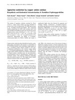
key in Table 1. The knees from naïve control animals were
unremarkable and had a mean damage score of 3.7 ± 0.3 (Fig-
ure 3). In contrast, in both no pump (12.7 ± 1.4) and PEG 300
vehicle alone (10.7 ± 1.4) treatment groups, portions of the
non-calcified cartilage had been worn down to the tidemark
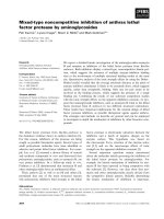
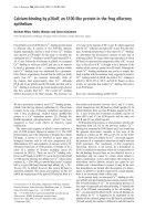
Figure 1
Clinical scores from murine collagen-induced arthritis study 1Clinical scores from murine collagen-induced arthritis study 1. Animals
were given initial injection of type II collagen on day -21 and disease
was induced with a second injection on day 0. On day 3 (arrow),
pumps were implanted and clinical signs measured twice a week from
day 10 to day 28. By day 10, a statistically significant, dose-dependent
decrease in the clinical score was observed with all doses of CP-
690550 and these remained significant throughout the remainder of
the study.
Arthritis Research & Therapy Vol 10 No 1 Milici et al.
Page 4 of 9
(page number not for citation purposes)
and significant cell influx and synovial hypertrophy were
observed. In regions where the non-calcified articular cartilage
was still present, it was extensively depleted of proteoglycan
and devoid of chondrocytes. Treatment with CP-690550
resulted in a dose dependent reduction in the inflammation
and damage to the articular cartilage (Figure 4). The average
histological damage scores in the CP-690550 treated mice
ranged from 9.8 at 1.5 mg/kg/day to 4.4 at 15 mg/kg/day (Fig-
ure 3). The histologically determined ED
50
dose of CP-
690550 was approximately 6.5 mg/kg/day.
In the second CIA study, the clinical score data correlated with
the histological results from the four paws in that the greatest
efficacy was observed with the 15 mg/g dose of CP-690550
(84% inhibition) while the mid and low doses of CP-690550
were statistically equivalent to treatment with anti-TNF (45 %
inhibition).
Serum IL-6 levels
Serum IL-6 levels were measured in the second CIA study and
were found to be elevated ~4.6-fold in diseased control mice
vs naïve mice (Figure 5). Whereas lower doses of CP-690550
trended towards a reduction in IL-6 levels, only the 15 mg/kg/
day group produced a statistically significant effect.
Administration of the anti-TNF was also significantly effective
at lowering serum IL-6 levels.
Rat AA
Clinical changes
By day 14 after adjuvant administration in the rat AA model,
paw swelling was evident in all rats except those receiving CP-
690550 at 15 mg/kg/day. Treatment with CP-690550 pro-
duced a dose-dependent inhibition of footpad swelling (Figure
6). Near complete inhibition was achieved at both the 5 and
15 mg/kg dose levels at all time points. Swelling in the 1.5 mg/
kg dose level was reduced relative to vehicle from days 7–14.
Histological changes
Histological evaluation of the hind paws revealed significant
inflammation and damage present in the vehicle dosed animals
(Figure 7). The bones and joint cavities from the first metatar-
sal to the tibia on the medial side of the foot were evaluated on
a 0–8 scale using a modified scoring key (Table 2). Only the
feet from the vehicle and CP-690550 15 mg/kg/day animals
were evaluated histologically. A significant reduction was
observed in the damage score in the CP-690550 15 mg/kg/
day treated group (2.4 ± 0.3 damage score) vs the vehicle
treated group (5.9 ± 0.6 damage score).
Drug levels in serum
In the first murine CIA study, serum levels of CP-690550 on
day 28 ranged from 6 ng/ml at 1.5 mg/kg/day to 70 ng/ml at
15 mg/kg/day (Table 3). In the second CIA study, equivalent
doses of CP-690550 produced approximately 50% less drug
in the serum on day 31. In the rat, equivalent doses of CP-
690550 produced greater than fourfold higher drug levels
than in the mouse (Table 3).
Discussion
CP-690550 produced significant dose-dependent attenua-
tion of inflammatory swelling, cell influx and cartilage damage
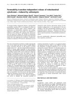
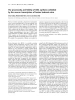
Figure 2
Clinical scores from murine collagen-induced arthritis study 2Clinical scores from murine collagen-induced arthritis study 2. Animals
were given initial injection of type II collagen on day -21 and disease
was induced with a second injection on day 0. On day 3 (arrow),
pumps were implanted and clinical signs measured twice a week from
day 6 to day 31. By day 9, a statistically significant, dose-dependent
decrease in the clinical score was observed with all doses of CP-
690550 and these remained significant to day 28. On day 31, only the
high and mid dose of CP-690550 contained a statistically significant
decrease in clinical score. At no time point during the course of the
study did the anti-tumor necrosis factor (TNF) antibody treatment result
in a statistically significant decrease in clinical score.
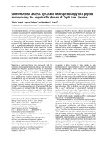
Figure 3
Histological evaluation of damage to murine kneesHistological evaluation of damage to murine knees. Histological sec-
tions of knee samples from murine collagen-induced arthritis study 1
were graded as described in Table 1. CP-690550 produced a dose-
dependent inhibition of knee damage that reached statistical signifi-
cance (p < 0.0001) at the 15 mg/kg/day dose relative to vehicle.
Available online />Page 5 of 9
(page number not for citation purposes)
in two well characterized rodent models. A T cell contribution
to disease has been demonstrated in both models [15-17]. In
murine CIA, the magnitude of effects observed at the highest
dose of the JAK3 inhibitor tested (15 mg/kg/d) were greater
than those following administration of anti-TNF antibody (TN-
1912) when assessing clinical scores and histology. The mag-
nitude of effect of anti-TNF that we observed on the clinical
arthritis score is consistent with that reported previously [10-
12] when animals were dosed with the same anti-TNF mAb.
Anti-TNF treatment is efficacious in murine CIA when dosed
before or immediately after the onset of CIA (see review of the
role of TNF and IL-1 in CIA; [18]). Even though we did begin
treating the mice immediately after disease induction, the fact
that anti-TNF treatment was not as efficacious as treatment
with CP-690550 in murine CIA could be due to the role of IL-
1 or other inflammatory mediators in this animal model.
CP-690550 doses/exposures that produced effects in this
model are consistent with those demonstrating immune sup-
pression in other murine models including delayed-type hyper-
sensitivity and cardiac allograft transplantation [5,6].
Interestingly, both CP-690550 (78% reduction vs control) and
the anti-TNF mAb (68% reduction vs control) significantly
reduced serum IL-6 levels. IL-6 has been proposed to play an
important role in the development of CIA based upon delay in
onset and reduction in disease magnitude observed in mice
genetically deficient in this cytokine [19]. The effects of anti-
TNF on IL-6 are consistent with other reports in which inhibi-
tion of TNF action, either via genetic ablation of its receptor
[20] or via anti-TNF mAb [21,22] were found to down-modu-
late levels of IL-6. However, in our studies, anti-TNF mAb treat-
ment reduced serum IL-6 by a similar magnitude as CP-
690550 but did not demonstrate the same degree of efficacy,
which suggests the JAK3 inhibitor, affected other inflammatory
mediators important for expression of disease in this model. A
role for IL-6 in rheumatoid arthritis has been proposed based
upon the ability of the cytokine to activate inflammatory
responses and osteoclastogenesis and is supported by posi-
tive clinical data obtained with the anti-IL-6 mAb tocilizumab in
this patient population [23].
The efficacy produced by CP-690550 in the rodent models of
arthritis may result from its ability to affect signaling of a
number of cytokines including IL-2, -7, -15 and -21 as a con-
sequence of JAK3 inhibition [5]. IL-2 mRNA was found to be
markedly increased in arthritic paws from mice with CIA during
the early phases of disease [24]. This may explain the efficacy
observed following prophylactic administration of an anti-IL2R
antibody in this model [25]. When mice with established
disease were treated with cyclosporine 50 or 75 mg/kg/day,
disease was also attenuated [26]. Tacrolimus is another, albeit
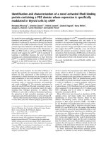
Figure 4
Representative histological sections from murine collagen-induced arthritis study 1Representative histological sections from murine collagen-induced
arthritis study 1. In all figures, the arrowheads point to the femoral con-
dyle (top) and tibial plateau (bottom) articular cartilages and the aster-
isks highlight the inflammatory cells in the soft tissue surrounding the
joint. Panel (a) is from a naïve control containing no damage to the
articular cartilage and few cells in the soft tissue surrounding the joint.
Panel (b) is from a vehicle treated animal, demonstrating significant
influx of inflammatory cells into the synovial tissue and cavity as well as
significant proteoglycan loss and erosion of the articular cartilage. Pan-
els (c) and (d) are from animals that have been dosed with CP-690550
at 15 and 1.5 mg/kg/day, respectively. At both dose levels, CP-
690550 decreased cell influx, synovial hypertrophy, articular cartilage
damage and proteoglycan loss. The knees from animals dosed with
CP-690550 15 mg/kg/day were very similar to the knees from the
naïve animals. Bar = 500 μm.
Table 1
Scoring key for murine knees
Score Edge Proteoglycans Open lacuna Non-calcified
layer
Synovial lining Blood in cavity Pannus
tongues
0 Smooth > 90% present None > 90% present 1 even Absent Absent
1 Rough > 50% present Some > 50% present < 50% 2 layers Present Present
2 < 50% present Many < 50% present > 50% 2 layers
3 < 10% present < 10% present 2 layers
4 > 2 layers
Arthritis Research & Therapy Vol 10 No 1 Milici et al.
Page 6 of 9
(page number not for citation purposes)
more potent, calcineurin inhibitor that has also demonstrated
efficacy in experimental models of rheumatoid arthritis [27]. In
rat arthritis models, tacrolimus suppressed paw inflammation,
type II collagen antibody formation and delayed-type hyper-
sensitivity to type II collagen [27,28]. While clinical trials of
tacrolimus in rheumatoid arthritis have been conducted, it
appears that the compound has a narrow therapeutic window
which limits its utility [29].
IL-15 is a cytokine with close homology to IL-2 whose receptor
shares signaling through the common gamma chain. Previous
studies from our lab have demonstrated that CP-690550
inhibits IL-15-mediated up-regulation of activation markers on
CD8+ T cells and NK cells [30]. Upon chronic treatment with
CP-690550, there is a preferential loss of these cells from the
circulation, which is consistent with a role for IL-15 in their sur-
vival [6,30]. Evidence is emerging for the importance of IL-15
in the pathogenesis of rheumatoid arthritis. Elevated serum
levels of the cytokine have been reported in arthritic patients,
the primary source of which may be macrophages residing in
the synovial lining layer of inflamed joints [31]. IL-15 produces
a number of effects which may be relevant to the pathogenesis
of arthritis including recruitment and activation of T lym-
phocytes into the synovial membrane and induction of TNFα
production [32,33]. A soluble fragment of the murine IL-15Rα
chain inhibited development of collagen-induced arthritis in
DBA/1 mice [34]. Administration of an IL-15 mutant/Fcγ2c
fusion protein in established murine CIA blocked disease pro-
gression and reduced long term articular inflammation and
destruction [33]. The therapeutic benefit achieved by inhibit-
ing IL-15 is supported by evidence that HuMax-IL-15, a fully
human anti-IL-15 mAb, produced encouraging signs of effi-
cacy in rheumatoid arthritis patients [35].
IL-21 is a cytokine produced by activated CD4+ T cells that
also signals through JAK3. It enhances T cell activation,
proliferation and secretion of pro-inflammatory cytokines such
as TNFα and IL-21R has been shown to be over-expressed in
inflamed synovial membrane and peripheral blood or synovial
fluid leukocytes of rheumatoid arthritis patients [36]. A recent
publication reported that blockade of IL-21 effects with a
murine IL-21 receptor Fc fusion protein attenuated disease in
both mouse and rat models of arthritis [22]. Effects in a 'semi-
therapeutic' murine CIA model (compound administration
begun when 10% of mice began to exhibit clinical signs of dis-
ease) included reduction in disease severity scores (including
histology) and serum IL-6 levels. Effects produced by IL-
21RFc were even more profound in a rat adjuvant-induced
arthritis model in which full amelioration of clinical signs was
achieved in conjunction with significant reduction in histologi-
cal damage [22]. Recent evidence demonstrates that IL-21 is
a key cytokine involved in the generation of Th17 cells which
have been shown to mediate tissue inflammation via produc-
tion of IL-17 [37,38]. Thus it is possible that CP-690550,
through inhibition of IL-21R signaling, may also be efficacious
in the CIA model by reducing IL-17 producing Th17 cells
which have been proposed to play an important role in the
pathogenesis of autoimmune diseases.
Figure 5
Serum interleukin (IL)-6 levels from murine collagen-induced arthritis study 2Serum interleukin (IL)-6 levels from murine collagen-induced arthritis
study 2. Blood was drawn from mice 15 days following the second type
II collagen injection and serum IL-6 measured by enzyme-linked immu-
nosorbent assay (ELISA). Data are mean ± standard error of the mean
of values from 6–8 animals/treatment group, except the naïve group (n
= 3).
Figure 6
Measurement of rat foot pad swelling in rat adjuvant-induced arthritis (AA) modelMeasurement of rat foot pad swelling in rat adjuvant-induced arthritis
(AA) model. Animals were injected at the base of the tail with Mycobac-
terium butyricum in mineral oil on day 0 to induce disease. On day 10
(arrow), pumps were implanted and foot swelling measured twice a
week from day 14 to day 24. CP-690550 decreased hind paw swelling
in a dose dependent manner. In the rat AA model, data were expressed
as percent control with the diseased vehicle treatment group on day 24
being set to 100%. As early as day 14, both the 5 and 15 mg/kg dose
of CP-690550 resulted in a statistically significant decrease in foot vol-
ume. By day 17, all doses of CP-690550 resulted in a statistically sig-
nificant decrease in foot volume that remained significant for the rest of
the study. Data are mean ± standard error of the mean of 10 animals
per group, except for the naïve group (n = 5).
Available online />Page 7 of 9
(page number not for citation purposes)
IL-7 represents another member of the IL-2 family that signals
through the common gamma chain. It plays a key role in T cell
homeostasis supporting growth, proliferation and survival of
developing and mature T cells. In mice, unlike humans, its
absence or blockade results in a diminution of B cell numbers
as was evident in our own studies that examined the effects of
chronic CP-690550 administration on circulating lymphocytes
[6]. IL-7 has also been suggested to play a role in rheumatoid
arthritis based upon the observation of increased levels of the
cytokine in this patient population, its ability to induce TNFα
and induction of bone loss by stimulation of RANKL-depend-
ent osteoclastogenesis [39].
The potential for CP-690550 to attenuate multiple cytokines
associated with rheumatoid arthritis by virtue of its ability to
inhibit JAK3 may provide improved efficacy vs a single agent.
For example, TNF antagonists rarely induce complete disease
remission and not all patients respond to TNF-blocking thera-
pies [3]. IL-1 antagonism also demonstrates some effective-
ness albeit to a lesser extent than TNF blockers [40]. However,
combined inhibition of these two cytokines has been shown to
provide increased benefit relative to inhibition of either alone
[41]. Hence, JAK3 inhibition provides a potentially beneficial
target for the treatment of RA based upon its ability to inhibit
multiple cytokines known to be involved in the pathogenesis of
the disease.
Conclusion
CP-690550, a potent inhibitor of JAK3, reduced the clinical
and histological manifestations of joint inflammation, including
bone and cartilage damage, when administered therapeuti-
cally in murine CIA and rat AA. The effects of CP-690550
were dose dependent and higher doses were required for sup-
pression of CIA histopathology than clinical manifestations.
These data support the evaluation of CP-690550 for DMARD
activity in RA patients.
Competing interests
All authors were, or currently are employed by Pfizer Global
Research and Development. Pfizer is financing the publication
of this manuscript.
Figure 7
Histological evaluation of damage to foot pad in rat adjuvant-induced arthritis (AA) modelHistological evaluation of damage to foot pad in rat adjuvant-induced arthritis (AA) model. Representative sections from vehicle (a) and 15 mg/kg/
day CP-690550 (b) treated rats on day 24. In the vehicle treated animal there was significant inflammation (asterisk) and destruction of the metatar-
sal bone (arrow) and joint spaces (arrowhead). In contrast, there was no inflammation and the metatarsal joint spaces appeared normal in the CP-
690550 treated animal.
Table 2
Modified scoring key for rat foot pad damage
Score Proteoglycan retention Inflammation Bone damage
0 > 90% present Few cells Normal
1 > 50% present Slight to moderate Questionable damage
2 Only chondrocytes stain Moderate to heavy Damaged
3 < 10% present Extremely heavy
Arthritis Research & Therapy Vol 10 No 1 Milici et al.
Page 8 of 9
(page number not for citation purposes)
Authors' contributions
AJM was responsible for histology, data analysis and manu-
script writing, EK for data analysis and manuscript writing, LA
for all Pfizer in vivo animal work, SZ for assistance with
manuscript writing, and PC for concept and assistance with
manuscript writing.
Acknowledgements
The authors would like to acknowledge Gretchen Beckius for histology
support, Brett Perry and Colleen Gibbons for in vivo animal work and
Mike Fisher, Chandra Prakash, Kwansik Yoon and Jian Lin for drug
metabolism support of this study.
References
1. Weyand C, Goronzy I, Takemura S, Kurtin P: Cell-cell interactions
in synovitis. Interactions between T cells and B cells in rheu-
matoid arthritis. Arthritis Res 2000, 2:457-463.
2. O'Dell J: Therapeutic strategies for rheumatoid arthritis. New
Eng J Med 2004, 350:2591-2602.
3. Olsen N, Stein M: New drugs for rheumatoid arthritis. New Eng
J Med 2004, 350:2167-2179.
4. Kitahara K, Kawai S: Cyclosporine and tacrolimus for the treat-
ment of rheumatoid arthritis. Curr Opin Rheumatol 2007,
19:238-245.
5. Changelian PS, Flanagan ME, Ball DJ, Kent CR, Magnuson KS,
Martin WH, Rizzuti BJ, Sawyer PS, Perry BD, Brissette WH,
McCurdy SP, Kudlacz EM, Conklyn MJ, Elliott EA, Koslov ER,
Fisher MB, Strelevitz TJ, Yoon K, Whipple DA, Sun J, Munchhof MJ,
Doty JL, Casavant JM, Blumenkopf TA, Hines M, Brown MF, Lillie
BM, Subramanyam C, Shang-Poa C, Milici AJ, et al.: Prevention
of organ allograft rejection by a specific janus kinase 3
inhibitor. Science 2003, 302:875-878.
6. Kudlacz E, Perry B, Sawyer P, Conklyn M, McCurdy S, Brissette
W, Flanagan M, Changelian P: The novel JAK-3 inhibitor CP-
690550 is a potent immunosuppressive agent in various
murine models. Am J Transplant 2004, 4:51-57.
7. O'Shea JJ, Gadina M, Schreiber R: Cytokine signaling in 2002:
new surprises in the Jak/Stat pathway. Cell 2002,
109:S121-131.
8. Walker J, Ahern M, Coleman M, Weedon H, Papangelis V, Berou-
kas D, Roberts-Thomson P, Smith M: Changes in synovial tissue
Jak-STAT expression in rheumatoid arthritis in response to
successful DMARD treatment. Ann Rheum Dis 2006,
65:1558-1564.
9. Sheehan K, Ruddle N, Schreiber R: Generation and characteri-
zation of hamster monoclonal antibodies that neutralize
murine tumor necrosis factors. J Immunol 1989,
142:3884-3893.
10. Graneto M, Kurumbail R, Vazquez M, Shieh H, Pawlitz J, Williams
J, Stallings W, Geng L, Naraian A, Koscyk F, Stealey MA, Xu XD,
Weier RM, Hanson GJ, Mourey RJ, Compton RP, Mnich SJ, Ander-
son GD, Monahan JB, Devraj R: Synthesis, crystal structure, and
activity of pyrazole-based inhibitors of p38 kinase. J Med
Chem 2007, 50:5712-5719.
11. Thorbecke G, Shah R, Kuruvilla A, Hardison A, Palladino M:
Involvement of endogenous tumor necrosis factor α and
transforming growth factor β during induction of collagen type
II arthritis in mice. Proc Natl Acad Sci USA 1992,
89:7375-7379.
12. Williams R, Feldmann M, Maini R: Cartilage destruction and
bone erosion in arthritis: the role of tumor necrosis factor α.
Ann Rheum Dis 2000, 59:i75-80.
13. Lubberts E, Koenders M, Oppers-Walgreen B, van den Bersselaar
L, Coenen-de Roo C, Joosten L, van den Berg W: Treatment with
a neutralizing anti-murine interleukin-17 antibody after the
onset of collagen-induced-arthritis reduces joint inflamma-
tion, cartilage destruction, and bone erosion. Arthritis Rheum
2004, 50:650-659.
14. Shay AK, Bliven ML, Scampoli DN, Otterness IG, Milici AJ: Effects
of exercise on synovium and cartilage from normal and
inflammed knees. Rheumatol Int 1995, 14:183-189.
15. Brand D, Kang A, Rosloniec E: Immunopathogenesis of colla-
gen arthritis. Springer Semin Immunopathol 2003, 25:3-18.
16. Holmdahl R, Lorentzen J, Lu S, Olofsson P, Wester L, Holmberg J,
Pettersson U: Arthritis induced in rats with non-immunogenic
adjuvants as models for rheumatoid arthritis. Immunol Rev
2001, 184:184-202.
17. Luross J, Williams N: The genetic and immunopathological
processes underlying collagen-induced arthritis. Immunology
2001, 103:407-416.
18. van den Berg W, Joosten L, Kollias G, van de Loo F: Role of tumor
necrosis factor α in experimental arthritis: separate activity if
IL-1β in chronicity and cartilage destruction. Ann Rheum Dis
1999, 58(Suppl I):40-48.
19. Sasai M, Saeki Y, Ohshima S, Nishioka K, Mima T, Tanaka T, Kat-
ada Y, Yoshizaki K, Suemura M, Kishimoto T: Delayed onset and
reduced severity of collagen-induced arthritis in interleukin-6-
deficient mice. Arthritis Rheum 1999, 42:1635-1643.
20. Bultinck J, Brouckaert P, Cauwels A: The in vivo contribution of
hematopoietic cells to systemic TNF and IL-6 production dur-
ing endotoxemia.
Cytokine 2006, 36:160-166.
21. Ghezzi P, Sacco S, Agnello D, Marullo A, Caselli G, Bertini R: LPS
induces IL-6 in the brain and in serum largely through TNF
production. Cytokine 2000, 12:1205-1210.
22. Young D, Hegen M, Ma H, Whitters M, Albert L, Lowe L, Senices
M, Wu P, Sibley B, Leathurby Y, Brown TP, Nickerson-Nutter C,
Keith JC Jr, Collins M: Blockade of the interleukin-21/inter-
leukin-21 receptor pathway ameliorates disease in animal
models of rheumatoid arthritis. Arthritis Rheum 2007,
56:1152-1163.
23. Smolen J, Maini R: Interleukin-6: a new therapeutic target.
Arthritis Res Ther 2006, 8:S2-5.
24. Thornton S, Duwel L, Boivin G, Ma Y, Hirsch R: Association of the
course of collagen-induced arthritis with distinct patterns of
cytokine and chemokine messenger RNA expression. Arthritis
Rheum 1999, 42:1109-1118.
25. Banerjee S, Wei B-Y, Hillman K, Luthra H, David C: Immunosup-
pression of collagen-induced arthritis in mice with an anti-IL-2
receptor antibody. J Immunol 1988, 141:1150-1154.
26. Hom J, Butler L, Riedl P, Bendele A: The progression of the
inflammation in established collagen-induced arthritis can be
altered by treatments with immunological or pharmacological
agents which inhibit T cell activities. Eur J Immunol 1988,
18:881-888.
Table 3
CP-690550 serum levels (ng/ml)
Dose (mg/kg/d) Mouse study 1
a
Mouse study 2
b
Rat study
15 70 ± 17 37 ± 5 259 ± 41
5 27 ± 6 17 ± 2 124 ± 16
1.5 6 ± 1 < LLOQ 24 ± 5
Data are mean ± standard error of the mean. All samples were collected at the termination of the study. n = 10 for all groups. LLOQ, lower limit of
quantification.
a
LLOQ = 5 ng/ml (Pfizer study).
b
LLOQ = 10 ng/ml (Boulder BioPath study).
Available online />Page 9 of 9
(page number not for citation purposes)
27. Miyata S, Ohkubo Y, Mutoh S: A review of the action of tac-
rolimus (FK506) on experimental models of rheumatoid
arthritis. Inflamm Res 2005, 54:1-9.
28. Magari K, Miyata S, Nishigaki F, Ohkubo Y, Mutoh S: Comparison
of anti-arthritic properties of leflunomide with methotrexate
and FK506: effect on T cell activation-induced inflammatory
cytokine production in vitro and rat adjuvant-induced arthritis.
Inflamm Res 2004, 53:544-550.
29. Fleischmann R, Iqbal I, Stern R: Tacrolimus in rheumatoid
arthritis. Expert Opin Pharmacother 2006, 7:91-98.
30. Conklyn M, Andresen C, Changelian P, Kudlacz E: The JAK3
inhibitor CP-690550 selectively reduces NK and CD8+ cell
numbers in cynomolgus monkey blood following chronic oral
dosing. J Leukoc Biol 2004, 76:1248-1255.
31. Gonzalez-Alvaro I, Ortiz A, Garcia-Vicuna R, Balsa A, Pascual-Sal-
cedo D, Laffon A: Increased serum levels of interleukin-15 in
rheumatoid arthritis with long term disease. Clin Exp
Rheumatol 2003, 21:639-642.
32. McInnes I, Al-Mughales J, Field M, Leung B, Huang F-P, Dixon R,
Sturrock R, Wilkinson P, Liew F: The role of interleukin-15 in T-
cell migration and activation in rheumatoid arthritis. Nat Med
1996, 2:175-182.
33. McInnes I, Leung B, Sturrock R, Field M, Liew F: Interleukin-15
mediates T cell-dependent regulation of tumor necrosis fac-
tor-α production in rheumatoid arthritis. Nat Med
1997:189-195.
34. Ruchatz H, Leung B, Wei X, McInnes I, Liew F: Soluble IL-15
receptor a-chain administration prevents murine collagen-
induced arthritis: a role for IL-15 in development of antigen-
induced immunopathology. J Immunol 1998, 160:5654-5660.
35. Baslund B, Tvede N, Danneskiold-Samsoe B, Larsson P, Panayi G,
Petersen J, Petersen L, Beurskens F, Schuurman J, van de Winkel
J, Parren PWHI, Gracie JA, Jongbloed S, Liew FY, McInnes IB:
Targeting interleukin-15 in patients with rheumatoid arthritis.
Arthritis Rheum 2005, 52:2686-2692.
36. Li J, Shen W, Kong K, Liu Z: Interleukin-21 induces T-cell acti-
vation and pro-inflammatory cytokine secretion in rheumatoid
arthritis. Scand J Immunol 2006, 64:515-522.
37. Korn T, Bettelli E, Gao W, Awasthi A, Jager A, Strom T, Oukka M,
Kuchroo V:
IL-21 initiates an alternative pathway to induce
proinflammatory TH17 cells. Nature 2007, 448:484-487.
38. Nurieva R, Yang X, Martinez G, Zhang Y, Panopoulos A, Ma L,
Schluns K, Tian Q, Watowich S, Jetten A, Dong C: Essential
autocrine regulation by IL-21 in the generation of inflammatory
T cells. Nature 2007, 448:480-483.
39. Hartgring S, Bijlsma J, Lafeber F, van Roon J: Interleukin-7
induced immunopathology in arthritis. Ann Rheum Dis 2006,
65:69-74.
40. Zwerina J, Redlich K, Schett G, Smolen J: Pathogenesis of rheu-
matoid arthritis: targeting cytokines. Ann NY Acad Sci 2005,
1051:716-729.
41. Zwerina J, Hayer S, Tohidast-Akrad M, Bergmeister H, Redlich K,
Feige U, Dunstan C, Kollias G, Steiner G, Smolen J, Schett G: Sin-
gle and combined inhibition of tumor necrosis factor, inter-
leukin-1, and RANKL pathways in tumor necrosis factor-
induced arthritis: effects on synovial inflammation, bone ero-
sion, and cartilage destruction. Arthritis Rheum 2004,
50:277-290.