Báo cáo Y học: Agmatine oxidation by copper amine oxidase Biosynthesis and biochemical characterization of N-amidino-2-hydroxypyrrolidine pdf
Bạn đang xem bản rút gọn của tài liệu. Xem và tải ngay bản đầy đủ của tài liệu tại đây (376.64 KB, 9 trang )
Agmatine oxidation by copper amine oxidase
Biosynthesis and biochemical characterization of
N
-amidino-2-hydroxypyrrolidine
Paolo Ascenzi
1,
*, Mauro Fasano
2,
*, Maria Marino
1
, Giorgio Venturini
1
and Rodolfo Federico
1
1
Department of Biology, University ÔRoma TreÕ, Rome, Italy;
2
Department of Structural and Functional Biology,
University of Insubria, Varese, Italy
The p roduct of agmatine oxidation catalyzed by Pisum
sativum L. copper amine oxidase has been identified by
means of one- and two-dimensional
1
H-NMR spectroscopy
to be N-amidino-2-hydroxypyrrolidine. This compound
inhibits competitively rat nitric oxide synthase type I and
type II (NOS-I and NOS-II, respectively) and bovin e t rypsin
(trypsin) activity, values of K
i
being (1.1 ± 0.1) · 10
)5
M
(at
pH 7.5 and 37.0 °C), (2.1 ± 0.1) · 10
)5
M
(at pH 7.5 and
37.0 °C), and (8.9 ± 0.4) · 10
)5
M
(at p H 6.8 and 21.0 °C),
respectively. Remarkably, the affinity o f N-amidino-
2-hydroxypyrrolidine f or NOS-I, NOS-II and trypsin is
significantly higher than that observed for agmatine and
clonidine binding. Furthermore, N-amidino-2-hydroxy-
pyrrolidine a nd agmatine are more efficient than clonidine in
displacing [
3
H]clonidine (¼ 1.0 · 10
)8
M
) from specific
binding sites in heart rat membranes, values of IC
50
being
(1.3 ± 0.4) · 10
)9
M
and (2.2 ± 0.4) · 10
)8
M
,respec-
tively (at pH 7.4 an d 37.0 °C).
Keywords: c opper amine oxidase; agmatine; N-amidino-2-
hydroxypyrrolidine; enzyme inhibition; type 1 imidazoline
receptor b ind ing.
Copper a mine oxidase has been identified in b acteria, yeasts,
fungi, plants, a nd animals. This enzyme is a homodimer o f
70- to 90-kDa subunits, each c ontaining a s ingle copper i on
and a covalently bound cofactor formed by the post-
translational modification of the catalytic tyrosyl residue
to 2, 4,5-trihydroxyphenylalanine quinone (TPQ) [ 1–4].
Copper amine oxidase catalyzes the oxidative deamination
of biogenic amines, including mono, di, and polyamines,
neurotransmitters such as catecholamines, histamine and
xenobiotic amines, with substrate p references depending
upon the enzyme source [1–5]. The copper amine oxidase
catalyzed reactions follow t he general s cheme:
E
ox
þ R-CH
2
-NH
2
! E
red
þ R-CHO ðreaction 1Þ
E
red
þ O
2
þ H
2
O ! E
ox
þ NH
3
þ H
2
O
2
ðreaction 2Þ
where E
ox
represents the enzyme–quinone, R-CH
2
-NH
2
is
the substrate, E
red
is the enzyme–aminoquinol, and R-CHO
is the product aldehyde. Substrate amines interact directly
with TPQ in the reductive part of the process forming a
Schiff base complex (reaction 1). Proton abstraction of the
substrate, catalyze d by an invariant Asp residue, leads to the
release of product aldehyde and leaves the enzyme in the
reduced aminoquinol form (reaction 1) [1–4]. The oxidative
part (reaction 2) leads to reoxidation of the aminoquinol
cofactor with the release of ammonia and hydrogen
peroxide [1–4].
Copper amine oxidase catalyzes also the oxidation of
agmatine [3–5], which has been recognized to be an impor-
tant bioactive molecule, b eing identified as a novel neuro-
transmitter and modulator of cardiovascular functions via
binding to type 1 imidazoline (I
1
-R) and a-adrenergic
receptors [6,7]. Interestingly, agmatine inhibits nitric oxide
synthase isoforms [8,9] and induces the release of some
peptide hormones [7]. To date, the product(s) of the copper
amine oxidase catalyzed oxidation of agmatine has not been
identified. Moreover, no information is available o n the role
played by the product(s) of agmatine metabolism on cell
function(s). Here, the b iosynthesis and the biochemical
characterization of N-amidino-2-hydroxypyrrolidine, the
product of agmatine oxidation by Pisum sativum L. copper
amine oxidase, is reported.
MATERIALS AND METHODS
Proteins
P. sativum copper amine oxidase was purified as previously
reported [10]. Rat nitric oxide synthase type I (NOS-I) was
prepared from the rat brain homogenate [11]. Rat nitric
oxide synthase type II (NOS-II) was prepared from the lung
homogenate of rats treated with E. coli lipopolysaccharide
(10 mgÆkg
)1
) [11]. NOS-I and NOS-II containing specimens
were homogenized at pH 7.5 (5.0 · 10
)2
M
Hepes buffer),
5.0 · 10
)4
M
EGTA, 1.0 · 10
)3
M
dithiothreitol, and
0.1 mgÆmL
)1
phenylmethanesulfonyl fluoride [11]. Then,
Correspondence to P. Ascenzi, Dipartimento di Biologia, Universita
`
ÔRoma TreÕ, Viale Guglielmo Marconi 446, I-00146 Rome, Italy.
Fax: + 39 06 551 76321, Tel.: + 39 06 55176329,
E-mail:
Abbreviations:I
1
-R, type 1 imidazoline receptor; MMFF, Merck
Molecular Force Field; NOS-I, rat nitric oxide synthase type I (neu-
ronal constitutive isoform); NOS-II, rat nitric oxide synthase type II
(inducible isoform); TPQ, 2,4,5-trihydroxyphenylalanine quinone;
trypsin, bovine trypsin.
Enzymes: bovine catalase (EC 1.11.1.6); bovine trypsin (EC 3.4.21.4);
Pisum sativum L. copper amine oxidase (EC 1.4.3.6); rat nitric oxide
synthase type I (EC 1.14.13.39); rat nitric oxide synthase type II
(EC 1.14.13.39).
*Note: These authors contributed equally to this work.
(Received 26 July 2 001, r evised 1 7 October 2001, acc epted 3 December
2001)
Eur. J. Biochem. 269, 884–892 (2002) Ó FEBS 2002
NOS-I and NOS-II containing homogenates were desalted
by chromatography over disposab le PD-10 columns packed
withSephadexG-25medium(AmershamPharmaciaBio-
tech, Uppsala, Sweden). Bovine calmodulin, bovine cata-
lase, bovine serum albumin, bovine trypsin (trypsin),
and horseradish peroxidase were purchased from Sigma
Chemical Co (St Louis, MO, USA). Proteins were of
reagent grade and used without further purification.
Chemicals
Agmatine, aminoantipyrine, N-a-benzoyl-
L
-arginine p-nitro-
anilide, clonidine, 3,5-dichloro-2-hydroxybenzenesulfonic
acid, epinephrine, phenylmethanesulfonyl fluoride, and
Escherichia coli lipopolysaccharide (serotype 0127:B8) were
obtained from Sigma Chemical Co. [
3
H]
L
-arginine (specific
activity 2.0 TBqÆmmol
)1
)and[
3
H]clonidine (specific activity
2.6 TBqÆmmo l
)1
) were purchased from NEN
TM
Life
Science Products (Boston, MA, USA). Deuterium oxide
(99.8% isotopic enrichment) was obtained f rom C ortec
(Paris, France). All the other chemicals were from Merck
AG (Darmstadt, Germany). All products were of analytical
or reagent grade and u sed without further purification.
Animals
Male Sprague–Dawley rats (from Morini, I taly), 4- to
5-month-old, were housedandacclimatized for 1 week un der
controlled temperature (20 ± 1 °C), humidity (55 ± 10%),
and light (from 7 a.m. to 7 p.m) conditions. T he rats were
anaesthetized with ether in a fume hood, and o rgans
removed and rapidly chilled in liquid nitrogen (brain
and lung) or i n ice-cold medium solution (2.0 · 10
)2
M
NaHCO
3
; heart). Animal experiments were p erformed accor-
ding to ethical guidelines for t he conduct o f animal r esearch.
P. sativum
copper amine oxidase assay
Oxidation of agmatine by P. sativu m copper amine oxi-
dase was investigated s pectrophotometrically by follo-
wing the formation of a pink adduct ( e
515nm
¼ 2.6 ·
10
4
M
)1
Æcm
)1
), as a result of the oxidation of aminoanti-
pyrine and 3,5-dichloro-2-hydroxybenzenesulfonic acid cat-
alyzed by horseradish peroxidase, at pH 7.0 (1.0 · 10
)1
M
phosphate buffer) and 25.0 °C [5,6,10]. In a typical experi-
ment, 20 lL of a buffered P. sativum copper amine
oxidase solution (1.0 · 10
)1
M
phosphate buffer, pH 7.0)
wereaddedtoabufferedsolution(1.0mL;1.0· 10
)1
M
phosphate buffer, pH 7.0) containing the substrate (i.e.
agmatine), aminoantipyrine ( 1.0 · 10
)4
M
), 3 ,5-dichloro-
2-hydroxybenzenesulfonic acid (1.0 · 10
)3
M
), and horse-
radish peroxidase (1.5 · 10
)6
M
). The initial velocity for the
enzymatic oxidation of agmatine was then measured.
P. sativum copper amine oxidase activity was also
assayed polarographically with a Clark electrode (Hansa-
tech Instruments Ltd, Norfolk, UK) by following the O
2
consumption, at pH 7.0 (1.0 · 10
)1
M
phosphate buffer)
and 25.0 °C [12]. In a typical experiment, 20 lLofa
buffered agmatine solution (1.0 · 10
)1
M
phosphate buffer,
pH 7.0) were added to a buffered s olution (1.0 mL;
1.0 · 10
)1
M
phosphate buffer, pH 7.0) containing
P. sativum copper amine oxidase. The initial velocity for
the enzymatic oxidation of agmatine was then measured.
In the e nzyme assay, t he P. sativum copper amine oxidase
concentration was 5.0 · 10
)9
M
and the agmatine concen-
tration ranged between 5.0 · 10
)5
M
and 5.0 · 10
)3
M
.The
enzyme activity was linear up to 5 min of incubation and
results were expressed as lmol productÆs
)1
Æ(lmo l e nzy me)
)1
.
Under all th e e xperimental c onditions, t he initial velocity for
the P. sativum copper amine oxidase catalyzed oxidation of
agmatine was unaffected by the enzyme/substrate incuba-
tion time. In fact, the enzyme/substrate equilibration time
was very short, being completed within the mixing time
(% 15 s).
Values of the first-order rate-limiting catalytic constant
(k
cat
) and of the Michaelis constant, as determined in the
absence of the inhibitor (K
0
m
)fortheP. sativum copper
amine oxidase catalyzed oxidation of agmatine, were
obtained from the dependence of the initial velocity for
agmatine oxidation ( v
i
) on the substrate (i.e. agmatine)
concentration ([S]), according to Eqn (1) [13]:
v
i
¼ k
cat
½S=ðK
0
m
þ½SÞ ð1Þ
Values of k
cat
and K
0
m
for the P. sativum copper amine
oxidase catalyzed oxidation of agmatine are 1.3 ± 0.1 s
)1
and (3.8 ± 0.3) · 10
)4
M
, r espectively, at pH 7.0 ( 1.0 ·
10
)1
M
phosphate buffer) and 25.0 °C (Fig. 1). Values of
k
cat
and K
0
m
are independent of the enzyme assay.
Biosynthesis of
N
-amidino-2-hydroxypyrrolidine
N-Amidino-2-hydroxypyrrolidine was synthesized as fol-
lows. Twenty micrograms of P. sativum copper a mine
oxidase were added to 1.0 mL of a buffered 2.0 · 10
)3
M
agmatine solution (5.0 · 10
)2
M
phosphate buffer, pH 7.4).
28 lg of bovine catalase were also added to the reaction
solution (1.0 mL) in order to remove H
2
O
2
, arising from the
P. sativum copper a mine oxidase catalyzed oxidation of
agmatine. The reaction solution was stirred vigorously at
25.0 °C for 20 min, and the product recovered by ultrafil-
tration on Amicon P M10 membranes (Amicon, Inc.,
Beverly, MA, USA).
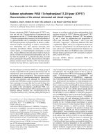
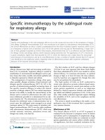
Fig. 1. Effect of substrate (i.e. agmatine) concentration on values of v
i
for the P. sativ um copper amine oxidase catalyzed oxidation of agma-
tine. T he c ontinuou s line was calculated according t o Eqn (1), with the
following values of k
cat
(¼ 1.3±0.1s
)1
)andK
0
m
[¼ (3.8 ± 0.3) ·
10
)4
M
]. Data were obtained at pH 7.0 a nd 25.0 °C, mean ± SD. Fo r
further details, see text.
Ó FEBS 2002 N-Amidino-2-hydroxypyrrolidine characterization (Eur. J. Biochem. 269) 885
The total conversion of agmatine to N-amidino-2-
hydroxypyrrolidine was detected by
1
H-NMR s pectroscopy.
Moreover, the agmatine/N-amidino-2-hydroxypyrrolidine
stoichiometry is 1 : 1 as shown by
1
H-NMR spectroscopy.
The N-amidino-2-hydroxypyrrolidine concentration was
determined from 100% conversion of agmatine to
N-amidino-2-hydroxypyrrolidine a s d emonstrated by
1
H-NMR spectroscopy.
Under a ll the experimental conditions, the formation of
free 4-guanidinobutyraldehyde was observed n either by
the o-aminobenzaldehyde assay [14] (data not shown) nor
1
H-NMR spectroscopy (Figs 2 and 3).
NMR spectroscopy
P. sativum copper amine oxidase catalyzed oxidation of
agmatine was conducted as described above, in deuterated
phosphate buffer (pD 7 .4; uncorrected pH-meter reading
7.0); residual oxygen was removed with a mild nitrogen
stream. A control s pectrum was recorded prior to addition
of P. sativum copper amine oxidase.
1
H-NMR one- and
two-dimensional spectra were recorded at 25.0 °Cona
Bruker AVANCE 600 NMR spectrometer (Bruker Ana-
lytik, Rheinstetten, Germany), operating at a magnetic field
strength of 14.1 T. The residual water signal was s uppressed
by a 2-s presaturation before the observation pulse. The
duration of the pulse corresponding to a flip angle of 90°
was 7 .4 ls. The spin system o f the agmatine oxidation
product was assigned by COSY, by setting the flip angle of
the second pulse to 35°. T o this purpose, 256 t
1
increments
were recorded (4096 points each). The resulting matrix was
zero-filled to 1024 · 4096 complex points and processed
with a 5 °-shifted squared sinebell in both dimensions [15].
Building of the
N
-amidino-2-hydroxypyrrolidine structure
Energy minimization of the proposed structure of
N-amidino-2-hydroxypyrrolidine w as performed on a
Silicon Graphics Octane workstation (SGI, Mountain
View, CA, USA) by using the program
SPARTAN
(Wave-
function Inc., Irvine, CA, USA).
NOS-I and NOS-II assay
NOS-I and NOS-II activity was assessed by evaluating the
conversion of [
3
H]
L
-arginine to [
3
H]
L
-citrulline at pH 7.5
(5.0 · 10
)2
M
Hepes buffer) and 37.0 °C, in the absence and
presence of N-amidino-2-hydroxypyrrolidine. In a typical
experiment, a NOS-I or NOS-II aliquot (50 lL) was a dded
to the reaction mixture (100 lL) containing 1.0 · 10
)3
M
NADPH, 1.2 · 10
)3
M
CaCl
2
,1.0lgÆmL
)1
calmodulin,
1.0 · 10
)5
M
FAD, 1.0 · 10
)5
M
FMN, [
3
H]
L
-arginine
(from 12 to 185 kBq) and
L
-arginine (from 1.0 · 10
)6
M
to 1. 0 · 10
)4
M
), in the absence and presence of N-ami-
dino-2-hydroxypyrrolidine (from 5.0 · 10
)6
M
and 5.0 ·
10
)5
M
). For the determination of NOS-II activity, CaCl
2
and calmodulin were omitted, and 1.0 · 10
)3
M
EGTA was
added to the reaction mixture. NOS-I and NOS-II activity
was assayed in the presence of 5.0 · 10
)5
M
BH
4
[16]. In the
enzyme assay, the NOS-I or NOS-II concentration was
2.0 · 10
)7
M
. After 15 min incubation, the reaction was
stopped by addition of an ice-cold 2.0 · 10
)2
M
Hepes
buffer solution (700 lL), pH 5.5, containing 2.0 · 10
)3
M
EDTA. [
3
H]
L
-citrulline was separated from [
3
H]
L
-arginine
by ion exchange chromatography on Dowex 50WX8
(Fluka Chemie AG) [11,16]. The enzyme activity was
linear up to 30 min of incubation and results were expressed
as pmol productÆmin
)1
Æ(mg protein)
)1
. U nder all the
experimental conditions, the initial velocity for NOS-I a nd
NOS-II catalyzed conversion of
L
-arginine to
L
-citrulline
was unaffected by the enzyme/inhibitor/substrate incuba-
tion time. In fact, the enzyme/inhibitor/substrate equilibra-
tion time was very short, being completed within the mixing
time (% 15 s).
Values of the first-order rate-limiting catalytic constant
(k
cat
) and of the Michaelis constant, as determined in the
absence and presence of the inhibitor (K
0
m
and K
app
m
,
respectively), for NOS-I and NOS-II catalyzed conversion
of
L
-arginine to
L
-citrulline were obtaine d from the depen-
dence of the initial velocity for substrate conversion ( v
i
)on
the
L
-arginine concentration ([S]), according to Eqn (1) [13].
Values of k
cat
and K
0
m
for the NOS-I catalyzed conversion
of
L
-arginine to
L
-citrulline were 1 .4 ± 10
2
pmol prod-
uctÆmin
)1
Æ(mg protein)
)1
and 4.0 · 10
)6
M
, respectively, at
pH 7.5 and 37.0 °C [11]. Values of k
cat
and K
0
m
for the
NOS-II catalyzed conversion of
L
-arginine to
L
-citrulline
were 4.7 · 10
1
pmol productÆmin
)1
Æ(mg protein)
)1
and
1.8 · 10
)5
M
, respectively, at pH 7.5 and 37.0 °C [17].
NO production was also monitored spectrophotometri-
cally (between 350 and 460 nm) following the NO-mediated
conversion of human oxy-hemoglobin (6.0 · 10
)6
M
), added
to the N OS-I and NOS-II preparations, to m et-hemoglobin,
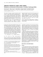
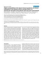
Fig. 2.
1
H-NMR spectra of 2.0 · 10
)3
M
agmatine before (A) and after (B) oxidation
catalyzed by P. sativum copper amine oxidase,
at pD 7.4 and 25.0 °C. Acquisition param-
eters: 4 scans, flip angle 45°, relaxation delay
2 s. T he residual water s ignal was suppressed
by presaturation. For further details, see text.
886 P. Ascenzi et al. (Eur. J. Biochem. 269) Ó FEBS 2002
in the presence of N-amidino-2-hydroxypyrrolidine as the
substrate instead of
L
-arginine, at pH 7.5 ( 5.0 · 10
)2
M
Hepes buffer) and 37.0 °C [18,19].
Trypsin assay
The trypsin catalyzed hydrolysis of N-a-benzoyl-
L
-arginine
p-nitroanilide was investigated spectrophotometrically (at
408 nm), at pH 6.8 (1.0 · 10
)1
M
phosphate buffer) and
21.0 °C [20], in the absence and presence of N-amidino-
2-hydroxypyrrolidine. In a typical experiment, 20 lLofa
buffered trypsin solution (1.0 · 10
)1
M
phosphate buffer,
pH 6.8) were added to 1.0 mL of a buffered solution
(1.0 · 10
)1
M
phosphate buffer, pH 6.8) containing the
substrate (i.e. N-a-benzoyl-
L
-arginine p-nitroanilide) and
the inhibitor (i.e. N-amidino-2-hydro xypyrrolidine). The
initial velocity for the enzymatic hydrolysis of N-a-benzoyl-
L
-arginine p-nitroanilide was then measured. In the enzyme
assay, the trypsin concentration was 1.0 · 10
)6
M
,the
N-a-benzoyl-
L
-arginine p-nitroanilide concentration ranged
between 1.0 · 10
)5
M
and 1.0 · 10
)3
M
,andtheN-ami-
dino-2-hydroxypyrrolidine concentration ranged between
2.0 · 10
)5
M
and 8.0 · 10
)5
M
. The enzyme activity was
linear up to 10 min of incubation an d results were expressed
as lmol productÆs
)1
Æ(lmol enzyme)
)1
.Underalltheexper-
imental conditions, the initial velocity for the trypsin
catalyzed hydrolysis of N-a-benzoyl-
L
-arginine p-nitroani-
lide w as unaffected by the enzyme/inhibitor/substrate
incubation time. In f act, the enzyme/inhibitor/substrate
equilibration time was very short, being completed within
the mixing time (% 15 s).
Values of the first-order rate-limiting catalytic constant
(k
cat
) a nd of the Michaelis constant determined in the
absence and presence of the inhibitor (K
0
m
and K
app
m
,
respectively) for the trypsin catalyzed hydrolysis of
N-a-benzoyl-
L
-arginine p-nitroanilide were obtained from
the d ependence of the initial velocity f or substrate
hydrolysis (v
i
)ontheN-a-benzoyl-
L
-arginine p-nitroani-
lide c oncentration ([S]), according to Eqn (1) [13]. Values
of k
cat
and K
0
m
for the trypsin catalyzed hydrolysis of
N-a-benzoyl-
L
-arginine p-n itroanilide were 0.70 s
)1
and
3.0 · 10
)4
M
, respectively, at pH 6.8 and 21.0 °C[20].
Determination of values of the inhibition
dissociation equilibrium constant (
K
i
)
for
N
-amidino-2-hydroxypyrrolidine binding
to NOS-I, NOS-II, and trypsin
Values of the inhibition dissociation equilibrium constant
(K
i
) for the competitive inhibition of the N OS-I and NOS-II
catalyzed conversion of
L
-arginine to
L
-citrulline (at pH 7.5
and 37.0 °C) and of the trypsin catalyzed hydrolysis of
N-a-benzoyl-
L
-arginine p-nitroanilide (at pH 6.8 and
21.0 °C) by N-amidino-2-hydroxypyrrolidine were deter-
mined from the linear dependence of the K
app
m
/K
0
m
ratio on
the inhibitor concentration (i.e. [I]), according to Eqn (2)
[13]:
K
app
m
=K
0
m
¼ K
À 1
i
½Iþ1 ð2Þ
As expected for a simple competitive inhibition system [13],
values of k
cat
for the NOS-I a nd NOS-II catalyzed
conversion of
L
-arginine to
L
-citrulline and for the trypsin
catalyzed hydrolysis of N-a-benzoyl-
L
-arginine p-nitroani-
lide were unaffected by the inhibitor concentration within
the standard deviation (± 5%).
Model building of the NOS-II: and trypsin:
N
-amidino-2-hydroxypyrrolidine complexes
Molecular models of the human NOS-II: and bovine
trypsin:N-amidino-2-hydroxypyrrolidine complexes were
built using the coordinates of the human NOS-II:S-ethyl-
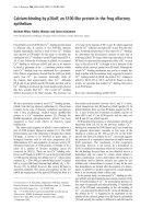
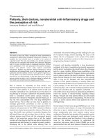
Fig. 3. Two-dimensional COSY spectrum of N-amidino-2-hydroxy-
pyrrolidine, the cyclic oxidation product of agmatine, at pD 7.4 and
25.0 °C (top) and ball-and-stick model of N-amidino-2-hydroxypyrro-
lidine (bottom). Acquisition parameters: 4 scans, 1 6 dummy scans,
relaxation delay 2 s. Labels refer to the resonance assignment in
Fig. 1B. For further details see text.
Ó FEBS 2002 N-Amidino-2-hydroxypyrrolidine characterization (Eur. J. Biochem. 269) 887
isothiourea complex (PDB accession no. 4NOS) [21] and the
bovine t rypsin:benzamidine adduct (PDB a ccession no.
1CE5) [22] as templates, respectively. The atomic coordi-
nates o f rat NOS-II are not yet ava ilable [23], the
homologous human enzyme was used instead. The confor-
mations of the N-amidino-2-hydroxypyrrolidine in the
enzyme:inhibitor complexes were obtained after 10 ps
molecular dynamics. Ene rgy minimization and molecu lar
dynamics were performed on a Silicon Graphics O
2
workstation ( SGI, Irvine, CA, USA) with
HYPERCHEM
4.5
for SGI (Hypercube Inc., Gainesville, FL, USA).
I
1
-R binding assay
Cardiac muscle (cleaned of connective t issue and fat) was
finely minced and homogenized in ice-cold medium solution
2.0 · 10
)2
M
NaHCO
3
, c ontaining 1.0 · 10
)4
M
phen-
ylmethanesulfonyl fluoride, with a wet weight to volume
ratio of 1 : 7, using a glass-Teflon homogenizer (10 · 30 s)
[24]. The homogenate was centrifuged at 1500 g for 15 min
(4.0 °C). The supernatant was centrifuged at 45 000 g for
5 min (at 4.0 °C). The pellet was washed twice, then
re-suspended i n 2 mL of ice-cold 5.0 · 10
)3
M
Hepes buffer,
containing 5.0 · 10
)4
M
EGTA, 5.0 · 10
)4
M
MgCl
2
,and
1.0 · 10
)4
M
ascorbic acid (pH 7.4) [25]. Membrane pre-
parations were f ree o f m itochondria and nuclei as confirmed
by subcellular enzymatic marker assays (data not shown).
Two-hundred and forty micrograms of membrane pro-
tein were incubated for 55 min with 1.3 nmol to 40 nmol
[
3
H]clonidine at 37.0 °C in a final volume of 0.5 mL of
5.0 · 10
)3
M
Hepes buffer, con taining 5.0 · 10
)4
M
EGTA,
5.0 · 10
)4
M
MgCl
2
,and1.0· 10
)4
M
ascorbic acid
(pH 7 .4). The reaction was stopped by rapid vacuum
filtration with a Millipore harvester throughWhatman GF/C
glass fiber filters (Whatman International Ltd Maidstone,
UK) p resoaked with 10% polyethyleneglycol in Tris/HCl
2.0 · 10
)2
M
, containing MgCl
2
1.0 · 10
)2
M
, followed by
rapid washing of filters with 10 mL ice-cold 5.0 · 10
)3
M
Hepes buffer, containing 5.0 · 10
)4
M
EGTA, 5.0 · 10
)4
M
MgCl
2
,and1.0· 10
)4
M
ascorbic acid (pH 7.4). Filters
were placed in a 6-mL scintillation fluid and the radio-
activity determined by liquid s cintillation counting. Epine-
phrine (1.0 · 10
)5
M
), which does not bind to imidazoline
sites [26,27], was added to the assay to prevent [
3
H]clonidine
from binding to a-adrenergic receptors. Nonspecific binding
wasdefinedas[
3
H]clonidine-binding (the [
3
H]clonidine
concentration ranged between 1.5 · 10
)4
M
and
5.0 · 10
)4
M
). Saturation studies were performed with
1.0 · 10
)8
M
[
3
H]clonidine and increasing concentrations
of the unl abelled ligand (i.e. N-amidino-2-hydroxypyrroli-
dine, agmatine, and clonidine; f rom 1.0 · 10
)9
M
to
1.0 · 10
)6
M
). Protein concentration was measured by the
method of Bradford [28], using bovine serum albumin as the
standa rd.
Values of IC
50
for [
3
H]clonidine displacement from I
1
-R
in heart rat membranes by N-amidino-2-hydroxypyrroli-
dine, agmatine, and clonidine were determined according to
Eqn (3):
a ¼ 1=f1 þð½L=IC
50
Þg ð3Þ
where a is the m olar fraction of [
3
H]clonidine bound to I
1
-R
present in heart rat membranes and [L] is the concentration
of the ligand (i.e. N-amidino-2-hydroxypyrrolidine, agma-
tine, or clonidine) [29].
RESULTS
Over the w hole substrate ( i.e. agmatine) concentration
range explored (i.e. between 5.0 · 10
)5
M
and
5.0 · 10
)3
M
), the P. sativum copper amine oxidase cata-
lyzed oxidation of agmatine follows simple Michaelis–
Menten kinetics (Fig. 1). According to the literature [30],
values of k
cat
and K
0
m
for the P. sativum copper amine
oxidase catalyzed oxidation of agmatine are 1.3 ± 0.1 s
)1
and (3.8 ± 0.3) · 10
)4
M
, respectively, at pH 7.0 and
25.0 °C. Moreover, values of k
cat
and K
0
m
were independent
of the enzymatic assay used (spectrophotometric vs. pola-
rographic). The stoichiometric analysis of the enzymatic
oxidation of agmatine yields a molar ratio of sub strate (i.e.
agmatine) to O
2
and H
2
O
2
of 1 : 1 : 1.
Figure 2 shows the
1
H-NMR s pectra of agmatine
before (Fig. 2 A) and after (Fig. 2B) oxidation catalyzed
by P. sativum copper amine oxidase, at pD 7.4 and
25.0 °C.Theagmatinesampleshowssomesignalsatthe
impurity level, which however do not hamper the
observation of the main component. The main features
of Fig. 2B with respect to Fig. 2A are: (a) the upset of a
downfield-shifted signal at d ¼ 5.5 p.p.m., and (b) the
splitting of CH
2
signals in magnetically unequivalent
components. On the basis of the general mechanism (see
reactions 1 and 2), one trip let (relative area 1) should
occur at about d ¼ 9 p.p.m., corresponding to the formyl
proton, one triplet at about d ¼ 3 p.p.m. (relative area 2),
and two multiplets at about d ¼ 2 p.p.m. (relative area 2
each). As the -CHO signal w as not observed, the
formation of the corresponding free aldehyde (i.e. 4-
guanidobutyraldehyde) was ruled out. To note t hat the
agmatine/N-amidino-2-hydroxypyrrolidine stoichiometry
is 1 : 1 as shown by
1
H-NMR spectroscopy.
A possible explanation for the resoluti on of the
magnetic equivalence of CH
2
groups would be the
formation of an intramolecular Sc hiff base in its emiac-
etalic form, deriving from nucleophilic attack of the
guanidinic
e
N nitrogen to the (transient) aldehydic
carbonyl. T his implies the formation of a chiral center
on the ring, with all CH
2
protons consequently becoming
diastereotopic and hence magnetically non equivalent (see
Scheme 1). As the presence of free 4-guanidobutyralde-
hyde was never detected, the formation of the cyclic
product N-amidino-2-hydroxypyrrolidine should o ccur
within the enzyme catalytic center (shown within square
brackets in Scheme 1).
Figure 3 (top panel) shows the magnitude COSY spec-
trum of the product of agmatine oxidation catalyzed by
P. sativum copper amine oxidase. Starting from the emiac-
etalic proton A, it is possible to walk over the whole spin
system and identify the connectivities on the basis of
3
J
scalar couplings [15]. As three-bond couplings were not
observed, it was assumed that the involved protons form
dihedral angles close to 90° [31]. I n other word s, the absence
of scalar coupling between A a nd, say, C identified the axial-
equatorial pairs. Figure 3 (bottom panel) shows the ball-
and-stick model of N-amidino-2-hydroxypyrrolidine (the
product of agmatine o xidation catalyzed by P. sativum
copper amine oxidase) after 200 cycles of energy minimi-
888 P. Ascenzi et al. (Eur. J. Biochem. 269) Ó FEBS 2002
zation in the MMFF force field [32], with torsion angles
constrained according to the results of the COSY spectrum
(Fig. 3, top panel).
AsshowninFig.4,N-amidino-2-hydroxypyrrolidine
inhibits competitively t he NOS-I and NOS-II catalyzed
conversion of
L
-arginine to
L
-citrulline and the trypsin
catalyzed hydrolysis of N-a-benzoyl-
L
-arginine p-nitroani-
lide. Table 1 gives K
i
values for N-amidino-2-hydroxy-
pyrrolidine (present study), agmatine [8,30], and clonidine
[16,30] binding to NOS-I, NOS-II, and trypsin. Remark-
ably, the affinity of N-amidino-2-hydroxypyrrolidine for
NOS-I, NOS-II, and trypsin is systematically higher than
that observed for agmatine and clonidine b inding (see
Table 1 ). As reported for agmatine [8] an d clonidine [16],
N-amidino-2-hydroxypyrrolidine is not a NO precursor. In
fact, human oxy-hemoglobin added to NOS-I and NOS-II
preparations is not converted to met-hemoglobin in the
presence of N-amidino-2-hydroxypyrrolidine as t he sub-
strate instead of
L
-arginine (data not shown).
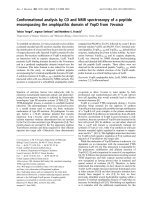
Figure 5 shows the molecular models of the human
NOS-II: and bovine trypsin:N-amidino-2-hydroxypyrro-
Scheme 1.
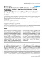
Fig. 4. Effect of N-amidino-2-hydroxypyrrolidine c oncentration (i.e.
[Inhibitor]) on the K
app
m
=K
0
m
ratio for the competitive inhibition of NOS-I
(squares) and NOS-II (triangles) catalyzed conversion of
L
-arginine
to
L
-citrulline, and of the trypsin (circles) c atalyzed hydrolysis of
N-a-benzoyl-
L
-arginine p-nitroanilide. The con tinuous lines were cal-
culated according to Eqn (2) with values of K
i
giveninTable1.Data
were obtained between pH 6.8 and 7.5 and between 21.0 °Cand
37.0 °C, mean ± SD, for further details, s ee text.
Table 1. Values of K
i
for N-amidino-2-hydroxypyrrolidine, agmatine, and clonidine binding to NOS-I, NOS-II, and trypsin.
Enzyme
K
i
(
M
)
N-Amidino-2-
hydroxypyrrolidine Agmatine Clonidine
NOS-I (1.1 ± 0.1) · 10
)5a
(6.6 ± 1.1) · 10
)4b
(5.0 ± 0.2) · 10
)3c
NOS-II (2.1 ± 0.1) · 10
)5a
(2.2 ± 0.2) · 10
)4b
>5 · 10
)2c
Trypsin (8.9 ± 0.4) · 10
)5d
>10
)2e
>10
)2e
a
pH 7.5 and 37.0 °C. Present study.
b
pH 7.8 and 37.0 °C. From [8].
c
pH 7.5 and 37.0 °C. From [16].
d
pH 6.8 and 21.0 °C. Present study.
e
pH 7.0 and 25.0 °C. From [30].
Ó FEBS 2002 N-Amidino-2-hydroxypyrrolidine characterization (Eur. J. Biochem. 269) 889
lidine co mplexes. I n human NOS-II (top panel), N-amidino-
2-hydroxypyrrolidine is hosted in the hydrophobic cavity
defined by the heme p rosthetic group and by the facing
hydrophobic residues Ala270 and Val271, as observed for a
number of nitrogen heterocycles [21,33] (note that N-ami-
dino-2-hydroxypyrrolidine is constrained in a semiboot
conformation, with the nitrogen lone pair directed towards
the heme iron). The positively charged amidino group of
N-amidino-2-hydroxypyrrolidine appears to be stabilized
by the negatively charged carboxylate of the Glu296 residue
which is required for
L
-arginine binding [21,33]. By homo-
logy, this residue corresponds to Glu597 and Glu371 in rat
NOS-I and NOS-II, respectively [23]. Moreover, as previ-
ously reported for the bovine trypsin:benzamidine complex
[22,34], N-amidino-2-hydroxypyrrolidine binds to the
enzyme primary s pecificity subsite S
1
(bottom p anel).
Interestingly, the alicyclic group is extended in a se michair
conformation, with the positively charged amidino g roup of
N-amidino-2-hydroxyp yrrolidine forming a salt bridge with
the negatively charged carboxylate of the trypsin Asp189
residue. The latter is required for recognition of the cationic
amino acid residue present at t he P
1
position of substrates
and inhibitors of trypsin-like serine proteinases [35,36].
N-Amidino-2-hydroxypyrrolidine, agmatine, and cloni-
dine bind to I
1
-binding sites (i.e. I
1
-R). In fact, the I
2
sites,
which are not considered as receptors and showing a
mitochondrial localization possibly corresponding to
monoamine oxidase [37,38], are removed from rat h eart
membrane preparations. Figure 6 shows [
3
H]clonidine
displacement from I
1
-R present in rat heart membranes
by N-amidino-2-hydroxypyrrolidine, agmatine, and cloni-
dine. As observed in other target tissues [25], the specific
binding of [
3
H]clonidine to rat h eart membranes is s aturable
(data not shown). Moreover, specific bindin g amounts to
3650 ± 294 d.p.m.Æh
)1
Æ(mg p rotein)
)1
, at saturating
[
3
H]clonidine concentration (¼ 1.0 · 10
)8
M
). N-amidino-
2-hydroxypyrrolidine and agmatine are more efficient than
clonidine in displacing [
3
H]clonidine from specific binding
sites in heart rat membranes, values o f IC
50
being
Fig. 5. N-Amidino-2-hydroxypyrrolidine binding mode tohuman NOS-II
(top) and bovine trypsin (bottom). The conformations of N-amidino-
2-hydroxypyrrolidine in the enzyme:inhibitor complexes were ob tained
after 10 ps molecular dynamics. For further details, see text.
Fig. 6. Competition of N-amidino-2-hydroxypyrrolidine (circles),
agmatine (triangles), and clonidine (squares) with [
3
H]clonidine for its
specific binding sites in rat heart membranes. The filled diamond indi-
cates [
3
H]clonidine saturating s pec ific binding ( a ¼ 1) in the a bsence of
the ligand (i.e. clonidine, or agmatine or N-amidino-2-hydroxypyrro -
lidine). The continuo us lines were calculated according to Eqn (3)
with the following IC
50
values: N-am idino-2-hydroxypyrrolidine
and agmatine, IC
50
¼ (1.3 ± 0.4) · 10
)9
M
, and clonidine, IC
50
¼
(2.2 ± 0.4) · 10
)8
M
. Data were obtained at pH 7.4 and 37.0 °C,
mean ± SD. For further details, see text.
890 P. Ascenzi et al. (Eur. J. Biochem. 269) Ó FEBS 2002
(1.3 ± 0.4) · 10
)9
M
and (2.2 ± 0.4) · 10
)8
M
, respec-
tively (at pH 7.4 and 37.0 °C) (Fig. 6).
DISCUSSION
For the first time, N-amidino-2-hydroxypyrrolidine, the
product of agmatine oxidation by P. sativum copper amine
oxidase, has been identified an d characterized from the
structural and biochemical viewpoints. Notably, the enzy-
matic oxidation of agmatine leads to the cyclic compound
N-amidino-2-hydroxypyrrolidine, as the only detectable
reaction product (Figs 2 and 3). In f act, the formation of
4-guanidinobutyraldehyde was never observed. Therefore,
4-guanidinobutyraldehyde, the best substrate of the alde-
hyde dehydrogenase that occurs in Fabaceae plants a nd rat
hepatocytes with copper amine oxidase [39–42], does not
appear to originate from the enzymatic cycling of agmatin e
to N-amidino-2-hydroxypyrrolidine.
N-Amidino-2-hydroxypyrrolidine inhibits competitively
NOS-I, NOS-II, and trypsin (Fig. 4). This compound
binds to the Glu597 and Glu371 carboxylate, present in
NOS-I and NOS-II, respectively (Glu296 in human NOS-
II; see Fig. 5), which is required for substrate ( i.e.
L
-arginine) recognition [21,33]. Moreover, N-amidino-2-
hydroxypyrrolidine binds to the trypsin primary specificity
subsite S
1
forming a salt bridge with the Asp 189
carboxylate (Fig. 5). The latter is required for recognition
of the cationic amino acid residue present at the P
1
position of substrates and inhibitors of trypsin-like serine
proteinases [35,36].
N-Amidino-2-hydroxypyrrolidine and agmatine displace
efficiently [
3
H]clonidine from I
1
-R present in heart rat
membranes (Fig. 6). Interestingly, d ifferent physiological
roles (i.e. neuronal neurotransmission and hypotensive
protection of cardiovascular system) have been linked to
agmatine, which has b een reported to be the endogenous
ligand for I-R
1
[7] and to represent the N-amidino-
2-hydroxypyrrolidine precursor. In this respect, pleiotropic
functional role(s) of N-amidino-2-hydroxypyrrolidine may
be envisaged, as reported for agmatine [7].
As a w hole, agmatine oxidation by P. sativum copper
amine oxidase may represent a new biocatalytic route for
the synthesis of N-amidino-2-hydroxypyrrolidine, possibly
representing a lead compound for the development of NOS
and trypsin-like s erine p rotease i nhibitors. Moreover,
N-amidino-2-hydroxypyrrolidine may represent a new
ligand for I
1
-R.
ACKNOWLEDGEMENTS
Authors wish to t hank Prof S. Aim e and Dr G. Rea for helpful
discussions and Dr L. Leone and Mr A. Merante for technical
assistance. This study was partially supported by grants from the
National Research Council of Italy (CNR, target oriented project
ÔBiotechnologyÕ, 99.00280.PF49 to P. A., a nd 99.00360.PF49 to M . F.).
Access to t he 6 00 MHz NMR facility h as be en g ranted b y Bioindustry
Park Canavese, Colleretto Giacosa, TO, Italy.
REFERENCES
1. McIntire, W.S. & Hartmann, C. (1993) Copper-containing amine
oxidases. In Principles an d Applications of Quinoproteins (Davison,
V.L., ed.), pp. 99–171. Marcel Dekker, New York.
2. Fontecave, M. & Eklund, H . (1995) Copper amine oxidase: a novel
use for a tyrosine. Structure 3, 1127–1129.
3. Klinman, J.P. (1996) Mechanisms whereby mononuclear copper
proteins functionalize organic substrates. Chem. Rev. 96, 2541–
2561.
4. Buffoni, F. & Ignesti, G. (2000) The copper-containin g amine
oxidases: biochemical aspects and functional role. Mol. Genet .
Metab. 71, 559–564.
5. Federico, R., Angelini, R ., Ercolini, L., Ventur ini, G., Mattevi, A.
& Ascenzi, P. (1997) Competitive inhibition of swine kidney
copper amine o xidase by drugs: amiloride , clonidine, a nd gabexate
mesylate. Biochem. Biophys. Res. Commun. 240, 150–152.
6. Holt, A . & Baker, G.B. (1995) Metabolism of a gmatine (clonidine-
displacing substance) by diamine oxidase and the possible impli-
cations f or stu dies o f i mi dazoline r ec eptors. Prog. Brain. Res. 106,
187–197.
7. Reis, D.J. & Regunathan, S. (2000) Is agmatine a n ovel neuro-
transmitter in brain? Trends Pharmacol. Sci. 21, 187–193.
8. Galea, E ., Regunathan, S., Eliopoulos, V., Feinstein, D.L. & R eis,
D.J. (1996) Inhibition of mammalian nitric oxide synthases by
agmatine, an endogenous polyamine formed by decarboxylation
of arginine. Biochem. J. 316, 247–249.
9. Demady, D.R., Jianmongkol, S., Vuletich, J.L., Bender, A.T. &
Osawa, Y. (2001) Agmatine enhances the NADPH oxidase
activity of neuronal NO synthase and leads to oxidative inacti-
vation of the enzyme. Mol. Pharmacol. 59, 24–29.
10. McGuirl, M.A., McCahon, C.D., McKeown, K.A. & Dooley,
D.M. (1994) Purification and characterization of pea seedling
amine o xidase for crystallization studies. Plant P hysiol. 106, 1205–
1211.
11. Stuehr, D.J. & Griffith, O.W. (1996) Purification, assay and
properties of mammalian nitric oxide synthases. In Methods in
Nitric Oxide Research (Feelisch, M. & Staml er, J.S., eds), pp. 177–
186. John Wiley & Sons Ltd, Chichester.
12.Rinaldi,A.,Floris,G.&Finazzi-Agro
`
, A. (1982) Purification
and properties of diamine oxidase from Euphorbia latex. Eur. J.
Biochem. 127, 417–422.
13. Ascenzi, P., Ascenzi, M.G. & Amiconi, G. (1987) Enzyme
competitive inhibition: graphical determination of K
i
and presen-
tation of d ata in comparative studies. Biochem. Educ. 15, 134–
135.
14. Holmestedt,B.,Larsson,L.&Tham,R.(1961)Furtherstudies
on spectrophotometric method for the determination of amine
oxidase activity. Biochim. Biophys. Acta 48, 182–186.
15. Braun, S., Kalinowski, H O. & Berger, S. (1998) 150 and More
Basic NMR Experiments. Wiley-VCH, Weinheim.
16. Venturini, M., Colasanti, M., Persichini, T., Fioravanti, E.,
Federico, R. & Ascenzi, P. (2000) Selective inhibition of nitric
oxide synthase type I by clonidine, an antihypertensive drug.
Biochem. Pharmacol. 60, 539–544.
17. Venturini, G., Colasanti, M., Fioravanti, E., Bianchini, A. &
Ascenzi, P . ( 1999) Direct effect of te mperature on the catalytic
activity o f nitric oxide synthases types I, II, and III. Nitric Oxide 3,
375–382.
18. Feelisch, M., Kubitzek, D. & W erringloer, J. (1996) The oxyhe-
moglobin assay. In Met hods in Nitric Oxide Research (Feelish, M .
& Stamler, J.S., eds), pp. 455–478. John Wiley & Sons Ltd,
Chichester.
19. Venturini, G., Menegatti, E. & Ascenzi, P. (1997) Competitive
inhibition of nitric oxide synthase by p-aminobenzamidine, a
serine proteinase inhibitor. Biochem. Biophys. Res. Commun. 232,
88–90.
20. Ascenzi,P.,Menegatti,E.,Guarneri,M.,Bortolotti,F.&Anto-
nini, E. (1982) Catalytic properties of serine proteases. 2. Com-
parison between human urinary kallikrein and human urokinase,
bovine b-trypsin, bovine thrombin, and bo vine a-chymo trypsin.
Biochemistry 21, 2483–2490.
Ó FEBS 2002 N-Amidino-2-hydroxypyrrolidine characterization (Eur. J. Biochem. 269) 891
21. Fischmann, T.O., Hruza, A., Niu, X.D., Fossetta, J.D., Lunn,
C.A., Dolphin, E., Prongay, A.J., Reichert, P., Lundell, D.J.,
Narula, S.K. & Weber, P.C. (1999) Structural characterization of
nitric oxide synthase isoforms r eveals striking active-site c onser-
vation. Nat. Struct. Biol. 6, 233–242.
22. Ota, N., Stroupe, C., Ferreira da Silva, J.M.S ., Shah, S.S., M ares-
Guia, M. & Brunger, A.T. (1999) Non-Boltzm ann Thermo-
dynamic In tegration (N BT I) fo r macromolecular systems: r elative
free energy of bind ing of trypsin to be nzam idine and ben zylamine.
Proteins 37, 641–653.
23. Berman, H .M., Westbrook, J., Feng, Z., Gilliland, G., Bhat, T.N.,
Weissig, H., Shindyalov, I.N. & Bourne, P.E. (2000) The Protein
Data Bank. Nucleic Acids Res. 28, 235–242.
24. Glossmann, H. & Ferry, D.R. (1985) Assay for calcium channels.
Methods Enzymol. 109, 513–550.
25. Molderings, G.J., Donecker, K. & Go
¨
thert, M. (1995) Charac-
terization of non-adrenergic [
3
H]clonidine binding sites in rat
stomach: high affinity of imidazolines, guanidines and sigma
ligands. Naunyn-Schmiedeberg’s Arch. Pharmacol. 351, 561–564.
26. Molderings, G.J., Moura, D., Fink, K., Bo
¨
nisch, H . & Go
¨
thert,
M. (1993) Binding of [
3
H]clonidine to I
1
-imidazoline sites in
bovine adrenal medullary membranes. Naunyn-Schmiedeberg’s
Arch. Pharmacol. 348, 70–76.
27. Molderings, G.J., Kundt, L . & Go
¨
thert, M. (1994) [
3
H]Idazoxan
binding to bovine adrenal medullary membranes: identification
and pharmacological characterization of I
2
-imidazoline sites.
Naunyn-Schmiedeberg’s Arch. Pharmacol. 350, 252–257.
28. Bradford, M.M. (1976) A rapid, sensitive method for the quanti-
fication of mg quantities of protein, utilizing the principle of
protein-dye binding. Anal. Biochem. 72 , 248–254.
29. Ascenzi, P., Desideri, A., Amiconi, G., Bertollini, A., Bolognesi,
M., Castagnola, M., Coletta, M. & Brunori, M. (1988) Effect of
inositol hexakisphosphate on the spectroscopic properties of the
nitric oxide derivative of ferrous naturally glycated human
hemoglobin HbA
1c
. J. Inorg. Biochem. 34, 19–24.
30. Federico, R., Leone, L., Botta, M., Bind a, C., Angelini, R.,
Venturini, G. & Ascenzi, P. (2001) Inh ibition of pig liver and Zea
mays L. polyamine o xidase: a comparative study. J. Enzyme Inh ib.
16, 147–155.
31. Friebolin, H. ( 1993) Basic O ne- and Two-Dimensional NMR
Spectroscopy. VCH, Weinheim.
32. Halgren, T.A. (1996) M erck Molecular F orce Field. I . Basis, form ,
scope, parame trization, and performance of MMFF 94. J. Com-
put. Chem. 17, 490–519.
33. Crane, B.R., Arvai, A.S., Gachhui, R., Wu, C., Ghosh, D.K.,
Getzoff, E.D., Stuehr, D.J. & Tainer, J.A. (1997) The structure of
nitric oxide synthase oxygenase domain and inhibitor complexes.
Science 278, 425–431.
34. Bode, W. & Schwager, P. (1975) The refined crystal structure of
bovine b-trypsin at 1.8 A
˚
resolution. II. Crystallographic refine-
ment, calcium binding site, benzamidine binding site and active
site at pH 7.0. J. Mol. Biol. 98, 693–717.
35. Bode, W. & Huber, R. (1992) Natural protein proteinase inhibi-
tors and their interactio n with proteinases. Eur. J. Biochem. 20 4,
433–451.
36. Bode, W. & Huber, R. (2000) Structural basis of the endopro-
teinase–protein inhibitor inte raction. Biochim. Biophys. Acta 1477,
241–252.
37. Tesson, F., Limon-B oulez, I., U rban, P., Puype, M., Van -
dekerckhove,J.,Coupry,I.,Pompon,D.&Parini,A.(1995)
Localization of I
2
-imidazoline binding site s on mon oamine oxi-
dases. J. Biol. Chem. 270, 9856–9861.
38. Ernsberger,P.&Haxhiu,M.A.(1997)TheI
1
-imidazoline-binding
site is a functional receptor mediating vasodepression via the
ventral medulla. Am. J. Physiol. 273, R1572–R1579.
39. Matsuda, H. & Suzuki, Y. (1981) Purification and properties of
the diamine oxidase from Vicia f aba le aves. Plant C ell P hysiol. 22,
737–746.
40. Matsuda, H. & Suzuki, Y. (1984) c-Guanidinobutyraldehyde
dehydrogenase of Vicia faba leaves. Plant Physiol. 76, 654–657.
41. Kaneoke,M.,Shimizu,E.&Yorifuji,T.(1994)Metabolismof
L-arginine, agmatine, and related-compounds in Nocardioides
simplex . Biosci. Biotechn Biochem. 58, 244–249.
42. Cabella, C., Gardini, G., Corpillo, D., Test ore, G., Bedino, S.,
Solinas, S.P., Cravanzola, C., Vargiu, C., Grillo, M.A. &
Colombatto, S. (2001) Transport and metabolism of agmatine in
rat hepatocyte cultures. Eur. J. Biochem. 268, 940–947.
892 P. Ascenzi et al. (Eur. J. Biochem. 269) Ó FEBS 2002