Báo cáo y học: "Prolonged, granulocyte–macrophage colony-stimulating factor-dependent, neutrophil survival following rheumatoid synovial fibroblast activation by IL-17 and TNFalpha" doc
Bạn đang xem bản rút gọn của tài liệu. Xem và tải ngay bản đầy đủ của tài liệu tại đây (1.3 MB, 12 trang )
Open Access
Available online />Page 1 of 12
(page number not for citation purposes)
Vol 10 No 2
Research article
Prolonged, granulocyte–macrophage colony-stimulating
factor-dependent, neutrophil survival following rheumatoid
synovial fibroblast activation by IL-17 and TNFalpha
Greg Parsonage, Andrew Filer, Magdalena Bik, Debbie Hardie, Sian Lax, Katherine Howlett,
Leigh D Church, Karim Raza, See-Heng Wong, Emily Trebilcock, Dagmar Scheel-Toellner,
Mike Salmon, Janet M Lord and Christopher D Buckley
Rheumatology Research Group, MRC Centre for Immune Regulation, Institute for Biomedical Research, University of Birmingham, Vincent Drive,
Edgbaston, Birmingham B15 2TT, UK
Corresponding author: Christopher D Buckley,
Received: 16 Nov 2007 Revisions requested: 20 Dec 2007 Revisions received: 31 Mar 2008 Accepted: 23 Apr 2008 Published: 23 Apr 2008
Arthritis Research & Therapy 2008, 10:R47 (doi:10.1186/ar2406)
This article is online at: />© 2008 Parsonage et al.; licensee BioMed Central Ltd.
This is an open access article distributed under the terms of the Creative Commons Attribution License ( />),
which permits unrestricted use, distribution, and reproduction in any medium, provided the original work is properly cited.
Abstract
Introduction A surprising feature of the inflammatory infiltrate in
rheumatoid arthritis is the accumulation of neutrophils within
synovial fluid and at the pannus cartilage boundary. Recent
findings suggest that a distinct subset of IL-17-secreting T-
helper cells (T
H
17 cells) plays a key role in connecting the
adaptive and innate arms of the immune response and in
regulating neutrophil homeostasis. We therefore tested the
hypothesis that synovial fibroblasts bridge the biological
responses that connect T
H
17 cells to neutrophils by producing
neutrophil survival factors following their activation with IL-17.
Methods IL-17-expressing cells in the rheumatoid synovium,
and IL-17-expressing cells in the peripheral blood, and synovial
fluid were examined by confocal microscopy and flow cytometry,
respectively. Peripheral blood neutrophils were cocultured
either with rheumatoid arthritis synovial fibroblasts (RASF) or
with conditioned medium from RASF that had been pre-exposed
to recombinant human IL-17, TNFα or a combination of the two
cytokines. Neutrophils were harvested and stained with the vital
mitochondrial dye 3,3'-dihexyloxacarbocyanine iodide before
being enumerated by flow cytometry.
Results T
H
17-expressing CD4
+
cells were found to accumulate
within rheumatoid synovial tissue and in rheumatoid arthritis
synovial fluid. RASF treated with IL-17 and TNFα (RASF
IL-17/TNF
)
effectively doubled the functional lifespan of neutrophils in
coculture. This was entirely due to soluble factors secreted from
the fibroblasts. Specific depletion of granulocyte–macrophage
colony-stimulating factor from RASF
IL-17/TNF
-conditioned
medium demonstrated that this cytokine accounted for
approximately one-half of the neutrophil survival activity.
Inhibition of phosphatidylinositol-3-kinase and NF-κB pathways
showed a requirement for both signalling pathways in RASF
IL-
17/TNF
-mediated neutrophil rescue.
Conclusion The increased number of neutrophils with an
extended lifespan found in the rheumatoid synovial
microenvironment is partly accounted for by IL-17 and TNFα
activation of synovial fibroblasts. T
H
17-expressing T cells within
the rheumatoid synovium are likely to contribute significantly to
this effect.
Introduction
In established rheumatoid arthritis (RA), highly differentiated
CD4
+
T lymphocytes persist within synovial tissue, and are
prevented from undergoing apoptosis by high local concentra-
tions of type I interferons [1]. Simplistically, the preponderance
of IFNγ-expressing T cells and the paucity of IL-4-expressing T
cells, in situ and ex vivo, has led to the description of RA as an
immune-mediated inflammatory disease that is associated
BSA = bovine serum albumin; ELISA = enzyme-linked immunosorbent assay; FCM = fibroblast-conditioned medium; FITC = fluorescein isothyocy-
anate; GM-CSF = granulocyte–macrophage colony-stimulating factor; HBSS = Hank's Buffered saline solution; IL = interleukin; IFN = interferon;
MEM = Eagle's "Minimal Essential Media"; NF = nuclear factor; PBS = phosphate-buffered saline; RA = rheumatoid arthritis; RASF = rheumatoid
arthritis synovial fibroblasts; RASF
IL-17/TNF
= rheumatoid arthritis synovial fibroblasts stimulated with TNFα and IL-17; rh = recombinant human TBST
= Tris-buffered saline containing 0.1% Tween 20; T
H
17 cells = IL-17-secreting T-helper cells; TNF = transforming growth factor.
Arthritis Research & Therapy Vol 10 No 2 Parsonage et al.
Page 2 of 12
(page number not for citation purposes)
with a predominantly T-helper type-1 T-cell cytokine profile [2-
4]. This T-helper type-1 T-cell paradigm, however, does not
adequately account for the large numbers of neutrophils that
also accumulate within the synovial space.
During active phases of disease, large numbers of activated
neutrophils are found in the synovial fluid of both very early RA
and established RA patients [5,6]. As a source of proinflamma-
tory mediators such as IL-1β, CXCL8 and TNFα, activated
neutrophils clearly contribute to the complex cytokine milieu of
the inflamed joint [5]. There is evidence to suggest that CD4
T cells and neutrophils may be engaged in complex cytokine
crosstalk. For example the T-helper type-1 T-cell-associated
cytokine IL-18 indirectly induces the recruitment of neutrophils
in a murine model of arthritis [7]. Similarly, IFNγ-stimulated
neutrophils have been shown to release potent chemoattract-
ants for T-helper type-1 T cells and NK cells [8].
Intermittent neutrophil accumulation within the synovial fluid of
RA patients results in the degradation of extracellular matrix
proteins that are crucial for the lubricative function of synovial
fluid. The release of reactive oxygen intermediates and broad-
acting proteases from the intracellular granules of neutrophils
is responsible for this. Activated neutrophils have also been
found at the cartilage pannus interface, where they may pro-
mote joint erosion more directly [9]. Furthermore an elegant
series of investigations has revealed an essential role for neu-
trophils (and other cells of the innate immune system) both in
the initiation and progression of catastrophic joint inflamma-
tion in the murine K/BxN spontaneous arthritis model [10].
Synovial neutrophils are therefore likely to make a significant
contribution to the pathology of RA.
We recently described a distinctive but transient synovial fluid
cytokine profile in patients with very early synovitis destined to
become RA that distinguished such patients from those who
did not progress to RA [11]. The T-cell-derived cytokine IL-17
formed part of this distinctive cytokine profile. IL-17 has been
implicated in the process of chronic inflammatory pathology at
various anatomical locations including the synovium via
release from a distinct subset of CD4 T cells termed IL-17-
secreting T-helper cells (T
H
17 cells) [12-14].
Consistent with their distinct developmental lineage, which
requires the presence of the transcriptional factor retinoic
acid-related orphan receptor γt isoform, T
H
17 cells appear to
have a unique function in bridging the adaptive and innate
arms of the immune system to promote host defence against
extracellular bacterial infections [15]. T
H
17 cells also play an
important homeostatic role in regulating neutrophil production
and blood neutrophil counts [16,17] and in promoting the
recruitment of neutrophils into tissues. In addition, recent stud-
ies have shown that these cells contribute to bone destruction
by activating osteoclasts in models of arthritis [14]. Human
T
H
17 cells were described very recently in large numbers in
the gut mucosa [18]. In addition, T
H
17 cells have been shown
to express the chemokine receptors CCR6 and CCR4 [19]
and to depend on IL-1 and IL-6 but not on transforming growth
factor beta for their production [20,21].
In the present study we tested the hypothesis that synovial
fibroblasts bridge the biological responses that connect the
presence of T
H
17 cells in the synovium with the increase in
neutrophils seen in RA by producing neutrophil survival factors
following their activation with IL-17.
Materials and methods
Reagents
All cell culture reagents were endotoxin-free and were pur-
chased from Sigma (St Louis, MO, USA). Recombinant human
(rh) granulocyte–macrophage colony-stimulating factor (GM-
CSF), rhIL-17A and rhTNFα were from Peprotech (London,
UK), monoclonal anti-human GM-CSF (MAB215) and polyclo-
nal anti-human GM-CSF (AF215NA) were from R&D Systems
(Abingdon, UK), and recombinant IFNβ was from Biosource
(Paisley, UK). Type I IFN receptor (CD118) blocking antibody
was obtained from Calbiochem (San Diego, CA), anti-IL-6R
and anti-TNFα were from R&D (Abingdon, UK), human GM-
CSF and IL-6 ELISA kits were from BDPharmingen (San
Diego, CA), and protein G-conjugated agarose was from
Upstate (Lake Placid, NY, USA). The 0.2 μm Transwell inserts
were obtained from Falcon (BD biosciences, San Jose, CA,
USA), rhIL-2 from Chiron (Middlesex, UK), phytohaemaglutinin
from Murex (Dartford, UK) and 3,3'-dihexyloxacarbocyanine
iodide was from Molecular Probes (Leiden, The Netherlands).
Additional reagents, including adenosine, adenosine deami-
nase, indomethacin, NS398 and MK886, were from Sigma.
The multiplex assay used Beadmate kits (Upstate) as specified
in the manufacturer's instructions, read with a Luminex 100
system (Upstate).
Patient samples, fibroblast culture and treatment
Rheumatoid synovial fibroblasts were isolated from the syn-
ovium of RA patients meeting the 1987 American College of
Rheumatology criteria [22] undergoing joint replacement sur-
gery. Clinical details of patients fulfilling the 1987 American
College of Rheumatology criteria who donated samples are
presented in Table 1. The Disease Activity Scores using 28
joint counts (DAS28) at the time of joint replacement are given
for patients donating samples from which fibroblasts were cul-
tured. Six of the eight patients undergoing joint replacement
who gave samples for fibroblast culture had DAS28 counts
>5.2, indicating active disease.
Peripheral blood and synovial fluid were also obtained from RA
patients meeting the 1987 American College of Rheumatol-
ogy criteria [22]. Ethical approval for the use of patient-derived
material was given by the local research ethics committee, and
Available online />Page 3 of 12
(page number not for citation purposes)
all patients provided written informed consent (LREC refer-
ence 5735).
Fibroblasts were maintained in fibroblast medium (consisting
of 81.3% RPMI 1640, 16.3% foetal calf serum, 0.81 × MEM
nonessential amino acids, 0.81 mM sodium orthopyruvate,
1.62 mM glutamine, 810 U/ml penicillin and 81 μg/ml strepto-
mycin) at 37°C in a humidified 5% carbon dioxide atmosphere.
The fibroblast phenotype was confirmed by morphology and
immunofluorescence microscopy as described previously
[23]. All cells expressed fibronectin and prolyl-4-hydroxylase,
while less than 0.5% cells stained positive for CD68, von Wil-
lebrand factor, CD31 or cytokeratin.
Synovial fibroblasts were used between passages 4 and 8.
Before the addition of exogenous cytokines, fibroblasts were
seeded into flat-bottomed 96-well plates at a density of 6 ×
10
3
cells/well and were cultured for 48 hours in coculture
medium (consisting of 88.9% RPMI 1640 medium supple-
mented with 8.9% heat-inactivated foetal calf serum, 1.62 mM
glutamine, 810 U/ml penicillin, 81 μg/ml streptomycin and 1.8
mM (pH 7.4) HEPES). The coculture medium was then
refreshed and the fibroblasts exposed to 10 ng/ml IL-17A and
10 ng/ml TNFα. Following cytokine treatment or coculture, the
fibroblasts were washed extensively with RPMI 1640 and
were cultured with or without 10
5
neutrophils/well for a further
24 hours. Cells or conditioned medium were then harvested.
Preparation of neutrophils
Peripheral blood of consented healthy volunteers was taken
into tubes containing the anticoagulant ethylenediamine
tetraacetic acid and was then mixed with T500 Dextran to sed-
iment the red blood cells. Mononuclear cells and granulocytes
were separated by centrifugation across a saline-based Per-
coll gradient (upper phase, 54% vol/vol; lower phase, 79%
vol/vol), and were then washed in PBS to remove residual Per-
coll. Neutrophil preparations were routinely >98% pure and
viable, as assessed by May–Grunwald Giemsa-stained
cytospins.
Cell survival assays
Flow cytometry was carried out using the 20 μl volume dump
facility of the Beckman Coulter Epics XL bench-top flow
cytometer (High Wycombe, UK). When used in combination
with vital dyes, this technique enables accurate enumeration of
live cells within a sample of defined volume. Neutrophils were
gated based on their forward scatter versus side scatter pro-
files, and apoptotic cells were excluded by loss of 3,3'-dihexy-
loxacarbocyanine iodide positivity. Survival is expressed as
percentage of the starting population. The percentage of cells
Table 1
Clinical details of rheumatoid arthritis patients
Age (years) Sex Rheumatoid Factor Erosive disease Therapy DAS28 score
Samples used to culture fibroblasts
62 Female + + Methotrexate 5.5
52 Female + + Methotrexate 4.4
66 Male + + Etanercept, oral prednisolone 5.5
71 Female + + D-penicillamine 5.3
62 Female + + 6.8
59 Female + + Methotrexate 6.8
30 Female + + Azathioprine 6.5
77 Male + + Sulphasalazine 4.0
Samples used for confocal microscopy and flow cytometry
72 Female + + Methotrexate
73 Female + + Methotrexate
73 Female + + Methotrexate, oral prednisolone
62 Female Methotrexate, infliximab
64 Female + +
66 Female + + Methotrexate
Arthritis Research & Therapy Vol 10 No 2 Parsonage et al.
Page 4 of 12
(page number not for citation purposes)
committed to apoptosis was confirmed with May–Grunwald
Giemsa-stained cytospins.
Cytokine depletion and blockade studies
Supernatants from cytokine-treated fibroblasts, and solutions
containing higher concentrations of recombinant target pro-
teins were depleted of cytokine in three rounds of incubation
with an excess of irrelevant or depleting antibody-coated pro-
tein G-conjugated agarose beads. Residual beads were
removed by centrifugation before exposing neutrophils to the
medium or performing ELISAs to verify that cytokine depletion
was successful (the lower detection limit of the GM-CSF
ELISA was 4 pg/ml). For lipopolysaccharide studies, 10 ng/ml
lipopolysaccharide (Sigma) was added to neutrophils in the
presence or absence of conditioned medium from IL-17A-
treated and TNFα-treated cells and of 50 μg/ml polymyxin B
sulphate.
Inhibitor studies
Freshly isolated neutrophils were pretreated with Ly294002
(20 μM) or Bay 11-7085 (1 μM) or vehicle control for 45 min-
utes at 37°C, washed, and cultured for 24 hours in fibroblast-
conditioned medium (FCM) from untreated rheumatoid arthri-
tis synovial fibroblasts (RASF) or from rheumatoid arthritis syn-
ovial fibroblasts stimulated with TNFα and IL-17 (RASF
IL-
17/TNF
). The optimal concentration of inhibitors used was
established by performing a dose–response analysis.
Neutrophil functional studies
To measure the functionality of neutrophils, superoxide release
upon stimulation with 200 nM f-Met-Leu-Phe was measured
using the substrate cytochrome c. Following their 24-hour
treatments, neutrophils were washed once in HBSS buffer
(HBSS pH 7.3 + 25 mM HEPES and 5 mM glucose), and
were resuspended in HBSS containing 1% human serum.
Then 5 × 10
4
neutrophils were added to triplicate wells of a
96-well plate in the presence or absence of f-Met-Leu-Phe.
Release of superoxide was quantified by measuring the colour
change of cytochrome c at 450 nm emission. The positive con-
trol was complete reduction of cytochrome c using the reduc-
ing agent sodium dithionite (100 mM).
Inhibitor of NFκB measurement
Degradation of inhibitor of NFκB in the presence of 10 ng/ml
TNFα or FCM was assessed by western blotting. Neutrophils
(at least 5 million cells/sample) were lysed with 10% trichloro-
acetic acid (Sigma) followed by 5 minutes incubation on ice.
The lysates were centrifuged for 5 minutes at 14,000 × g at
4°C and the supernatant was discarded. The pellet was
washed twice with ice-cold acetone, centrifuging each time as
before. The pellet was dissolved in SDS-PAGE sample load-
ing buffer (0.125 M Tris pH 6.8 containing 20% glycerol, 2%
SDS, 5% mercaptoethanol and 25 μg/ml bromophenol blue).
The sample was boiled, then cooled on ice for 5 minutes each,
before performing gel electrophoresis using a 12% SDS gel.
Proteins were then transferred to a polyvinylidene difluoride
membrane (Flowgen Ltd, Sittingbourne, UK) for 2 hours at
0.45 A using a wet blotting system (Biorad Hemel Hempstead,
UK). The blots were blocked for 1 hour with 5% skimmed milk
in Tris-buffered saline containing 0.1% Tween 20 (TBST) for
1 hour. Blots were incubated with primary antibodies (IkBα
Rabbit IgG; Upstate) diluted in Tris-buffered saline containing
1% skimmed milk overnight at 4°C, washed four times with
TBST for 10 minutes each, and then incubated with secondary
antibodies (horseradish peroxidase-donkey anti-rabbit; Amer-
sham, Bucks, UK) diluted in TBST. Blots were washed three
times with TBST for 10 minutes each and developed for 5 min-
utes with enhanced chemiluminescence reagents
(Amersham).
Flow cytometry analysis of peripheral blood and synovial
fluid cells for IL-17 expression
Peripheral blood mononuclear cells and synovial fluid mononu-
clear cells were isolated by density gradient centrifugation on
Ficoll-paque™-Plus (GE Healthcare, Slough, UK). Following
centrifugation, peripheral blood mononuclear cells and syno-
vial fluid mononuclear cells were isolated from the buffy coat
and were added to fresh RPMI 1640 medium. Cells were
washed twice with RPMI 1640 medium by centrifugation at
600 × g for 8 minutes. Cells were then counted and resus-
pended at 1 × 10
6
cells/ml in fresh medium. Intracellular IL-17
was detected by flow cytometry after stimulation of cells with
50 ng/ml phorbol 12-myristate 13-acetate (PMA) (Sigma) and
250 ng/ml ionomycin (Sigma) in the presence of 10 μg/ml
Breveldin A (Sigma) for 4 hours in a 24-well plate (Sarstedt,
Leicester, UK).
Cells were removed from culture and resuspended in 2%
BSA/PBS. Cell surface staining with fluorescein isothyocy-
anate (FITC)-labelled anti-CD4 (Immunotools, Friesoythe, Ger-
many), phycoerythrin-cyanine 5-labelled anti-CD3
(Immunotools), FITC-labelled IgG
1
isotype control (Immunoto-
ols), and phycoerythrin-cyanine 5-labelled IgG
1
isotype control
(Serotec, Oxfordshire, UK) was performed by incubating cells
on ice with the appropriate antibody combination for 20 min-
utes. Cells were subsequently fixed and permeabilised with
Cytoperm according to manufacturer's instructions
(Caltag/Invitrogen, Towcester, UK).
Cells were incubated with either phycoerythrin-labelled anti-IL-
17 (eBiosciences, San Diego, CA, USA) or phycoerythrin-
labelled IgG
1
isotype control (Immunotools) for 20 minutes,
and were then washed and acquired on a Beckman Coulter
Epics XL bench-top flow cytometer. T cells were gated based
on their forward scatter versus side scatter profiles and their
CD3 expression. CD4
+
IL-17
+
T cells are expressed as a per-
centage of the CD3
+
population.
Available online />Page 5 of 12
(page number not for citation purposes)
Confocal microscopy
Full thickness (5 μm) sections of human rheumatoid synovium
were prepared and fixed in 100% acetone at 4°C for 20 min-
utes. The sections were rehydrated with PBS pH 7.4 with 5%
foetal calf serum (Biosera Ltd, Ringmer, UK), and indirect
immunofluorescence was performed using the following pri-
mary antibody combinations: IgG
2b
mouse anti-human CD3
(UCHT-1, 17 μg/ml; gift from Peter Beverley, University
College Hospital, London, UK) with rabbit anti-human von
Willibrand factor (A0082, 6.2 μg/ml; Dako, Glostrup, Den-
mark) and IgG
1
mouse anti-human IL-17 (12-7179, 1.25
μg/ml; eBiosciences); or IgG
2b
mouse anti-human CD4
(OKT4) with IgG
2a
mouse anti-human CD8 (OKT8, both OKT
clones used as ascitic fluid 1/100; American Type Culture
Collection, Middlesex, UK) and rabbit anti-human IL-17
(AHP455G, 20 μg/ml; AbD Serotec, Oxfordshire, UK).
The secondary antibody combinations used were: goat anti-
mouse IgG
2b
-cyanine 5 (1090-15, 4 μg/ml; Southern Biotech
Inc., Birmingham, AL, USA) with donkey anti-rabbit-tetrame-
thyl-rhodamine (711-026-152, 6 μg/ml; Jackson Immuno-
Research, Newmarket, UK) and goat anti-mouse IgG
1
-FITC
(1070-02, 20 μg/ml; Southern Biotech Inc.); or goat anti-
mouse IgG
2b
-cyanine 5 (1090-15, 4 μg/ml; Southern Biotech
Inc.) with goat anti-mouse IgG
2a
-FITC (1080-02, 20 μg/ml;
Southern Biotech Inc.) and donkey anti-rabbit-tetramethyl-
rhodamine (711-026-152 6 μg/ml; Jackson Immuno-
Research).
Irrelevant control antibodies (X0931 and X0903; Dako) were
used to confirm IL-17 staining, while primary antibodies were
omitted as controls for other stains. Sections were counter-
stained with nuclear DNA dye Hoechst 33258 (20 μg/ml;
Riedel De Haenag, Seelze, Hannover, Germany) and were
subsequently mounted in 2.4% 1,4-diazabicyclo [2.2.2]octane
(Aldrich, Gillingham, UK) in glycerol pH 8.6 (Fisons Scientific,
Loughborough, UK). A Zeiss 510 laser-scanning confocal
microscope (Zeiss, Welwyn Garden City, UK) was used to vis-
ualise staining with images captured and processed using the
Zeiss LSM Image Examiner software (Zeiss).
Statistical analysis
A nonparametric distribution was assumed for all assays. Sta-
tistical analysis of differences in neutrophil survival induced by
fibroblasts or FCM across multiple groups was performed
using Kruskal–Wallis one-way analysis of variance and Dunn's
post test. For the difference between paired samples (deple-
tions, blocking experiments, experiments with and without
transwells), significance was assessed using the Wilcoxon
signed rank test with two-tailed P values. Results are pre-
sented as the mean ± standard deviation or standard error
where appropriate.
Results
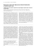
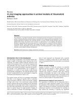
IL-17-expressing CD4 T cells accumulate within the
rheumatoid synovium
We first determined whether T
H
17 cells are present within the
rheumatoid synovium. IL-17 was expressed on CD3
+
T cells
within the synovium, and in particular on T cells found in a
perivascular distribution (Figure 1a). Expression of IL-17 was
confined to CD4 T cells, with no expression on CD8 T cells,
although there were some IL-17-expressing cells in the syn-
ovium that did not express CD4 or CD8 (Figure 1b). Equally
some CD4 T cells did not express IL-17 (Figure 1a,b). IL-17-
expressing CD4
+
CD3
+
cells were also found at low frequency
within rheumatoid synovial fluid (Figure 1c).
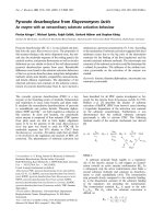
IL-17 and TNFα synergise to significantly enhance
fibroblast-mediated neutrophil survival
Previous studies have shown that IL-17 and TNFα synergise
with each other to produce potent biological effects in leuco-
cytes. We therefore cross-titrated recombinant human IL-17A
and TNFα on RASF to determine whether these cytokines,
either individually or in combination, could enhance the ability
of RASF to support neutrophil viability in cocultures of neu-
trophils with fibroblasts.
Figure 2a shows that the basal level of neutrophil survival in
the presence of RASF increases significantly and in a dose-
dependent manner when RASF are pretreated with a combi-
nation of IL-17 and TNFα. As would be expected, the increase
in survival is mirrored precisely by an inhibition of apoptosis
(data not shown). Optimal neutrophil survival was achieved at
a dose of 1 to 10 ng/ml IL-17 and 10 ng/ml TNFα. Pretreat-
ment of RASF with the highest doses (10 ng/ml) of IL-17 or
TNFα alone did not significantly enhance survival at 24 hours
(Figure 2b). Neutrophils cocultured with fibroblasts that had
been pretreated with IL-17 and TNFα (RASF
IL-17/TNF
), how-
ever, remained viable for 24 hours. Indeed, the level of viability
exceeded that obtained with a 100 ng/ml dose of rhGM-CSF
– a prototypical neutrophil survival factor added directly to
neutrophil monoculture (data not shown). Direct microscopic
analysis of fibroblast-mediated neutrophil survival by cellular
morphology and active caspase-3 immunostaining confirmed
these results (Figure 2c,d and data not shown).
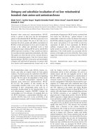
Coculture with RASF
IL-17/TNF
doubles the functional
lifespan of neutrophils
We next determined the kinetics of rescue from apoptosis,
mediated by coculture of neutrophils with RASF
IL-17/TNF
. Fig-
ure 3a shows that, in the presence of RASF
IL-17/TNF
, the neu-
trophil lifespan is effectively doubled. Furthermore, rescued
neutrophils were functionally active and capable of generating
the superoxide radical to the same extent as freshly isolated
neutrophils upon treatment with f-Met-Leu-Phe (Figure 3b).
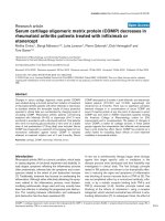
Delayed neutrophil apoptosis is attributable to soluble,
temperature-sensitive factors secreted by RASF
IL-17/TNF
We next investigated the mechanism by which cytokine-
treated synovial fibroblasts prolonged neutrophil survival by
Arthritis Research & Therapy Vol 10 No 2 Parsonage et al.
Page 6 of 12
(page number not for citation purposes)
Figure 1
IL-17 expression on T cells within rheumatoid synovium and in synovial fluid CD4
+
CD3
+
T cellsIL-17 expression on T cells within rheumatoid synovium and in synovial fluid CD4
+
CD3
+
T cells. (a) Rheumatoid synovial tissue was examined
by immunohistochemistry. IL-17 (red) was found to colocalise with CD3
+
T cells (blue) in perivascular cuffs (purple). Blood vessels were localised
with von Willebrand factor (vWF) (green). Nuclear staining is shown in grey. (b) IL-17 (red) expression is associated with CD4
+
T cells (blue) but not
CD8
+
(green) T cells. Nuclear staining is shown in grey. (c) flow cytometric analysis of peripheral blood (PB) and synovial fluid (SF) CD3
+
T cells
demonstrates that IL-17 is expressed in SF CD4
+
T cells. PE, phycoerythrin; FITC, fluorescein isothyocyanate.
Available online />Page 7 of 12
(page number not for citation purposes)
comparing the ability of FCM and transwell-separated neu-
trophil–synovial cocultures to influence neutrophil survival
(Figure 4a). In addition to exposing neutrophils to the high
local concentrations of cytokines released from fibroblasts,
coculture allows direct cell–cell and cell–matrix interactions to
occur. Separating neutrophils from fibroblasts by a transwell
filter or using FCM prohibits direct cell contact between the
two cell types, but allows soluble factors to be shared.
We observed no significant differences between the rescue
afforded by direct coculture, FCM or transwell-separated cul-
tures, indicating that soluble factors released from
Figure 2
Rheumatoid arthritis synovial fibroblasts stimulated with TNFα and IL-17 efficiently extend neutrophil survivalRheumatoid arthritis synovial fibroblasts stimulated with TNFα and
IL-17 efficiently extend neutrophil survival. (a) Peripheral blood neu-
trophils were cocultured with rheumatoid arthritis synovial fibroblasts
(RASF) pretreated for 24 hours with the indicated concentrations of
cytokines. Recombinant human (rh)TNFα concentrations: open circle,
0 pg/ml; open square, 1 pg/ml; open triangle, 10 pg/ml; open inverted
triangle, 100 pg/ml; filled circle, 1,000 pg/ml; filled square, 10,000
pg/ml. *P < 0.05 versus rhTNFα = 0 pg/ml. (b) Peripheral blood neu-
trophils were cultured alone or cocultured with RASF pretreated for 24
hours with the indicated cytokines both at a concentration of 10 ng/ml.
**P < 0.01. Data represent mean ± standard deviation from at least five
independent experiments. Absolute neutrophil survival was determined
by flow cytometry using fixed volume dumping, with exclusion of apop-
totic cells by gating on cells with a maintained mitochondrial membrane
potential as assessed by 3,3'-dihexyloxacarbocyanine iodide staining.
Neutrophil morphology was examined on cytospins after 24 hours of
coculture with (c) untreated RASF or (d) RASF stimulated with TNFα
and IL-17.
Figure 3
Coculture with stimulated rheumatoid arthritis synovial fibroblasts dou-bles the functional lifespan of peripheral blood neutrophilsCoculture with stimulated rheumatoid arthritis synovial fibroblasts
doubles the functional lifespan of peripheral blood neutrophils. (a)
Peripheral blood neutrophils were cocultured with untreated rheuma-
toid arthritis synovial fibroblasts (RASF) (open squares) or with RASF
stimulated with IL-17 and TNFα (RASF
IL-17/TNF
) (closed squares), and
their survival was assessed every 24 hours by flow cytometry. ***P <
0.001. (b) The ability to produce superoxide radical in response to f-
Met-Leu-Phe was determined in freshly isolated neutrophils (open bars)
and neutrophils cocultured with RASF
IL-17/TNF
for 24 hours (filled bars).
**P < 0.01 versus unstimulated cells.
Arthritis Research & Therapy Vol 10 No 2 Parsonage et al.
Page 8 of 12
(page number not for citation purposes)
RASF
IL-17/TNF
are both necessary and sufficient to extend the
functional lifespan of neutrophils in cocultures with activated
synovial fibroblasts. Furthermore, at least 50% of the survival
effect mediated by the soluble factor was temperature sensi-
tive as activity was destroyed following the treatment of condi-
tioned medium at 92°C (Figure 4b).
GM-CSF partially accounts for neutrophil rescue by
cytokine-activated fibroblasts and CD4 T-cell-RASF
cocultures
In an attempt to identify which soluble factor(s) might be
responsible for neutrophil survival, we screened the FCM from
RASF
IL-17/TNF
for a range of cytokines and chemokines using
multiplex bead ELISAs. This screening revealed elevated lev-
els of GM-CSF, granulocyte colony-stimulating factor, CCL2
and CXCL8 in FCM from RASF
IL-17A/TNF
compared with
untreated RASF or with RASF treated with IL-17 or TNF alone
(Table 1).
Since GM-CSF is a well-characterised neutrophil survival fac-
tor, we tested whether this was the factor responsible for neu-
trophil survival by specific immunodepletion from FCM using
depleting antibodies. We consistently detected approximately
100 pg/ml GM-CSF in FCM from RASF
IL-17/TNF
. Following
antibody-mediated depletion, the GM-CSF concentrations fell
to levels that were essentially undetectable by ELISA (Figure
5b). We found that GM-CSF only accounted for approximately
50% of the neutrophil rescue activity released by synovial
fibroblasts in response to IL-17 and TNFα (Figure 5a). Com-
bined immunodepletion of GM-CSF and other candidate sur-
vival factors detected in FCM from RASF
IL-17/TNF
– such as
granulocyte colony-stimulating factor, CCL2 and CXCL8 –
failed to inhibit neutrophil survival any further.
Using an inhibitor of the phosphatidylinositol-3-kinase signal-
ling pathway (Ly294002) and an inhibitor of the NF-κB path-
way (Bay11-7085), we found that both phosphatidylinositol-3-
kinase and NF-κB signalling pathways contributed signifi-
cantly to the RASF
IL-17/TNF
-mediated neutrophil survival –
implying that in addition to GM-CSF, another factor that uti-
lises phosphatidylinositol-3-kinase and/or NF-κB also plays a
role (Figure 5b). We confirmed the role of the NF-κB pathway
by demonstrating inhibitor of NF-κB (I-κB) degradation on a
western blot of cell lysates from neutrophils treated with FCM
from RASF
IL-17/TNF
(Figure 5d).
Involvement of NF-κB suggested further possible neutrophil
survival candidates, including TNFα, IL-6 and IFNβ. No TNFα
was present, however, in the multiplex assay of cytokine-pre-
treated fibroblast supernatants (Table 2). IL-6 is produced in
large quantities by activated fibroblasts (Table 2). IFNβ is a
well described, fibroblast-derived neutrophil survival factor.
Using an effective blocking antibody to the IFNβ receptor, we
showed that blockade of IFNβ within neutrophil survival exper-
iments had no effect on the enhanced survival seen in the
presence of conditioned medium (Figure 6a). We found that
recombinant IL-6 does not induce a significant delay in
neutrophil apoptosis in this system (data not shown), On the
basis that synergy between multiple survival candidates could
be leading to enhanced survival, however, we performed a
combined experiment in which GM-CSF and TNFα were
depleted from conditioned media, before adding to neu-
trophils pretreated with blocking antibodies to interferon and
IL-6 receptors (Figure 6b). Although recombinant TNFα
induced some survival, the combined effect of all depletions
and blockade was no greater than the effect of GM-CSF
blockade alone.
We hypothesised that the temperature-insensitive component
of neutrophil rescue might result from the presence of adeno-
Figure 4
Soluble, temperature-sensitive factors released by stimulated rheuma-toid arthritis synovial fibroblasts extend neutrophil survivalSoluble, temperature-sensitive factors released by stimulated
rheumatoid arthritis synovial fibroblasts extend neutrophil survival.
(a) Peripheral blood neutrophils were either cocultured with fibroblasts
(Fb), with conditioned medium from IL-17 and TNFα pretreated fibrob-
lasts (FCM), or on a transwell filter suspended above fibroblasts (Tw)
for 24 hours. Error bars show the mean ± standard deviation from three
independent experiments. **P < 0.01, *P < 0.05; ns, nonsignificant. (b)
Culture supernatant from rheumatoid arthritis synovial fibroblasts stimu-
lated with IL-17 and TNFα was heated to 92°C for the times indicted
before culture with neutrophils for 24 hours, and neutrophil survival was
measured. Error bars show the mean ± standard deviation from three
independent experiments
Available online />Page 9 of 12
(page number not for citation purposes)
sine, arachidonic acid derivatives, or contaminating lipopoly-
saccharide. Neither adenosine nor adenosine deaminase,
however, affected neutrophil survival. Furthermore, neither the
cyclooxygenase-2 inhibitors indomethacin and NS398 nor the
5-lipooxygenase inhibitor MK-886 inhibited survival induced
by FCM from RASF
IL-17/TNF
(data not shown). To rule out an
effect of contaminating lipopolysaccharide, we used polymyxin
B to bind lipopolysaccharide, but this did not inhibit neutrophil
rescue (Figure 6c).
Discussion
We have previously shown that IL-17 can be detected by mul-
tiplex-bead ELISA in the synovial fluid of patients with early
synovitis destined to develop RA [11]. Here we show that an
important biological consequence of IL-17, produced by CD4
T
H
17 cells found in the rheumatoid synovium, is enhanced
neutrophil survival. This survival effect resulting from inhibition
of spontaneous neutrophil apoptosis is mediated in part by
synovial fibroblast-derived GM-CSF. When stimulated with IL-
17 and TNFα, synovial fibroblasts produced soluble survival
factors that effectively doubled the functional lifespan of neu-
trophils. This activity was significantly reduced by pretreat-
ment of neutrophils with the phosphatidylinositol-3-kinase
inhibitor Ly294002 and the NF-κB inhibitor Bay 11-7085. Our
findings demonstrate that T
H
17 cells are found in the rheuma-
toid synovium, and extend the observations of T
H
17 cells in
mice models of autoimmune arthritis to human RA.
Other studies have measured a slight increase in GM-CSF
and IL-6 secretion from human bronchial epithelial cells,
human umbilical vein endothelial cells and RASF in response
to IL-17 treatment alone [24,25]. We, however, observed no
reproducible enhancement of RASF-mediated neutrophil sur-
vival after pretreatment with IL-17 alone. This suggests that, at
least in the rheumatoid synovium, there appears to be a strin-
gent requirement for cytokine synergism (IL-17 and TNFα) to
produce functionally relevant levels of neutrophil survival fac-
tors. Together with recent data suggesting a role for IFNγ in
the resolution phase of inflammation [26], the description of a
role for T
H
17 cells in murine models of arthritis, and our finding
of T
H
17 cells in the rheumatoid synovium, this raises the ques-
tion of how useful it is to view RA as a T-helper type-1 T-cell-
associated pathology.
Laan and coworkers found that systemic administration of a
function-blocking anti-GM-CSF antibody to mice prevented
the accumulation of neutrophils in bronchoalveolar fluid follow-
ing intranasal treatment with IL-17A and TNFα [24]. The
authors did not attribute this effect to blockade of neutrophil
survival in lung tissues, but to blockade of granulopoiesis.
Interestingly it has been reported that a RA patient receiving
rhGM-CSF in order to treat concomitant agranulocytosis
(Felty's syndrome) suffered a flare in arthritis as a direct result
of treatment [27]. Our data suggest that stromal cell-derived
GM-CSF is likely to be important for IL-17 and TNFα-induced
Figure 5
GM-CSF in conditioned medium from stimulated rheumatoid arthritis synovial fibroblasts maintains neutrophil viabilityGM-CSF in conditioned medium from stimulated rheumatoid
arthritis synovial fibroblasts maintains neutrophil viability. Condi-
tioned medium from rheumatoid arthritis synovial fibroblasts stimulated
with IL-17 and TNFα (RASF
IL-17/TNF
) maintains neutrophil viability in part
through the release of granulocyte–macrophage colony-stimulating fac-
tor (GM-CSF) and via phosphatidylinositol-3-kinase-dependent and
NF-κB-dependent pathways. (a) Using either an irrelevant control anti-
body (open bars) or specific GM-CSF antibodies (filled bars) conju-
gated to agarose beads, serum-free conditioned medium (unstimulated
or IL-17A/TNFα stimulated) was depleted of GM-CSF and added to
freshly isolated peripheral blood neutrophils for 24 hours. Error bars
show the mean ± standard deviation from three independent experi-
ments. *P < 0.05. (b) The degree of depletion of GM-CSF was deter-
mined by ELISA in fibroblast-conditioned medium (FCM) from
unstimulated or IL-17A/TNFα-stimulated FCM, before (open bars) and
after (filled bars) depletion with anti-GM-CSF antibodies/agarose
beads. A fixed dose of 100 pg/ml recombinant human (rh)GM-CSF
was used as a positive control for the ELISA and to check the efficiency
of GM-CSF depletion (filled bars). ND, not detectable. (c) Freshly iso-
lated neutrophils were pretreated with vehicle control (open bars), 20
μM Ly294002 (filled bars) or 1 μM Bay 11-7085 (filled bars) before
being cultured for 24 hours in FCM from unstimulated or IL-17/TNFα-
stimulated fibroblasts. *P < 0.05. (d) Neutrophils that had been
exposed to medium alone (cont), TNFα (as a positive control), or IL-
17/TNFα-stimulated FCM were subjected to western blotting and were
labelled using primary antibodies to inhibitor of NF-κB (IκB) and, as a
loading control, β-actin.
Arthritis Research & Therapy Vol 10 No 2 Parsonage et al.
Page 10 of 12
(page number not for citation purposes)
neutrophil survival at sites of inflammation, including the RA
synovium.
It is clear that other unknown factors within FCM are also
required to achieve efficient prolongation of neutrophil
survival. We have also eliminated granulocyte colony-stimulat-
ing factor as a candidate for neutrophil survival, despite
detecting very high concentrations of this protein in the super-
natant from IL-17 and TNFα-stimulated fibroblasts. This elimi-
nation is consistent with the fact that granulocyte colony-
stimulating factor acts at an earlier stage to promote granulo-
poiesis [28], whereas GM-CSF has an effect both to promote
granulopoiesis in the bone marrow and to prevent neutrophil
apoptosis in tissues. We also detected very high concentra-
tions of the neutrophil chemokine CXCL8 in FCM. We
observed that neutrophils in coculture with RASF exhibited a
highly motile phenotype, yet blockade of CXCL8 had no effect
on neutrophil survival (data not shown). In this context, CXCL8
is reported to have a more significant role in the recruitment
and priming of neutrophils than in their protection from apop-
tosis, especially in the context of GM-CSF-mediated rescue
[24,29].
Conclusion
Taken together, our data suggest that the presence of CD4
+
T
H
17 cells and TNFα is capable of perpetuating a neutrophil
infiltrate through an interaction with synovial fibroblasts within
the rheumatoid synovium. Granulocyte colony-stimulating fac-
tor, and perhaps GM-CSF acting systemically to promote
granulopoiesis, combined with local release and endothelial
presentation of CXCL8 may be responsible for increasing the
production and release of neutrophils from the bone marrow
and their subsequent recruitment to inflamed tissues. Once
neutrophils arrive in the tissue, however, we propose that local
production of GM-CSF, by IL-17A and TNFα-stimulated
fibroblasts, prolongs their functional lifespan. The cytokine
profiles in synovial fluid from early RA patients and established
RA patients are therefore consistent with a microenvironment
that contains freely diffusible survival factors for neutrophils,
produced by synovial fibroblasts. Our findings provide a
potential molecular explanation for the persistently high levels
of neutrophils found in the inflamed rheumatoid microenviron-
ment, by linking T
H
17 cells to neutrophil survival via synovial
fibroblasts.
Competing interests
The authors declare that they have no competing interests.
Authors' contributions
GP and AF contributed equally to this work. GP and AF con-
ceived of the study, participated in its design and coordination,
carried out the coculture experiments, and helped to draft the
manuscript. DH and SL carried out confocal microscopy. LDC
performed flow cytometry of IL-17 cells. MB, KH, ET and
Table 2
Cytokine and stromal factor levels in cell culture supernatants
Analyte Analyte concentration
Medium alone TNFα IL-17A IL-17A and TNFα
IL-1β
IL-2
IL-6 ***** ****** ****** *******
IL-7 * *
CXCL8 ***** ******
IL-10
IL-15
Granulocyte–macrophage colony-stimulating factor * **
Granulocyte colony-stimulating factor * ** * *****
CCL2 *****
CCL3 * *** * *
CCL5 *** *
CXCL10 * * * *
TNFα
Supernatant from rheumatoid arthritis synovial fibroblasts cultured in medium alone, IL-17A, TNFα or both IL-17 and TNFα were assayed by
multiplex ELISA for cytokines and chemokines, and by ELISA for IL-6 (lower detection limit, 15 pg/ml). Duplicate wells for each sample were
analysed and the data shown are after subtracting background levels from wells. , undetectable and <1 pg/ml; *, 1 to 50 pg/ml; **, 50.1 to 100
pg/ml; ***, 100.1 to 500 pg/ml; ****, 500.1 to 1,000 pg/ml; *****, 1,000.1 to 5,000 pg/ml; ******, 5,000.1 to 10,000 pg/ml; *******, 10,000.1 to
50,000 pg/ml.
Available online />Page 11 of 12
(page number not for citation purposes)
Figure 6
Lack of contribution of IFNβ, TNFα, IL-6 and lipopolysaccharide to survival induced by conditioned mediumLack of contribution of IFNβ, TNFα, IL-6 and lipopolysaccharide to survival induced by conditioned medium. (a) Recombinant IFNβ or fibro-
blast-conditioned medium (FCM) from rheumatoid arthritis synovial fibroblasts stimulated with TNFα and IL-17 (RASF
IL-17/TNF
) were added to neu-
trophils in the presence or absence of an anti-CD118 (type I interferon receptor) blocking antibody (filled bars) or irrelevant control. (b) Using either
irrelevant control antibodies, specific granulocyte–macrophage colony-stimulating factor (GM-CSF) and/or TNFα antibodies conjugated to agarose
beads, serum-free conditioned medium (unstimulated or IL-17A/TNFα stimulated) was depleted of GM-CSF and/or TNFα and added to freshly iso-
lated peripheral blood neutrophils for 24 hours. In some experiments, additional blockade of IFNβ receptors (CD118) and IL-6 receptors was
employed after depletion steps. Error bars show the mean ± standard deviation from three independent experiments. (c) Lipopolysaccharide (10
ng/ml) or FCM from RASF
IL-17/TNF
was added to neutrophils in the presence or absence of polymyxin B (50 μg/ml, filled bars). **P < 0.01, *P < 0.05;
ns, nonsignificant.
Arthritis Research & Therapy Vol 10 No 2 Parsonage et al.
Page 12 of 12
(page number not for citation purposes)
S-HW performed coculture and inhibitor experiments. DS-T,
KR, MS and JML participated in the study design and coordi-
nation, and revised the manuscript critically. CDB conceived
of the study, participated in its design and coordination, and
helped to draft the manuscript. All authors read and approved
the final manuscript.
Acknowledgements
This work was supported by grants from the Medical Research Council
and Arthritis Research Campaign. The authors are members of the Euro-
pean Autocure Consortium.
References
1. Pilling D, Akbar AN, Girdlestone J, Orteu CH, Borthwick NJ, Amft
N, Scheel-Toellner D, Buckley CD, Salmon M: Interferon-beta
mediates stromal cell rescue of T cells from apoptosis. Eur J
Immunol 1999, 29:1041-1050.
2. Simon AK, Seipelt E, Sieper J: Divergent T-cell cytokine patterns
in inflammatory arthritis. Proc Natl Acad Sci USA 1994,
91:8562-8566.
3. Morita Y, Yamamura M, Kawashima M, Harada S, Tsuji K, Shibuya
K, Maruyama K, Makino H: Flow cytometric single-cell analysis
of cytokine production by CD4+ T cells in synovial tissue and
peripheral blood from patients with rheumatoid arthritis.
Arthritis Rheum 1998, 41:1669-1676.
4. Firestein GS: Evolving concepts of rheumatoid arthritis. Nature
2003, 423:356-361.
5. Edwards SW, Hallett MB: Seeing the wood for the trees: the
forgotten role of neutrophils in rheumatoid arthritis. Immunol
Today 1997, 18:320-324.
6. Raza K, Scheel-Toellner D, Lee CY, Pilling D, Curnow SJ, Falciani
F, Trevino V, Kumar K, Assi LK, Lord JM, Gordon C, Buckley CD,
Salmon M: Synovial fluid leukocyte apoptosis is inhibited in
patients with very early rheumatoid arthritis. Arthritis Res Ther
2006, 8:R120.
7. Canetti CA, Leung BP, Culshaw S, McInnes IB, Cunha FQ, Liew
FY: IL-18 enhances collagen-induced arthritis by recruiting
neutrophils via TNF-alpha and leukotriene B4. J Immunol
2003, 171:1009-1015.
8. Gasperini S, Marchi M, Calzetti F, Laudanna C, Vicentini L, Olsen
H, Murphy M, Liao F, Farber J, Cassatella MA: Gene expression
and production of the monokine induced by IFN-gamma
(MIG), IFN-inducible T cell alpha chemoattractant (I-TAC), and
IFN-gamma-inducible protein-10 (IP-10) chemokines by
human neutrophils. J Immunol 1999, 162:4928-4937.
9. Bromley M, Woolley DE: Histopathology of the rheumatoid
lesion. Identification of cell types at sites of cartilage erosion.
Arthritis Rheum 1984, 27:857-863.
10. Wipke BT, Allen PM: Essential role of neutrophils in the initia-
tion and progression of a murine model of rheumatoid
arthritis. J Immunol 2001, 167:1601-1608.
11. Raza K, Falciani F, Curnow SJ, Ross EJ, Lee CY, Akbar AN, Lord
JM, Gordon C, Buckley CD, Salmon M: Early rheumatoid arthritis
is characterized by a distinct and transient synovial fluid
cytokine profile of T cell and stromal cell origin. Arthritis Res
Ther 2005, 7:R784-R795.
12. Chabaud M, Durand JM, Buchs N, Fossiez F, Page G, Frappart L,
Miossec P: Human interleukin-17: A T cell-derived proinflam-
matory cytokine produced by the rheumatoid synovium.
Arthritis Rheum 1999, 42:963-970.
13. Chabaud M, Miossec P: The combination of tumor necrosis fac-
tor alpha blockade with interleukin-1 and interleukin-17 block-
ade is more effective for controlling synovial inflammation and
bone resorption in an ex vivo model. Arthritis Rheum 2001,
44:1293-1303.
14. Sato K, Suematsu A, Okamoto K, Yamaguchi A, Morishita Y,
Kadono Y, Tanaka S, Kodama T, Akira S, Iwakura Y, Cua DJ, Takay-
anagi H: Th17 functions as an osteoclastogenic helper T cell
subset that links T cell activation and bone destruction. J Exp
Med 2006, 203:2673-2682.
15. Ye P, Rodriguez FH, Kanaly S, Stocking KL, Schurr J, Schwarzen-
berger P, Oliver P, Huang W, Zhang P, Zhang J, Shellito JE, Bagby
GJ, Nelson S, Charrier K, Peschon JJ, Kolls JK: Requirement of
interleukin 17 receptor signaling for lung CXC chemokine and
granulocyte colony-stimulating factor expression, neutrophil
recruitment, and host defense. J Exp Med 2001, 194:519-527.
16. Veldhoen M, Hocking RJ, Atkins CJ, Locksley RM, Stockinger B:
TGFbeta in the context of an inflammatory cytokine milieu
supports de novo differentiation of IL-17-producing T cells.
Immunity 2006, 24:179-189.
17. Schwarzenberger P, Huang W, Ye P, Oliver P, Manuel M, Zhang
Z, Bagby G, Nelson S, Kolls JK: Requirement of endogenous
stem cell factor and granulocyte-colony-stimulating factor for
IL-17-mediated granulopoiesis. J Immunol 2000,
164:4783-4789.
18. Annunziato F, Cosmi L, Santarlasci V, Maggi L, Liotta F, Mazzinghi
B, Parente E, Fili L, Ferri S, Frosali F, Giudici F, Romagnani P, Par-
ronchi P, Tonelli F, Maggi E, Romagnani S: Phenotypic and func-
tional features of human Th17 cells. J Exp Med 2007.
19. Acosta-Rodriguez EV, Rivino L, Geginat J, Jarrossay D, Gattorno
M, Lanzavecchia A, Sallusto F, Napolitani G: Surface phenotype
and antigenic specificity of human interleukin 17-producing T
helper memory cells. Nat Immunol 2007, 8:639-646.
20. Chen Z, Tato CM, Muul L, Laurence A, O'Shea JJ: Distinct regu-
lation of interleukin-17 in human T helper lymphocytes. Arthri-
tis Rheum 2007, 56:2936-2946.
21. Acosta-Rodriguez EV, Napolitani G, Lanzavecchia A, Sallusto F:
Interleukins 1beta and 6 but not transforming growth factor-
beta are essential for the differentiation of interleukin 17-pro-
ducing human T helper cells. Nat Immunol 2007, 8:942-949.
22. Arnett FC, Edworthy SM, Bloch DA, McShane DJ, Fries JF, Cooper
NS, Healey LA, Kaplan SR, Liang MH, Luthra HS: The American
Rheumatism Association 1987 revised criteria for the classifi-
cation of rheumatoid arthritis. Arthritis Rheum 1988,
31:315-324.
23. Filer A, Parsonage G, Smith E, Osborne C, Thomas AM, Curnow
SJ, Rainger GE, Raza K, Nash GB, Lord J, Salmon M, Buckley CD:
Differential survival of leukocyte subsets mediated by syno-
vial, bone marrow, and skin fibroblasts: Site-specific versus
activation-dependent survival of T cells and neutrophils.
Arthritis Rheum 2006, 54:2096-2108.
24. Laan M, Prause O, Miyamoto M, Sjostrand M, Hytonen AM, Kaneko
T, Lotvall J, Linden A: A role of GM-CSF in the accumulation of
neutrophils in the airways caused by IL-17 and TNF-alpha. Eur
Respir J 2003, 21:387-393.
25. Fossiez F, Djossou O, Chomarat P, Flores-Romo L, Ait-Yahia S,
Maat C, Pin JJ, Garrone P, Garcia E, Saeland S, Blanchard D, Gail-
lard C, Das MB, Rouvier E, Golstein P, Banchereau J, Lebecque s:
T cell interleukin-17 induces stromal cells to produce proin-
flammatory and hematopoietic cytokines. J Exp Med 1996,
183:2593-2603.
26. Seo SK, Choi JH, Kim YH, Kang WJ, Park HY, Suh JH, Choi BK,
Vinay DS, Kwon BS: 4-1BB-mediated immunotherapy of rheu-
matoid arthritis. Nat Med 2004, 10:1088-1094.
27. de Vries EG, Willemse PH, Biesma B, Stern AC, Limburg PC, Vel-
lenga E: Flare-up of rheumatoid arthritis during GM-CSF treat-
ment after chemotherapy. Lancet 1991, 338:517-518.
28. Lieschke GJ, Grail D, Hodgson G, Metcalf D, Stanley E, Cheers C,
Fowler KJ, Basu S, Zhan YF, Dunn AR: Mice lacking granulocyte
colony-stimulating factor have chronic neutropenia, granulo-
cyte and macrophage progenitor cell deficiency, and impaired
neutrophil mobilization. Blood 1994, 84:1737-1746.
29. Cowburn AS, Deighton J, Walmsley SR, Chilvers ER: The survival
effect of TNF-alpha in human neutrophils is mediated via NF-
kappa B-dependent IL-8 release. Eur J Immunol 2004,
34:1733-1743.