Báo cáo y học: "Cartilage degradation is fully reversible in the presence of aggrecanase but not matrix metalloproteinase activity" pdf
Bạn đang xem bản rút gọn của tài liệu. Xem và tải ngay bản đầy đủ của tài liệu tại đây (1.92 MB, 12 trang )
Open Access
Available online />Page 1 of 12
(page number not for citation purposes)
Vol 10 No 3
Research article
Cartilage degradation is fully reversible in the presence of
aggrecanase but not matrix metalloproteinase activity
Morten A Karsdal
1
, Suzi H Madsen
1
, Claus Christiansen
1
, Kim Henriksen
1
, Amanda J Fosang
2
and
Bodil C Sondergaard
1
1
Nordic Bioscience A/S, Herlev Hovedgade 207, DK-2730 Herlev, Denmark
2
University of Melbourne Department of Paediatrics and Murdoch Childrens Research Institute, Royal Children's Hospital, Flemington Road, Parkville,
3052, Melbourne, Victoria, Australia
Corresponding author: Morten A Karsdal,
Received: 27 Nov 2007 Revisions requested: 27 Feb 2008 Revisions received: 11 May 2008 Accepted: 30 May 2008 Published: 30 May 2008
Arthritis Research & Therapy 2008, 10:R63 (doi:10.1186/ar2434)
This article is online at: />© 2008 Karsdal et al.; licensee BioMed Central Ltd.
This is an open access article distributed under the terms of the Creative Commons Attribution License ( />),
which permits unrestricted use, distribution, and reproduction in any medium, provided the original work is properly cited.
Abstract
Introduction Physiological and pathophysiological cartilage
turnover may coexist in articular cartilage. The distinct enzymatic
processes leading to irreversible cartilage damage, compared
with those needed for continuous self-repair and regeneration,
remain to be identified. We investigated the capacity of repair of
chondrocytes by analyzing their ability to initiate an anabolic
response subsequent to three different levels of catabolic
stimulation.
Methods Cartilage degradation was induced by oncostatin M
and tumour necrosis factor in articular cartilage explants for 7,
11, or 17 days. The catabolic period was followed by 2 weeks
of anabolic stimulation (insulin growth factor-I). Cartilage
formation was assessed by collagen type II formation (PIINP).
Cartilage degradation was measured by matrix
metalloproteinase (MMP) mediated type II collagen degradation
(CTX-II), and MMP and aggrecanase mediated aggrecan
degradation by detecting the
342
FFGVG and
374
ARGSV
neoepitopes. Proteoglycan turnover, content, and localization
were assessed by Alcian blue.
Results Catabolic stimulation resulted in increased levels of
cartilage degradation, with maximal levels of
374
ARGSV (20-fold
induction), CTX-II (150-fold induction), and
342
FFGVG (30-fold
induction) (P < 0.01). Highly distinct protease activities were
found with aggrecanase-mediated aggrecan degradation at
early stages, whereas MMP-mediated aggrecan and collagen
degradation occurred during later stages. Anabolic treatment
increased proteoglycan content at all time points (maximally,
250%; P < 0.001). By histology, we found a complete
replenishment of glycosaminoglycan at early time points and
pericellular localization at an intermediate time point. In contrast,
only significantly increased collagen type II formation (200%; P
< 0.01) was observed at early time points.
Conclusion Cartilage degradation was completely reversible in
the presence of high levels of aggrecanase-mediated aggrecan
degradation. After induction of MMP-mediated aggrecan and
collagen type II degradation, the chondrocytes had impaired
repair capacity.
Introduction
Osteoarthritis (OA) most likely results from altered biomechan-
ical stress that leads to alterations in chondrocyte metabolism
[1]. Cartilage turnover may be a more dynamic process than
traditionally thought, with continuous remodeling of both the
collagen and proteoglycan components of the articular matrix
[2], although proteoglycans under physiological conditions
may be more remodeled than collagens [3,4].
Cartilage turnover normally is maintained by a balance
between catabolic and anabolic processes in which compen-
satory mechanisms in response to altered biomechanical
stresses such as altered gait, weight distribution, or traumatic
injury [1] ensure homeostasis in normal healthy individuals.
ADAMTS = a disintegrin and metalloproteinase with thrombospondin motifs; CTX-II = crosslinked C-terminal neo-epitopes of type II collagen; DMEM
= Dulbecco's modified Eagle's medium; ELISA = enzyme-linked immunosorbent assay; GAG = glycosaminoglycan; IGF = insulin growth factor; MI
= metabolically inactive; MMP = matrix metalloproteinase; OA = osteoarthritis; OSM = oncostatin M; PBS = phosphate-buffered saline; PBS-BTB =
phosphate-buffered saline with bovine serum albumin and Tween; PIINP = N-terminal pro-peptide of pro-collagen type II; S-GAG = sulphated gly-
cosaminoglycan; TNF = tumour necrosis factor.
Arthritis Research & Therapy Vol 10 No 3 Karsdal et al.
Page 2 of 12
(page number not for citation purposes)
This continuous turnover of cartilage may be an integrated part
of reversible and physiologically important turnover. In con-
trast, a disturbance in the metabolism leading to an increase in
the metabolic activity and activation of the pathological proc-
esses could lead to irreversible cartilage destruction [2,4]. Ide-
ally, novel drugs designed to promote articular cartilage health
should attenuate only pathological turnover and stimulate or
maintain physiological turnover. However, at present, these
processes have not been dissociated, most likely due to the
lack of experimental systems and molecular tools for assess-
ing cartilage turnover.
Studies in dogs have shown that proteoglycan loss from artic-
ular cartilage is reversible and that proteoglycan levels are
restored after limited times of joint immobilization [4]. Further-
more, studies in animal models of cartilage degradation in
which repair mechanisms can be studied, such as zymosan-
induced arthritis and antigen-induced arthritis, demonstrated
that cartilage damage was reversible only if the level of colla-
gen II degradation was low [2]. However, these studies did not
analyze aggrecanolysis mediated by the aggrecanases and
matrix metalloproteinases (MMPs) separately or in detail.
Cartilage is composed predominantly of collagen type II (60%
to 70% of dry weight) and proteoglycans (10% of dry weight);
aggrecan is the most abundant proteoglycan in cartilage [5].
The key mediators of cartilage degradation include the MMPs
and the closely related ADAMTS (a disintegrin and metallopro-
teinase with thrombospondin motifs) [6-12]. Aggrecan is
degraded by both MMPs and ADAMTS, whereas collagen
type II is degraded by MMPs, including MMP-1, -8, -13, and -
14 [7,13-18]. These proteases release specific aggrecan or
collagen II fragments that can be measured in vitro and in vivo
[19]. Several of these molecular tools for assessing in situ car-
tilage degradation are new and have not been widely available.
Only assays for measuring collagen type II degradation have
been available in enzyme-linked immunosorbent assay (ELISA)
formats [6,20-22]. Although assays for measuring sulphated
glycosaminoglycans (S-GAGs) are available, these assays do
not distinguish between synthesis and degradation of the pro-
teoglycans [19]. Furthermore, they do not distinguish MMP-
mediated degradation that generates DIPEN
341
and
342
FFGVG fragments [23] from aggrecanase-mediated degra-
dation that generates ITEGE
373
and
374
ARGSV fragments
[24]. Thus, these more specific markers of aggrecanolysis may
further assist our understanding of cartilage turnover and
repair.
Articular cartilage explants exposed to catabolic cytokines
such as oncostatin M (OSM) and tumour necrosis factor (TNF)
are useful ex vivo models of cartilage degradation with a high
in vivo likeness, since the extracellular matrix is intact and con-
tains all the regulators and natural structural components of
articular cartilage [25]. In the present study, we investigated
the enzymatic processes leading to irreversible cartilage
destruction compared with continuous self-repair and regen-
eration with the aim of assessing when cartilage repair capac-
ity was exhausted and reversibility was lost. We hypothesized
that cartilage loss may be reversible if the catabolic period is
short. We used OSM and TNF as catabolic stimulators to
drive time- and concentration-dependent degradation of the
cartilage matrix under standardized conditions [6]. Secondary
to the catabolic induction, we investigated cartilage repair
mechanisms after insulin growth factor (IGF)-I stimulation. IGF
is a powerful anabolic growth factor that stimulates formation
of type II collagen synthesis [26,27] and aggrecan synthesis
[22,28] in cartilage explants in vitro.
Materials and methods
Reagents
All reagents were of analytical grade. The culture medium
comprised 1:1 Dulbecco's modified Eagle's medium (DMEM)
+ Ham's F-12 with penicillin and streptomycin (all from Invitro-
gen Corporation, Carlsbad, CA, USA). Human recombinant
OSM and recombinant human IGF were obtained from Sigma-
Aldrich (Poole, UK), and human recombinant TNF-α was
obtained from R&D Systems (Abingdon, UK).
Tissue preparation
Bovine articular cartilage explants were carefully harvested by
cutting with a scalpel the outermost layer of articular cartilage
without adherent calcified cartilage from bovine heifer stifle
joints between 1 and 1.5 years of age. The cartilage explants
(12 to 14 mg) were washed three times in phosphate-buffered
saline (PBS), placed in 96-well plates, incubated at 37°C, 5%
CO
2
, and cultured under serum-free conditions in 200 μL of
DMEM/F-12 containing cytokines in five replicates. As a con-
trol, articular cartilage explants and metabolically inactivated
explants were cultured in DMEM/F-12. To deactivate the
metabolism of the articular cartilage explants used for the 'met-
abolically inactive' (MI) condition (to investigate non-chondro-
cyte-mediated release of fragments), the explants were placed
in cryo-tubes (Nunc, Roskilde, Denmark) and then frozen in liq-
uid N
2
and thawed at 37°C in a water bath for three repeated
freeze-thaw cycles.
Experimental design
All cell cultures with bovine articular cartilage explants were
approved by the local ethics committee. Articular cartilage
explants were stimulated for 7, 11, or 17 days with the
cytokines OSM (10 ng/mL) and TNF (20 ng/mL). Each cata-
bolic period was followed by either (a) no stimulation or (b)
(100 ng/mL) IGF stimulation for 2 weeks, resulting in total cul-
ture times of 21, 25, or 31 days (Figure 1c). Between the cat-
abolic and anabolic phases, the explants were washed three
times in PBS. On the last day of culture, samples from each
treatment were either formaldehyde-fixed or snap-frozen. For
other controls, additional samples were cultured for either 7,
11, or 17 days and treated without stimulation (vehicle), OSM
+ TNF, and IGF, and these samples were also formaldehyde-
Available online />Page 3 of 12
(page number not for citation purposes)
fixed and snap-frozen. Control treatments were analyzed in
parallel on the same plate for vehicle, MI, (100 ng/mL) IGF,
and OSM (10 ng/mL) + TNF (20 ng/mL) for 21 days and, on
the last day, were formaldehyde-fixed or frozen for protein
extraction. All treatment conditions were refreshed three times
a week with freshly prepared medium plus stimulants. The con-
ditioned medium was collected and stored at -20°C for further
analysis. The use of MI cartilage as a control serves to control
for the passive physical-chemical release of proteins and other
molecules into the culture medium. Thereby, the difference
between MI and vehicle is the cell-mediated release.
Biochemical markers of cartilage degradation
a) Detection of CTX-II fragments
Crosslinked C-terminal neo-epitopes of type II collagen, CTX-
II, is an MMP-mediated degradation fragment of collagen type
II. CTX-II fragments were measured in the pre-clinical Carti-
Laps ELISA (IDS Ltd., Boldon, UK), which is an enzyme-linked
immunoassay based on a mouse monoclonal antibody recog-
nizing the six-amino acid epitope (EKGPDP) at the C-terminal
telo-peptide of collagen type II. The assay can be used for
measuring levels of CTX-II in conditioned media of explants
cultures.
b) Detection of MMP-derived aggrecan fragment
342
FFGVG-G2
Monoclonal antibody AF-28 recognizing the N-terminal neo-
epitope generated by MMP cleavage at the amino acid
sequence DIPEN
341
-
342
FFGVG localized in the inter-globular
domain of aggrecan has been described previously [29] and
manufactured by IDS Ltd., Boldon, UK. The
342
FFGVG-G2
assay combines two monoclonal antibodies in a sandwich
ELISA; the other antibody, F78, recognizes epitopes in the G1
and G2 globular domains of aggrecan [24].
c) Detection of aggrecanase-derived aggrecan fragment
374
ARGSV
The ELISA detecting the aggrecanase-derived fragments of
the N-terminal
374
ARGSV combines two monoclonal antibod-
ies in a sandwich ELISA system. The BC3 antibody (Abcam
plc, Cambridge, UK) is used as the capturing antibody and the
other antibody, F78, recognizes epitopes in the G1 and G2
globular domains of aggrecan [24]. In more detail, reagents
and buffer were Rb × mouse IgG F(ab)
2
from Chemicom Inter-
national, Temecula, CA, USA and mouse monoclonal (BC-3)
to Aggrecan ARGxx (ab3773) (Abcam plc). Stock standards
were: Aggrecan from bovine articular cartilage (cat. no.
A1960; Sigma-Aldrich) digested with ADAMTS-4. Recom-
binant Human ADAMTS-4 (Aggrecanase 1) (cat. no.
CC1028; Millipore Corporation, Billerica, MA, USA). Peroxi-
dase (POD)-conjugated F78 Ab (IDS Ltd, Bolton, UK). Normal
Mouse Serum (Calbiochem, now part of EMD Biosciences,
Inc., San Diego, CA, USA). Maxisorp plate cat. no. 438172,
(Nunc). Coating solution: 10 mL of Na
2
CO
3
buffer combined
with 100 μL of 1 mg/mL of Rb × mouse IgG F(ab)
2
. Mono-
clonal buffer: 1:100 dilution of mouse monoclonal (BC-3) to
aggrecan ARGS (ab3773) in PBS with bovine serum albumin
and Tween (PBS-BTB) buffer. POD solution: 1:3,300 dilution
of POD-conjugated F78 Ab dilution in PBS-BTB buffer con-
taining 2.5% normal mouse serum. Standard dilution of
ADAMTS-4 cleaved aggrecan, 12,500, 3,250, 3,125, 1,563,
781, 390, 195, and 0 ng/mL. Assay procedures: Maxisorp
plates are coated with 100 μL of coating buffer overnight at
4°C without shaking. Washing five times, in PBS-BTB buffer.
100 μL of 1:100 dilution of mouse monoclonal (BC-3) to
aggrecan ARGS antibody into each well, incubated for 1 hour
at 20°C with 300 rpm shaking. Washing five times. 100 μL of
diluted standards and samples into wells is added and incu-
bated for 1 hour at 20°C with 300 rpm shaking. Washing five
times. 100 μL of 300 ng/mL POD-conjugated F78 Ab contain-
ing 2.5% normal mouse serum is added and incubated for 1
hour at 20°C with 300 rpm shaking. Washing five times. 100
μL of TMB is added, incubated for 15 minutes at 20°C with
300 rpm shaking. After 15 minutes, the reaction is stopped
with 100 μL of 0.18 M H
2
SO
4
stopping solution. Optical den-
sity at 450 nm with 650 nm as reference is measured. The intra
and inter-assay variations of the assay were 9.6% and 11.2%,
respectively.
d) Detection of S-GAG
The concentration of S-GAG in conditioned medium and car-
tilage extracts was measured using the Alcian blue-binding
assay (Euro-Diagnostica, Malmö, Sweden) according to the
manufacturer's instructions.
Biochemical markers of cartilage synthesis
Newly synthesized type II collagen was quantified as a marker
of cartilage formation using a novel ELISA-based system [26].
This ELISA detects an internal amino acid sequence (GPQG-
PAGEQGPRGDR) in the pro-peptide from the N-terminal of
collagen type II, the pre-clinical PIINP (IDS Ltd, Bolton, UK),
and the assay was used for the assessment of cartilage forma-
tion from the conditioned medium according to the manufac-
turer's instructions.
Extraction of the cartilage explants
The amount of S-GAG in the cartilage explants after termina-
tion of the culture was determined by extraction of the proteins
by liquid N
2
pulverization in quadruplicates. The explants were
individually snap-frozen in liquid N
2
and transferred to frozen
stainless-steel pulverization aggregates and, by means of the
Bessman tissue pulverizer (Spectrum Laboratories, Inc., Ran-
cho Dominguez, CA, USA), were pulverized and solubilized in
10 volumes of ice-cold buffer: 50 mM Tris-HCl, pH 7.4, con-
taining 0.1 M NaCl and 0.1% Triton X-100 with 1:100 pro-
tease inhibitor cocktail III (Calbiochem UK, now part of Merck,
Darmstadt, Germany) and 20 μM GM6001 (Biomol Interna-
tional L.P., Plymouth Meeting, PA, USA), a general MMP inhib-
itor. Compared with that of the traditional procedure, this
procedure, using guanidine extraction and papain digestion,
Arthritis Research & Therapy Vol 10 No 3 Karsdal et al.
Page 4 of 12
(page number not for citation purposes)
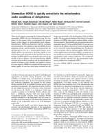
Figure 1
Quantification of aggrecan within the articular cartilage explantsQuantification of aggrecan within the articular cartilage explants. The proteins of the cultured explants were extracted by liquid N
2
pulverization. (a)
Cartilage was extracted immediately after isolation (t = 0) or after culture for 21 days with vehicle, insulin growth factor (IGF), oncostatin M plus
tumour necrosis factor (OSM + TNF), or metabolically inactive (MI) control for assessing passive physiochemical release. (b) Cartilage was
extracted after the three different levels of cytokine treatment followed by an identical 14 days with either vehicle or IGF. IGF significantly stimulated
proteoglycan content within the cartilage explants at all time points. (c) Quantification of sulphated glycosaminoglycan (S-GAG) from all treatments
over the entire experimental period. S-GAG released from cartilage explants to the conditioned medium was quantified by the Alcian blue-binding
assay. The curves represent the release at days when the conditioned medium was fully replaced, and the values were accumulated over the entire
period. MI, metabolically inactive; O + T, oncostatin M plus tumour necrosis factor; W/O, without stimulation (vehicle control). (d) Quantification of
S-GAG turnover 2 weeks after the catabolic induction. The aggrecan release in the identical 14-day period, with or without IGF stimulation following
three different periods of catabolic stimulation, was measured by the Alcian blue-binding assay. The results show the accumulated release of S-
GAG during the 2 weeks with anabolic stimulation (IGF) and without stimulation (vehicle). *P < 0.05, **P < 0.01, ***P < 0.001.
Available online />Page 5 of 12
(page number not for citation purposes)
results in 95% of the total yield of S-GAG. This approach was
specifically chosen as it allows for measurement of the pro-
teins and neo-epitopes. Papain or other digestions destroy
peptide sequences. We detected neither pro-peptides nor
neo-epitopes in normal unstimulated cartilage.
Zymography
MMP-2 and MMP-9 expression and activity were determined
by gelatinase zymography as described previously [6]. This
technique allows for assessment of both pro-enzymes and
active enzymes, which migrate differently according to their
molecular weight during SDS-PAGE electrophoresis. This is
important as all MMPs are synthesized as pro-enzymes, which
then later are activated. The pro-enzyme is not activated under
SDS-PAGE nor preparation but during overnight incubation in
the activation buffer [6,30]. Briefly, 5 μL of the samples was
loaded onto 7.5% SDS-polyacrylamide gels containing 0.5
mg/mL gelatin. After electrophoresis, the gels were incubated
overnight at 37°C in 0.1% Triton X-100, 5 mM CaCl
2
, 1 mM
ZnCl
2
, 3 mM NaN
3
, and 50 mM Tris pH 7.4 in a closed con-
tainer, and then stained with coomassie blue, and finally
destained, dried, and scanned for documentation.
Histology
One cartilage explant from each treatment was taken out of
culture on the appropriate day, fixed in formaldehyde, and
processed for standard histology. Alcian blue was used to
stain the proteoglycans in 5-μm sections. The sections were
stained in a 1% solution of Alcian blue (Sigma-Aldrich) in 3%
acetic acid (pH 2.5) for 30 minutes and rinsed in tap water for
2 minutes, and the nuclei were counterstained with Ehrlich's
hematoxylin. The sections were dehydrated and mounted in
DPX. Digital histographs were captured using an Olympus
BX60 microscope with × 60 magnification and an Olympus
C5050-zoom digital camera (Olympus, Tokyo, Japan).
Statistics
All graphs show one representative experiment of at least
three, each with at least four replicates. Mean values and
standard error of the mean were calculated using GraphPad
Prism (GraphPad Software, Inc., San Diego, CA, USA) and
compared by the Student two-tailed unpaired t test of statisti-
cal significance assuming normal distribution. Asterisks indi-
cate the significance levels (*P < 0.05, **P < 0.01, ***P <
0.001).
Results
OSM and TNF induce cartilage degradation, whereas IGF
induces cartilage formation
A number of studies in different animal species have shown
that OSM and TNF in combination induce cartilage degrada-
tion in vitro, in part through upregulation of both MMP and
aggrecanase activities [6-11]. IGF induces cartilage formation
with regard to both collagen type II and proteoglycan synthesis
[26-28]. To investigate the repair and formation potential of
distinct levels of pathological chondrocytes, we used these
well-described cytokines to induce three different levels of cat-
abolic activity followed by anabolic stimulation. The experi-
ments were designed such that different levels of chondrocyte
catabolism were induced (OSM + TNF) for 7, 11, and 17 days
followed by identical lengths of culture with either IGF or vehi-
cle (14 days) to investigate the capacity for repair.
Anabolic stimulation indicates that cartilage
degradation is completely reversible after short-term
catabolic stimulation
The total content of proteoglycan retained in the articular car-
tilage explants was measured to determine whether aggrecan
lost from the explants during the catabolic phase could be
replaced during the subsequent anabolic phase. As seen in
Figure 1a, IGF treatment increased total S-GAG content by
approximately 125% compared with the vehicle control, in
agreement with previous reports [28,31]. OSM + TNF activa-
tion alone resulted in more than 95% (P < 0.001) depletion of
the proteoglycan content. Interestingly, articular cartilage cul-
tured alone in the absence of cytokine induction lost 50% of
proteoglycan compared with that to the MI control. compared
to the levels of the negative control, metabolic inactive (MI).
Compared with t = 0, the MI control lost 40% (P < 0.001) of
total S-GAG content, suggesting a substantial physical-chem-
ical diffusion from the culture compared with that of the cell-
mediated release when comparing the vehicle with the MI
control.
Chondrocytes in the articular cartilage explants exposed to the
different levels of catabolic treatment responded differently to
IGF treatment. IGF significantly increased the proteoglycan
content in all the catabolically depleted explants (Figure 1b).
Furthermore, we found that anabolic stimulation restored the
S-GAG content in the explants completely when initiated after
7 days of catabolic treatment (comparing Figure 1a vehicle
with Figure 1b IGF-stimulated), whereas at later stages only
incomplete anabolic responses were obtained. These results
indicate that cartilage degradation until day 7 is close to fully
reversible, whereas proteoglycan depletion at days 11 and 17
is less reversible. One important limitation of the extraction
experiments is that extracted S-GAG may be the result of both
newly synthesized proteoglycans and the inhibition of loss of
proteoglycans. However, as presented below, retained prote-
oglycans in the presence of IGF are positioned as circles
around the chondrocytes, suggesting new synthesis, although
this needs to be documented further.
Proteoglycan degradation, in addition to the extraction of pro-
teoglycan from the cartilage plugs, can be measured by S-
GAG release into the conditioned medium, although S-GAG
release is more the result of turnover, in contrast to the MMP-
and aggrecanase-generated neo-epitopes discussed previ-
ously. Figure 1c shows accumulated S-GAG release in the
conditioned medium from all treatments. Stimulation with
Arthritis Research & Therapy Vol 10 No 3 Karsdal et al.
Page 6 of 12
(page number not for citation purposes)
OSM and TNF resulted in substantially increased S-GAG
release until day 7 compared with non-stimulated and MI
explants. However, after the first 7 days of stimulation with
OSM and TNF, there were negligible changes in S-GAG
release, with or without subsequent anabolic stimulation, most
likely because nearly all of the S-GAG was released by day 7.
IGF stimulation, without previous catabolic stimulation,
decreased S-GAG loss into the conditioned medium, consist-
ent with its anabolic actions in cartilage.
To investigate the anabolic potential of chondrocytes following
the catabolic periods, we accumulated the S-GAG levels
released to the conditioned medium for the anabolic periods
(days 7 to 21, 11 to 25, and 17 to 31) (that is, during the 14-
day anabolic period subsequent to the catabolic insult). As
seen in Figure 1d, when the different levels of pathologies
were investigated, only small differences in S-GAG release
were detected, in contrast to the measurements performed on
the cartilage matrix itself.
Collagen type II synthesis can be induced only after
short-term degradation
To further investigate the anabolic response of chondrocytes
to IGF after different levels of catabolic stimuli, we measured
the release of the N-terminal pro-peptide of pro-collagen type
II, PIINP, as a marker of collagen type II synthesis [26]. As
expected, metabolically inactivated explants showed no type II
collagen synthesis, whereas IGF stimulation throughout the
culture period resulted in a 4-fold induction of collagen type II
synthesis compared with the vehicle control (Figure 2a). In
addition, the OSM + TNF-stimulated explants did not synthe-
size or release collagen II pro-peptides.
To investigate the anabolic potential of chondrocytes following
the catabolic periods, we accumulated the PIINP levels
released to the conditioned medium for the anabolic periods
(days 7 to 21, 11 to 25, and 17 to 31) (that, is during the 14-
day anabolic period subsequent to the catabolic insult). After
7 days of catabolic stimulation, collagen type II synthesis
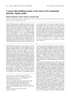
Figure 2
Quantification of pro-peptides of collagen type IIQuantification of pro-peptides of collagen type II. (a) Quantification of collagen type II synthesis from all treatments over the entire experimental
period. Collagen type II synthesis in cartilage explants was measured by the concentration of N-terminal pro-peptides of type II collagen in the condi-
tioned medium using the PIINP enzyme-linked immunosorbent assay (ELISA). The curves represent the release found at the specific day, where the
conditioned medium was fully replaced, and the values were accumulated over the entire period. Vehicle control, metabolically inactive (MI), O + T,
oncostatin M plus tumour necrosis factor. (b) Quantification of collagen type II formation 2 weeks after the catabolic induction. The collagen type II
synthesis in the identical 14-day periods with or without IGF stimulation following the three different periods of catabolic stimulation was measured
by the PIINP ELISA. The conditioned medium was fully replaced three times a week. The results show the accumulated release of collagen type II
pro-peptide during the two weeks with anabolic stimulation (insulin growth factor, IGF) and without stimulation (vehicle). IGF-I significantly induced
collagen type II formation at low and intermediate catabolic insult, but not at maximal insult. *P < 0.05. PIINP, N-terminal pro-peptide of pro-collagen
type II.
Available online />Page 7 of 12
(page number not for citation purposes)
increased in response to IGF treatment. However, we
observed a lower level of IGF-induced collagen II synthesis
after 11 days of cytokine treatment and no IGF-induced colla-
gen II synthesis after 17 days of cytokine treatment (Figure
2b).
Interestingly, under the current culture conditions, the carti-
lage did not lose the IGF-I responsiveness during prolonged
culture periods. When IGF-I was added after 7, 11, or 17 days
of culture, a similar induction of cartilage synthesis was
observed (data not shown). In addition, these data suggest
that cartilage has low levels of continuous collagen type II for-
mation measured by the PIINP assay, however these levels
could potently be stimulated by IGF-I exposure.
To further investigate the amount of PIINP that was retained in
the cartilage compared with that which was released, we
extracted articular cartilage either non-stimulated or stimulated
with either catabolic or anabolic stimulation. We are not able
to detect PIINP under any conditions (data not shown). These
data suggest that n-telo-peptides of pro-collagen type II under
the current culture condition are almost exclusively released
during synthesis and thereby may be valid markers for collagen
type II formation. These data further support our hypothesis
that cartilage loss is reversible if the catabolic stimulation is
short. Similarly, the potential for reversing cartilage degrada-
tion diminishes if cytokine treatment is extensive.
Assessment of aggrecanase- and MMP-mediated
cartilage degradation indicates that loss of repair
mechanisms occurs after induction of MMP activity
To further characterize the molecular mechanism underlying
the loss of repair capacity, we measured levels of the catabolic
biomarkers
374
ARGSV,
342
FFGVG, and CTX-II after the indi-
vidual catabolic treatments. We found that OSM + TNF-stim-
ulated degradation, mediated by aggrecanases and measured
using the
374
ARGSV-G2 assay, was high at day 7, intermedi-
ate at day 10, and almost absent at day 17 (Figure 3a). This is
consistent with the S-GAG release data showing that the
majority of S-GAGs are released at the early stages of cata-
bolic stimulation. The levels of the MMP-generated fragment
342
FFGVG-G2 showed that MMP-mediated aggrecan was
undetectable at days 7 and 10 and high at day 17 (Figure 3b).
The high aggrecanase activity at the early stages of culture
may mask the MMP-mediated aggrecan epitope (
342
FFGVG-
G2) by further processing in generating the aggrecanase
(
374
ARGSV) site; however, Fosang and colleagues [32] have
found that further processing of
342
FFGVG to generate
374
ARGSV cannot occur, at least not in vitro. High levels of
MMP activity should have generated CTX-II fragments that are
not further processed by other proteases, suggesting that
MMP activity is present only at a lower level at early culture
time points. This was verified by the use of a fluorescence sub-
strate technique, in which MMP levels were detectable only in
the presence of catabolic stimulation and only at late time
points (data not shown), which correlate well with previous
findings, documenting extensive MMP activities at later stages
of catabolic induction but not at early stages [6,9,32]. Interest-
ingly, most S-GAG is released at earlier time points than the
342
FFGVG release, indicating that aggrecan loss is due prima-
rily to aggrecanase activity, but later, aggrecanolysis shifts to
an MMP-mediated degradation mode. Finally, we found that
the release of the collagen type II degradation fragment CTX-
II (Figure 3c) occurred with a pattern similar to that of
342
FFGVG (Figure 3b), consistent with the fact that the CTX-
II fragment is MMP-generated [33].
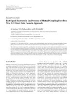
Figure 3
Quantification of aggrecan and collagen degradation products at days 7, 11, and 17Quantification of aggrecan and collagen degradation products at days
7, 11, and 17. Articular cartilage explants were cultured in the presence
or absence of oncostatin M plus tumour necrosis factor (OSM + TNF).
Conditioned medium was collected at days 7, 11, and 17. (a) Aggreca-
nase-mediated aggrecan degradation was measured by the
374
ARGSV-G2 enzyme-linked immunosorbent assay (ELISA), (b) matrix
metalloproteinase (MMP)-mediated aggrecan degradation was quanti-
fied by the
342
FFGVG-G2 ELISA, and (c) MMP-mediated collagen type
II degradation was quantified in the CTX-II ELISA. **P < 0.01, ***P <
0.001. CTX-II, crosslinked C-terminal neo-epitopes of type II collagen.
Arthritis Research & Therapy Vol 10 No 3 Karsdal et al.
Page 8 of 12
(page number not for citation purposes)
In summary, these data appear to mimic cartilage degradation
in arthritis where aggrecanase activity on aggrecan precedes
MMP mediated aggrecan degradation that is subsequently fol-
lowed by MMP degradation of collagen, which has been
reported with various techniques from other labs [32]. In addi-
tion, these data show that there is a positive correlation
between MMP activity (evidenced by the
342
FFGVG-G2 and
CTX biochemical markers) and the inability of cytokine-treated
chondrocytes to initiate and/or maintain anabolic activity.
Switching to anabolic stimulation after short-term
catabolic stimulation can reduce MMP activity
To further investigate the protease levels during anabolic and
catabolic phases of chondrocyte stimulation, we measured
MMP activity by gelatine zymography (Figure 4). The
342
FFGVG-G2 and CTX-II peptides are generated by an array
of MMPs, of which MMP-2 and MMP-9 are only a subset. On
other occasions, the presence of these gelatinases has been
a valid indication of total MMP activity and thereby the cata-
bolic potential of the culture [6]. Gelatinase activity at 7, 11,
and 17 days after catabolic treatment was compared with
gelatinase activity after 7 days of IGF treatment, correspond-
ing to the middle of the anabolic stimulation period. We found
that gelatinase activity and expression were attenuated by IGF,
but not completely reversed, compared with gelatinase activity
after 7 days with vehicle alone (Figure 4). The results with sam-
ples analyzed after 14 days of IGF or vehicle were similar to
those for 7 days of IGF or vehicle (data not shown). The pres-
ence of active MMP-2 and MMP-9 at days 11 to 17 corre-
sponds to the period when high levels of
342
FFGVG-G2 and
CTX-II are detected in Figures 3a and 3c. These data also indi-
cate that, even in the presence of substantial MMP activity,
chondrocytes are able to synthesize new aggrecan and prote-
oglycans (Figure 1b), but not collagen type II (Figure 2b).
Proteoglycan staining confirms the pattern of
reversibility
To visualize the repair enhanced by IGF treatment, cultured
cartilage was harvested at different time points. Proteoglycans
in the cartilage were visualized using Alcian blue staining, the
same dye used in the S-GAG assay. The control articular car-
tilage explants (shown in the bottom row of Figure 5) were cul-
tured for 21 days with vehicle, OSM + TNF, IGF, or MI control
for 21 days. In complete agreement with the S-GAG quantifi-
cations in Figure 2, IGF increased whereas OSM + TNF
decreased GAG content compared with vehicle. MI control
contained more GAG compared with vehicle as the cell-medi-
ated loss of proteoglycan content was abrogated. With regard
to the dynamics in the reversibility experiments presented in
the upper panels, the vehicle control explants gradually lost S-
GAG content over time, whereas the explants treated with IGF
maintained the S-GAGs, even after 17 days in culture. OSM +
TNF treatment depleted proteoglycans from the matrix maxi-
mally by day 7, consistent with the results in Figure 1c which
show that S-GAG release into the medium is also maximal by
day 7. Treatment with IGF stimulated GAG synthesis in the
explants that were treated with cytokines for 7 and 11 days,
but not for 17 days. IGF treatment of explants after 7 days of
catabolic stimuli restored proteoglycan content throughout the
entire cartilage matrix. IGF treatment after 11 days of catabolic
treatment showed new proteoglycan synthesis around
chondrocytes, indicative of repair. There was also evidence of
repair in the absence of IGF treatment after 11 days of cata-
bolic stimuli; however, the repair was substantially improved in
the presence of IGF. Chondrocytes treated with catabolic
cytokines for 17 days reinitiated, only to a very minor extent,
proteoglycan synthesis in the presence of IGF compared with
that of vehicle. These data support the idea that cartilage deg-
radation may be more reversible before induction of MMP
activity.
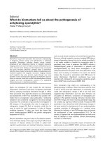
Figure 4
Gelatinase activity is attenuated but not abrogated during insulin growth factor (IGF) stimulationGelatinase activity is attenuated but not abrogated during insulin growth factor (IGF) stimulation. Gelatinase activity in conditioned medium from
bovine articular cartilage explants was investigated by zymography. Lane 1 shows standards for matrix metalloproteinase (MMP)-9 + MMP-2. Condi-
tioned medium at the end of each catabolic culture period (7, 11, or 17 days) was used as a reference (lanes 2 to 4). Conditioned medium from cul-
tures treated with IGF (lanes 8 to 10) or vehicle (lanes 5 to 7) 7 days after the catabolic period was analyzed. Compared with vehicle and baseline
measurements, IGF only attenuated MMP production and activation.
Available online />Page 9 of 12
(page number not for citation purposes)
Discussion
OA is the most common degenerative disease of the joints
[34,35] and this multifactorial and diverse disease is charac-
terized by increased activity of at least two groups of enzymes,
the MMPs and the ADAMTS, which mediate the degradation
of the type II collagen and aggrecan-containing matrix [19].
However, the molecular sequence of events leading to irre-
versible damage and the level of cartilage destruction at which
the damage becomes irreversible remain to be investigated.
With the recent development of assays for the detection of
type II collagen synthesis ex vivo, as well as both MMP- and
aggrecanase-mediated degradation of aggrecan [24], carti-
lage turnover can be assessed in more molecular detail.
By using a combination of OSM and TNF (which is known to
induce pathological degradation [6]) and anabolic stimulation
by IGF (which is a known powerful anabolic growth factor for
chondrocytes [26]), we assessed the anabolic potential of the
three stages of pathologically activated chondrocytes. We
found that once MMP-mediated degradation was in progress,
the capacity for repair was completely lost with regard to col-
lagen type II synthesis, whereas proteoglycan synthesis was
strongly attenuated. In contrast, at the time of maximal aggre-
canase activity, the proteoglycan loss was fully reversible.
These findings correlate well with previous in vivo studies indi-
cating that aggrecan loss was reversible as long as the pro-
gression was not too advanced [2,4,36]. In further support of
these findings are studies in inflammatory arthritis models
which indicated that only low levels of type II collagen degra-
dation could be reversed [2,4,36]. The present data further
support these findings, and demonstrate that even in this sim-
ple ex vivo system, the molecular mechanism of action under-
lying the irreversible degeneration of cartilage involves the
Figure 5
Insulin growth factor (IGF) stimulates local replenishment of cartilageInsulin growth factor (IGF) stimulates local replenishment of cartilage. Articular cartilage explants were cultured with either oncostatin M plus tumour
necrosis factor (OSM + TNF) or vehicle for 7, 11, and 17 days. Subsequently, cartilage explants were paraffin-embedded and stained for aggrecan
content as described in Materials and methods. Aggrecan is completely depleted from the tissue at 7, 11, and 17 days. Other cultures were treated
with either OSM + TNF or vehicle for 7, 11, and 17 days followed by stimulation with either IGF or vehicle control for 14 days. Subsequently, carti-
lage explants were paraffin-embedded and stained for aggrecan content as described in Materials and methods. As a control experiment, articular
cartilage explants were cultured for 21 days with vehicle, OSM + TNF, IGF, or metabolically inactive (MI) control for 21 days as controls (lower
panel). W/O, without stimulation.
Arthritis Research & Therapy Vol 10 No 3 Karsdal et al.
Page 10 of 12
(page number not for citation purposes)
induction of MMP activities, whereas the aggrecanases mainly
are involve in reversible processes.
To examine whether the anabolic growth factor IGF could
affect protease activities, we investigated MMP expression at
the end of the catabolic stimulation and after the anabolic
period (Figure 4). Surprisingly, anabolic induction after the cat-
abolic period did not result in a complete abrogation of MMP
activity, but only a reduction as seen in Figure 4. This suggests
that, even in the presence of increased protease activities,
chondrocytes are able to start making new matrix. The ex vivo
studies presented here indicate that cartilage degradation
may be more reversible than previously thought.
Studies have elucidated that chondrocytes in a series of com-
plicated events involving gene transcription lose their IGF
responsiveness and thereby potentially lose their repair capac-
ity, in part through nitric oxide exposure and upregulation of
SOCS3 (suppressor of cytokine signaling 3) [27,37,38]. This
might contribute in part to the loss of reversibility, as reversibil-
ity in the current studies was investigated as IGF responsive-
ness. The current studies showed complete reversibility after
7 days of cytokine treatment and showed attempted repair
(aggrecan pericellular staining after 14 days), though under
different experimental conditions. Even after extensive cata-
bolic insult, some proteoglycan synthesis was seen when
exposed to IGF-I. Interestingly, the articular cartilage under the
current culture conditions did not lose its IGF-I responsive-
ness. When articular cartilage was exposed to IGF stimulation
at days 7, 11, and 17 in the absence of catabolic stimulation,
similar inductions of PIINP syntheses were observed (data not
shown).
With regard to the possible continuous turnover of collagen
type II and proteoglycans in the articular cartilage matrix, the
current experiments may provide some additional information.
We observed a continuous synthesis of collagen type II even
in non-stimulated conditions (Figure 2a). Thus, these data fur-
ther support the notion that both collagen type II and prote-
oglycans are continuously turned over in the articular matrix,
although the proteoglycan turnover may be superior to that of
the collagen turnover. In the current experiments, this is best
visualized by the nanogram quantities of pro-collagen epitopes
compared with the microgram quantities of S-GAG and the
aggrecanase-generated epitopes of aggrecan, ARGS-G2.
These data are in agreement with those of previous investiga-
tors concluding that aggrecanases are the major mediators of
aggrecan turnover [12,39] and that proteoglycans are remod-
eled to a higher degree compared with that of collagen type II
[40-42].
The current experiments have measured the release of degra-
dation products from the articular matrix as markers of pro-
tease activities. The sequential timing, coordination, individual
roles, and the interactions between MMP and aggrecanase
activities are highly researched topics that are only beginning
to be partly understood. The data do not provide the complete
answer but hopefully add a piece of the highly complicated
puzzle. Most interestingly, aggrecanase-mediated aggrecan
degradation was virtually absent at the end of the study period;
instead, the release of MMP-derived aggrecan fragments was
detected at this time. We have verified that there indeed are
high levels of aggrecanase activity present at later stages of
the culture period (data not shown). The results in Figure 3
suggest that there is a population of aggrecan that is resistant
to aggrecanase cleavage. This population 'survives' high levels
of aggrecanase activity for up 17 days but is then cleaved by
MMPs. This separate pool of aggrecan molecules that have a
different protein degradation profile needs to be investigated
in more molecular detail and may allow for further understand-
ing of the molecular events leading to cartilage destruction.
Many alternative hypotheses and conditions, including but not
limited to the following, need to be investigated: (a) whether
the aggrecanases have been processed, altering their sub-
strate specificity (possibly by MMPs), (b) whether the lack of
aggrecanase-mediated aggrecanolysis is due to limited availa-
bility of aggrecan for ADAMTS-mediated turnover (possibly
due to processing at the cell surface of newly synthesized
aggrecan molecules by membrane-type MMPs), and (c)
whether the extensive aggrecanase activity early in the cul-
tures masks the MMP-generated fragments of aggrecan,
which in theory could be possible as aggrecanase activity
would shed the MMP site from the aggrecan molecule. With
regard to whether aggrecanase activities mask the MMP-gen-
erated extracellular matrix fragments of aggrecan, additional
information may be found in the present data. If aggrecanase
activities should have masked the MMP-mediated activity on
aggrecan as a consequence of high levels of MMP activity, the
MMP-generated collagen type II epitope CTX-II should have
been generated, as the CTX-II epitope is a promiscuous site
generated by most MMPs [6,33]. The absence of both CTX-II
and the MMP-mediated aggrecan fragment at the early culture
days suggests lower levels of MMP activity at these time
points compared with those of later time points. The low level
of MMP activity early in the cultures compared with the exten-
sive activity later under catabolic induction was verified by the
use of a fluorescence substrate (data not shown), which was
in complete agreement with previous findings using other
techniques [6].
This and other studies begin to suggest that OA may be
approached differently depending on the level of disease pro-
gression, in which each stage would require different interven-
tion strategies. Our studies suggest that interventions of OA
by anabolic therapies may be useful. These possible anabolic
strategies should be able, at best, to regenerate cartilage or at
least to replenish lost aggrecan in the articular cartilage. Since
the method developed in this study corresponds well to the sit-
uations seen in vivo, with respect to generation and regenera-
tion of cartilage damage, we speculate that it should be
Available online />Page 11 of 12
(page number not for citation purposes)
implemented for testing the chondro-anabolic effect of differ-
ent drugs.
This is based, in particular, on the fact that damaged cartilage
or cytokine-primed cartilage responds differently than normal
cartilage [43] and has less of an anabolic response [37]. The
latter study indicated that, in particular, the anabolic response
in chondrocytes to IGF was dependent on the cytokine milieu
[27]. Therefore, if OA is diagnosed sufficiently early, more car-
tilage than traditionally thought may be regenerated or pre-
served. The current data indicate that chondrocytes are
responsive to anabolic stimulation even at significant MMP
activity levels and that aggrecanase activities have very little
effect, if any, on the level of repair capacity.
The current study has some important limitations, which
include the use of young bovine cartilage and the use of the
synchronous cultures. The synchronous induction of cartilage
degradation may be different from that seen overall in a
weight-bearing joint, although the same processes are
entailed. Furthermore, the current experiments were con-
ducted under ex vivo conditions, in which cartilage is cultured
under non-weight-bearing conditions, in which cutting of the
cartilage may induce alternative metabolism. This may influ-
ence the cartilage metabolism and thereby allow for skewed
interpretations of the turnover compared with that of the in vivo
conditions.
Our results show that, under the influence of anabolic stimuli,
cartilage explants depleted of aggrecan by aggrecanases can
restore their aggrecan content provided that the catabolic
stimulation has not been too severe. We have yet to explore
the precise mechanism of how this is achieved, but it is likely
to reflect the imbalance between aggrecan synthesis/reten-
tion and aggrecan degradation. The balance is more likely to
be tipped in favour of retention after a short catabolic period
than a long one. When chondrocytes that have received the
short cytokine treatment commence new matrix synthesis, they
might do so more effectively because the cells are 'healthier'
than cells exposed to a long cytokine treatment. The results
show that chondrocytes exposed to both 7 days and 11 days
of catabolic treatment are able to compensate by self-repair,
but the rate at which repair is initiated and then continued is
less in explants receiving the longer treatment. Very little pro-
teoglycan synthesis was possible in explants exposed to the
longest treatment (17 days), suggesting that the exposure of
chondrocytes in these explants was chronic and interfered
substantially with normal chondrocyte function.
Conclusion
We have developed a model and molecular tools that allowed
us to investigate the repair-capacity potential of pathologically
activated chondrocytes. We found that once MMP-mediated
type II collagen and aggrecan degradation was induced, the
reversibility was lost as determined by collagen type II synthe-
sis, whereas proteoglycan synthesis was strongly attenuated.
Interestingly, even in the presence of extensive aggrecanase
activities, cartilage degradation seems completely reversible.
Competing interests
MAK and CC are stockholders of Nordic Bioscience. AJF
declares that she has no competing interests. All other authors
are full-time employees of Nordic Bioscience.
Authors' contributions
MAK designed the study, wrote the manuscript, and partici-
pated in all parts of the experiments. AJF and CC critically
reviewed the manuscript, provided expert advice, and partici-
pated in the drafting of the manuscript. SHM, KH, and BCS
carried out cartilage explants cultures, histology, proteoglycan
extraction, and measurements. All authors read and approved
the final manuscript.
Acknowledgements
The authors thank Eren U Sumer for measuring aggrecanase-mediated
aggrecan assays. No independent funding was obtained for these
studies.
References
1. Abramson SB, Attur M, Yazici Y: Prospects for disease modifi-
cation in osteoarthritis. Nat Clin Pract Rheumatol 2006,
2:304-312.
2. van Meurs JB, van Lent PL, Holthuysen AE, Singer II, Bayne EK,
Berg WB Van den: Kinetics of aggrecanase- and metalloprotei-
nase-induced neoepitopes in various stages of cartilage
destruction in murine arthritis. Arthritis Rheum 1999,
42:1128-1139.
3. Karsdal MA, Henriksen K, Sørensen MG, Gram J, Schaller S, Dzi-
egiel MH, Heegaard AM, Christophersen P, Martin TJ, Christiansen
C, Bollerslev J: Acidification of the osteoclastic resorption com-
partment provides insight into the coupling of bone formation
to bone resorption. Am J Pathol 2005, 166:467-476.
4. Behrens F, Kraft EL, Oegema TR Jr: Biochemical changes in
articular cartilage after joint immobilization by casting or
external fixation. J Orthop Res 1989, 7:335-343.
5. Kiani C, Chen L, Wu YJ, Yee AJ, Yang BB: Structure and function
of aggrecan. Cell Res 2002, 12:19-32.
6. Sondergaard BC, Henriksen K, Wulf H, Oestergaard S, Schurigt
U, Bräuer R, Danielsen I, Christiansen C, Qvist P, Karsdal MA: Rel-
ative contribution of matrix metalloprotease and cysteine pro-
tease activities to cytokine-stimulated articular cartilage
degradation. Osteoarthritis Cartilage 2006, 14:738-748.
7. Hui W, Rowan AD, Richards CD, Cawston TE: Oncostatin M in
combination with tumor necrosis factor alpha induces carti-
lage damage and matrix metalloproteinase expression in vitro
and in vivo. Arthritis Rheum 2003, 48:3404-3418.
8. Cawston TE, Curry VA, Summers CA, Clark IM, Riley GP, Life PF,
Spaull JR, Goldring MB, Koshy PJ, Rowan AD, Shingleton WD:
The role of oncostatin M in animal and human connective tis-
sue collagen turnover and its localization within the rheuma-
toid joint. Arthritis Rheum 1998, 41:1760-1771.
9. Karsdal MA, Sumer EU, Wulf H, Madsen SH, Christiansen C,
Fosang AJ, Sondergaard BC: Induction of increased cAMP lev-
els in articular chondrocytes blocks matrix metalloproteinase-
mediated cartilage degradation, but not aggrecanase-medi-
ated cartilage degradation. Arthritis Rheum 2007,
56:1549-1558.
10. Caterson B, Flannery CR, Hughes CE, Little CB: Mechanisms
involved in cartilage proteoglycan catabolism.
Matrix Biol
2000, 19:333-344.
11. Little CB, Flannery CR, Hughes CE, Mort JS, Roughley PJ, Dent C,
Caterson B: Aggrecanase versus matrix metalloproteinases in
Arthritis Research & Therapy Vol 10 No 3 Karsdal et al.
Page 12 of 12
(page number not for citation purposes)
the catabolism of the interglobular domain of aggrecan in
vitro. Biochem J 1999, 344(Pt 1):61-68.
12. Glasson SS, Askew R, Sheppard B, Carito B, Blanchet T, Ma HL,
Flannery CR, Peluso D, Kanki K, Yang Z, Majumdar MK, Morris EA:
Deletion of active ADAMTS5 prevents cartilage degradation in
a murine model of osteoarthritis. Nature 2005, 434:644-648.
13. Little CB, Mittaz L, Belluoccio D, Rogerson FM, Campbell IK,
Meeker CT, Bateman JF, Pritchard MA, Fosang AJ: ADAMTS-1-
knockout mice do not exhibit abnormalities in aggrecan turn-
over in vitro or in vivo. Arthritis Rheum 2005, 52:1461-1472.
14. Stanton H, Rogerson FM, East CJ, Golub SB, Lawlor KE, Meeker
CT, Little CB, Last K, Farmer PJ, Campbell IK, Fourie AM, Fosang
AJ: ADAMTS5 is the major aggrecanase in mouse cartilage in
vivo and in vitro. Nature 2005, 434:648-652.
15. Little CB, Meeker CT, Hembry RM, Sims NA, Lawlor KE, Golub
SB, Last K, Fosang AJ: Matrix metalloproteinases are not
essential for aggrecan turnover during normal skeletal growth
and development. Mol Cell Biol 2005, 25:3388-3399.
16. Collins-Racie LA, Flannery CR, Zeng W, Corcoran C, Annis-Free-
man B, Agostino MJ, Arai M, DiBlasio-Smith E, Dorner AJ, Georgi-
adis KE, Jin M, Tan XY, Morris EA, LaVallie ER: ADAMTS-8
exhibits aggrecanase activity and is expressed in human artic-
ular cartilage. Matrix Biol 2004, 23:219-230.
17. Mort JS, Flannery CR, Makkerh J, Krupa JC, Lee ER: Use of anti-
neoepitope antibodies for the analysis of degradative events
in cartilage and the molecular basis for neoepitope specificity.
Biochem Soc Symp 2003:107-114.
18. Tang BL: ADAMTS: a novel family of extracellular matrix
proteases. Int J Biochem Cell Biol 2001, 33:33-44.
19. Schaller S, Henriksen K, Hoegh-Andersen P, Søndergaard BC,
Sumer EU, Tanko LB, Qvist P, Karsdal MA: In vitro, ex vivo, and in
vivo methodological approaches for studying therapeutic tar-
gets of osteoporosis and degenerative joint diseases: how
biomarkers can assist? Assay Drug Dev Technol
2005,
3:553-580.
20. Oestergaard S, Sondergaard BC, Hoegh-Andersen P, Henriksen
K, Qvist P, Christiansen C, Tankó LB, Karsdal MA: Effects of ova-
riectomy and estrogen therapy on type II collagen degradation
and structural integrity of articular cartilage in rats: implica-
tions of the time of initiation. Arthritis Rheum 2006,
54:2441-2451.
21. Billinghurst RC, Wu W, Ionescu M, Reiner A, Dahlberg L, Chen J,
van Wart H, Poole AR: Comparison of the degradation of type
II collagen and proteoglycan in nasal and articular cartilages
induced by interleukin-1 and the selective inhibition of type II
collagen cleavage by collagenase. Arthritis Rheum 2000,
43:664-672.
22. Billinghurst RC, Dahlberg L, Ionescu M, Reiner A, Bourne R,
Rorabeck C, Mitchell P, Hambor J, Diekmann O, Tschesche H,
Chen J, Van Wart H, Poole AR: Enhanced cleavage of type II col-
lagen by collagenases in osteoarthritic articular cartilage. J
Clin Invest 1997, 99:1534-1545.
23. Fosang AJ, Neame PJ, Last K, Hardingham TE, Murphy G, Hamil-
ton JA: The interglobular domain of cartilage aggrecan is
cleaved by PUMP, gelatinases, and cathepsin B. J Biol Chem
1992, 267:19470-19474.
24. Sumer EU, Sondergaard BC, Rousseau JC, Delmas PD, Fosang
AJ, Karsdal MA, Christiansen C, Qvist P: MMP and non-MMP-
mediated release of aggrecan and its fragments from articular
cartilage: a comparative study of three different aggrecan and
glycosaminoglycan assays. Osteoarthritis Cartilage 2007,
15:212-221.
25. Ishikawa T, Nishigaki F, Christgau S, Noto T, Mo J, From N, Min-
oura K, Hirayama Y, Ohkubo Y, Mutoh S: Cartilage destruction in
collagen induced arthritis assessed with a new biochemical
marker for collagen type II C-telopeptide fragments. J
Rheumatol 2004, 31:1174-1179.
26. Olsen AK, Sondergaard BC, Byrjalsen I, Tanko LB, Christiansen C,
Müller A, Hein GE, Karsdal MA, Qvist P: Anabolic and catabolic
function of chondrocyte ex vivo is reflected by the metabolic
processing of type II collagen. Osteoarthritis Cartilage 2007,
15:335-342.
27. Studer RK, Levicoff E, Georgescu H, Miller L, Jaffurs D, Evans CH:
Nitric oxide inhibits chondrocyte response to IGF-I: inhibition
of IGF-IRbeta tyrosine phosphorylation. Am J Physiol Cell
Physiol 2000, 279:C961-C969.
28. Curtis AJ, Devenish RJ, Handley CJ: Modulation of aggrecan and
link-protein synthesis in articular cartilage.
Biochem J 1992,
288(Pt 3):721-726.
29. Fosang AJ, Last K, Gardiner P, Jackson DC, Brown L: Develop-
ment of a cleavage-site-specific monoclonal antibody for
detecting metalloproteinase-derived aggrecan fragments:
detection of fragments in human synovial fluids. Biochem J
1995, 310(Pt 1):337-343.
30. Kleiner DE, Stetler-Stevenson WG: Quantitative zymography:
detection of picogram quantities of gelatinases. Anal Biochem
1994, 218:325-329.
31. McQuillan DJ, Handley CJ, Campbell MA, Bolis S, Milway VE, Her-
ington AC: Stimulation of proteoglycan biosynthesis by serum
and insulin-like growth factor-I in cultured bovine articular
cartilage. Biochem J 1986, 240:423-430.
32. Fosang AJ, Last K, Stanton H, Weeks DB, Campbell IK, Hard-
ingham TE, Hembry RM: Generation and novel distribution of
matrix metalloproteinase-derived aggrecan fragments in por-
cine cartilage explants. J Biol Chem 2000, 275:33027-33037.
33. Oestergaard S, Chouinard L, Doyle N, Karsdal MA, Smith SY,
Qvist P, Tankó LB: The utility of measuring C-terminal telopep-
tides of collagen type II (CTX-II) in serum and synovial fluid
samples for estimation of articular cartilage status in experi-
mental models of destructive joint diseases. Osteoarthritis
Cartilage 2006, 14:670-679.
34. Elders MJ: The increasing impact of arthritis on public health.
J Rheumatol Suppl 2000, 60:6-8.
35. Wieland HA, Michaelis M, Kirschbaum BJ, Rudolphi KA: Osteoar-
thritis – an untreatable disease? Nat Rev Drug Discov 2005,
4:331-344.
36. Stoop R, Kraan PM van der, Buma P, Hollander AP, Poole AR,
Berg WB Van den: Denaturation of type II collagen in articular
cartilage in experimental murine arthritis. Evidence for colla-
gen degradation in both reversible and irreversible cartilage
damage. J Pathol 1999, 188:329-337.
37. Smeets RL, Veenbergen S, Arntz OJ, Bennink MB, Joosten LA,
Berg WB Van den, Loo FA van de: A novel role for suppressor
of cytokine signaling 3 in cartilage destruction via induction of
chondrocyte desensitization toward insulin-like growth factor.
Arthritis Rheum 2006, 54:1518-1528.
38. Loo FA van de, Arntz OJ, van Enckevort FH, van Lent PL, Berg WB
Van den: Reduced cartilage proteoglycan loss during
zymosan-induced gonarthritis in NOS2-deficient mice and in
anti-interleukin-1-treated wild-type mice with unabated joint
inflammation. Arthritis Rheum 1998, 41:634-646.
39. Glasson SS, Askew R, Sheppard B, Carito BA, Blanchet T, Ma HL,
Flannery CR, Kanki K, Wang E, Peluso D, Yang Z, Majumdar MK,
Morris EA: Characterization of and osteoarthritis susceptibility
in ADAMTS-4-knockout mice. Arthritis Rheum 2004,
50:2547-2558.
40. Lark MW, Bayne EK, Flanagan J, Harper CF, Hoerrner LA, Hutch-
inson NI, Singer II, Donatelli SA, Weidner JR, Williams HR, Mum-
ford RA, Lohmander LS: Aggrecan degradation in human
cartilage. Evidence for both matrix metalloproteinase and
aggrecanase activity in normal, osteoarthritic, and rheumatoid
joints. J Clin Invest 1997, 100:93-106.
41. Lohmander LS, Atley LM, Pietka TA, Eyre DR: The release of
crosslinked peptides from type II collagen into human synovial
fluid is increased soon after joint injury and in osteoarthritis.
Arthritis Rheum 2003, 48:3130-3139.
42. Struglics A, Larsson S, Pratta MA, Kumar S, Lark MW, Lohmander
LS: Human osteoarthritis synovial fluid and joint cartilage con-
tain both aggrecanase- and matrix metalloproteinase-gener-
ated aggrecan fragments. Osteoarthritis Cartilage 2006,
14:101-113.
43. Fan Z, Bau B, Yang H, Soeder S, Aigner T: Freshly isolated oste-
oarthritic chondrocytes are catabolically more active than nor-
mal chondrocytes, but less responsive to catabolic stimulation
with interleukin-1beta. Arthritis Rheum 2005, 52:136-143.