Báo cáo y học: " High avidity autoreactive T cells with a low signalling capacity through the T-cell receptor: central to rheumatoid arthritis pathogenesis" pot
Bạn đang xem bản rút gọn của tài liệu. Xem và tải ngay bản đầy đủ của tài liệu tại đây (688.35 KB, 9 trang )
Page 1 of 9
(page number not for citation purposes)
Available online />Abstract
Self-reactive T cells with low signalling capacity through the T-cell
receptor were recently observed in the SKG mouse model of
rheumatoid arthritis (RA) and have been linked to a spontaneous
mutation in the ZAP-70 signal transduction molecule. Here we
hypothesize that similar mechanisms also drive RA, associated with
an abnormal innate and adaptive immune response driven by
nuclear factor-κB activation and tumour necrosis factor secretion.
Similar to the essential role played by pathogens in SKG mice, we
propose that HLA-associated immunity to chronic viral infection is
a key factor in the immune dysregulation and joint inflammation that
characterize RA.
Introduction
In 1996, Thomas and Lipsky [1] proposed a model for
rheumatoid arthritis (RA) pathogenesis in which endogenous
self-antigens were presented by activated peripheral
dendritic cells (DCs) to autoreactive T cells that had escaped
thymic selection. Synovial DCs were shown to be activated,
probably as a consequence of proinflammatory signals
derived from the RA joint environment, including cytokines
and T-cell derived CD40 ligand [1,2]. The model stemmed
from observations that autologous peripheral blood T cells
proliferated strongly in vitro in response to RA synovial DCs
presenting endogenous antigenic peptide (known as the
autologous mixed lymphocyte response). At that time it was
unclear how T cells with the capacity to respond strongly to
self-antigen might escape thymic deletion and enter the
peripheral repertoire. However, the subsequent discovery by
Sakaguchi and colleagues [3] of a spontaneous mouse
mutant, known as ‘SKG’, which developed inflammatory
arthritis resembling RA, has provided a possible mechanism.
Thymic selection and the predisposition to
autoimmunity
Central (or thymic) tolerance defects are important and
probably essential contributors to spontaneous autoimmune
disease [4]. T cells are selected in the thymus according to
their affinity for self-MHC (major histocompatibility complex)
bearing endogenous self-antigens displayed by the thymic
cortical epithelial cells. Negative selection then deletes those
T cells that are reactive to self-antigen above a threshold of
affinity for self-antigen/MHC complexes expressed and
presented by medullary antigen-presenting cells (APCs),
notably medullary epithelial cells and medullary DCs [5].
In the medulla, medullary epithelial cells express the highest
levels of autoimmune regulator (AIRE), a transcription factor
that controls the expression of peripheral tissue antigens. In
the absence of AIRE, glandular (salivary and lacrimal glands,
liver, pancreas and thyroid) organ-specific autoimmunity
develops [6]. Interestingly, neither mice nor humans with
AIRE mutations develop autoimmune arthritis, possibly
because AIRE does not directly regulate the expression of
joint-specific self-proteins in the thymus.
Medullary DCs have also been shown to delete self-reactive T
cells in the thymus in experimental settings [7], but
abnormalities in these cells have not yet been implicated in
any spontaneous autoimmune model. Although the spectrum
of self-antigen presentation by medullary DCs is unknown,
they can capture antigen from peripheral tissues - presumably
including synovial joints - and delete self-antigen-specific
thymocytes in the medulla.
Review
High avidity autoreactive T cells with a low signalling capacity
through the T-cell receptor: central to rheumatoid arthritis
pathogenesis?
Ranjeny Thomas
1
, Malcolm Turner
1
and Andrew P Cope
2
1
Diamantina Institute for Cancer, Immunology and Metabolic Medicine, University of Queensland, Princess Alexandra Hospital, Brisbane, Queensland,
4102, Australia
2
The Kennedy Institute of Rheumatology, Faculty of Medicine, Imperial College, 1 Aspenlea Road, Hammersmith, London W6 8LH, UK
Corresponding author: Ranjeny Thomas,
Published: 24 July 2008 Arthritis Research & Therapy 2008, 10:210 (doi:10.1186/ar2446)
This article is online at />© 2008 BioMed Central Ltd
ACPA = antibody to citrullinated proteins; AIRE = autoimmune regulator; APC = antigen-presenting cell; CTL = cytotoxic T lymphocyte; DC =
dendritic cell; EBV = Epstein-Barr virus; HA = haemagglutin antigen; HLA = human leucocyte antigen; IFN = interferon; IL = interleukin; LPS =
lipopolysaccharide; MHC = major histocompatibility complex; NF-κB = nuclear factor-κB; RA = rheumatoid arthritis; RF = rheumatoid factor; SNP =
single nucleotide polymorphism; TCR = T-cell receptor; TLR = Toll-like receptor; TNF = tumour necrosis factor; ZAP-70 = ζ-associated protein of
70 kDa.
Page 2 of 9
(page number not for citation purposes)
Arthritis Research & Therapy Vol 10 No 4 Thomas et al.
Although an affinity threshold applies for central deletion of
self-reactive T cells, this threshold varies according to the
susceptibility of thymocytes to death and the capacity of the
T-cell receptor (TCR) and downstream pathways to transmit
an activation signal. Moreover, the efficiency of self-antigen
presentation depends on the ability of thymic APCs to
process and present self-antigen, and the density of MHC
and co-stimulatory molecules on the APC surface.
A number of well established spontaneous animal models of
autoimmunity are characterized by defects in the normal
process of either positive or negative selection, thus
permitting the entry of autoreactive T cells into the peripheral
repertoire. In the periphery, subsequent genetic or environ-
mental proinflammatory events more readily trigger the activa-
tion of these T cells, and thus the development of auto-
immune disease [8]. Does this scenario fit the SKG RA
model or human RA itself?
TCR signalling is dramatically attenuated in the SKG mouse
model of spontaneous arthritis. This is due to a mutation in
the SH2 domain of the gene encoding ζ-associated protein
of 70 kDa (ZAP-70), a TCR proximal protein tyrosine kinase
that is essential for T-cell activation after the TCR engages
antigen [3]. Experiments using TCR transgenic mice show
that high-affinity self-reactive T cells escape negative
selection in these mice. At the same time, defective TCR
signalling also attenuates positive selection, reducing the
peripheral T-cell pool compared with wild-type mice (Figure 1).
The abnormal peripheral T-cell repertoire, comprising a higher
proportion of self-reactive T cells than in wild-type mice, is
demonstrable ex vivo, because peripheral SKG T cells
incubated with autologous APC proliferate vigourously in
spite of the ZAP-70 mutation, and secrete IL-17 in the autolo-
gous mixed lymphocyte response [9]. SKG mice develop
spontaneous rheumatoid factor (RF)-positive inflammatory
arthritis, resembling RA in patients, when housed in a conven-
tional animal facility where environmental pathogen exposure
might occur at low levels. Conversely, in a microbiologically
clean facility, mice do not develop joint disease, although RF
and other autoantibodies are still detectable [3,9].
In an elegant follow-up study, Sakaguchi and coworkers [9]
showed that subclinical fungal infection is predominantly
responsible for the inflammatory signals that drive spon-
taneous joint disease in SKG mice. β-Glucan molecules
derived from the fungal cell wall signal through the dectin-1
cell surface C-type lectin receptor on the cell surface of
antigen-presenting DCs. Reis e Sousa and colleagues [10]
demonstrated that signalling of murine DCs though the
dectin-1 receptor promotes the secretion of proinflammatory
cytokines, including IL-6, tumour necrosis factor (TNF) and IL-
23, but little IL-12. In SKG mice, such DCs activated by
dectin-1 promote the in vitro and in vivo differentiation of
CD4
+
T-effector cells secreting IL-17 [9]. Lymphopenia may
be an important contributor to the self-reactive response in
this case because it promotes homeostatic proliferation of
effector T cells, similar to that demonstrated in other auto-
immune models [11,12].
T-cell phenotype and function
CD4
+
SKG T cells in the periphery exhibit a phenotype
characteristic of antigen-experienced, post-activated cells, as
are typically observed in autoimmune arthritis. There are
increased proportions of CD44
hi
, CD25
+
, CD69
+
, OX40
+
and CD45RB
dim
cells, as compared with the proportions in
wild-type BALB/c littermates [3]. When adoptively transferred
to lymphopenic hosts, SKG T cells proliferate just as
efficiently as wild-type T cells [9]. Although both SKG and
wild-type T-cell subsets produce similar proportions of
T-helper-17 and T-helper-1 effectors under these conditions,
SKG T cells are more strongly self-reactive than wild-type
T cells [9].
Another murine model of spontaneous inflammatory arthritis
that fits this paradigm was reported very recently. In the F
1
progeny of BALB/c mice containing both haemagglutin
antigen (HA)-specific TCR-transgenic CD4
+
T cells and HA
driven by a MHC class II-specific promoter (known as
TS1×HACII mice) [13], high-affinity HA-specific T cells are
negatively selected in the thymus, but low-affinity HA-specific
T cells bearing low levels of cell surface TCR expand in the
periphery over time. Similar to SKG T cells, these CD4
+
T cells exhibit a post-activated memory phenotype, with low
proliferative capacity but high capacity for cytokine produc-
tion in response to antigen stimulation ex vivo. The mice
develop a T-cell-dependent and B-cell-independent peripheral
arthritis, pneumonitis and cardiac inflammation from around
6 weeks of age, with a gradual progression in severity. The
disease phenotype is similar to other spontaneous arthritis
models (but unlike autoimmune models in which AIRE is
deficient), which lack endocrine or glandular multi-organ
inflammatory pathology.
It is striking that autoantigen-experienced memory CD4
+
cells
with low TCR signalling capacity are particularly associated
with autoimmune arthritis. However, the relative joint
specificity arising from immunity toward an antigen whose
expression is not joint restricted is puzzling. We speculate
that the capacity of such T cells to secrete relevant cytokines
(including IFN-γ, IL-17 and TNF [13]), in concert with tissue-
specific homing properties, might underlie the induction of
arthritis. The extent to which joint stromal cells (including
synovial fibroblasts) are exquisitely sensitive to cytokine
stimulation, as compared with stromal cells from other
tissues, remains a matter of debate.
In RA, antigen-experienced synovial T cells, with a similar
CD45RB
dim
phenotype to SKG T cells, have an acquired
TCR signalling deficiency. We previously showed that
synovial T cells proliferated poorly and secreted low levels of
IL-2 in vitro [14]. The reduced T-cell proliferation seen in RA
Page 3 of 9
(page number not for citation purposes)
is also associated with reduced TCR signal intensity, reduced
calcium signalling and reduced expression of TCR-ζ. It has
been shown that TCR-ζ chains are either not expressed or
lack phosphorylation in RA synovial fluid T cells. TCR-ζ chain
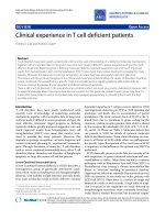
Available online />Figure 1
Pathogenesis of inflammatory arthritis. (a) The SKG model and (b) a model for rheumatoid arthritis (RA) suggested by the skg mouse. As a result
of altered thymic selection, the peripheral T-cell repertoire responds to self-antigen with higher affinity compared with the healthy situation,
facilitating self-specific activation and population of the periphery with post-activated memory T cells. These T cells produce proinflammatory
cytokines and provide efficient help for autoantibody production, but they have limited capacity for infection control. Antigen-presenting dendritic
cells (DCs) are activated directly by fungal β-glucans (panel a) or indirectly through T cells or proinflammatory cytokines (panels a and b). ACPA,
antibodies to citrullinated protein; CTL, cytotoxic T lymphocyte; EBV, Epstein-Barr virus; IFN, interferon; IL, interleukin; RF, rheumatoid factor; TCR,
T-cell receptor; TNF, tumour necrosis factor; WT, wild-type.
expression levels correlated with RA T-cell responsiveness
[15]. We previously defined populations of TCR-ζ
dim
T cells
in peripheral blood with characteristics of prior antigen
experience, based on cell surface phenotype, cytokine
expression and migratory competence [16]. In chronic inflam-
matory diseases (for example, RA and systemic lupus erythe-
matosus) it has been proposed that an inflammatory milieu
contributes to reduction in TCR-ζ expression in antigen-
experienced T cells. Inflammatory factors that could contribute
to this process in predisposed individuals include nutrient
depletion, increased expression of reactive oxygen inter-
mediates such as H
2
O
2
, and induction of stress pathways [17].
Genetic, acquired and age-related factors could thus contri-
bute to a state of chronic TCR signalling deficiency in RA.
In contrast, IFN-γ and IL-17 production by RA T cells appears
to be spared [16,18,19]. In addition, synovial T cells potently
induce B cells to secrete autoantibodies [14] and activate
synovial macrophages, DCs and resident stromal cells. These
cells, in turn, express inflammatory cytokines and chemokines
through cell contact-dependent mechanisms [20]. Thus, in
spite of their TCR signalling deficiencies, synovial T cells can
promote chronic inflammation within the synovial lesion,
stimulating B cells, and promoting macrophage and DC
activation and robust secretion of cytokines. Beyond these
acquired signalling defects, is there any evidence that low
TCR signalling capacity might precede RA?
Genetic provocation of autoreactive T cells
with low TCR signalling capacity
The primary genetic defect in the SKG autoimmune arthritic
mouse model is a point mutation in the TCR proximal protein
tyrosine kinase ZAP-70. This mutation does not alter ZAP-70
expression, but nevertheless it dramatically reduces the
affinity of the carboxyl-terminal SH2 domain of ZAP-70 in
binding phosphorylated tyrosine residues in the immuno-
receptor tyrosine-based activation motif (ITAM) modules of
the TCR-ζ chain [3]. This mutation can therefore entirely
account for the thymic selection shift and the generation of a
repertoire of autoreactive T cells with a high avidity for self-
antigen/MHC complexes in SKG mice. However, the
question arises as to whether there are similar (or functionally
related) mutations in RA.
To date, no allelic variants of the human ZAP70 gene have
been described in association with RA or in association with
any other known immune-mediated inflammatory disease. In
contrast, attention has recently focused on elucidating the
function of the PTPN22 gene that encodes a protein tyrosine
phosphatase called LYP (lymphocyte tyrosine phosphatase)
[21]. The R620W variant of this gene is, somewhat
unexpectedly, a gain-of-function mutant that reduces TCR
signalling capacity. Functional data from healthy donors
homozygous or heterozygous for the R620W mutation
confirm that peripheral blood T cells are hyporesponsive to
antigen receptor stimulation. This polymorphism would thus
be predicted to impair positive and negative selection of
autoreactive T cells [22,23]. Within the context of SKG and
RA T cells, it is interesting that carriage of the variant allele
was also associated with reduced IL-10 production and an
increase in the numbers of CD4
+
memory T cells, potentially
associated with increased self-reactivity. Expression of TNF-α
and IFN-γ was unaffected [23]. As a result of altered thymic
selection, this phenomenon might arise through increased
intrinsic responsiveness and augmented generation of effector
T cells that recognize endogenous self-peptides presented
by APCs in vivo. A complementary possibility is that gain-of-
function PTPN22 mutants suppress TCR signalling in natural
regulatory T cells and thus impair peripheral tolerance. RA
has also been associated with single nucleotide poly-
morphisms (SNPs) in the MHC class II transactivator gene
(MHC2TA). These SNPs are predicted to reduce the
efficiency of self-antigen presentation by APCs in the thymus
and periphery, with effects on the T-cell repertoire similar to
those associated with PTPN22 R620W [24]. These altera-
tions in the repertoire of healthy individuals with PTPN22
R620W suggest that a low TCR signalling capacity may
predispose otherwise healthy individuals to RA, just as SKG
mice are predisposed to (but do not develop) arthritis in the
absence of infection.
Presentation of self-antigen to autoreactive
T cells promoting rheumatoid arthritis
depends on activation of dendritic cells
Activated DCs play several roles in autoimmune arthritis. They
serve as APCs for T-cell priming, as accessory cells in the
generation of primary antibody responses, and as producers
of proinflammatory cytokines (alongside synoviocytes and
macrophages) [25-27]. DCs infiltrate inflamed tissue, take up
and process antigen locally, and then activate MHC-restric-
ted naïve T cells in draining lymph nodes [1,27-30]. In turn,
autoreactive primed T cells co-stimulate DC activation par-
ticularly through CD40 ligand, reinforcing the autoimmune
response that eventually leads to excessive autoantibody
production and chronic inflammation associated with RA [2].
DCs are activated by the uptake of immunogenic antigen,
pathogen and damage recognition ligands, a role played - at
least in part - by fungal β-glucan signalling through dectin-1 in
SKG mice [31-33]. Proinflammatory cytokines also activate
DCs, although evidence is emerging that the gene activation
programme is in this instance different from that activated by
pathogen or lipopolysaccharide (LPS) [34]. Are DCs
activated in RA, how does this come about, and how do high-
avidity autoreactive T cells respond?
SKG, TS1×HACII mice and RA DCs and macrophages share
a capacity for ‘hyper-activation’. This activation is enhanced
by strong positive feedback from post-activated memory T cells,
by immune complex ligation of Fc receptors and by
proinflammatory cytokines [9,13]. DCs and macrophages
from the synovial fluid of RA patients exhibit an unusual and
persistent drive for LPS-induced nuclear factor-κB (NF-κB)
Arthritis Research & Therapy Vol 10 No 4 Thomas et al.
Page 4 of 9
(page number not for citation purposes)
activation ex vivo [35,36], apparently in the face of strong
signals for exhaustion and counter-regulation that would
normally halt activation [37,38]. This hyper-activation
contrasts with monocytes and DCs isolated from patients
with type 1 diabetes, which we have shown shut down
NF-κB in response to LPS [39]. Although it has only been
technically feasible to examine peripheral blood DCs from
patients with diabetes, when we compared peripheral blood
DCs from RA patients we did not find a similar exhausted
response to LPS in RA [39]. In a murine model, a Toll-like
receptor (TLR)4-mediated signalling pathway blocked TLR
ligand responsiveness and promoted an exhausted pheno-
type. In the absence of TLR4 signalling, DCs exposed to
proinflammatory cytokines in vivo could be further activated
ex vivo by other TLR ligands [34]. Although the mechanism
distinguishing the responsiveness of RA and diabetes DCs to
LPS is not yet clear, the implication is that DCs would
present antigen more efficiently in the face of infection or
other proinflammatory events in RA, whereas they would be
less effective in response to the same stimuli in diabetes. DC
hyperactivity appears to be characteristic of the pathogenesis
of autoimmune arthritis in both RA and the described murine
models.
MHC-peptide interactions with T cells in RA
Variation in the HLA-DRB1 gene of the MHC is more strongly
associated with RA than variation in any other locus. The
variation maps to the third hypervariable region of the DRβ-
chain and is found in many different human leucocyte antigen
(HLA)-DR molecules linked to RA [40]. The locus encodes a
conserved susceptibility sequence - known as the ‘shared
epitope’ - that is positively charged and forms the fourth
anchoring pocket (P4) in the HLA-DR peptide binding groove
[41]. Antibodies to citrullinated proteins (ACPAs) and RF are
more likely in RA patients with the shared epitope and who
smoke [42-44]. Thus, it has been proposed, in view of
evidence that smoking promotes citrullination of self-proteins
in the lung, that smoking promotes ACPAs in those with at-
risk HLA genotypes [43]. We found that peripheral blood
T cells from patients with RA susceptibility HLA-DR alleles
and ACPAs proliferated poorly in response to specific shared
epitope-associated citrullinated peptides, consistent with low
signal capacity through the TCR. However, the T cells
strongly induced proinflammatory cytokine secretion in
response to these peptides as well as the native form of
these epitopes. Surprisingly, these responses occurred at
very low concentrations of peptide, suggestive of high-affinity
anti-self-responses (Capini C and coworkers, unpublished
data). We therefore propose that subsets of self-reactive
T cells that interact with high-avidity with peptide-MHC may
compensate for attenuated TCR signalling, which is
consistent with our ex vivo observations that T cells from RA
patients respond with high avidity to citrullinated and
noncitrullinated self-antigens. Expression of CD70 by
antigen-experienced T cells may be at least one mechanism
by which antigen-specific responses may be augmented [45].
This ongoing autoreactivity would result in the contraction of
the T-cell repertoire and highly selective expansion of self-
reactive T-cell clones.
Chronic inflammation and the tumour
necrosis factor/nuclear factor-
κκ
B drive in
rheumatoid arthritis
Based on human and animal data, what are the key factors
that drive chronic inflammation in RA? Experiments in
different animal arthritic models, including TNF transgenic
mice, and IL-1 receptor antagonist knockout and p50 knock-
out mice, indicate that proinflammatory stimuli driving the
expression of TNF, IL-1, or NF-κB p50 are sufficient to drive
the development of autoimmune polyarthritis in susceptible
strains [46-49]. NF-κB stimulates the transcription of genes
important for cellular responses to stress, injury and
inflammation [50], and thus NF-κB signalling simultaneously
sustains synovial inflammation and promotes DC and
monocyte activation and differentiation, resulting in priming of
autoreactive lymphocytes. We and others have provided
additional evidence that TNF and IL-1 directly enhance B-cell
and T-cell autoreactivity through effects on regulatory T cells
[51-53]. Nicotine, lactation, mineral oil exposure and Epstein-
Barr virus (EBV) - environmental factors associated with RA -
all promote NF-κB activity, associated with TNF and IL-1
secretion by myeloid and stromal cells, and DC and B-cell
activation [54-57].
On the other hand, combinations of disease-modifying anti-
rheumatic drugs and biologic therapies that suppress the
activity of NF-κB can induce RA remission [58,59]. Thus,
both human and murine evidence indicates that NF-κB
activation is required to drive RA, and that factors that
suppress this activity are disease suppressive [48,60,61].
TNF clearly plays a critical role in RA perpetuation, activating
and being activated by NF-κB in a positive feedback loop.
Genetic and environmental provocation of
strong activation of innate immunity and
antigen presentation
There are links between RA and NF-κB driven genes of the
innate immune response involved in pathogen recognition,
proinflammatory cytokine production and modulation of the
strength of cellular signalling in response to activation
signals. RA-associated SNPs have been detected in
complement-5-TRAF1, STAT 4 and in DCIR, another lectin
receptor that is expressed on the surface of DCs [62-65].
Identification of these SNPs has potential implications for the
way in which we assess the impact of environmental RA risk
factors - such as infection and tobacco smoke - in individuals
genetically predisposed to RA. Apart from direct cellular
effects, tissue damage caused by tobacco smoke or infection
also provoke the release of endogenous pathogen recog-
nition receptor ligands derived from host cellular debris (also
known as damage-associated molecular patterns or DAMPs).
These have been shown to function as auto-adjuvants, which
Available online />Page 5 of 9
(page number not for citation purposes)
both perpetuate and reinforce the inflammatory response and
stimulate the APC function of DCs.
The role of viral pathogens in driving nuclear
factor-
κκ
B
EBV, which infects about 98% of the world’s population, has
the strongest viral association with RA [66,67]. Almost all the
arthritogenic viruses, including EBV, rubella, parvovirus B19,
hepatitis B and C, HIV, HTLV1 and Ross River Fever, activate
NF-κB in order to replicate, suggesting the possibility that
arthritis develops as a side-effect of NF-κB activation. These
viruses manipulate the NF-κB pathway to enhance their
replication and host cell survival, while blocking apoptosis
and immune recognition [68]. The EBV latent membrane
protein-1 activates NF-κB through interaction with TNF
receptor 1 and the TNF receptor 1-associated death domain.
Activation bypasses the cytoplasmic TNF signalling pathway
[68]. NF-κB activation by EBV allows it to evade the normal
host responses and leads to a persistent low-grade B-cell
infection. EBV DNA has been detected in synovial tissue from
RA patients, using polymerase chain reaction, in situ hybridi-
zation and immunohistochemical staining [69]. EBV latent
membrane protein-1 has also been demonstrated in RA
synoviocytes and lymphocytes. The EBV Epstein-Barr nuclear
antigen (EBNA)-1 protein also undergoes citrullination. Thus,
EBV can induce antibodies to citrullinated peptides [70,71].
The EBV capsid protein gp110 also contains the shared
epitope sequence [72]. The evidence suggests there is a
deficiency in viral control coincident with RA, which is
consistent with a host immunodeficient state. In RA patients,
there are increased numbers of EBV-infected B lymphocytes,
higher specific antibody titres, and impaired EBV-specific
cytotoxic T lymphocyte (CTL) activity, as compared with
otherwise healthy EBV-infected individuals [73,74].
We propose that simultaneous NF-κB stimulation by viral
infection and RA results in a ‘mutually permissive’ state, with
viral infection promoting RA disease, and vice versa, through
NF-κB. The key question is whether patients at risk for RA are
also at greater risk for immune dysregulation during EBV
infection. For us, the evidence is in favour. Hijacking of B
lymphocyte cellular machinery by EBV promotes chronic dys-
regulated immune activation with increased NF-κB activity,
and the propensity both for B-cell autoantibody secretion and
lymphoma development [69]. Because EBV infection
activates the NF-κB pathway in B lymphocytes, they are
prone to apoptotic cell death in response to NF-κB inhibition
during RA treatment [75]. Furthermore, in those predisposed
to RA, EBV infection may persist through a state of relative
immunodeficiency imposed by attenuated TCR signals,
reducing the efficacy of EBV-specific CTLs. Functional CTLs
are essential for effective control of EBV-associated lympho-
proliferative disease in post-transplant settings [76]. This
immune dysregulation associated with failure of normal T-cell-
mediated infection control in RA might explain how RA
inflammatory disease can appear T-cell independent, as
indicated by poor clinical responses to T-cell-depleting
therapies. On the other hand, strategies such as CTLA4-Ig
(CTL antigen 4-immunoglobulin), which specifically target a
T-cell-dependent pathway, are effective because they
probably confer desirable immuno-regulation on the multiple
sites of T-cell action.
Synthesis: similarity and differences in
pathogenesis of arthritis in SKG mice and RA
Pathogenic T cells from both SKG and TS1×HACII mice and
RA patients appear to share the following characteristics: a
reduced capacity for TCR signalling; increased proportions of
T cells with a post-activated differentiated memory pheno-
type; a reduced capacity for proliferation and IL-2 production,
despite their capacity for IL-17 and IFN-γ secretion;
enhanced B-cell help and a strong capacity for autoantibody
production; and an enhanced response to self-antigens.
Figure 1 depicts models of disease pathogenesis in SKG
mice and RA patients, highlighting their similarities and some
differences.
Clearly, in the SKG model it is easier to ascertain that low
TCR signalling capacity underlies arthritis development. In
RA, although we have argued that secondary TCR signalling
deficiencies provide a positive feedback loop for
inflammation, it will be of interest to determine whether similar
TCR signalling deficiencies precede inflammatory disease, for
instance whether they are evident in otherwise healthy
individuals who are ACPA positive and at risk for RA. Further
evidence could be obtained from patients achieving drug-free
remission from chronic inflammation, such as after allogeneic
stem cell transplantation. Although we have argued that
infection plays a role in SKG mice and RA patients, the
nature of this role appears to be different in each setting, with
more direct inflammatory signalling of DCs in SKG mice.
Indeed, we believe that if infectious or TLR-mediated damage
signals are involved in driving DC and macrophage activation
in RA, as appears to be the case in SKG mice, then the usual
counter-regulatory response to TLR activation must be
attenuated. The development of arthritis in TS1×HACII mice
even in a microbiologically clean facility [13] indicates that
infectious signals are not required to drive arthritis within the
context of autoantigenic T cells with reduced TCR signalling
capacity. We propose that arthritis in this model develops
independent of a pathogen drive because of the very high
precursor frequency of autoantigen-specific T cells. In
contrast, the reduced frequency of T cells specific for arthrito-
genic autoantigen among the polyclonal T-cell repertoire in
the SKG mice, or indeed in RA, is less likely to provide suf-
ficient feedback to DCs to drive spontaneous inflammation.
In RA, we propose that infection is intimately associated with
the HLA susceptibility locus. Shared epitope alleles are
common in the Caucasian population but they are strongly
associated with RA, along with the development of both RF
and ACPAs, and with severe erosive clinical disease. Why
Arthritis Research & Therapy Vol 10 No 4 Thomas et al.
Page 6 of 9
(page number not for citation purposes)
does shared epitope-associated RA persist at a frequency of
around 1% in the population? We propose that the HLA
susceptibility illuminates a bigger picture than the unfortunate
side effect of joint autoimmunity. The polymorphic HLA genes
evolved as a result of selection pressure by infection, and the
shared epitope alleles thus identify individuals with particular
immunity to infection. Our hypothesis is that EBV infection
sets up a particularly ‘cosy’ symbiotic relationship with hosts
bearing HLA susceptibility alleles and primary TCR signalling
deficiency. As a result of EBV infection, persistent presen-
tation of viral antigens could impose pressure on the T-cell
repertoire, contributing with self-antigen presentation to drive
expansion of an activated memory population, which further
acquires inflammation-associated TCR signalling defects.
This phenomenon may underlie the observed thymic and
bone marrow stem cell deficiency, excessive production of
CD28
null
and other post-activated, terminally differentiated
memory T cell phenotypes, hyper-activated DCs and B cells,
and excess numbers of EBV-associated lymphomas and
other tumours in RA patients [77]. Indeed, when synovial fluid
T cells from RA patients were analyzed using EBV MHC
class I tetramers, they were found to contain a high propor-
tion of virus-specific T cells with an activated phenotype [78].
As might have been predicted, it was the differentiated
CD8
+
CD28
null
T-cell population that could be isolated from
RA patients after stimulation with immunodominant lytic
peptide EBV epitopes [79]. It is likely that EBV is not the only
infection to result in a mutually permissive state of auto-
reactivity in RA. Other examples include the increased
probability of RF production in patients with chronic HCV or
with ageing, because the T-cell repertoire is progressively
populated with a higher proportion of post-activated memory
T cells, creating a positive feedback loop as TCR signalling
capacity decreases.
Conclusion
Although the SKG mouse model is by no means identical to
human RA, it does mirror aspects of pathogenesis relating to
gene-environment interactions that are involved in promoting
autoimmune arthritis. This forces us to confront the paradox
of how T cells with low TCR signalling capacity nevertheless
interact with APCs and thus play initiating and continuing
roles in the generation of autoimmune inflammation in RA
patients. An improved understanding of the primary
pathogenetic mechanisms of T cells in RA will probably have
important implications for the design of effective and safe
immunotherapies.
Competing interests
The authors declare that they have no competing interests.
Acknowledgements
We thank Caetano Reis e Sousa (funded by Cancer Research UK) for
helpful discussions, and William Burns and Ian Frazer (both funded by
University of Queensland) for critical reading of the manuscript.
Ranjeny Thomas is supported by Arthritis Queensland and Andrew
Cope by Wellcome Trust UK and the Arthritis Research Campaign UK.
References
1. Thomas R, Lipsky PE: Could endogenous self-peptides pre-
sented by dendritic cells initiate rheumatoid arthritis?
Immunol Today 1996, 17:559-564.
2. MacDonald KPA, Nishioka N, Lipsky PE, Thomas R: Functional
CD40-ligand is expressed by T cells in rheumatoid arthritis. J
Clin Invest 1997, 100:2404-2414.
3. Sakaguchi N, Takahashi T, Hata H, Nomura T, Tagami T, Yamazaki
S, Sakihama T, Matsutani T, Negishi I, Nakatsuru S, Sakaguchi S:
Altered thymic T-cell selection due to a mutation of the ZAP-
70 gene causes autoimmune arthritis in mice. Nature 2003,
426:454-460.
4. Ardavin C: Thymic dendritic cells. Immunol Today 1997, 18:
350-361.
5. Kappler JW, Roehm N, Marrack P: T cell tolerance by clonal
elimination in the thymus. Cell 1987, 49:273-280.
6. Mathis D, Benoist C: A decade of AIRE. Nat Rev Immunol 2007,
7:645-650.
7. Bonasio R, Scimone ML, Schaerli P, Grabie N, Lichtman AH, von
Andrian UH: Clonal deletion of thymocytes by circulating den-
dritic cells homing to the thymus. Nat Immunol 2006, 7:1092-
1100.
8. Yoshitomi H, Sakaguchi N, Kobayashi K, Brown GD, Tagami T,
Sakihama T, Hirota K, Tanaka S, Nomura T, Miki I, Gordon S, Akira
S, Nakamura T, Sakaguchi S: A role for fungal {beta}-glucans
and their receptor Dectin-1 in the induction of autoimmune
arthritis in genetically susceptible mice. J Exp Med 2005, 201:
949-960.
9. Hirota K, Hashimoto M, Yoshitomi H, Tanaka S, Nomura T, Yam-
aguchi T, Iwakura Y, Sakaguchi N, Sakaguchi S: T cell self-reac-
tivity forms a cytokine milieu for spontaneous development of
IL-17
+
Th cells that cause autoimmune arthritis. J Exp Med
2007, 204:41-47.
10. LeibundGut-Landmann S, Gross O, Robinson MJ, Osorio F, Slack
EC, Tsoni SV, Schweighoffer E, Tybulewicz V, Brown GD, Ruland
J, Reis e Sousa C: Syk- and CARD9-dependent coupling of
innate immunity to the induction of T helper cells that
produce interleukin 17. Nat Immunol 2007, 8:630-638.
11. Koh WP, Chan E, Scott K, McCaughan G, France M, Fazekas de
St Groth B: TCR-mediated involvement of CD4
+
transgenic T
cells in spontaneous inflammatory bowel disease in lym-
phopenic mice. J Immunol 1999, 162:7208-7216.
12. Cozzo C, Larkin J, 3rd, Caton AJ: Self-peptides drive the periph-
eral expansion of CD4
+
CD25
+
regulatory T cells. J Immunol
2003, 171:5678-5682.
13. Rankin AL, Reed AJ, Oh S, Cozzo Picca C, Guay HM, Larkin J
3rd, Panarey L, Aitken MK, Koeberlein B, Lipsky PE, Tomaszewski
JE, Naji A, Caton AJ: CD4
+
T cells recognizing a single self-
peptide expressed by APCs induce spontaneous autoimmune
arthritis. J Immunol 2008, 180:833-841.
14. Thomas R, McIlraith M, Davis LS, Lipsky PE: Rheumatoid syn-
ovium is enriched in CD45RBdim mature memory T cells that
are potent helpers for B cell differentiation. Arthritis Rheum
1992, 35:1455-1465.
15. Romagnoli P, Strahan D, Pelosi M, Cantagrel A, van Meerwijk JP:
A potential role for protein tyrosine kinase p56(lck) in
rheumatoid arthritis synovial fluid T lymphocyte hyporespon-
siveness. Int Immunol 2001, 13:305-312.
16. Zhang Z, Gorman CL, Vermi AC, Monaco C, Foey A, Owen S,
Amjadi P, Vallance A, McClinton C, Marelli-Berg F, Isomäki P,
Russell A, Dazzi F, Vyse TJ, Brennan FM, Cope AP: TCRzetadim
lymphocytes define populations of circulating effector cells
that migrate to inflamed tissues. Blood 2007, 109:4328-4335.
17. Zhang Z, Gorman C, Clark JM, Cope AP: Rheumatoid arthritis: a
disease of chronic, low-amplitude signals transduced through
T cell antigen receptors? Wien Med Wochenschr 2006, 156:2-
10.
18. Allen ME, Young SP, Michell RH, Bacon PA: Altered T lympho-
cyte signaling in rheumatoid arthritis. Eur J Immunol 1995, 25:
1547-1554.
19. Maurice MM, Lankester AC, Bezemer AC, Geertsma MF, Tak PP,
Breedveld FC, van Lier RA, Verweij CL: Defective TCR-mediated
signaling in synovial T cells in rheumatoid arthritis. J Immunol
1997, 159:2973-2978.
20. Dayer JM, Burger D: Cytokines and direct cell contact in syn-
ovitis: relevance to therapeutic intervention. Arthritis Res
1999, 1:17-20.
Available online />Page 7 of 9
(page number not for citation purposes)
21. Bottini N, Vang T, Cucca F, Mustelin T: Role of PTPN22 in type 1
diabetes and other autoimmune diseases. Semin Immunol
2006, 18:207-213.
22. Vang T, Congia M, Macis MD, Musumeci L, Orrú V, Zavattari P,
Nika K, Tautz L, Taskén K, Cucca F, Mustelin T, Bottini N: Autoim-
mune-associated lymphoid tyrosine phosphatase is a gain-of-
function variant. Nat Genet 2005, 37:1317-1319.
23. Rieck M, Arechiga A, Onengut-Gumuscu S, Greenbaum C, Con-
cannon P, Buckner JH: Genetic variation in PTPN22 corre-
sponds to altered function of T and B lymphocytes. J Immunol
2007, 179:4704-4710.
24. Swanberg M, Lidman O, Padyukov L, Eriksson P, Akesson E,
Jagodic M, Lobell A, Khademi M, Börjesson O, Lindgren CM,
Lundman P, Brookes AJ, Kere J, Luthman H, Alfredsson L, Hillert J,
Klareskog L, Hamsten A, Piehl F, Olsson T: MHC2TA is associ-
ated with differential MHC molecule expression and suscepti-
bility to rheumatoid arthritis, multiple sclerosis and
myocardial infarction. Nat Genet 2005, 37:486-494.
25. Kitamura H, Iwakabe K, Yahata T, Nishimura S, Ohta A, Ohmi Y,
Sato M, Takeda K, Okumura K, Van Kaer L, Kawano T, Taniguchi
M, Nishimura T: The natural killer T (NKT) cell ligand alpha-
galactosylceramide demonstrates its immunopotentiating
effect by inducing interleukin (IL)-12 production by dendritic
cells and IL-12 receptor expression on NKT cells. J Exp Med
1999, 189:1121-1128.
26. Cavanagh LL, Boyce A, Smith L, Padmanabha J, Filgueira L,
Pietschmann P, Thomas R: Rheumatoid arthritis synovium con-
tains plasmacytoid dendritic cells. Arthritis Res Ther 2005, 7:
R230-R240.
27. Leung BP, Conacher M, Hunter D, McInnes IB, Liew FY, Brewer
JM: A novel dendritic cell-induced model of erosive inflamma-
tory arthritis: distinct roles for dendritic cells in T cell activa-
tion and induction of local inflammation. J Immunol 2002, 169:
7071-7077.
28. Thomas R, Davis LS, Lipsky PE: Rheumatoid synovium is
enriched in mature antigen-presenting dendritic cells. J
Immunol 1994, 152:2613-2623.
29. Dittel BN, Visintin I, Merchant RM, Janeway CA Jr: Presentation
of the self antigen myelin basic protein by dendritic cells
leads to experimental autoimmune encephalomyelitis. J
Immunol 1999, 163:32-39.
30. Ludewig B, Odermatt B, Landmann S, Hengartner H, Zinkernagel
RM: Dendritic cells induce autoimmune diabetes and maintain
disease via de novo formation of local lymphoid tissue. J Exp
Med 1998, 188:1493-1501.
31. Sallusto F, Lanzavecchia A: Understanding dendritic cell and T-
lymphocyte traffic through the analysis of chemokine recep-
tor expression. Immunol Rev 2000, 177:134-140.
32. Caux C, Massacrier C, Vanbervliet B, Dubois B, van Kooten C,
Durand I, Banchereau J: Activation of human dendritic cells
through CD40 cross-linking. J Exp Med 1994, 180:1263-1272.
33. O’Sullivan BJ, Thomas R: CD40 Ligation conditions dendritic
cell antigen-presenting function through sustained activation
of NF-kappaB. J Immunol 2002, 168:5491-5498.
34. Nolte MA, Leibundgut-Landmann S, Joffre O, Reis e Sousa C:
Dendritic cell quiescence during systemic inflammation driven
by LPS stimulation of radioresistant cells in vivo. J Exp Med
2007, 204:1487-1501.
35. Pettit AR, MacDonald KPA, O’Sullivan B, Thomas R: Differenti-
ated dendritic cells expressing nuclear RelB are predomi-
nantly located in rheumatoid synovial tissue perivascular
mononuclear cell aggregates. Arthritis Rheum 2000, 43:791-
800.
36. Huang Q, Ma Y, Adebayo A, Pope RM: Increased macrophage
activation mediated through toll-like receptors in rheumatoid
arthritis. Arthritis Rheum 2007, 56:2192-2201.
37. Yoza BK, Hu JY, Cousart SL, Forrest LM, McCall CE: Induction
of RelB participates in endotoxin tolerance. J Immunol 2006,
177:4080-4085.
38. Napolitani G, Rinaldi A, Bertoni F, Sallusto F, Lanzavecchia A:
Selected Toll-like receptor agonist combinations synergisti-
cally trigger a T helper type 1-polarizing program in dendritic
cells. Nat Immunol 2005, 6:769-776.
39. Mollah ZUA, Pai S, Moore C, O’Sullivan BJ, Harrison MJ, Peng J,
Phillips K, Prins JB, Cardinal J, Thomas R: Abnormal NF-kappa B
function characterizes human type 1 diabetes dendritic cells
and monocytes. J Immunol 2008, 180:3166-3175.
40. du Montcel ST, Michou L, Petit-Teixeira E, Osorio J, Lemaire I,
Lasbleiz S, Pierlot C, Quillet P, Bardin T, Prum B, Cornelis F,
Clerget-Darpoux F: New classification of HLA-DRB1 alleles
supports the shared epitope hypothesis of rheumatoid arthri-
tis susceptibility. Arthritis Rheum 2005, 52:1063-1068.
41. Gregersen PK, Silver J, Winchester RJ: The shared epitope
hypothesis: an approach to understanding the molecular
genetics of suseptibility to rheumatoid arthritis. Arthritis
Rheum 1987, 30:1205-1213.
42. Silman AJ, Newman J, MacGregor AJ: Cigarette smoking
increases the risk of rheumatoid arthritis. Results from a
nationwide study of disease-discordant twins. Arthritis Rheum
1996, 39:732-735.
43. Klareskog L, Stolt P, Lundberg K, Källberg H, Bengtsson C,
Grunewald J, Rönnelid J, Harris HE, Ulfgren AK, Rantapää-
Dahlqvist S, Eklund A, Padyukov L, Alfredsson L: A new model
for an etiology of rheumatoid arthritis: smoking may trigger
HLA-DR (shared epitope)-restricted immune reactions to
autoantigens modified by citrullination. Arthritis Rheum 2006,
54:38-46.
44. Padyukov L, Silva C, Stolt P, Alfredsson L, Klareskog L: A gene-
environment interaction between smoking and shared
epitope genes in HLA-DR provides a high risk of seropositive
rheumatoid arthritis. Arthritis Rheum 2004, 50:3085-3092.
45. Lee WW, Yang ZZ, Li G, Weyand CM, Goronzy JJ: Unchecked
CD70 expression on T cells lowers threshold for T cell activa-
tion in rheumatoid arthritis. J Immunol 2007, 179:2609-2615.
46. Keffer J, Probert L, Cazlaris H, Georgopoulos S, Kaslaris E, Kious-
sis D, Kollias G: Transgenic mice expressing human tumour
necrosis factor: a predictive genetic model of arthritis. EMBO
J 1991, 10:4025-4031.
47. Horai R, Saijo S, Tanioka H, Nakae S, Sudo K, Okahara A, Ikuse T,
Asano M, Iwakura Y: Development of chronic inflammatory
arthropathy resembling rheumatoid arthritis in interleukin 1
receptor antagonist-deficient mice. J Exp Med 2000, 191:313-
320.
48. Campbell IK, Gerondakis S, O’Donnell K, Wicks IP: Distinct roles
for the NF-kappaB1 (p50) and c-Rel transcription factors in
inflammatory arthritis. J Clin Invest 2000, 105:1799-1806.
49. Tak PP, Gerlag DM, Aupperle KR, van de Geest DA, Overbeek M,
Bennett BL, Boyle DL, Manning AM, Firestein GS: Inhibitor of
nuclear factor kappaB kinase beta is a key regulator of syn-
ovial inflammation. Arthritis Rheum 2001, 44:1897-1907.
50. O’Sullivan B, Thompson AG, Thomas R: NF-kappa B as a thera-
peutic target in autoimmune disease. Curr Opin Ther Targets
2007, 11:111-122.
51. O’Sullivan B, Thomas HE, Pai S, Santamaria P, Iwakura Y,
Steptoe RJ, Kay TW, Thomas R: IL-1 breaks tolerance through
expansion of CD25
+
effector T cells. J Immunol 2006, 176:
7278-7287.
52. Nakae S, Asano M, Horai R, Sakaguchi N, Iwakura Y: IL-1
enhances T cell-dependent antibody production through
induction of CD40 ligand and OX40 on T cells. J Immunol
2001, 167:90-97.
53. Ehrenstein MR, Evans JG, Singh A, Moore S, Warnes G, Isenberg
DA, Mauri C: Compromised function of regulatory T cells in
rheumatoid arthritis and reversal by anti-TNFalpha therapy. J
Exp Med 2004, 200:277-285.
54. Izumi KM, Kieff ED: The Epstein-Barr virus oncogene product
latent membrane protein 1 engages the tumor necrosis factor
receptor-associated death domain protein to mediate B lym-
phocyte growth transformation and activate NF-kappaB. Proc
Natl Acad Sci USA 1997, 94:12592-12597.
55. Yang SR, Chida AS, Bauter MR, Shafiq N, Seweryniak K, Maggir-
war SB, Kilty I, Rahman I: Cigarette smoke induces proinflam-
matory cytokine release by activation of NF-kappaB and
posttranslational modifications of histone deacetylase in
macrophages. Am J Physiol Lung Cell Mol Physiol 2006, 291:
L46-L57.
56. Brand JM, Frohn C, Cziupka K, Brockmann C, Kirchner H, Luhm J:
Prolactin triggers pro-inflammatory immune responses in
peripheral immune cells. Eur Cytokine Netw 2004, 15:99-104.
57. Pai S, O’Sullivan B, Abdul-Jabbar I, Peng J, Connoly G, Khanna R,
Thomas R: Nasopharyngeal carcinoma-associated Epstein-
Barr virus-encoded oncogene latent membrane protein 1
potentiates regulatory T-cell function. Immunol Cell Biol 2007,
85:370-377.
Arthritis Research & Therapy Vol 10 No 4 Thomas et al.
Page 8 of 9
(page number not for citation purposes)
58. Quinn MA, Conaghan PG, O’Connor PJ, Karim Z, Greenstein A,
Brown A, Brown C, Fraser A, Jarret S, Emery P: Very early treat-
ment with infliximab in addition to methotrexate in early,
poor-prognosis rheumatoid arthritis reduces magnetic reso-
nance imaging evidence of synovitis and damage, with sus-
tained benefit after infliximab withdrawal: results from a
twelve-month randomized, double-blind, placebo-controlled
trial. Arthritis Rheum 2005, 52:27-35.
59. Palanki MS: Inhibitors of AP-1 and NF-kappa B mediated tran-
scriptional activation: therapeutic potential in autoimmune
diseases and structural diversity. Curr Med Chem 2002, 9:
219-227.
60. Foxwell B, Browne K, Bondeson J, Clarke C, de Martin R, Brennan
F, Feldmann M: Efficient adenoviral infection with IkappaB
alpha reveals that macrophage tumor necrosis factor alpha
production in rheumatoid arthritis is NF-kappaB dependent.
Proc Natl Acad Sci USA 1998, 95:8211-8215.
61. Tomita T, Takeuchi E, Tomita N, Morishita R, Kaneko M,
Yamamoto K, Nakase T, Seki H, Kato K, Kaneda Y, Ochi T: Sup-
pressed severity of collagen-induced arthritis by in vivo trans-
fection of nuclear factor kappaB decoy oligodeoxynucleotides
as a gene therapy. Arthritis Rheum 1999, 42:2532-2542.
62. Robinson MJ, Sancho D, Slack EC, LeibundGut-Landmann S,
Reis e Sousa C: Myeloid C-type lectins in innate immunity. Nat
Immunol 2006, 7:1258-1265.
63. Remmers EF, Plenge RM, Lee AT, Graham RR, Hom G, Behrens
TW, de Bakker PI, Le JM, Lee HS, Batliwalla F, Li W, Masters SL,
Booty MG, Carulli JP, Padyukov L, Alfredsson L, Klareskog L,
Chen WV, Amos CI, Criswell LA, Seldin MF, Kastner DL,
Gregersen PK: STAT4 and the risk of rheumatoid arthritis and
systemic lupus erythematosus. N Engl J Med 2007, 357:977-
986.
64. Plenge RM, Seielstad M, Padyukov L, Lee AT, Remmers EF, Ding
B, Liew A, Khalili H, Chandrasekaran A, Davies LR, Li W, Tan AK,
Bonnard C, Ong RT, Thalamuthu A, Pettersson S, Liu C, Tian C,
Chen WV, Carulli JP, Beckman EM, Altshuler D, Alfredsson L,
Criswell LA, Amos CI, Seldin MF, Kastner DL, Klareskog L,
Gregersen PK: TRAF1-C5 as a risk locus for rheumatoid arthri-
tis: a genomewide study. N Engl J Med 2007, 357:1199-1209.
65. Lorentzen JC, Flornes L, Eklöw C, Bäckdahl L, Ribbhammar U,
Guo JP, Smolnikova M, Dissen E, Seddighzadeh M, Brookes AJ,
Alfredsson L, Klareskog L, Padyukov L, Fossum S: Association of
arthritis with a gene complex encoding C-type lectin-like
receptors. Arthritis Rheum 2007, 56:2620-2632.
66. Balandraud N, Meynard JB, Auger I, Sovran H, Mugnier B, Reviron
D, Roudier J, Roudier C: Epstein-Barr virus load in the periph-
eral blood of patients with rheumatoid arthritis: accurate
quantification using real-time polymerase chain reaction.
Arthritis Rheum 2003, 48:1223-1228.
67. Balandraud N, Roudier J, Roudier C: Epstein-Barr virus and
rheumatoid arthritis. Autoimmun Rev 2004, 3:362-367.
68. Hiscott J, Kwon H, Genin P: Hostile takeovers: viral appropria-
tion of the NF-kappaB pathway. J Clin Invest 2001, 107:143-
151.
69. Toussirot E, Roudier J: Pathophysiological links between
rheumatoid arthritis and the Epstein-Barr virus: an update.
Joint Bone Spine 2007, 74:418-426.
70. Pratesi F, Tommasi C, Anzilotti C, Chimenti D, Migliorini P: Deimi-
nated Epstein-Barr virus nuclear antigen 1 is a target of anti-
citrullinated protein antibodies in rheumatoid arthritis. Arthritis
Rheum 2006, 54:733-741.
71. Anzilotti C, Riente L, Pratesi F, Chimenti D, Delle Sedie A, Bom-
bardieri S, Migliorini P: IgG, IgA, IgM antibodies to a viral citrul-
linated peptide in patients affected by rheumatoid arthritis,
chronic arthritides and connective tissue disorders. Rheuma-
tology (Oxford) 2007, 46:1579-1582.
72. Roudier J, Petersen J, Rhodes GH, Luka J, Carson DA: Suscepti-
bility to rheumatoid arthritis maps to a T-cell epitope shared
by the HLA-Dw4 DR b-1 chain and the Ebstein-Barr virus gly-
coprotein gp110. Proc Natl Acad Sci USA 1989, 86:5104-
5108.
73. Sawada S, Takei M: Epstein-Barr virus etiology in rheumatoid
synovitis. Autoimmun Rev 2005, 4:106-110.
74. Gaston JS, Rickinson AB, Yao QY, Epstein MA: The abnormal
cytotoxic T cell response to Epstein-Barr virus in rheumatoid
arthritis is correlated with disease activity and occurs in other
arthropathies. Ann Rheum Dis 1986, 45:932-936.
75. Cahir-McFarland ED, Carter K, Rosenwald A, Giltnane JM, Hen-
rickson SE, Staudt LM, Kieff E: Role of NF-kappa B in cell sur-
vival and transcription of latent membrane protein
1-expressing or Epstein-Barr virus latency III-infected cells. J
Virol 2004, 78:4108-4119.
76. Khanna R, Bell S, Sherritt M, Galbraith A, Burrows SR, Rafter L,
Clarke B, Slaughter R, Falk MC, Douglass J, Williams T, Elliott SL,
Moss DJ: Activation and adoptive transfer of Epstein-Barr
virus-specific cytotoxic T cells in solid organ transplant
patients with posttransplant lymphoproliferative disease. Proc
Natl Acad Sci USA 1999, 96:10391-10396.
77. Weyand CM, Goronzy JJ, Kurtin PJ: Lymphoma in rheumatoid
arthritis: an immune system set up for failure. Arthritis Rheum
2006, 54:685-689.
78. Tan LC, Mowat AG, Fazou C, Rostron T, Roskell H, Dunbar PR,
Tournay C, Romagné F, Peyrat MA, Houssaint E, Bonneville M,
Rickinson AB, McMichael AJ, Callan MF: Specificity of T cells in
synovial fluid: high frequencies of CD8
+
T cells that are spe-
cific for certain viral epitopes. Arthritis Res 2000, 2:154-164.
79. Klatt T, Ouyang Q, Flad T, Koetter I, Buhring HJ, Kalbacher H,
Pawelec G, Muller CA: Expansion of peripheral CD8
+
CD28
-
T
cells in response to Epstein-Barr virus in patients with
rheumatoid arthritis. J Rheumatol 2005, 32:239-251.
Available online />Page 9 of 9
(page number not for citation purposes)