Báo cáo y học: "Novel splice variants derived from the receptor tyrosine kinase superfamily are potential therapeutics for rheumatoid arthritis" pdf
Bạn đang xem bản rút gọn của tài liệu. Xem và tải ngay bản đầy đủ của tài liệu tại đây (1.83 MB, 16 trang )
Open Access
Available online />Page 1 of 16
(page number not for citation purposes)
Vol 10 No 4
Research article
Novel splice variants derived from the receptor tyrosine kinase
superfamily are potential therapeutics for rheumatoid arthritis
Pei Jin
1
, Juan Zhang
1
, Percy F Sumariwalla
2
, Irene Ni
1
, Brett Jorgensen
1
, Damian Crawford
2
,
Suzanne Phillips
3
, Marc Feldmann
2
, H Michael Shepard
1
and Ewa M Paleolog
2
1
Receptor BioLogix, Inc., Palo Alto, CA 94303, USA
2
Kennedy Institute of Rheumatology, Faculty of Medicine, Imperial College London, London W6 8LH, UK
3
Gentris Corporation, Morrisville, NC 27560, USA
Corresponding author: Pei Jin,
Received: 13 May 2008 Revisions requested: 9 Jun 2008 Revisions received: 25 Jun 2008 Accepted: 1 Jul 2008 Published: 1 Jul 2008
Arthritis Research & Therapy 2008, 10:R73 (doi:10.1186/ar2447)
This article is online at: />© 2008 Jin et al.; licensee BioMed Central Ltd.
This is an open access article distributed under the terms of the Creative Commons Attribution License ( />),
which permits unrestricted use, distribution, and reproduction in any medium, provided the original work is properly cited.
Abstract
Introduction Despite the advent of biological therapies for the
treatment of rheumatoid arthritis, there is a compelling need to
develop alternative therapeutic targets for nonresponders to
existing treatments. Soluble receptors occur naturally in vivo,
such as the splice variant of the cell surface receptor for
vascular endothelial growth factor (VEGF) – a key regulator of
angiogenesis in rheumatoid arthritis. Bioinformatics analyses
predict that the majority of human genes undergo alternative
splicing, generating proteins – many of which may have
regulatory functions. The objective of the present study was to
identify alternative splice variants (ASV) from cell surface
receptor genes, and to determine whether the novel proteins
encoded exert therapeutic activity in an in vivo model of arthritis.
Methods To identify novel splice variants, we performed RT-
PCR using an mRNA pool representing major human tissue
types and tumors. Novel ASV were identified by alignment of
each cloned sequence to its respective genomic sequence in
comparison with full-length transcripts. To test whether these
ASV have biologic activity, we characterized a subset of them
for ligand binding, and for efficacy in an animal model of arthritis.
The in vivo study was accomplished using adenoviruses
expressing secreted ASV.
Results We cloned 60 novel human ASV from 21 genes,
encoding cell surface receptors – many of which are known to
be important in the regulation of angiogenesis. The ASV were
characterized by exon extension, intron retention and alternative
exon utilization. Efficient expression and secretion of selected
ASV – corresponding to VEGF receptor type 1, VEGF receptor
type 2, VEGF receptor type 3, angiopoietin receptor Tie1, Met
(receptor for hepatocyte growth factor), colony-stimulating
factor 1 receptor, platelet-derived growth factor receptor beta,
fibroblast growth factor receptor 1, Kit, and RAGE – was
demonstrated, together with binding to their cognate ligands.
Importantly, ASV derived from VEGF receptor type 1 and Tie1,
and to a lesser extent from VEGF receptor type 2 and fibroblast
growth factor receptor 1, reduced clinical signs of arthritis in
vivo. The reduction was paralleled by decreased joint
inflammation and destruction.
Conclusion The present study shows that unique ASV derived
from receptors that play key roles in angiogenesis – namely,
VEGF receptor type 1 and, for the first time, Tie1 – can markedly
reduce arthritis severity. More broadly, our results demonstrate
that ASV are a source of novel proteins with therapeutic
potential in diseases in which angiogenesis and cellular
hyperplasia play a central role, such as rheumatoid arthritis.
Introduction
Rheumatoid arthritis (RA) has a prevalence of about 1% in
most parts of the world. While targeting TNFα using biological
inhibitors has been an undoubted success, efficacy does not
usually approach remission. Moreover, increasing usage of
anti-TNFα biological agents in RA is associated with an aug-
mented risk of infections, including tuberculosis [1-5]. As a
consequence, initiatives to develop alternative targets in RA
Adv = adenovirus; Ang-1 = angiopoietin-1; ASV = alternative splice variants; CIA = collagen-induced arthritis; CSF = colony-stimulating factor; Fc =
crystallizable fragment; FGFR = fibroblast growth factor receptor; H & E = hematoxylin and eosin; HUVEC = human umbilical vein endothelial cells;
PBS = phosphate-buffered saline; PCR = polymerase chain reaction; PDGF = platelet-derived growth factor; PDGFRβ = platelet-derived growth
factor receptor beta; RA = rheumatoid arthritis; RT = reverse transcriptase; RTK = receptor tyrosine kinase; Tie = tyrosine kinase with immunoglobulin
and epidermal growth factor homology domains; TNF = tumor necrosis factor; VEGF = vascular endothelial growth factor; VEGFR = vascular
endothelial growth factor receptor.
Arthritis Research & Therapy Vol 10 No 4 Jin et al.
Page 2 of 16
(page number not for citation purposes)
are desirable, especially for use in combination with TNFα
inhibitors.
Cell surface receptors such as receptor tyrosine kinases
(RTKs) mediate ligand-induced signal transduction from the
extracellular to the intracellular environment. Dysregulation of
RTK signaling is implicated in the pathogenesis of many
human diseases, including cancer and autoimmune diseases
[6,7]. The discovery that soluble forms of receptors can abro-
gate receptor–ligand interaction has fueled substantial inter-
est in their potential application as biotherapeutics.
Etanercept, a molecularly engineered fusion protein com-
posed of the extracellular domain of TNF receptor type II, is an
example of a clinically effective soluble receptor-based thera-
peutic, with potent activity in RA [8].
Soluble receptors are known to occur naturally in vivo [9]. Two
major mechanisms involved in the formation of naturally occur-
ring soluble receptors are proteolytic cleavage of membrane
receptors and alternative pre-mRNA splicing. The latter is a
process in which multiple proteins are created from a single
pre-mRNA [10-13]. Bioinformatics analyses predict that the
majority of human genes undergo alternative splicing, sug-
gesting that alternative splicing is a significant component in
generating diversity of function in the human genome [11]. The
protein products of alternative splicing may serve as homeo-
static regulators in physiology and disease [14-16]. This is
illustrated by the splice variant of vascular endothelial growth
factor receptor (VEGFR) type 1 (sVEGFR1 or sFlt-1). Vascular
endothelial growth factor (VEGF) plays a pivotal role in regu-
lating angiogenesis, and binds sFlt-1 in vivo. Suppression of
endogenous sFlt-1 was found to abolish corneal avascularity
in mice [17]. Conversely, sFlt-1 has been shown to modulate
disease in other in vivo models, including animal models of RA
[18-22].
To determine the frequency of functional soluble splice forms
of cell surface receptors, we have developed a high-through-
put method for gene scanning, cloning, and characterization
that identified functional alternative splice variants (ASV). The
present work describes the RT-PCR selection and molecular
cloning of 60 novel soluble receptors as splice variants of 21
RTKs and other cell surface receptor genes, including VEGF
and TNF receptors. These cell surface receptor-derived ASV
differ from transmembrane proteins, or shed receptors, by the
deletion or addition of unique amino acids as a result of alter-
native splicing events, including exon extensions and dele-
tions. The novel ASV that we identified included splice variants
of receptors for VEGF (VEGFR1, VEGFR2 and VEGFR3) and
for angiopoietin-1 (Ang-1) receptor Tie1 (tyrosine kinase with
immunoglobulin and epidermal growth factor homology
domains 1), as well as for platelet-derived growth factor recep-
tor beta (PDGFRβ) and fibroblast growth factor receptors
(FGFRs).
We selected 10 ASV for further analysis, chosen on the basis
of their potential effects on angiogenesis, which represent an
attractive target for therapy in RA [23-28]. We confirmed that
ASV derived from cell surface receptors retained their ligand
binding ability and were transcribed in human normal and
malignant tissues. Furthermore, using adenoviruses express-
ing secreted ASV, we demonstrated that these ASV exhibit
differential effects in a murine model of RA – namely, collagen-
induced arthritis (CIA), which is in widespread use as a tool for
developing new therapeutics. Work in the acute CIA model
formed the basis for the widespread clinical use of TNFα inhib-
itors for treatment of RA [29-32]. Moreover, we and other
workers have shown that inhibition of angiogenesis amelio-
rates disease [18,20,33-38]. We observed that ASV corre-
sponding to VEGFR1, and to a lesser extent VEGFR2,
reduced arthritis severity, in agreement with our earlier findings
using sFlt-1 [18,20]. We also observed for the first time that
ASV corresponding to Tie1 significantly reduced arthritis
severity and joint destruction. While expression of Ang-1
[39,40] and of Tie receptors [41-43] has been reported in RA,
this is the first demonstration that Tie1 is effective in an in vivo
model of arthritis. We also observed a modest effect of
FGFR1 ASV in acute CIA.
These data establish that ASV derived from receptors that play
key roles in angiogenesis – VEGFR1 and, for the first time,
Tie1 – can reduce arthritis severity. More broadly, ASV are a
source of novel proteins with therapeutic potential in diseases
in which angiogenesis and cellular hyperplasia play a central
role, such as RA.
Materials and methods
Materials
Human umbilical vein endothelial cells (HUVEC) and endothe-
lial cell medium-2 were obtained from Cambrex (East Ruther-
ford, NJ, USA). Tie1-751 was
125
I-custom-labeled by GE-
Amersham (Piscataway, NJ, USA). Anti-human Tie1 (C18) and
Tie2 (C-20) rabbit polyclonal antibodies specific to the C-ter-
minal receptor domains were obtained from Santa Cruz Bio-
technology (Santa Cruz, CA, USA). Mouse penta-His antibody
was obtained from Qiagen (Valencia, CA, USA). Anti-Myc
mouse monoclonal antibody (9E10) was obtained from Roche
Diagnostics (Indianapolis, IN, USA). Antibodies detecting
extracellular domains of soluble receptors, human VEGFR1/
Fc and VEGFR3/Fc chimeras, human VEGF-C, VEGF-D and
VEGF
165
, and anti-human VEGF-D polyclonal antibody were
obtained from R&D Systems (Minneapolis, MN, USA).
RT-PCR cloning of novel alternative splice variants and
generation of alternative splice variant adenoviruses
mRNAs that represent major human tissue types from healthy
or diseased tissues and from cell lines were purchased from
Clontech (Mountain View, CA, USA) and from Strategene (La
Jolla, CA, USA), and were pooled. Synthesis of the first-strand
cDNA was performed using STRATASCRIPT reverse
Available online />Page 3 of 16
(page number not for citation purposes)
transcriptase (Stratagene) following the manufacturer's
instructions. For PCR amplification, gene-specific PCR prim-
ers were selected. The forward primers flanked the start
codon. The reverse primers were selected from the transmem-
brane region of the receptors. PCR conditions were 35 cycles
of 95°C for 45 seconds, 60°C for 50 seconds, and 72°C for
5 minutes. The reaction was terminated with an elongation
step of 72°C for 10 minutes.
PCR products were electrophoresed on 1% agarose gel, and
were stained with Gelstar (BioWhittaker, Walkersville, MD,
USA). The DNA bands were extracted with the QiaQuick
®
gel
extraction kit (Qiagen), ligated into the pDrive UA-cloning vec-
tor (Qiagen), and transformed into Escherichia coli. Recom-
binant plasmids were selected on bacterial agar plates
containing 100 μg/ml carbenicillin. For each transfection, 200
to 1,000 colonies were randomly picked and their cDNA insert
sizes were determined by PCR with M13 forward vector and
reverse vector primers. Representative clones from PCR prod-
ucts with distinguishable molecular masses as visualized by
fluorescence imaging (Alpha Innotech, San Leandro, CA,
USA) were completely sequenced.
For the bioinformatics analyses, computational analysis of
alternative splicing was performed by alignment of each cDNA
sequence to its respective genomic sequence using SIM4
(software for analysis of splice variants; Pennsylvania State
University, Centre County, Pennsylvania, USA). Only tran-
scripts with canonical (for example, GT–AG) donor–acceptor
splicing sites were considered for further analysis.
The replication-deficient adenoviral expression system ViraP-
ower was used for subcloning and expression of the ASV pro-
teins following the manufacturer's instructions (Invitrogen,
Carlsbad, CA, USA). Recombinant ASV-expressing adenovi-
ruses were produced and amplified in HEK293A cells (Invitro-
gen), purified through a double-cesium chloride centrifugation
procedure, and titrated by measuring the plaque-forming units
or the infectious particle units in HEK293 cells. The Adv-Fc
control virus, expressing a murine IgG
2a
Fc fragment, has been
previously described [44]. Adv-LacZ virus was purchased
from Welgen (Worcester, MA, USA).
Alternative splice variant mRNA expression
Expression of ASV mRNA was analyzed using RT-PCR and
quantitative RT-PCR. Human normal RNA and tumor RNA
(Total RNA Master Panel II) was purchased from Clontech and
was DNase treated. First-strand cDNA was synthesized using
the ABI High Fidelity Kit (Applied Biosystems, Foster City, CA,
USA). For PCR amplification, the primers were designed using
Oligo 6 (Molecular Biology Insights, Inc., Cascade, CO, USA).
The condition for PCR amplification of FGFR4 and FGFR4-
ASV was 30 cycles of 95°C for 45 seconds, 60°C for 50 sec-
onds, and 72°C for 1 minute. The reaction was terminated with
an elongation step of 72°C for 10 minutes.
For quantitative RT-PCR, gene-specific primers and probes
were designed and assayed for specificity and efficiency using
a human universal RNA sample. Quantitative RT-PCR was per-
formed using an ABI 7900 HT sequence detection system
(Applied Biosystems, Foster City, CA, USA) and TaqMan
®
chemistries. cDNA was amplified in triplicate wells for both the
normal and variant gene on the same plate. Cycle threshold
values were determined and the average cycle threshold val-
ues were calculated and analyzed using The Institute for
Genomic Research, TIGR Multiexperiment Viewer hierarchical
clustering module [45].
Protein expression and secretion
Splice variant cDNAs were subcloned into pcDNA3.1 (Invitro-
gen) with a Myc-His tag fused at the C-terminus of the pro-
teins. To facilitate secretion, the native signal sequences of
ASV derived from Met, FGFR1, VEGFR1, and RAGE were
replaced by the tissue plasminogen activator signal/pro
sequence (GenBank accession number NM_000930
) by
PCR cloning. All constructs were sequence verified, and were
transiently expressed in HEK293 cells using LipofectAmine
2000 following the manufacturer's instruction (Invitrogen).
Cell culture supernatants were collected 48 hours after trans-
fection. To analyze expression of the recombinant proteins,
equal amounts (20 μl) of supernatants were separated on
SDS-PAGE gels. The separated proteins were transferred to
nitrocellulose membranes, and were probed with anti-Myc
antibody.
Purification of recombinant Tie1-751
Tie1-751 was subcloned into pcDNA3.1 as described above
with a Myc-His tag fused at the C-terminus of the proteins
(Tie1-751(6His)). To construct Tie1-751-Fc, the Fc fragment
of human IgG
1
(from Pro100 to Lys330) was PCR amplified
and fused inframe to the 3' end of Tie1-751 in the pcDNA 3.1
vector via restriction digestion using the XhoI-AgeI site. Tie1-
751(6His) and Tie1-751-Fc were transiently expressed in
HEK 293 cells. Conditioned media were collected 72 hours
later. Tie1-751(6His) was purified using a Ni-Sepharose 6
Fast Flow column (GE-Amersham, Piscataway, NJ, USA) and
Tie1-751-Fc was purified using a Protein-A Sepharose col-
umn (GE-Amersham), following the manufacturer's instruc-
tions. Purity of the recombinant proteins was >95% as
determined by SDS-PAGE and Coomassie Blue staining.
Ligand binding
To determine whether the ASV bound their cognate ligands,
96-well assay plates were coated with VEGF-A, VEGF-C,
platelet-derived growth factor (PDGF)-AB, hepatocyte growth
factor, colony-stimulating factor (CSF), and Ang-1, respec-
tively, at 4 μg/ml in PBS. The immobilized ligand-coated plates
were used for binding of matched ASV in the same order, as
follows: VEGFR1-541, VEGFR2-712, PDGFRβ-336, Met-
877, CSF1R-306, and Tie1-751. In the case of VEGFR1-541,
VEGFR2-712, PDGFRβ-336, Met-877, and CSF1R-306,
Arthritis Research & Therapy Vol 10 No 4 Jin et al.
Page 4 of 16
(page number not for citation purposes)
supernatants from the ASV-expressing HEK293 cells were
used for binding assays. The purified Tie1-751(6His) was
used for Ang-1 binding. Binding was performed for 1.5 hours
at room temperature followed by three rapid rinses in PBS/
0.05% Tween-20. Bound ASV were detected using biotin-
labeled, extracellular domain-specific antibodies.
Binding of Tie1-751 to human umbilical vein endothelial
cells
For cell surface binding of
125
I-Tie1-751(6His), HUVEC were
seeded into a 96-well plate at 1.4 × 10
4
cell/well in endothelial
growth medium-2. Next day, medium was replaced with an ice-
cold binding buffer (Hanks' balanced salt solution supple-
mented with 20 mM Hepes and 0.25% bovine serum albumin,
pH 7.5).
125
I-Tie1-751 was added to the binding buffer in the
presence or absence of unlabeled Tie1-751. Binding was per-
formed at 4°C for 1 hour followed by four washes with ice-cold
PBS/0.05% Tween-20. A scintillation cocktail OptiPhase
'SuperMix' (PerkinElmer, Waltham, MA, USA) was added to
each well, and the plates were read by Microbeta Trilux
(PerkinElmer).
For direct binding of Tie1-751 to transmembrane Tie1 and
Tie2, HUVEC were seeded into a six-well plate at 0.5 × 10
6
/
well in endothelial growth medium-2. Next day, binding was
carried out at 4°C for 1 hour in an ice-cold binding buffer (as
above) containing 1 μM purified Tie1-751(6His). At the end of
the binding, cells were treated with or without the membrane-
impermeable chemical amine-reactive cross-linking agent
DTSSP (3,3'-dithiobis [sulfosuccinimidylpropionate] (Pierce
Biotechnology Inc., Rockford, IL, USA) at 1 mM for 30 min-
utes. This treatment was followed by inactivation of 3,3'-dithio-
bis(sulfosuccinimidylpropionate) with 20 mM Tris buffer, pH
7.5, for 15 minutes. Cells were subsequently lysed and immu-
noprecipitated using a C-terminal-specific anti-Tie1 or anti-
Tie2 antibody. The immunoprecipitated proteins were ana-
lyzed by western blotting using anti-His antibody that recog-
nizes the His-tagged Tie1-751.
Evaluation of the therapeutic potential of alternative
splice variants in a mouse model of arthritis
Ten-week-old DBA/1-Ola/Hsd mice (H-2
q
haplotype; Harlan
Laboratories UK Limited, Bicester, Oxon, UK) were immunized
with purified bovine type II collagen prepared inhouse, and
were emulsified with Freund's complete adjuvant, containing
paraffin oil, and lyophilized Mycobacterium tuberculosis H37
Ra (Difco Becton Dickinson, Oxford, UK) [46]. Onset of
arthritic disease was around 2 to 3 weeks later. ASV adenovi-
ruses were administered intravenously (10
7
plaque-forming
units/0.1 ml per mouse) via tail vein injection to mice on day 1
of arthritis.
All limbs were assessed daily and scored as follows: 1 = slight
edema or erythema; 1.5 = edema and erythema involving at
least some digits; 2 = frank edema/erythema involving the
entire paw; and 2.5 = pronounced edema and erythema lead-
ing to incapacitated mobility [37,38]. A spring-loaded caliper
(least detectable measure = 0.1 mm; Rohm GB Limited, King-
ston-Upon-Thames, UK) was employed to measure the hind-
paw thickness (mm) daily, which was expressed as the degree
of paw swelling from day 1 of arthritis (Δ
mm
).
All murine work procedures had the approval of the local ethi-
cal review process committee, which followed the Helsinki
Declaration Principles, and were carried out under Project
Licence 70/5446.
For pharmacokinetic analysis, mice received tail vein injection
of 1 × 10
9
plaque-forming units of Adv-Tie1-751(6His). Sera
were taken after injection at the indicated times and were ana-
lyzed by SDS-PAGE followed by western blotting with anti-
Tie1 antibody. Signals exposed onto an X-ray film in a visually
estimated linear range were scanned and quantitated using
Typhoon Trio instrument (GE-Amersham) and were compared
with a known concentration of purified Tie1-751(6His).
Histological evaluation of joint architecture
At the end of the 10-day period of monitoring, the hind feet of
the mice were fixed in 10% buffered formalin solution, decal-
cified (Rapid-Cal™; BBC Biochemical, Dallas, TX, USA),
embedded in paraffin wax positioned laterally and sagittally
sectioned. Serial sections of 5 to 6 μm thickness were
obtained, dewaxed and stained with H & E or toluidine blue.
The stained sections were scored for changes to joint archi-
tecture by an observer blinded to the study groups. Each sec-
tion was screened for changes to the joint architecture, and
every joint was scored as follows: normal; mild (minimal syno-
vitis, some cartilage loss, shrinkage in the size of cartilage
chondrocytes with denucleation, and bone erosions limited to
discrete foci); moderate (more extensive synovial hyperplasia,
destruction of large segments of the cartilage and considera-
ble bone erosions caused by an invasive pannus front); and
severe (complete destruction of the joint architecture).
Statistical analysis
P values were determined using a two-tailed t test assuming
unequal variances. Data on the progression of arthritic disease
were analyzed using two-way analysis of variance. Histology
data were analyzed by the chi-square test for trend.
Results
Cloning of novel alternative splice variants coding for
secreted receptor isoforms
To identify novel splice variants from cell surface receptor
genes, we performed RT-PCR using a complex mRNA pool
representing major human tissue types and tumors. We
intended to identify novel splice patterns that lead to the for-
mation of secreted receptor isoforms. To do so, we selected
forward PCR primers that flank the start codon and reverse
Available online />Page 5 of 16
(page number not for citation purposes)
primers that are located in the transmembrane regions. The
amplified PCR products were separated on agarose gels and
the DNA bands were extracted, purified, and individually
cloned to generate gene-specific plasmid cDNA libraries. Two
hundred to 1,000 random recombinant clones within each
library were screened using PCR amplification to analyze the
insert sizes. Clones with subtle differences in insert sizes on
agarose gel electrophoresis were selected for complete DNA
sequencing. Novel splice variants were identified by alignment
of each cloned sequence to its respective genomic sequence
in comparison with full-length transcripts of sequence data-
bases of National Center for Biotechnology Information
(NCBI) using the splice variant analysis software SIM4 [47].
Only transcripts with canonical donor–acceptor splicing sites
(for example, GT–AG) were considered for further analysis, so
that potential PCR artifacts were excluded. We defined a
novel splice variant as an alteration in splice patterns to the
existing full-length transcript sequences from available
sequence databases, including Geneseq and other public
databases.
A total of 60 full-length splice variants, derived from the extra-
cellular domains of the 21 type 1 receptor genes, were con-
firmed to be novel – with variants from the c-Met proto-
oncogene being the most diverse (Table 1). Sequences of the
60 full-length novel splice variants were deposited with Gen-
Bank (accession numbers EU826561
to EU826620; see also
Additional files 1 and 2). Alignment of the cloned splice variant
cDNA sequences with the corresponding genomic and known
transcript sequences in available databases revealed that a
total of 83 alternative splice events occurred in the 60 novel
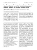
variants (Figure 1). We categorized the alternative splice
events, and found that 67.5% led to intron fusion (intron
sequences inserted into mature mRNA). These include novel
exon insertion, exon extension, and intron retention. The
remaining 32.5% of alternative splice events resulted in exon
loss (a portion or whole exon was skipped). A total of 18% of
the exon extensions and 50% of the exon truncations identified
in this study occurred at the 5' end of the alternatively spliced
exons. All of the 60 transcript variants encounter a stop codon
within the extracellular regions. As a result, these variants
encode soluble receptor isoforms, and were subsequently
referred to as ASV.
Detection of alternative splice variant mRNA expression
Expression of ASV mRNA relative to their corresponding con-
stitutively spliced transcripts was analyzed by both RT-PCR
and quantitative RT-PCR. Amplification of each target
sequence was performed across 29 distinct normal tissues as
well as cancer tissues including two cancer cell lines. For PCR
amplification of ASV, one primer was selected within the intron
fusion sequence and the other from a remote exon encom-
passing several introns. This approach ensured that only the
variant-specific mRNA transcript was amplified. An example of
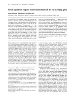
typical ASV mRNA expression (FGFR4) detected by RT-PCR
is shown in Figure 2a.
For a better comparison of mRNA expression and tissue distri-
bution, quantitative RT-PCR was performed to analyze ASV
and their corresponding constitutively spliced transcripts. Our
results demonstrated that expression of seven alternative
splice variant mRNAs (VEGFR1, VEGFR3, Met, RAGE, Tie1,
FGFR1, and Kit) is present in multiple normal and tumor tis-
sues (Figure 2b). Levels of expression varied among tissues,
with the ASV derived from VEGFR1, Met, and FGFR1 being
predominantly expressed in tumor tissues. In contrast, ASV
derived from VEGFR3 had the most restricted expression, and
were observed only in a few normal tissues and cancer cell
lines. These preliminary results indicate that expression of ASV
is tissue specific and occurs more frequently in tumor than nor-
mal tissues.
Table 1
Cloned alternative splice variant mRNAs
Receptor (n = 21) NCBI accession
number
Novel alternative splice
variants
VEGFR1 NM_002019 2
VEGFR2 NM_002253 1
VEGFR3 NM_002020 3
Met NM_000246 15
Ron NM_002447 4
Tie1 NM_005424 5
Tie2 NM_000459 2
CSF1R NM_005211 1
Kit NM_000222 1
PDGFRβ NM_002609 1
FGFR1 M34641 2
FGFR2 NM_000141 4
FGFR4 NM_002011 2
EPHA1 NM_005232 2
EPHA2 NM_004431 1
EPHB1 NM_004441 1
EPHB4 NM_004444 3
IGFR1 NM_000875 2
DDR1 NM_013993 2
TNFR1β NM_001066 1
RAGE NP_001127 5
Total 60
Sixty novel alternative splice variants were cloned from 21 cell
surface receptor genes by RT-PCR amplification followed by
extensive colony screening. The number of novel alternative splice
variants is presented for each receptor tested. NCBI, National
Center for Biotechnology Information.
Arthritis Research & Therapy Vol 10 No 4 Jin et al.
Page 6 of 16
(page number not for citation purposes)
Ligand binding potential of recombinant alternative
splice variants
Among the 60 ASV cloned, we selected 10 for initial func-
tional testing (Table 2). The selected ASV (corresponding to
ASV derived from VEGFR1, VEGFR2, VEGFR3, Tie1, Met, Kit,
CSF1R, PDGFRβ, FGFR1, and RAGE) represent diverse
members of gene families, possess known functional domains
such as ligand binding domains, and encode novel amino
acids compared with previously reported splice variant
sequences.
Efficient expression and secretion of the selected 10 recom-
binant ASV (VEGFR1, VEGFR2, VEGFR3, Tie1, Met, Kit,
CSF1R, PDGFRβ, FGFR1, and RAGE) from HEK293 cells
was confirmed by western blot analysis of the cell culture
supernatants, using anti-Myc antibody to detected the Myc-
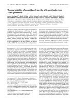
tagged ASV (Figure 3a). Furthermore, we observed ligand
binding by ASV proteins derived from VEGFR1, VEGFR2,
PDGFRβ, Met, and CSF1R – which bound to VEGF-A, VEGF-
C, PDGF, hepatocyte growth factor, and CSF, respectively
(Figure 3b). For evaluation of Tie1-751, purified recombinant
protein was used for binding to Ang-1, and a dissociation con-
stant (Kd) of approximately 89nM was measured (Figure 3b).
Not all receptor–ligand interactions could be detected by
plate-based binding, which may be a consequence of steric
issues associated with binding receptor or ligand to the sur-
face of the plate. Binding of VEGF-D to VEGFR3-765, for
example, was demonstrated only when the assay was per-
formed in solution (Figure 3c). Specificity of VEGF-D binding
to VEGFR3-765 was confirmed using a soluble VEGFR3/Fc
chimera, which was able to compete with VEGFR3-765 bind-
ing to VEGF-D – unlike a soluble VEGFR1/Fc chimera (Figure
3c).
Tie1-751 binds to membrane Tie1 and Tie2 on human
umbilical vein endothelial cells
Some soluble receptor splice variants have been shown to
bind cognate cell surface receptors and to modulate response
to ligand [48]. Tie1-751 comprises most of the extracellular
domain of Tie1 plus 11 C-terminal intron-derived amino acids.
To begin understanding the functionality of Tie1-751, we
tested whether Tie1-751 binds to endothelial cells. Proliferat-
ing endothelial cells (HUVEC) were incubated with
125
I-
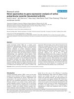
labeled Tie1-751. Our results showed that
125
I-Tie1-751 spe-
cifically bound to HUVEC, with an estimated dissociation con-
stant (Kd) of 121 nM (Figure 4a). Binding of
125
I-Tie1-751 to
HUVEC was competed by increasing amounts of unlabeled
Tie1-751 (Figure 4b).
Direct binding of Tie1-751(6His) to Tie1 and Tie2 on HUVEC
was also examined. Our results demonstrated interaction of
Tie1-751(6His) with the transmembrane Tie1, as well as with
the transmembrane Tie2 (Figure 4c).
Evaluation of alternative splice variant activity in an in
vivo model of arthritis
Since angiogenesis plays a key role in RA, we next evaluated
the therapeutic potential of ASV in an extensively validated
mouse model of arthritis – namely, acute CIA. On the day of
disease onset, replication-incompetent alternative splice vari-
ant-expressing adenoviruses were administered as a single
dose of 1 × 10
7
plaque-forming units. The severity of arthritis
in the mice was consecutively recorded for the following 10
days.
Control adenovirus (LacZ) was without significant effect on
disease severity (Table 3 and Figures 5 and 6). In contrast,
treatment with either Tie1-751 (Table 3 and Figure 5) or
VEGFR1-541 (Table 3 and Figure 6) alternative splice variant
adenoviruses significantly reduced disease severity, as evi-
denced by decreased clinical scores (P < 0.001), reduced
paw thickness (P < 0.001), and reduced joint inflammation
Figure 1
Splice events categorized by typeSplice events categorized by type. A total of 83 alternative splicing events were identified in the 21-gene array (Table 1). The identified splicing
events fell into five listed types. The splice pattern of the known transcript is depicted as type 1.
Available online />Page 7 of 16
(page number not for citation purposes)
and destruction (P < 0.01 and P < 0.001 for VEGFR1-541
and Tie1-751, respectively). An example of the joint histology
for untreated, LacZ ASV-treated and Tie1-751 ASV-treated
mice is shown in Figure 5c, with quantitative analysis of the
histology depicted in Table 3.
The presence of Tie1-751 in mouse sera was confirmed by
western blotting (Figure 5d). The effectiveness of Tie1-751 in
CIA was confirmed using recombinant Tie1-751-Fc protein
(Figure 5e).
A less marked disease-modifying effect was seen with the
adenovirus encoding FGFR1-320 (Table 3 and Figure 6),
which reduced clinical scores and paw thickness (P < 0.01)
but without achieving a statistically significant improvement of
joint histological evaluation (P < 0.057). Similarly, VEGFR2-
712 reduced the clinical score (P < 0.001) but failed to affect
the paw thickness and the histological scores (Table 3 and
Figure 6).
Figure 2
Alternative splice variant mRNA expressionAlternative splice variant mRNA expression. (a) RT-PCR detection of mRNA expression of FGFR4 (top panels) and FGFR4-ASV (bottom panels)
across 20 normal tissues and nine cancers, including two cancer cell lines. The amplified RT-PCR products were separated on 1% agarose gels
and visualized by ethidium bromide staining. bp, base pairs. (b) Expression profile heat map of the constitutively expressed (C) and matched splice
variant (V) mRNAs. Transcripts were analyzed across 20 normal tissues and nine cancers, including two cancer cell lines. Amplification of the consti-
tutive and splice variant sequences was performed using real-time PCR. Bar shows a color shift from green (high-level expression) to red (low-level
expression), with the corresponding cycle threshold values indicated.
Arthritis Research & Therapy Vol 10 No 4 Jin et al.
Page 8 of 16
(page number not for citation purposes)
Treatment with ASV derived from VEGFR3, RAGE, Met, c-Kit,
PDGFRβ, and CSF1R adenoviruses did not generate a signif-
icant effect on any of the disease parameters (Table 3 and Fig-
ure 6).
Discussion
The proliferative and invasive nature of RA synovium has fre-
quently led to comparisons with tumor development, and
therefore the usefulness of VEGF blockade for treatment of
certain cancers might be extrapolated to RA. Heterologous
CIA in mice shares many features with RA, and has been
widely used to study mechanisms involved in the arthritic proc-
ess and to identify new strategies for RA treatment, such as
TNFα inhibitors.
VEGF inhibition has been the focus of considerable clinically
oriented research, and angiogenesis blockade has been
shown to be effective in different in vivo models of arthritis,
including CIA [18,20,36,49,50]. VEGF inhibition in vivo,
however, is associated with side effects, such as impaired
wound healing, hemorrhage, and gastrointestinal perforation.
This is not surprising, given the heterozygous lethal phenotype
of VEGF knockout mice [51], which suggests a strategic role
for this molecule. Other positive regulators of angiogenesis
expressed in RA include hepatocyte growth factor and PDGF
[52,53]. To date, however, there have been no concerted
efforts to compare a range of different antiangiogenic
approaches side by side in a single study.
Bioinformatics surveys [11] and exon profiling [13,54] reveal
that the majority of pre-mRNAs are alternatively spliced. As
such, use of these soluble receptor variants might prove inval-
uable in designing new therapeutic strategies. We report here
that, using an efficient approach, we cloned 60 novel ASV of
21 genes encoding RTKs and other cell surface receptors.
The discovery of so many novel splice variants from a small
group of well-characterized drug target genes is consistent
with reports suggesting that alternative splicing is one of the
most significant components generating protein and func-
tional diversity in the human genome [13,54,55].
In vivo, soluble receptors are generated by both alternative
pre-mRNA splicing and proteolytic cleavage (shedding) of
membrane-anchored receptors, resulting in truncated mole-
cules lacking a transmembrane domain and an intracellular
segment. Soluble receptors may retain their ability to bind
ligands and function as ligand antagonists [9]; for example,
soluble TNF receptors [8] and soluble VEGFR1 [56]. Soluble
receptors are often generated through rational engineering. A
major difference between splice variant-derived soluble recep-
tors and engineered soluble receptors is that the former con-
tains novel amino acids and domain structures typically
derived from intron fusion. These alterations may subsequently
alter the functionality of the ASV as compared with the engi-
neered or metalloprotease-generated soluble receptors. An
example of altered function via alternative splicing is
VEGF
165
b, an antiangiogenic factor derived from the alterna-
tive splicing of VEGF pre-mRNA [57]. VEGF
165
b antagonizes
the angiogenic effect of VEGF
165
, which is also encoded by
the VEGF gene. Further studies are required to elucidate the
endogenous expression and function of the ASV described in
this report.
Inhibiting angiogenesis is a promising strategy for treatment of
neovascularization-related diseases [58], including RA [26].
Prior to anti-TNF therapeutics, 50% of RA patients become
Table 2
Alternative splice variants selected for functional testing
Splice variant Clone Length of ORF
a
Length of ECD
a
C-terminal novel amino acids
b
1 VEGFR1-541 018C02 541 758 LPPANSSFML PPTSFSSNYF HFLP*
2 VEGFR2-712 015F01 712 764 E*
3 VEGFR3-765 015G09 765 775 REGGPGEGQV RRPARPTIPN PGGPAPPPHP LQESTWRTPT RS*
4 Met-877 020H07 877 932 VRNALNTVLN HQLKLN*
5 Tie1-751 016G03 751 759 ERAGPTGPPG L*
6 CSF1R-306 005A06 306 512 GTPSPSLCPA *
7 c-Kit-413 002H01 413 520 SL*
8PDGFRβ-336 007C09 336 531 RAATCGSWER WAHYNLLSCI GAGHCR*
9 FGFR1-320 022C02 320 374 GTHCNFSSRC PALATGTGGA CISRLGETQR QESWKNGLLP
AWCHILPQL*
10 RAGE-387 021C06 387 342 IGETSPQALQ TLGLGCRTAQ ALISCPILAL SLTATPPLPP CTHTQASPAP
PAFCQESSQA SPFFPLS*
Ten alternative splice variants were selected for functional testing.
a
Lengths of the alternative splice variant open-reading frames (ORF) and
lengths of the wildtype receptor extracellular domains (ECD) are indicated by the numbers of amino acids.
b
Novel C-terminal amino acids of each
alternative splice variant are shown. *Stop codon.
Available online />Page 9 of 16
(page number not for citation purposes)
moderately disabled within 2 years and become severely
disabled within 10 years of disease onset. The increasing use
of anti-TNFα biological agents in RA is a major step forward,
but its use is restricted by an associated risk of infection,
including tuberculosis [1]. Most importantly, efficacy in long-
standing treatment does not usually result in remission. As a
Figure 3
Expression and ligand binding of recombinant alternative splice variantsExpression and ligand binding of recombinant alternative splice variants. (a) HEK293 cells were transiently transfected with the indicated
cDNA constructs. Conditioned media of HEK293 cells were collected after 48 hours, separated on SDS-PAGE gels and probed with an anti-Myc
antibody to detect the Myc-tagged alternative splice variants (ASV). Molecular weights (kDa) are indicated. (b) For VEGFR1-541, VEGFR2-712,
PDGFRβ-336, Met-877 and CSF1R-306, conditioned media from untransfected (Control, dashed lines) or ASV-transfected (Specific, solid lines)
HEK293 cells were applied to plates precoated with the receptor-specific ligands. Unbound ASV were detected using antibodies against the extra-
cellular domains of the receptors. Purified Tie1-751(6His) was used for Ang-1 binding, as above. Kd, dissociation constant. (c) Solution binding of
VEGF-D to VEGFR3-765-Myc. Binding was carried out by combining VEGF-D with conditioned medium from either VEGFR3-765-Myc-expressing
cells (lanes 1 to 3) or untransfected cells (lane 4). Subsequent immunoprecipitation was performed using anti-VEGF-D antibody and detected using
anti-Myc antibody. To confirm the specificity of interaction between VEGF-D and VEGFR3-765-Myc, binding was performed in the presence of five-
fold molar excess of either recombinant human VEGFR3/Fc chimera (lane 2) or soluble recombinant human VEGFR1/Fc chimera (lane 3). Molecular
weights (kDa) are indicated. CM, Conditioned medium; IP, Immunprecipitation; WB, Western blot.
Arthritis Research & Therapy Vol 10 No 4 Jin et al.
Page 10 of 16
(page number not for citation purposes)
consequence, initiatives to develop alternative treatments that
control disease progression in RA are desirable.
A well-documented feature of RA is an alteration in the density
of synovial blood vessels. Several angiogenic factors are
expressed in RA, including VEGF, PDGF, fibroblast growth
factor 1, and fibroblast growth factor 2, as well as Ang-1. Ang-
iogenesis is a multistep process, however, and – while VEGF
is important – other proangiogenic factors are also expressed
in RA and CIA. The contribution of other proangiogenic factors
to arthritic disease progression has not been well defined or
compared directly within the same disease model. In the
present study, 10 RTK-derived ASV were screened side by
side in the high-throughput CIA model, using replication-
incompetent adenoviruses as a delivery and in vivo expression
system. This method allows for screening many candidate bio-
logics quickly in a relevant disease model, without first
expressing and purifying the target molecules, and will select
for proteins that are significantly expressed and are bioactive
across species barriers. Some candidate proteins may give
false negative results because of issues related to expression
and stability in vivo, a species barrier, or a lack of activity in the
particular disease model.
In vivo screening of the ASV demonstrated clear differential
effects. Among them, ASV derived from VEGFR1 and Tie1
were found to be the most potent. The effect of VEGFR1-541
ASV confirms our own previous data and that of others, dem-
onstrating the effectiveness of VEGFR1 blockade in models of
arthritis [18,20,33,50]. In contrast, blockade of VEGFR2 in
models of arthritis has in general not been effective [33,50].
The effect of VEGFR2-712 ASV was modest in our study, with
inhibition of clinical score but not of paw swelling or histologi-
cal change. As the ultimate benefit of a potential therapeutic in
RA would be joint protection and reduced edema, the fact that
VEGFR2-712 ASV does not affect either paw swelling or joint
inflammation/destruction supports the view that VEGFR2
blockade is not likely to be beneficial in RA.
Expression of both Ang-1 [39,40] and angiopoietin receptors
Tie1 and Tie2 [41-43] in RA synovial tissue has been
described. Ang-1 is chemotactic and weakly mitogenic for
HUVEC [59,60], promotes formation of endothelial sprouts
[61], and has been proposed to act in concert with VEGF to
promote vascular network maturation [62,63]. Furthermore,
Ang-1 was found to be a survival factor for endothelial cells,
protecting HUVEC from apoptosis induced by serum with-
drawal [64]. Angiopoietin signaling was until recently
considered to be mediated via Tie2. The embryonic lethality of
Tie1 knockout mice, however, suggested that Tie1 signaling is
important in vascular network formation. It is now thought that
Tie1 may modulate signaling through Tie2 [65-67]. Marron
and colleagues reported that activation of Tie1 ectodomain
cleavage increased activation of Tie2, which could potentially
control signaling via Tie2 [68].
Figure 4
Tie1-751 interacts with Tie1 and Tie2Tie1-751 interacts with Tie1 and Tie2. (a) Specific binding of
125
I-
Tie1-751(6His) to human umbilical vein endothelial cells (HUVEC).
Nonspecific binding was determined in the presence of 100-fold
excess of unlabelled Tie1-751 and was subtracted from the total bind-
ing. CPM, counts per minute; Kd, dissociation constant. (b) Binding of
125
I-Tie1-751(6His) to HUVEC was competed by increasing amounts
of cold Tie1-751. Data are the mean ± standard error of the mean. (c)
Binding of Tie1-751(6His) to HUVEC. At the end of binding, cells were
treated with or without the cross-linker 3,3'-dithiobis(sulfosuccinimidyl-
propionate) (DTSSP), immunoprecipitated using a C-terminal-specific
anti-Tie1 (top panel) or anti-Tie2 (middle panel) antibody, and were ana-
lyzed by western blotting using anti-His antibody. To confirm equal
loading, cell lysates were blotted with anti-Tie1 antibody (bottom
panel). IP, Immunprecipitation; WB, Western blot.
Available online />Page 11 of 16
(page number not for citation purposes)
The novel activity of Tie1-751 in the CIA model [35,69] moti-
vated us to further examine its mechanism of action. Our
results demonstrate that Tie1-751 directly binds to Tie1 and
Tie2 on the surface of endothelial cells. Binding of Tie1-Fc to
transmembrane Tie1 and the interaction of transmembrane
Tie1 and Tie2 at the cell surface have recently been reported
[66]. The mechanism of binding Tie1-751 to Tie1 and to Tie2,
however, is currently unknown. Our initial characterization also
revealed that Tie1-751 inhibits Ang-1-induced Tie1
phosphorylation and the prosurvival effect of Ang-1 on
HUVEC (data not shown). These results suggest that Tie1-
751 may inhibit activation of the angiopoietin–Tie system by
both sequestering ligand and forming nonsignaling heterodim-
ers with cell surface receptors. It is possible that the C-termi-
nal intron-encoded domain of Tie1-751 expands its
functionality. Blocking Tie2 has been reported effective in CIA,
but no such data are available for Tie1 inhibition [70].
Further work is needed to confirm the function of novel
domains generated by alternative splicing. The differential
effects of the 10 ASV in arthritis in vivo, however, suggest that
selected ASV may have potential therapeutic application in RA
and in other angiogenesis-dependent conditions.
Conclusion
In summary, we describe an efficient method for the identifica-
tion and determination of biologic activity of novel ASV derived
from the cell surface receptor genes. Sixty ASV were identi-
fied. The variants identified commonly include unique amino
acids forming additional protein domains. Those ASV tested
were shown to bind cognate ligand. An alternative splice vari-
ant derived from Tie1 (Tie1-751) was shown to bind not only
Ang-1 but also cell surface Tie1 and Tie2. Using replication-
deficient adenoviruses as a means of screening for biologic
activity, we showed that RTK-derived ASV have selective
potential therapeutic activity in a murine model of RA. Further-
more, we have shown for the first time that inhibition of the
angiopoietin–Tie axis can markedly reduce arthritis severity.
The present work demonstrates that ASV are a potential
source of novel regulatory proteins, which may have therapeu-
tic potential in animal models of disease and warrant testing in
humans.
Competing interests
PJ, JZ, IN, BJ, and HMS are employees of Receptor BioLogix,
Inc. and hold stocks in the company, and declare competing
financial interests. MF and EMP have acted as consultants for
Receptor BioLogix, Inc. The other authors declare that they
have no competing interests. Receptor BioLogix, Inc. holds
the patents related to the content of the manuscript.
Authors' contributions
HMS designed the study. PJ assisted in the study design,
oversaw the project running and data analysis, and drafted the
manuscript. PJ, JZ, IN, and BJ performed the alternative splice
variant cloning, sequence analysis, protein expression and
purification, and ligand/receptor binding assays. SP per-
formed and analyzed the quantitative PCR experiment. EMP
Table 3
Effect of alternative splice variant-expressing adenoviruses on joint inflammation and destruction
Alternative splice variant adenovirus Mice per group (n) Joints assessed (n) P value
Clinical score Paw swelling Histological evaluation
Untreated 6 120 - - -
LacZ 6 164 0.4549 0.3759 0.3797
VEGFR1-541 5 53 <0.0001 <0.0001 0.0096
VEGFR2-712 5 44 <0.0001 0.1762 0.7340
VEGFR3-765 5 68 0.9366 0.2228 0.8148
Tie1-751 6 63 <0.0001 <0.0001 <0.001
Met-877 6 64 0.2924 0.6603 0.5038
c-Kit-413 6 55 0.0587 0.1501 0.1046
CSF1R-306 6 50 0.2448 0.5581 0.1510
PDGFRβ-336 6 41 0.8498 0.0632 0.8258
FGFR1-320 6 55 0.0044 0.0087 0.0568
RAGE-387 6 53 0.8543 0.1141 0.9799
Following onset of arthritis, mice were treated with the alternative splice variant adenovirus indicated. Data presented as P values of mice treated
with the indicated recombinant alternative splice variant adenoviruses as compared with untreated mice, and are expressed as the P value of
clinical scores, paw swelling, and histological evaluation. For clinical scores and paw swelling, data were analyzed using two-way analysis of
variance versus untreated mice. For histological evaluation, H & E and toluidine blue stained sections were scored for pannus formation, synovitis,
and bone and cartilage erosion. Data were analyzed using the chi-square test for trend versus untreated mice.
Arthritis Research & Therapy Vol 10 No 4 Jin et al.
Page 12 of 16
(page number not for citation purposes)
and MF assisted in the study design and coordination, and
oversaw the data analysis and drafting of the manuscript. PFS
and DC designed and carried out the in vivo arthritis studies.
All authors read and approved the final manuscript.
Figure 5
Inhibition of murine collagen-induced arthritis by Tie1-751Inhibition of murine collagen-induced arthritis by Tie1-751. On the day of arthritis onset, mice received intravenously 1 × 10
7
plaque-forming
units of adenoviruses expressing either LacZ (❍) or Tie1-751 alternative splice variants (ASV) (●), or remained untreated (ᮀ) as indicated. (a) Clin-
ical score was recorded daily, and data were analyzed by two-way analysis of variance versus untreated mice. LacZ, not significant (P = 0.3734);
Tie1-751, P < 0.001; n = 6 per group. (b) Paw swelling was recorded using calipers daily, and data were analyzed by two-way analysis of variance
versus untreated mice. LacZ, not significant (P = 0.5134); Tie1-751, P < 0.001. Data are means of n = 6. (c) Serial sections of mouse hind feet
were stained with either H & E (left panels) or toluidine blue (right panels). Figure shows tibia–tarsus joint sections from untreated mice (top panels),
from LacZ adenovirus-treated mice (middle panels), and from Tie1-751 ASV adenovirus-treated mice (bottom panels). Sections are shown at 40×
magnification; scale bar = 20 μm. (d) Pharmacokinetics of Tie1-751 from the ASV-expressing adenovirus. Sera from untreated mice or mice treated
intravenously with 1 × 10
9
plaque-forming units of Tie1-751 ASV adenovirus were analyzed after the indicated times by western blot, followed by
scanning and quantitation using Tie1-751 standard. (e) Effect of recombinant Tie1-751-Fc protein on clinical score. Results are from mice on day 10
of arthritis. Filled bars, untreated mice; empty bars, mice treated with recombinant Tie1-751-Fc 30 mg/kg, three times weekly. Data are means of n =
6. **P < 0.01 for Tie-751-Fc treated mice versus untreated mice.
Available online />Page 13 of 16
(page number not for citation purposes)
Figure 6
Differential effects of alternative splice variant-expressing adenoviruses on collagen-induced arthritis miceDifferential effects of alternative splice variant-expressing adenoviruses on collagen-induced arthritis mice. On the day of arthritis onset,
mice received intravenously 1 × 10
7
plaque-forming units of the indicated alternative splice variant (ASV)-expressing adenoviruses. Clinical scores
((a), (c), and (e))and paw thickness measured by calipers ((b), (d), and (f))were recorded daily. Data were analyzed by two-way analysis of variance
versus untreated mice (Table 3). (a) and (b) Mice received adenoviruses expressing either LacZ (❍), VEGFR1-541 (■), VEGFR2-712 (▲) or
VEGFR3-765 (●), or remained untreated (ᮀ). Data are means of n = 5 per group. (c) and (d) Mice received adenoviruses expressing either LacZ
(❍), Met-877 (■), Tie1-751 (▲) or FGFR1-320 (●), or remained untreated (ᮀ). Data are means of n = 6 per group. (e) and (f) Mice received aden-
oviruses expressing either LacZ (❍), RAGE-387 (■), PDGFRβ-366 (▲), c-Kit-413 (●), or CSF1R-306 (᭜), or remained untreated (ᮀ). Data are
means of n = 6 per group.
Arthritis Research & Therapy Vol 10 No 4 Jin et al.
Page 14 of 16
(page number not for citation purposes)
Additional files
Acknowledgements
The authors sincerely thank Scott Patton for editing this manuscript, and
the RBLX research team for support, discussion, and critical reading of
this manuscript. They are grateful to the staff of the Biological Services
Unit (Kennedy Institute of Rheumatology, Imperial College, London, UK)
for help in the care and maintenance of the laboratory mice used in our
studies; to the Histopathology Department, Charing Cross Hospital,
London – particularly David Essex, David Peston, and Ann Sandison –
for help in the sectioning and staining of mice hind feet specimens; and
to Kerri Reilly and Yvonne Raatz for advice with the molecular biology
studies. MF, EMP, and PFS would like to thank the Arthritis Research
Campaign of Great Britain, which provided support for this work.
References
1. Keane J, Gershon S, Wise RP, Mirabile-Levens E, Kasznica J,
Schwieterman WD, Siegel JN, Braun MM: Tuberculosis associ-
ated with infliximab, a tumor necrosis factor alpha-neutralizing
agent. N Engl J Med 2001, 345:1098-1104.
2. Cunnane G, Doran M, Bresnihan B: Infections and biological
therapy in rheumatoid arthritis. Best Pract Res Clin Rheumatol
2003, 17:345-363.
3. Gomez-Reino JJ, Carmona L, Valverde VR, Mola EM, Montero MD:
Treatment of rheumatoid arthritis with tumor necrosis factor
inhibitors may predispose to significant increase in tuberculo-
sis risk: a multicenter active-surveillance report. Arthritis
Rheum 2003, 48:2122-2127.
4. Giles JT, Bathon JM: Serious infections associated with anticy-
tokine therapies in the rheumatic diseases. J Intensive Care
Med 2004, 19:320-334.
5. Bongartz T, Sutton AJ, Sweeting MJ, Buchan I, Matteson EL, Mon-
tori V: Anti-TNF antibody therapy in rheumatoid arthritis and
the risk of serious infections and malignancies: systematic
review and meta-analysis of rare harmful effects in rand-
omized controlled trials. JAMA 2006, 295:2275-2285.
6. Gschwind A, Fischer OM, Ullrich A: The discovery of receptor
tyrosine kinases: targets for cancer therapy. Nat Rev Cancer
2004, 4:361-370.
7. Paleolog EM: Angiogenesis: a critical process in the pathogen-
esis of RA – a role for VEGF? Br J Rheumatol 1996,
35:917-919.
8. Cole P, Rabasseda X: The soluble tumor necrosis factor recep-
tor etanercept: a new strategy for the treatment of autoim-
mune rheumatic disease. Drugs Today (Barc) 2004,
40:281-324.
9. Fernandez-Botran R, Crespo FA, Sun X: Soluble cytokine recep-
tors in biological therapy. Expert Opin Biol Ther 2002,
2:585-605.
10. Black D: Protein diversity from alternative splicing: a challenge
for bioinformatics and post-genome biology. Cell 2000,
103:367-370.
11. Modrek B, Lee C:
A genomic view of alternative splicing. Nat
Genet 2002, 30:13-19.
12. Venables JP: Aberrant and alternative splicing in cancer. Can-
cer Res 2004, 64:7647-7654.
13. Zhu J, Shendure J, Mitra RD, Church GM: Single molecule profil-
ing of alternative pre-mRNA splicing. Science 2003,
301:836-838.
14. Foxwell BM, Yoshimura S, Bondeson J, Brennan FM, Feldmann M:
High efficiency gene transfer is an efficient way of defining
therapeutic targets: a functional genomics approach. Ann
Rheum Dis 2001, 60(Suppl 3):iii13-iii17.
15. Naor D, Nedvetzki S, Walmsley M, Yayon A, Turley EA, Golan I,
Caspi D, Sebban LE, Zick Y, Garin T, Karussis D, Assayag-Asherie
N, Raz I, Weiss L, Slavin S: CD44 involvement in autoimmune
inflammations: the lesson to be learned from CD44-targeting
by antibody or from knockout mice. Ann N Y Acad Sci 2007,
1110:233-247.
16. Vijayakrishnan L, Slavik JM, Illes Z, Rainbow D, Peterson LB,
Sharpe AS, Wicker LS, Kuchroo VK: An autoimmune disease-
associated CTLA4 splice variant lacking the B7 binding
domain signals negatively in T cells. Novartis Found Symp
2005, 267:200-212. discission 212–208.
17. Ambati BK, Nozaki M, Singh N, Takeda A, Jani PD, Suthar T, Albu-
querque RJ, Richter E, Sakurai E, Newcomb MT, Kleinman ME,
Caldwell RB, Lin Q, Ogura Y, Orecchia A, Samuelson DA, Agnew
DW, St Leger J, Green WR, Mahasreshti PJ, Curiel DT, Kwan D,
Marsh H, Ikeda S, Leiper LJ, Collinson JM, Bogdanovich S,
Khurana TS, Shibuya M, Baldwin ME, Ferrara N, Gerber HP, De
Falco S, Witta J, Baffi JZ, Raisler BJ, Ambati J: Corneal avascular-
ity is due to soluble VEGF receptor-1. Nature 2006,
443:993-997.
18. Miotla J, Maciewicz R, Kendrew J, Feldmann M, Paleolog E: Treat-
ment with soluble VEGF receptor reduces disease severity in
murine collagen-induced arthritis. Lab Invest 2000,
80:1195-1205.
19. Bainbridge JW, Mistry A, De Alwis M, Paleolog E, Baker A,
Thrasher AJ, Ali RR: Inhibition of retinal neovascularisation by
gene transfer of soluble VEGF receptor sFlt-1. Gene Ther
2002, 9:320-326.
20. Afuwape AO, Feldmann M, Paleolog EM: Adenoviral delivery of
soluble VEGF receptor 1 (sFlt-1) abrogates disease activity in
murine collagen-induced arthritis. Gene Ther 2003,
10:1950-1960.
21. Rota R, Riccioni T, Zaccarini M, Lamartina S, Gallo AD, Fusco A,
Kovesdi I, Balestrazzi E, Abeni DC, Ali RR, Capogrossi MC:
Marked inhibition of retinal neovascularization in rats follow-
ing soluble-flt-1 gene transfer. J Gene Med 2004, 6:992-1002.
22. Ideno J, Mizukami H, Kakehashi A, Saito Y, Okada T, Urabe M,
Kume A, Kuroki M, Kawakami M, Ishibashi S, Ozawa K: Prevention
of diabetic retinopathy by intraocular soluble flt-1 gene trans-
fer in a spontaneously diabetic rat model. Int J Mol Med 2007,
19:75-79.
23. Paleolog EM: Angiogenesis in rheumatoid arthritis. Arthritis
Research Supplement 2002, 3(Suppl 3):S81-90.
24. Ballara SC, Taylor PC, Reusch P, Marmé D, Feldmann M, Maini
RN, Paleolog EM: Raised serum vascular endothelial growth
factor levels are associated with destructive change in inflam-
matory arthritis. Arthritis Rheum 2001, 44:2055-2064.
25. Sivakumar B, Harry LE, Paleolog EM: Modulating angiogenesis:
more vs less. JAMA 2004, 292:972-977.
26. Bainbridge J, Sivakumar B, Paleolog E: Angiogenesis as a thera-
peutic target in arthritis: lessons from oncology. Curr Pharm
Des 2006, 12:2631-2644.
27. Larsen H, Akhavani MA, Raatz Y, Paleolog EM: Gene expression
studies to investigate disease mechanisms in rheumatoid
arthritis: does angiogenesis play a role? Curr Rheumatol Rev
2007, 3:243-251.
The following Additional files are available online:
Additional file 1
A Word file Summarizing the information of the 60 full-
length novel splice variants with GenBank accession
numbers.
See />supplementary/ar2447-S1.doc
Additional file 2
A Word file containing a table presenting the cDNA
sequences of the 60 full-length novel splice variants that
have been deposited with GenBank.
See />supplementary/ar2447-S2.doc
Available online />Page 15 of 16
(page number not for citation purposes)
28. Khong TL, Larsen H, Raatz Y, Paleolog E: Angiogenesis as a
therapeutic target in arthritis: learning the lessons of the color-
ectal cancer experience. Angiogenesis 2007, 4:243-258.
29. Williams RO, Feldmann M, Maini RN: Anti-tumor necrosis factor
ameliorates joint disease in murine collagen-induced arthritis.
Proc Natl Acad Sci USA 1992, 89:9784-9788.
30. Williams RO, Ghrayeb J, Feldmann M, Maini RN: Successful ther-
apy of collagen-induced arthritis with TNF receptor-IgG fusion
protein and combination with anti-CD4. Immunology 1995,
84:433-439.
31. Williams RO, Marinova-Mutafchieva L, Feldmann M, Maini RN:
Evaluation of TNF-alpha and IL-1 blockade in collagen-
induced arthritis and comparison with combined anti-TNF-
alpha/anti-CD4 therapy. J Immunol 2000, 165:7240-7245.
32. Williams RO, Mason LJ, Feldmann M, Maini RN: Synergy between
anti-CD4 and anti-tumor necrosis factor in the amelioration of
established collagen-induced arthritis. Proc Natl Acad Sci
USA 1994, 91:2762-2766.
33. de Bandt M, Ben Mahdi MH, Ollivier V, Grossin M, Dupuis M,
Gaudry M, Bohlen P, Lipson KE, Rice A, Wu Y, Gougerot-Pocidalo
MA, Pasquier C: Blockade of vascular endothelial growth factor
receptor I (VEGF-RI), but not VEGF-RII, suppresses joint
destruction in the K/BxN model of rheumatoid arthritis. J
Immunol 2003, 171:4853-4859.
34. Kim JM, Ho SH, Park EJ, Hahn W, Cho H, Jeong JG, Lee YW, Kim
S: Angiostatin gene transfer as an effective treatment strategy
in murine collagen-induced arthritis. Arthritis Rheum 2002,
46:793-801.
35. Geva E, Jaffe RB: Role of angiopoietins in reproductive tract
angiogenesis. Obstet Gynecol Surv 2000, 55:511-519.
36. Sone H, Kawakami Y, Sakauchi M, Nakamura Y, Takahashi A, Shi-
mano H, Okuda Y, Segawa T, Suzuki H, Yamada N: Neutralization
of vascular endothelial growth factor prevents collagen-
induced arthritis and ameliorates established disease in mice.
Biochem Biophys Res Commun 2001, 281:562-568.
37. Sumariwalla P, Cao Y, Wu H, Feldmann M, Paleolog E: The ang-
iogenesis inhibitor protease-activated kringles 1–5 reduces
the severity of murine collagen-induced arthritis. Arthritis Res
Ther 2003, 5:R32-R39.
38. Bainbridge J, Madden L, Essex D, Binks M, Malhotra R, Paleolog
EM: Methionine aminopeptidase-2 blockade reduces chronic
collagen-induced arthritis: potential role for angiogenesis
inhibition. Arthritis Res Ther 2007, 9:R127.
39. Scott BB, Zaratin PF, Colombo A, Hansbury MJ, Winkler JD, Jack-
son JR: Constitutive expression of angiopoietin-1 and -2 and
modulation of their expression by inflammatory cytokines in
rheumatoid arthritis synovial fibroblasts. J Rheumatol 2002,
29:230-239.
40. Gravallese EM, Pettit AR, Lee R, Madore R, Manning C, Tsay A,
Gaspar J, Goldring MB, Goldring SR, Oettgen P: Angiopoietin-1
is expressed in the synovium of patients with rheumatoid
arthritis and is induced by tumour necrosis factor alpha. Ann
Rheum Dis 2003, 62:100-107.
41. DeBusk LM, Chen Y, Nishishita T, Chen J, Thomas JW, Lin PC:
Tie2 receptor tyrosine kinase, a major mediator of tumor
necrosis factor alpha-induced angiogenesis in rheumatoid
arthritis. Arthritis Rheum 2003, 48:2461-2471.
42. Shahrara S, Volin MV, Connors MA, Haines GK, Koch AE: Differ-
ential expression of the angiogenic Tie receptor family in
arthritic and normal synovial tissue. Arthritis Res 2002,
4:201-208.
43. Uchida T, Nakashima M, Hirota Y, Miyazaki Y, Tsukazaki T, Shindo
H: Immunohistochemical localisation of protein tyrosine
kinase receptors Tie-1 and Tie-2 in synovial tissue of rheuma-
toid arthritis: correlation with angiogenesis and synovial
proliferation. Ann Rheum Dis 2000, 59:607-614.
44. Kuo CJ, Farnebo F, Yu EY, Christofferson R, Swearingen RA,
Carter R, von Recum HA, Yuan J, Kamihara J, Flynn E, D'Amato R,
Folkman J, Mulligan RC: Comparative evaluation of the antitu-
mor activity of antiangiogenic proteins delivered by gene
transfer. Proc Natl Acad Sci USA 2001, 98:4605-4610.
45. Saeed AI, Sharov V, White J, Li J, Liang W, Bhagabati N, Braisted
J, Klapa M, Currier T, Thiagarajan M, Sturn A, Snuffin M, Rezantsev
A, Popov D, Ryltsov A, Kostukovich E, Borisovsky I, Liu Z, Vinsavich
A, Trush V, Quackenbush J: TM4: a free, open-source system for
microarray data management and analysis. Biotechniques
2003, 34:374-378.
46. Miller EJ: Structural studies on cartilage collagen employing
limited cleavage and solubilization with pepsin. Biochemistry
1972, 11:4903-4909.
47. Florea L, Hartzell G, Zhang Z, Rubin GM, Miller W: A computer
program for aligning a cDNA sequence with a genomic DNA
sequence. Genome Res 1998, 8:967-974.
48. Jostock T, Mullberg J, Ozbek S, Atreya R, Blinn G, Voltz N, Fischer
M, Neurath MF, Rose-John S: Soluble gp130 is the natural inhib-
itor of soluble interleukin-6 receptor transsignaling
responses. Eur J Biochem 2001, 268:160-167.
49. Lu J, Kasama T, Kobayashi K, Yoda Y, Shiozawa F, Hanyuda M,
Negishi M, Ide H, Adachi M: Vascular endothelial growth factor
expression and regulation of murine collagen-induced
arthritis. J Immunol 2000, 164:5922-5927.
50. Luttun A, Tjwa M, Moons L, Wu Y, Angelillo-Scherrer A, Liao F,
Nagy JA, Hooper A, Priller J, De Klerck B, Compernolle V, Daci E,
Bohlen P, Dewerchin M, Herbert JM, Fava R, Matthys P, Carmeliet
G, Collen D, Dvorak HF, Hicklin DJ, Carmeliet P: Revasculariza-
tion of ischemic tissues by PlGF treatment, and inhibition of
tumor angiogenesis, arthritis and atherosclerosis by anti-Flt1.
Nat Med 2002, 8:831-840.
51. Carmeliet P, Ferreira V, Breier G, Pollefeyt S, Kieckens L, Gertsen-
stein M, Fahrig M, Vandenhoeck A, Harpal K, Eberhardt C,
Declercq C, Pawling J, Moons L, Collen D, Risau W, Nagy A:
Abnormal blood vessel development and lethality in embryos
lacking a single VEGF allele. Nature 1996, 380:435-439.
52. Koch AE, Halloran MM, Hosaka S, Shah MR, Haskell CJ, Baker SK,
Panos RJ, Haines GK, Bennett GL, Pope RM, Ferrara N: Hepato-
cyte growth factor. A cytokine mediating endothelial migration
in inflammatory arthritis. Arthritis & Rheumatism 1996,
39:1566-1575.
53. Remmers EF, Sano H, Lafyatis R, Case JP, Kumkumian GK, Hla T,
Maciag T, Wilder RL: Production of platelet derived growth fac-
tor B chain (PDGF-B/c-sis) mRNA and immunoreactive PDGF
B-like polypeptide by rheumatoid synovium: coexpression
with heparin binding acidic fibroblast growth factor-1. J
Rheumatol 1991, 18:7-13.
54. Yeakley JM, Fan JB, Doucet D, Luo L, Wickham E, Ye Z, Chee MS,
Fu XD: Profiling alternative splicing on fiber-optic arrays. Nat
Biotechnol 2002, 20:353-358.
55. Holmes WE, Sliwkowski MX, Akita RW, Henzel WJ, Lee J, Park
JW, Yansura D, Abadi N, Raab H, Lewis GD, Shepard HM, Kuang
WJ, Wood WI, Goeddel DV, Vandlen RL: Identification of hereg-
ulin, a specific activator of p185erbB2. Science 1992,
256:
1205-1210.
56. Holash J, Davis S, Papadopoulos N, Croll SD, Ho L, Russell M,
Boland P, Leidich R, Hylton D, Burova E, Ioffe E, Huang T, Radzie-
jewski C, Bailey K, Fandl JP, Daly T, Wiegand SJ, Yancopoulos
GD, Rudge JS: VEGF-Trap: a VEGF blocker with potent antitu-
mor effects. Proc Natl Acad Sci USA 2002, 99:11393-11398.
57. Qiu Y, Bevan H, Weeraperuma S, Wratting D, Murphy D, Neal CR,
Bates DO, Harper SJ: Mammary alveolar development during
lactation is inhibited by the endogenous antiangiogenic
growth factor isoform, VEGF165b. FASEB J 2008,
22:1104-1112.
58. Ferrara N, Kerbel RS: Angiogenesis as a therapeutic target.
Nature 2005, 438:967-974.
59. Davis S, Aldrich TH, Jones PF, Acheson A, Compton DL, Jain V,
Ryan TE, Bruno J, Radziejewski C, Maisonpierre PC, Yancopoulos
GD: Isolation of angiopoietin-1, a ligand for the TIE2 receptor,
by secretion-trap expression cloning. Cell 1996,
87:1161-1169.
60. Witzenbichler B, Maisonpierre PC, Jones P, Yancopoulos GD,
Isner JM: Chemotactic properties of angiopoietin-1 and -2, lig-
ands for the endothelial-specific receptor tyrosine kinase Tie2.
J Biol Chem 1998, 273:18514-18521.
61. DeBusk LM, Hallahan DE, Lin PC: Akt is a major angiogenic
mediator downstream of the Ang1/Tie2 signaling pathway.
Exp Cell Res 2004, 298:167-177.
62. Asahara T, Chen D, Takahashi T, Fujikawa K, Kearney M, Magner
M, Yancopoulos GD, Isner JM: Tie2 receptor ligands, angiopoi-
etin-1 and angiopoietin-2, modulate VEGF-induced postnatal
neovascularization. Circ Res 1998, 83:233-240.
63. Koblizek TI, Weiss C, Yancopoulos GD, Deutsch U, Risau W:
Angiopoietin-1 induces sprouting angiogenesis in vitro. Curr
Biol 1998, 8:529-532.
Arthritis Research & Therapy Vol 10 No 4 Jin et al.
Page 16 of 16
(page number not for citation purposes)
64. Papapetropoulos A, Garcia-Cardena G, Dengler TJ, Maisonpierre
PC, Yancopoulos GD, Sessa WC: Direct actions of angiopoie-
tin-1 on human endothelium: evidence for network stabiliza-
tion, cell survival, and interaction with other angiogenic growth
factors. Lab Invest 1999, 79:213-223.
65. Kontos CD, Cha EH, York JD, Peters KG: The endothelial recep-
tor tyrosine kinase Tie1 activates phosphatidylinositol 3-
kinase and Akt to inhibit apoptosis. Mol Cell Biol 2002,
22:1704-1713.
66. Saharinen P, Kerkela K, Ekman N, Marron M, Brindle N, Lee GM,
Augustin H, Koh GY, Alitalo K: Multiple angiopoietin recom-
binant proteins activate the Tie1 receptor tyrosine kinase and
promote its interaction with Tie2. J Cell Biol 2005,
169:239-243.
67. Yuan HT, Venkatesha S, Chan B, Deutsch U, Mammoto T,
Sukhatme VP, Woolf AS, Karumanchi SA: Activation of the
orphan endothelial receptor Tie1 modifies Tie2-mediated
intracellular signaling and cell survival. FASEB J 2007,
21:3171-3183.
68. Marron MB, Singh H, Tahir TA, Kavumkal J, Kim HZ, Koh GY, Brin-
dle NP: Regulated proteolytic processing of Tie1 modulates
ligand responsiveness of the receptor-tyrosine kinase Tie2. J
Biol Chem 2007, 282:30509-30517.
69. Eklund L, Olsen BR: Tie receptors and their angiopoietin lig-
ands are context-dependent regulators of vascular
remodeling. Exp Cell Res 2006, 312:630-641.
70. Chen Y, Donnelly E, Kobayashi H, Debusk LM, Lin PC: Gene ther-
apy targeting the Tie2 function ameliorates collagen-induced
arthritis and protects against bone destruction. Arthritis
Rheum 2005, 52:1585-1594.