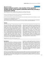
Báo cáo y học: "Human infrapatellar fat pad-derived stem cells express the pericyte marker 3G5 and show enhanced chondrogenesis after expansion in fibroblast growth factor-2" potx
Bạn đang xem bản rút gọn của tài liệu. Xem và tải ngay bản đầy đủ của tài liệu tại đây (2.05 MB, 11 trang )
Open Access
Available online />Page 1 of 11
(page number not for citation purposes)
Vol 10 No 4
Research article
Human infrapatellar fat pad-derived stem cells express the
pericyte marker 3G5 and show enhanced chondrogenesis after
expansion in fibroblast growth factor-2
Wasim S Khan, Simon R Tew, Adetola B Adesida and Timothy E Hardingham
United Kingdom Centre for Tissue Engineering at the Wellcome Trust Centre for Cell Matrix Research, Faculty of Life Sciences, Michael Smith
Building, University of Manchester, Oxford Road, Manchester, M13 9PT, UK
Corresponding author: Wasim S Khan,
Received: 5 Jul 2007 Revisions requested: 6 Sep 2007 Revisions received: 18 Jun 2008 Accepted: 3 Jul 2008 Published: 3 Jul 2008
Arthritis Research & Therapy 2008, 10:R74 (doi:10.1186/ar2448)
This article is online at: />© 2008 Khan et al.; licensee BioMed Central Ltd.
This is an open access article distributed under the terms of the Creative Commons Attribution License ( />),
which permits unrestricted use, distribution, and reproduction in any medium, provided the original work is properly cited.
Abstract
Introduction Infrapatellar fat pad (IPFP) is a possible source of
stem cells for the repair of articular cartilage defects. In this
study, adherent proliferative cells were isolated from digests of
IPFP tissue. The effects of the expansion of these cells in
fibroblast growth factor-2 (FGF-2) were tested on their
proliferation, characterisation, and chondrogenic potential.
Methods IPFP tissue was obtained from six patients undergoing
total knee replacement, and sections were stained with 3G5,
alpha smooth muscle actin, and von Willebrand factor to identify
different cell types in the vasculature. Cells were isolated from
IPFP, and both mixed populations and clonal lines derived from
them were characterised for cell surface epitopes, including
3G5. Cells were expanded with and without FGF-2 and were
tested for chondrogenic differentiation in cell aggregate
cultures.
Results 3G5-positive cells were present in perivascular regions
in tissue sections of the IPFP, and proliferative adherent cells
isolated from the IPFP were also 3G5-positive. However, 3G5
expression was on only a small proportion of cells in all
populations and at all passages, including the clonally expanded
cells. The cells showed cell surface epitope expression similar
to adult stem cells. They stained strongly for CD13, CD29,
CD44, CD90, and CD105 and were negative for CD34 and
CD56 but were also negative for LNGFR (low-affinity nerve
growth factor receptor) and STRO1. The IPFP-derived cells
showed chondrogenic differentiation in cell aggregate cultures,
and prior expansion with FGF-2 enhanced chondrogenesis.
Expansion in FGF-2 resulted in greater downregulation of many
cartilage-associated genes, but on subsequent chondrogenic
differentiation, they showed stronger upregulation of these
genes and this resulted in greater matrix production per cell.
Conclusion These results show that these cells express
mesenchymal stem cell markers, but further work is needed to
determine the true origin of these cells. These results suggest
that the expansion of these cells with FGF-2 has important
consequences for facilitating their chondrogenic differentiation.
Introduction
Cartilage is frequently damaged by trauma and in disease and
has a poor ability to heal. Cartilage defects that extend into the
subchondral bone show some signs of repair with the forma-
tion of neocartilage [1], probably due to the infiltration of the
defect with bone marrow-derived stem cells from the underly-
ing subchondral bone [2]. This principal is employed in the sur-
gical technique of subchondral drilling and microfracture to
stimulate cartilage repair. However, this can result in the for-
mation of fibrocartilage with properties mechanically inferior to
articular hyaline cartilage [3]. Autologous chondrocytes har-
vested from low-weight-bearing areas of articular cartilage and
expanded ex vivo are being used for the repair of focal hyaline
cartilage defects [4], but evidence suggests that this may fail
to halt progression of degenerative changes in the joint [5].
There has been a recent interest in cell-based therapies for
cartilage repair using adult stem cells or undifferentiated
αSMA = alpha smooth muscle actin; BSA = bovine serum albumin; DPBS = Dulbecco's phosphate-buffered solution; FGF-2 = fibroblast growth
factor-2; GAG = glycosoaminoglycan; IPFP = infrapatellar fat pad; LNGFR = low-affinity nerve growth factor receptor; NCAM = neural cell adhesion
molecule; PCR = polymerase chain reaction; TGF = transforming growth factor; vWF = von Willebrand factor.
Arthritis Research & Therapy Vol 10 No 4 Khan et al.
Page 2 of 11
(page number not for citation purposes)
progenitor cells. Stem cells have been reported to be present
in many adult human tissue types, including bone marrow, sub-
cutaneous adipose tissue, and the infrapatellar fat pad (IPFP)
[6-9]. Compared with bone marrow, IPFP is reported to give a
higher yield of stem cells and there is reduced pain and mor-
bidity associated with the harvest of cells [8]. In preliminary
work, we identified perivascular cells in the IPFP tissue which
stained with a monoclonal antibody, 3G5 [10]. The antigen
recognised by 3G5 is a cell surface ganglioside characteristic
of retinal vascular pericytes, which have been shown to have
multidifferentiation potential [11-15]. It has been suggested
that, if distributed widely with vascular capillaries, pericytes
may account for stem cells in other tissues [16-18]. In support
of this theory, a subendothelial network of pericyte-like cells
has been identified using 3G5 in the vascular bed in many
human tissues [19], and indeed many of the tissues from
which stem cells have been isolated have good vascularisa-
tion. A minor population of bone marrow-derived mesenchymal
stem cells has also been found to be positive for 3G5 [20].
The defining properties of stem cells are self-renewal and
multipotency. Unfortunately, these crucial properties in adult
stem cells show donor variability and may become limited on
expansion in monolayer culture [21,22]. As expansion is invar-
iably needed to increase the cell number for clinical applica-
tions, it is important to achieve expansion without a significant
compromise of differentiation potential. Fibroblast growth fac-
tor-2 (FGF-2) is a potent mitogen for a variety of cell types
derived from the mesoderm, including chondrocytes [23,24].
It has been shown to enhance proliferation and differentiation
of bone marrow-derived stem cells [25-28]. FGF produces
diverse and sometimes paradoxical effects on cell proliferation
and differentiation which are cell-type-dependent [29]. This
highlights the need for caution in extrapolating the effects of
FGF-2 from one cell type to another. We have previously
shown that IPFP-derived cells are able to undergo chondro-
genic differentiation [30], but the effect of FGF-2 on the
expansion and subsequent chondrogenesis in these cells has
not been previously investigated.
In our investigation of the potential of IPFP-derived cells from
elderly osteoarthritic patients undergoing joint replacement,
we characterised the cells and investigated the chondrogenic
response to expansion in FGF-2 in chondrogenic cultures. To
further explore the cell surface characterisation, single cells
were clonally expanded and stained for a panel of stem cell
markers, including 3G5. To allow for the effect of inherent var-
iability in the differentiation potential of cells between individu-
als [31], we carried out a patient-matched comparison of the
chondrogenic potential of cells expanded with and without
FGF-2.
Materials and methods
The IPFP was obtained with ethical approval and fully informed
consent from six patients undergoing total knee replacement
for osteoarthritis.
Immunohistochemical staining of tissue sections and
cell aggregates
The IPFP tissue and cell aggregates were fixed for 2 hours in
4% formaldehyde (BDH Ltd, Poole, UK)/Dulbecco's phos-
phate-buffered solution (DPBS) (Cambrex, Wokingham, UK).
The samples were then washed in 70% industrial methylated
spirit (BDH Ltd) and placed in a Shandon Citadel 2000 tissue
processor (Thermo Electron Corporation, Runcorn, UK). Par-
affin-embedded sections (5 μm) were taken and mounted on
slides precoated with Superfrost Plus (Menzel Glaser GmbH,
Braunschweig, Germany), dried in air, and left at 37°C over-
night. All incubations were performed in a humidity chamber at
20°C to 21°C, and all washes and dilutions were done in
DPBS unless otherwise stated.
3G5 staining of tissue sections
The slides were placed in 0.01 mmol citrate buffer (BDH Ltd)
for 10 minutes in a microwave at mid-power followed by cool-
ing to 30°C on ice. Sections were immunostained for 1 hour
in undiluted mouse anti-3G5 IgM prepared from a 3G5 hydri-
doma line (courtesy of Ann Canfield, University of Manchester,
UK) followed by washing and incubation for 1 hour in rabbit
anti-mouse biotin-conjugated secondary antibody (1:40 with
1% bovine serum albumin [BSA]; Dako, Ely, UK). Mouse IgG
antibody was used as a control (Santa Cruz Biotechnology,
Santa Cruz, CA, USA). Endogenous peroxidase activity was
quenched for 5 minutes with 3% hydrogen peroxide (Sigma-
Aldrich, Poole, UK) in methanol (BDH Ltd). Nonspecific bind-
ing was blocked with 10% normal rabbit serum (Sigma-
Aldrich) diluted in 1% BSA for 1 hour.
Alpha smooth muscle actin staining of tissue sections
Wash 1 was made up with 500 mL DPBS, 0.15 M NaCl, and
0.5% BSA, and wash 2 was made up with 500 mL DPBS,
0.15 M NaCl, and 0.1% BSA. Sections were immunostained
for 1 hour in mouse anti-human alpha smooth muscle actin
(αSMA) (1:400 in wash 1; courtesy of A. Canfield) followed by
washing in wash 1 for 1 hour and incubation for 1 hour in rab-
bit anti-mouse biotin-conjugated secondary antibody (1:50 in
wash 1). Mouse IgG antibody was used as a control. The
slides were then placed in wash 2 for 1 hour. Endogenous per-
oxidase activity was quenched for 30 minutes by placing the
slides in wash 1.
von Willebrand factor staining of tissue sections
Blocking solution was made up with 20% normal donkey
serum (Sigma-Aldrich). Sections were immunostained for 1
hour in serum-protein-absorbed rabbit anti-human von Wille-
brand factor (vWF) IgG (1:250 with 0.1% BSA in blocking
solution; Dako) followed by washing and incubation for 1 hour
Available online />Page 3 of 11
(page number not for citation purposes)
in donkey anti-rabbit biotin-conjugated antibody (1:300 with
0.1% BSA in blocking solution; Dako). Rabbit IgG was used
as a control (Santa Cruz Biotechnology). Endogenous peroxi-
dase activity was quenched for 30 minutes with 0.3% hydro-
gen peroxide in methanol. Nonspecific binding was blocked
for 10 minutes with the blocking solution.
Collagen type I, type II, and aggrecan staining of cell
aggregate sections
Sections were preincubated at 37°C with 0.1 U/mL chondroi-
tinase ABC (Sigma-Aldrich) for 1 hour and then immunos-
tained for 16 hours at 4°C with goat anti-human collagen type
I (C-18 polyclonal), collagen type II (N-19 polyclonal) (both
from Santa Cruz Biotechnology), or rabbit anti-human aggre-
can (BR1) (all at 1:100 dilution) followed by washing and incu-
bation for 30 minutes in donkey anti-goat IgG biotin-
conjugated secondary antibody (Santa Cruz Biotechnology)
for collagen type I and collagen type II and donkey anti-rabbit
IgG biotin-conjugated secondary antibody for aggrecan (all at
1:250 dilution). Goat IgG antibody (Santa Cruz Biotechnol-
ogy) was used as a control for collagen, and rabbit IgG was
used as a control for aggrecan. Endogenous peroxidase activ-
ity was quenched for 5 minutes with 3% hydrogen peroxide in
methanol. Nonspecific binding was blocked for 1 hour with
10% normal donkey serum diluted in 1% BSA.
For visualisation, sections were incubated for 30 minutes in
streptavidin-peroxidase complex (1:500; Dako), rinsed in dis-
tilled water, and incubated in fast-DAB (3,3'-diaminobenzi-
dine) peroxidase substrate (Sigma-Aldrich) for 5 minutes and
counterstained in diluted filtered haematoxylin (Sigma-Aldrich)
for 15 seconds. Images were then taken with an Axioplan 2
microscope with the use of an Axiocam HRc camera and Axio-
Vision 4.3 software (all from Carl Zeiss Ltd, Welwyn Garden
City, UK).
Cell isolation and culture
The IPFP tissue was dissected and cells were isolated by
digestion with 0.2% (vol/vol) collagenase I (Invitrogen, Paisley,
UK) for 3 hours at 37°C with constant agitation. The released
cells were sieved (70-μm mesh) and washed in basic medium,
namely Dulbecco's modified Eagle's medium supplemented
with 20% (vol/vol) foetal calf serum, 100 U/mL penicillin, and
100 μg/mL streptomycin (all from Cambrex), with
L-glutamine
(2 mM). The stromal cells were separated from the adipocytes
(floating) by centrifugation at 300 g for 5 minutes and were
counted and plated at 100,000 cells per square centimetre in
monolayer culture in basic medium with and without 10 ng/mL
rhFGF-2 (Sigma-Aldrich) supplementation. Cultures were
maintained at 37°C with 5% CO
2
and normal oxygen (20%).
Cultured cells from passage 2 were used for cell proliferation
rate studies, cell surface epitope characterisation, and cell
aggregate culture.
Cell proliferation rates
Cell proliferation rates were measured for passage 2 cells
plated with and without FGF-2-supplemented medium at
10,000 cells per square centimetre in a six-well plate. Cells
were trypsinised and collected at days 2, 4, 6, 8, and 10 after
plating, and the cell number was determined by counting with
a haemacytometer. The viability of the cells was determined by
staining with Trypan blue.
Isolation of clonal populations
Clonal cell populations were derived from single cells obtained
by limiting dilution. Freshly isolated cells obtained from a single
mixed parent IPFP population (mixed parent population is the
original, supposedly heterogenous, population of cells from
which the clonal cell lines were derived) were plated at a den-
sity of 0.33 cells per well in two polystyrene 96-well flat-bot-
tomed cell culture microplates (Corning Inc., supplied through
Fisher Scientific, Loughborough, UK). Based on Poisson dis-
tribution statistics, the probability of a clonal population being
derived from a single cell at this density is greater than 95%
[32]. Thirteen wells where a single cell had been noted initially
were identified, and the cell progressed to form a single col-
ony. These colonies were selected as they were thought to
arise from a single cell. Wells containing more than one colony
were excluded. The selected cell populations were trypsinised
on confluence and serially plated in a well of a six-well plate
(9.6 cm
2
), a T75 cell culture flask (75 cm
2
), and later a T225
cell culture flask (225 cm
2
) (all from Corning Inc.). Only 4 of
these 13 expandable clones reached confluence in T225
flasks. The remaining cells from the mixed parent IPFP-derived
population were plated at a concentration of 100,000 cells per
square centimetre in a T75 flask followed by a T225 flask on
confluence.
Cell surface epitope characterisation
Confluent passage 2 cells expanded with and without FGF-2,
and the four clonal and mixed parent populations were stained
with a panel of antibodies for cell surface epitopes. This
included antibodies against the following: CD13 (aminopepti-
dase N), CD44 (hyaluronan receptor), CD90 (Thy-1), LNGFR
(low-affinity nerve growth factor receptor), STRO1 (marker for
bone marrow-derived stem cell), and CD56 (neural cell adhe-
sion molecule, NCAM) from BD Biosciences (Oxford, UK);
CD29 (β1 integrin), CD105 (SH2 or endoglin), and CD34
(marker for haematopoetic cells) from Dako; and 3G5 (marker
for vascular pericytes). The cells were incubated for 1 hour
with the primary mouse antibodies (undiluted 3G5 and 1:100
dilution for others) followed by fluorescein isothiocyanate-con-
jugated anti-mouse IgM secondary antibody (1:40 dilution;
Dako). For controls, nonspecific monoclonal mouse IgG anti-
body was substituted for the primary antibody. The cells were
incubated with 4',6-diamidino-2-phenylindole stain (1:100
dilution) for 5 minutes, and images were captured with an Axi-
oplan 2 microscope using an Axiocam HRc camera and Axio-
Vision 4.3 software.
Arthritis Research & Therapy Vol 10 No 4 Khan et al.
Page 4 of 11
(page number not for citation purposes)
Cell aggregate culture
Three-dimensional cell aggregates (500,000 cells [33]) were
cultured at 37°C in 1 mL of chondrogenic media for 14 days
(medium changed every 2 days) in a normoxic humidified envi-
ronment. The chondrogenic culture media contained basic
media (as above, but without serum) with 1 × insulin-transfer-
rin-selenium supplement (ITS+1; final concentration 10 μg/mL
bovine insulin, 5.5 μg/mL transferrin, 5 ng/mL sodium selenite,
4.7 μg/mL linoleic acid, and 0.5 mg/mL BSA), 37.5 μg/mL
ascorbate 2-phosphate, 100 nM dexamethasone, 10 ng/mL
transforming growth factor (TGF)-β3, and 100 ng/mL insulin-
like growth factor-1 (all from Sigma-Aldrich).
Gene expression analysis
Quantitative real-time gene expression analysis was per-
formed for the following: aggrecan, versican, perlecan, colla-
gen type I (COL1A2), collagen type II (COL2A1), collagen
type IX (COL9A1), collagen type X (COL10A1), collagen type
XI (COL11A2), L-SOX5, SOX6, and SOX9. Total RNA was
extracted with Tri Reagent (Sigma-Aldrich) from passage 2
cells in monolayer and from cell aggregates at 14 days which
had been ground with Molecular Grinding Resin (Geno Tech-
nology Inc., St. Louis, MO, USA). cDNA was generated from
10 to 100 ng of total RNA by using reverse transcription fol-
lowed by poly(A) polymerase chain reaction (PCR) global
amplification [34]. Globally amplified cDNAs were diluted
1:1,000 and a 1-μL aliquot of the diluted cDNA was amplified
by quantitative real-time PCR in a final reaction volume of 25
μL by using an MJ Research Opticon with an SYBR Green
Core Kit (Eugentec, Seraing, Belgium). Gene-specific primers
were designed within 300 base pairs of the 3' region of the rel-
evant gene with the use of ABI Primer Express software
(Applied Biosystems, Foster City, CA, USA). Gene expression
analyses were performed relative to β-actin and calculated
using the 2
-ΔΔCt
method [35]. All primers (Invitrogen) were
based on human sequences: aggrecan, 5'-AGGGCGAGT-
GGAATGATGTT-3' (forward) and 5'-GGTGGCTGT-
GCCCTTTTTAC-3' (reverse); β-actin, 5'-AAGCCACCC
CACTTCTCTCTAA-3' (forward) and 5'-AATGCTATCAC-
CTCCCCTGTGT-3' (reverse); COL1A2, 5'-TTGCCCAAA
GTTGTCCTCTTCT-3' (forward) and 5'-AGCTTCTGT-
GGAACCATGGAA-3' (reverse); COL2A1, 5'-
CTGCAAAATAAAATCTCGGTGTTCT-3' (forward) and 5'-
GGGCATTTGACTCACACCAGT-3' (reverse); COL9A1, 5'-
CGGTTTGCCAGGAGCTATAGG-3' (forward) and 5'-
TCTCGGCCATTTTTCCCATA-3' (reverse); COL10A1, 5'-
TACCTTGTGCCTCCCATTCAA-3' (forward) and 5'-TACAG-
TACAGTGCATAAATAAATAATATATCTCCA-3' (reverse);
COL11A2, 5'-CCTGAGCCACTGAGTATGTTCATT-3' (for-
ward) and 5'-TTGCAGGATCAGGGAAAGTGA-3' (reverse);
L-SOX5, 5'-GAATGTGATGGGACTGCTTATGTAGA-3' (for-
ward) and 5'-GCATTTATTTGTACAGGCCCTACAA-3'
(reverse); SOX6, 5'-CACCAGATATCGACAGAGTGGTCTT-
3' (forward) and 5'-CAGGGTTAAAGGCAAAGGGATAA-3'
(reverse); SOX9, 5'-CTTTGGTTTGTGTTCGTGTTTTG-3'
(forward) and 5'-AGAGAAAGAAAAAGGGAAAGGTAAG
TTT-3' (reverse); and versican, 5'-TGCTAAAGGCTGCGAAT
GG-3' (forward) and 5'-AAAAAGGAATGCAGCA AAGAAG
A-3' (reverse).
DNA and glycosaminoglycan assays
The wet mass of cell aggregates was recorded at 14 days and
the aggregates were digested overnight at 60°C in 20 μL of
10 U/mL papain (Sigma-Aldrich), 0.1 M sodium acetate, 2.4
mM EDTA (ethylenediaminetetraacetic acid), and 5 mM
L-
cysteine at pH 5.8. DNA in the papain digest was measured
with PicoGreen (Invitrogen) with standard double-stranded
DNA (Invitrogen), and sulphated glycosoaminoglycan (GAG)
was assayed with 1,9-dimethylmethylene blue (Sigma-Aldrich)
with shark chondroitin sulphate (Sigma-Aldrich) as standard
[33,36].
Statistical analysis
Experiments were performed separately with cells from six
patients and all experiments were in triplicate. Cell proliferation
data, gene expression data, wet mass, GAG assay, and GAG
per DNA results are presented as a mean and standard error
of the mean. Student paired t test and a one-way analysis of
variance followed by Bonferroni correction were used to ana-
lyse the results from two and four culture conditions, respec-
tively, and determine the level of significance. Statistical
analyses were conducted with SPSS statistical software (ver-
sion 11.5) (SPSS Inc., Chicago, IL, USA). Significance was
set at a P value of less than 0.05.
Results
Immunohistochemical staining of the vasculature in
infrapatellar fat pad
IPFP tissue contained large areas of fat-rich adipocytes per-
meated by a vascular bed of arterioles, venules, and capillar-
ies, which were easily identified in the histological sections.
The antibody recognising vascular pericytes, 3G5, predomi-
nantly stained cells in the tunica adventitia, which formed the
supporting layer of the arterioles (Figure 1a,b), whereas anti-
vWF (endothelial cell marker) stained endothelial cells in the
tunica intima (Figure 1c,d) and anti-αSMA (smooth muscle cell
marker) stained cells in the tunica media, forming the muscular
wall of the arteriole (Figure 1e,f). All three antibodies were
therefore localised to cells in different regions of the small arte-
rioles. The positive staining for 3G5 in the perivascular cells
suggested the presence of pericytes in the IPFP tissue.
Cell isolation and expansion
Typically, the dissected IPFP tissue from one patient weighed
about 20 g, from which 5 g was usually taken to isolate 7.5 mil-
lion cells. Many of these died in early culture but others
attached and proliferated, and at 10 days it was clear that the
cells expanded with FGF-2 proliferated more rapidly to give
1.6 times more cells than those without FGF-2 (Figure 2a).
Passage 2 flasks without FGF-2 contained 8.6 ± 1.6 million
Available online />Page 5 of 11
(page number not for citation purposes)
cells, and flasks expanded with FGF-2 contained 13.6 ± 0.5
million cells (P = 0.02). The proliferation rate of cells without
FGF-2 was 0.13 ± 0.02 doublings per day, and with FGF-2 it
was 0.18 ± 0.01 doublings per day (P = 0.04). In spite of the
faster growth rate, the cells with FGF-2 did not become con-
fluent earlier than the control flasks, which appeared to be due
to the smaller size of the FGF-2-supplemented cells (Figure
2b,c). These results appeared to be comparable to those of
Wickham and colleagues [9] (2003), who reported 10 to 30
mL of tissue yielding 20 to 35 million cells after two passages.
Surface epitope characterisation of infrapatellar fat pad
cells
IPFP cells at passage 2 expanded with and without FGF-2
stained strongly for CD13, CD44, CD90, and CD105 (mark-
ers for mesenchymal stem cells) and for CD29 (β1 integrin)
(Figure 3). The cells stained poorly for LNGFR and STRO1,
which are markers on freshly isolated bone marrow-derived
stem cells, and stained sparsely for 3G5, the marker for vascu-
lar pericytes. Staining for the haematopoetic cell marker CD34
and for the neural marker CD56 (NCAM) was negative. This
pattern of cell surface staining showed the IPFP cell
population to be fairly homogeneous and to express a group
of epitopes commonly found on other adult stem cells.
Clonally expanded infrapatellar fat pad cells
Freshly isolated IPFP cells were cultured at clonal densities,
and four selected clones survived expansion to beyond 20 cell
doublings. These cells retained cell surface staining similar to
the original parent population, with consistent staining for the
various markers identified above (data not shown). The stain-
ing for 3G5 was very characteristic as, even in the clonally
expanded cells, the proportion of cells positive for 3G5 varied
between 1% and 20% (Figure 4). This suggested that the con-
ditions in monolayer culture did not favour 3G5 epitope
expression.
Figure 1
3G5, von Willebrand factor (vWF), and alpha smooth muscle actin (αSMA) staining in the infrapatellar fat pad (IPFP) tissue vasculature3G5, von Willebrand factor (vWF), and alpha smooth muscle actin
(αSMA) staining in the infrapatellar fat pad (IPFP) tissue vasculature.
3G5 (a, b) staining predominantly the tunica adventitia consisting of
supporting tissue in the vasculature, vWF (c, d) staining predominantly
the tunica intima consisting of the endothelial layer and the basement
membrane, and αSMA (e, f) staining predominantly the tunica media
consisting of the muscular layer of the arteriole are shown at × 10 (left
panels) and × 40 (right panels) magnifications in the IPFP tissue.
Figure 2
Effects of fibroblast growth factor-2 (FGF-2) expansion on cell prolifer-ation rates and morphologyEffects of fibroblast growth factor-2 (FGF-2) expansion on cell prolifer-
ation rates and morphology. (a) Cell proliferation rates for passage 2
infrapatellar fat pad-derived cells expanded in normal medium (black
bars) and FGF-2-supplemented medium (white bars) at days 2, 4, 6, 8,
and 10 are shown. Data are mean ± standard error of the mean (n = 6).
*P <0.05 (Student paired t test). Phase-contrast microscopy of cells
expanded in normal (b) and FGF-2-supplemented (c) media shows that
the latter were smaller, more fibroblastic, and less flattened.
Arthritis Research & Therapy Vol 10 No 4 Khan et al.
Page 6 of 11
(page number not for citation purposes)
Effect of fibroblast growth factor-2 expansion on
subsequent chondrogenic differentiation
In monolayer culture, the expression of genes characteristic of
chondrocytes, such as aggrecan, collagen type II, IX, and XI,
SOX5, and SOX9, was significantly lower in cells expanded
with FGF-2 compared with those without (P < 0.05) (Figure
5). On subsequent chondrogenic culture, cells expanded with
or without FGF-2 showed a chondrogenic response with
increased levels of the chondrogenic genes (P < 0.05). How-
ever, the cells expanded with FGF-2 showed greater
increases in gene expression for collagen type I, II, X, and XI
compared with cells expanded without FGF-2 (P < 0.05). The
Figure 3
Cell surface characterisation of infrapatellar fat pad (IPFP) cellsCell surface characterisation of infrapatellar fat pad (IPFP) cells. Cell surface staining on passage 2 IPFP cells expanded in the absence (a) and
presence (b) of fibroblast growth factor-2 (FGF-2) was performed using a panel of antibodies and fluorescein isothiocyanate-conjugated secondary
antibody (green) and DAPI (4'-6-diamidino-2-phenylindole) (blue). Results showed strong staining for CD13, CD29, CD44, CD90, and CD105,
weak staining for 3G5, and negative staining for LNGFR, STRO1, CD34, CD56, and the IgG control. The FGF-2-expanded cells are morphologically
different from cells expanded in the absence of FGF-2 but show a similar cell surface expression.
Available online />Page 7 of 11
(page number not for citation purposes)
chondrogenic cultures showed that the cell aggregates from
the FGF-2-expanded cells were heavier (Figure 6a) and the
GAG content (13.9 ± 1.2 μg) was twofold greater than the
non-FGF-2 controls (7.1 ± 1.3 μg) (P = 0.01) (Figure 6b). The
GAG per DNA ratios were also higher for the FGF-2-
expanded cells (P = 0.02) (Figure 6c). Immunohistochemical
analysis showed significant production of cartilage-like matrix,
including collagen type II and aggrecan, in all cell aggregates
placed in chondrogenic medium for 14 days, whether
expanded in FGF-2 or not (Figure 7). Staining for collagen type
I and II and aggrecan was slightly more enhanced for cells
expanded in the presence of FGF-2. Although cell aggregates
derived from cells expanded in the presence of FGF-2 stained
for collagen type I, the immunostaining was increased at the
peripheries and was less homogeneously distributed than for
collagen type II or aggrecan.
Discussion
Cell culture and characterisation of infrapatellar fat pad-
derived cells
The rate of proliferation of the IPFP-derived cells in monolayer
culture was significantly increased by FGF-2. A comparison of
their proliferation rate with other studies is difficult as the only
previous study plated cells at lower densities than those used
here (10,000 cells per square centimetre [37]) and it was
shown that the proliferation rate varied with cell density. No
previous study has reported cell surface staining of IPFP-
derived cells. It was therefore interesting that they showed a
pattern of expression on a high proportion of the IPFP-derived
cells and of epitopes commonly abundant on adult stem cells
derived from bone marrow and other tissues [20,21,38,39]
and that this expression was unaffected by FGF-2 and was
maintained in extended culture.
The pericyte marker 3G5 showed a consistent pattern of
expression as it was only ever present on a small proportion of
cells (typically less than 20%). As this was true even on the
progeny derived from a clonally expanded single cell, it sug-
gested that it did not reflect heterogeneity in the cell popula-
tion but was an epitope expressed by all cells, but only during
part of the cell cycle. It is thus possible that IPFP-derived cells
were a homogenously 3G5-positive population but that the
signals required for consistent expression of 3G5 were absent
from monolayer culture. It has previously been noted that the
expression of the 3G5 ganglioside varies in culture [40]. The
pattern of 3G5 expression has some similarities with STRO1
and LNFGR expression on bone marrow-derived stem cells,
which are positive when 'fresh' but become negative with fur-
ther culture [41-44]. Another possibility is that the expression
of 3G5 could be due to culture conditions and not the reminis-
cence and the demonstration of a cell origin, and further work
is needed before any firm conclusions are drawn. The pattern
of expression of CD13, CD29, CD44, CD90, and CD105 was
consistent during the initial culture on plastic and with pas-
sage, suggesting a fairly homogenous population of cells. The
effects of FGF-2 were interesting as, although FGF-2 resulted
in altered morphological appearance, the cell surface epitope
characterisation remained unaltered.
Clonal IPFP-derived cells retained the cell surface
characteristics of the parent IPFP cells, which were
similar to mesenchymal stem cells
The clonal populations of IPFP-derived cells appeared pheno-
typically homogenous and expressed a cell surface epitope
profile similar to that of the parent population. It was also nota-
ble that the clonal cells continued to express these markers
during a long period of cell expansion in culture involving at
least 20 cell doublings. The results showed that primary cul-
tures from IPFP-derived cells contained cells that can be
grown as clones after limiting dilution and that some clonally
expanded cells had high proliferative potential. The lack of
CD34 and CD56 expression suggested that none of the
Figure 4
Cell surface characterisation for 3G5 in clonally expanded infrapatellar fat pad (IPFP) cellsCell surface characterisation for 3G5 in clonally expanded infrapatellar
fat pad (IPFP) cells. Cell surface staining of four clonally expanded IPFP
cells (a-d) and the parent IPFP population (e) using 3G5 and fluores-
cein isothiocyanate-conjugated secondary antibody (green) and DAPI
(4'-6-diamidino-2-phenylindole) (blue) is shown. Results show a heter-
ogenous expression of 3G5 in the mixed IPFP population and also in
the clonal IPFP cells.
Arthritis Research & Therapy Vol 10 No 4 Khan et al.
Page 8 of 11
(page number not for citation purposes)
Figure 5
Gene expression in chondrogenic cultures of infrapatellar fat pad (IPFP) cellsGene expression in chondrogenic cultures of infrapatellar fat pad (IPFP) cells. Relative gene expression for proteoglycans (a), collagens (b), and
SOX genes (c) in monolayer with and without FGF-2-supplemented medium to determine basal levels and following subsequent chondrogenesis for
14 days is shown. Data are mean ± standard error of the mean (n = 6). *P < 0.05, **P < 0.001 (analysis of variance with Bonferroni correction).
Available online />Page 9 of 11
(page number not for citation purposes)
clonal cell lines was derived from haematopoetic, neural, or
myogenic progenitors or stem cells.
Evidence for pericytes in the IPFP tissue and IPFP-
derived cells
3G5 distinctively stains pericytes and these cells have been
shown to have multidifferentiation potential [14]. Histological
analyses showed that the IPFP tissue was well vascularised
and 3G5 stained cells around small blood vessels but not
endothelial cells or smooth muscle cells in sections of the
IPFP. These results provided prima facia evidence in support
of the hypothesis that cells comparable to vascular pericytes
were present in the IPFP tissue.
Chondrogenic differentiation of infrapatellar fat pad
cells
The in vitro chondrogenic differentiation in IPFP-derived cells
has not previously been analysed using quantitative RT-PCR
[8,9,37]. This revealed the massive induction of gene expres-
sion in going from monolayer culture through chondrogenic
differentiation in cell aggregates. It was not surprising to see
increased gene expression for collagen type X in chondro-
genic culture as the presence of TGF-β in cell culture media
has previously been associated with increased collagen type
X expression in mesenchymal stem cells [45]. This occurred
despite the fact that TGF-β inhibits the terminal differentiation
of chondrocytes in vivo [46].
Figure 6
Chondrogenic cultures of infrapatellar fat pad (IPFP) cells and effects of fibroblast growth factor-2 (FGF-2) expansionChondrogenic cultures of infrapatellar fat pad (IPFP) cells and effects
of fibroblast growth factor-2 (FGF-2) expansion. Wet weight (a), gly-
cosoaminoglycan (GAG) content (b), and GAG per DNA (c) per cell
aggregate in chondrogenic cultures after 14 days are shown. Data are
mean ± standard error of the mean (n = 6). *P < 0.05 (Student paired t
test).
Figure 7
Immunohistochemistry of chondrogenic cultures of infrapatellar fat pad (IPFP) cellsImmunohistochemistry of chondrogenic cultures of infrapatellar fat pad
(IPFP) cells. Immunohistochemical staining for collagen type I and II,
aggrecan, and control IgG in cell aggregates following chondrogenic
differentiation for 14 days in IPFP cells expanded with and without
fibroblast growth factor-2 (FGF-2)-supplemented medium is shown.
Arthritis Research & Therapy Vol 10 No 4 Khan et al.
Page 10 of 11
(page number not for citation purposes)
FGF-2-supplemented expansion potentiated subsequent
chondrogenic differentiation as the FGF-2-expanded cells
showed a much greater increase in type II collagen expression
than non-FGF-2-expanded cells. Previous studies on in vitro
cartilage formation have resulted in tissue of low collagen
content [47]. The use of FGF-2 may therefore be of particular
benefit in increasing the production of matrix in cartilage tis-
sue-engineered in vitro. The inhibition of actin stress fibres by
FGF-2 in adult human chondrocytes results in an upregulation
of SOX9 (S.R. Tew and T.E. Hardingham, unpublished data)
and, although there was no direct effect of FGF-2 expansion
on SOX9 expression in IPFP-derived cells in monolayer, it may
have suppressed subsequent actin stress fibre formation dur-
ing chondrogenesis.
Chondrogenic differentiation resulted in an increase in total
GAG and a greater GAG per DNA ratio in the cell aggregates
formed from cells cultured with FGF-2. This is comparable to
reports of the effects of FGF-2 on bone marrow-derived mes-
enchymal stem cells [25-27] and has important implications
for the role of FGF-2 in tissue engineering applications of
these cells. There was some decrease in total DNA in the
chondrogenic cultures, which was previously reported during
in vitro chodrogenesis in mesenchymal stem cells [48]. FGF-
2 is routinely used in the culture of bone marrow-derived mes-
enchymal stem cells, and we have determined a baseline for
the use of FGF-2 in the culture of fat pad-derived cells.
Conclusion
The present study showed that IPFP tissue contained cells
that expressed markers in common with other mesenchymal
stem cell markers. The study suggested that pericytes are can-
didate stem cells in human IPFP tissue, but further work is
needed to determine the true origin of these cells. Expansion
of these cells with FGF-2 has important consequences for
facilitating their chondrogenic differentiation.
Competing interests
The authors declare that they have no competing interests.
Authors' contributions
WSK conceived, designed, and performed the experiments
described in this study, was responsible for tissue procure-
ment and processing, and produced the initial version of this
manuscript. SRT and ABA helped perform the gene expres-
sion analyses. TEH supervised and oversaw the experiments
and writing of this manuscript. All authors read and approved
the final manuscript.
Acknowledgements
The authors (WSK) are grateful to the UK Medical Research Council
(MRC) and the Royal College of Surgeons of Edinburgh for funding a
Clinical Research Fellowship and to David S Johnson, Stepping Hill
Hospital, Stockport, UK, for support and assistance with tissue procure-
ment. The authors thank Ann Canfield, University of Manchester, UK, for
the supply of 3G5 and αSMA antibody and Julie Morris, Statistics
Department, Wythenshawe Hospital, Manchester, UK, for advising on
the statistical analyses. The research councils (Biotechnology and Bio-
logical Sciences Research Council, MRC, and Engineering and Physical
Sciences Research Council) are thanked for funding UK Centre for Tis-
sue Engineering and The Wellcome Trust for support for The Wellcome
Trust Centre for Cell-Matrix Research.
References
1. Newman A: Articular cartilage repair. Am J Sports Med 1998,
26:309-324.
2. Shapiro F, Koide S, Glimcher MJ: Cell origin and differentiation
in the repair of full thickness defects of articular cartilage. J
Bone Joint Surg Am. 1993, 75A:532-553.
3. Hunziker EB: Articular cartilage repair: basic science and clini-
cal progress; a review of the current status and prospects.
Osteoarthritis Cartilage. 2001, 10:432-463.
4. Brittberg M, Lindahl A, Nilsson C, Isaksson O, Patterson L: Treat-
ment of deep cartilage defects in the knee with autologous
chondrocyte transplantation. N Engl J Med 1994, 331:889-895.
5. Lee CR, Grodzinsky AJ, Hsu HP, Martin SD, Spector M: Effects of
harvest and selected cartilage repair procedures on the phys-
ical and biochemical properties of articular cartilage in the
canine knee. J Orthop Res 2000, 18:790-799.
6. Johnstone B, Hering TM, Caplan AI, Goldberg VM, Yoo JU: In vitro
chondrogenesis of bone marrow-derived mesenchymal pro-
genitor cells. Exp Cell Res 1998, 238:265-272.
7. Zuk PA, Zhu M, Mizino H, Huang J, Futrell JW, Katz AJ, Benhaim P,
Lorenz HP, Hedrick MH: Multilineage cells from human adipose
tissue: implications for cell-based therapies. Tissue Eng 2001,
7:211-228.
8. Dragoo JL, Samimi B, Zhu M, Hame SL, Thomas BJ, Lieberman JR,
Hedrick MH, Benhaim P: Tissue-engineered cartilage and bone
using stem cells from human infrapatellar fat pads. J Bone
Joint Surg Br. 2003, 85:740-747.
9. Wickham MQ, Erickson GR, Gimble JM, Vail TP, Guilak F:
Multipotent stromal cells derived from the infrapatellar fat pad
of the knee. Clin Orthop 2003, 412:196-212.
10. Khan WS, Andrews JG, Hardingham TE: Immunohistochemical
staining of the infrapatellar fat pad vasculature with 3G5, alpha
smooth muscle actin and von Willebrand factor [abstract]. Int
J Exp Path 2005, 86:A76-A77.
11. Rhodin JA: Ultrastructure of mammalian venous capillaries,
venules, and small collecting veins.
J Ultrastruct Res 1968,
25:452-500.
12. Meyrick B, Fujiwara K, Reid L: Smooth muscle myosin in precur-
sor and mature smooth muscle cells in normal pulmonary
arteries and the effect of hypoxia. Exp Lung Res 1981,
2:303-313.
13. Brighton CT, Lorich DG, Kupcha R, Reilly TM, Jones AR, Wood-
bury RA 2nd: The pericyte as a possible osteoblast progenitor
cell. Clin Orthop Relat Res 1992, 275:287-299.
14. Doherty M, Ashton B, Walsh S, Beresford J, Grant M, Canfield A:
Vascular pericytes express osteogenic potential in vitro and in
vivo. J Bone Miner Res 1998, 13:828-838.
15. Farrington-Rock C, Crofts NJ, Doherty MJ, Ashton BA, Griffin-
Jones C, Canfield AE: Chondrogenic and adipogenic potential
of microvascular pericytes. Circulation 2004, 110:2226-2232.
16. Gronthos S, Franklin DM, Leddy HA, Robey PG, Storms RW, Gim-
ble JM: Surface protein characterisation of human adipose tis-
sue-derived stromal cells. J Cell Physiol 2001, 189:54-63.
17. Chiou M, Xu Y, Longaker MT: Mitogenic and chondrogenic
effects of fibroblast growth factor-2 in adipose-derived mes-
enchymal cells. Biochem Biophys Res Commun 2006,
343:644-652.
18. Tare RS, Babister JC, Kanczler J, Oreffo RO: Skeletal stem cells:
phenotype, biology and environmental niches informing tissue
regeneration. Mol Cell Endocrinol 2008, 288:11-21.
19. Andreeva ER, Pugach IM, Gordon D, Orekhov AN: Continuous
subendothelial network formed by pericyte-like cells in human
vascular bed. Tiss Cell 1998, 30:127-135.
20. Shi S, Gronthos S: Perivascular niche of postnatal mesenchy-
mal stem cells in human bone marrow and dental pulp. J Bone
Miner Res 2003, 18:696-704.
21. Pittenger MF, Mbalaviele G, Black M, Mosca JD, Marshak DR:
Mesenchymal stem cells. In Primary Mesenchymal Cells Edited
Available online />Page 11 of 11
(page number not for citation purposes)
by: Koller MR, Palsson BO, Masters JRW. Dordrecht; Boston: Klu-
wer Academic Publishers; 2001:189-207.
22. Cancedda R, Dozin B, Giannoni P, Quarto R: Tissue engineering
and cell therapy of cartilage and bone. Matrix Biol 2003,
22:81-91.
23. Kato Y, Gospodarowicz D: Sulfated proteoglycan synthesis by
confluent cultures of rabbit costal chondrocytes grown in the
presence of fibroblast growth factor. J Cell Biol 1985,
100:477-485.
24. Zannettino AC, Paton S, Arthur A, Khor F, Itescu S, Gimble JM,
Gronthos S: Multipotential human adipose-derived stromal
stem cells exhibit a perivascular phenotype in vitro and in vivo.
J Cell Physiol 2008, 214:413-421.
25. Martin I, Muraglia A, Campanile G, Cancedda R, Quarto R: Fibrob-
last growth factor-2 supports ex vivo expansion and mainte-
nance of osteogenic precursors from human bone marrow.
Endocrinol 1997, 138:4456-4462.
26. Bianchi G, Banfi A, Mastrgiacoma M, Notaro R, Luzzatto L, Can-
cedda R, Quarto R: Ex vivo enrichment of mesenchymal cell
progenitors by fibroblast growth factor 2. Exp Cell Res 2003,
287:98-105.
27. Solchaga LA, Penick K, Porter JD, Goldberg VM, Caplan AI,
Welter JF: FGF-2 enhances the mitotic and chondrogenic
potentials of human adult bone marrow-derived mesenchymal
stem cells. J Cell Physiol 2005, 203:398-409.
28. Rider DA, Dombrowski C, Sawyer AA, Ng GH, Leong D, Hut-
macher DW, Nurcombe V, Cool SM: Autocrine fibroblast growth
factor 2 increases the multipotentiality of human adipose-
derived mesenchymal stem cells. Stem Cells 2008,
26:1598-1608.
29. Dailey L, Ambrosetti D, Mansukhani A, Basilico C: Mechanisms
underlying differential responses to FGF signaling. Cytokine
Growth Factor Rev. 2005, 16:233-247.
30. Khan WS, Adesida AB, Hardingham TE: Hypoxic conditions
increase HIF2alpha and enhance chondrogenesis in stem
cells from the infrapatellar fat pad of osteoarthritis patients.
Arthritis Res Ther 2007, 9:R55.
31. Huang JI, Kazmi N, Durbhakula MM, Hering TM, Yoo JU, Johnstone
B: Chondrogenic potential of progenitor cells derived from
human bone marrow and adipose tissue: a patient matched
comparison. J Orthop Res 2005, 23:
1383-1389.
32. Taswell C: Limiting dilution assays for the determination of
immunocompetent cell frequencies. J Immunol 1981,
126:1614-1619.
33. Tew SR, Li Y, Pothacharoen P, Tweats LM, Hawkins RE, Hard-
ingham TE: Retroviral transduction with SOX9 enhances re-
expression of the chondrocyte phenotype in passaged oste-
oarthritic human articular chondrocytes. Osteoarthritis
Cartilage 2005, 13:80-89.
34. Al Taher A, Bashein A, Nolar T, Hollingsworth M, Brady G: Global
cDNA amplification combined with real-time RT-PCR: accurate
quantification of multiple human potassium channel genes at
the single cell level. Yeast 2000, 17:201-210.
35. Livak KJ, Schmittgen TD: Analysis of relative gene expression
data using real-time quantitative PCR and the 2(-Delta Delta
C(T)) method. Methods 2001, 25:402-408.
36. Singer VL, Jones LJ, Yue ST, Haugland RP: Characterization of
PicoGreen reagent and development of a fluorescence-based
solution assay for double-stranded DNA quantitation. Anal
Biochem 1997, 249:228-238.
37. Mochizuki T, Muneta T, Sakaguchi Y, Nimura A, Yokoyama A, Koga
H, Sekiya I: Higher chondrogenic potential of fibrous syn-
ovium- and adipose synovium-derived cells compared with
subcutaneous fat-derived cells. Arthritis Rheum 2006,
54:843-853.
38. Haynesworth SE, Baber MA, Caplan AI: Cell surface antigens on
human marrow derived mesenchymal cells are detected by
monoclonal antibodies. Bone 1992, 13:69-80.
39. Baddoo M, Hill K, Wilkinson R, Gaupp D, Hughes C, Kopen GC,
Phinney DG: Characterisation of mesenchymal stem cells iso-
lated from murine bone marrow by negative selection. J Cell
Biochem 2003, 89:1235-1249.
40. Nayak RC, Berman AB, George KL, Eisenbarth GS, King GL: A
monoclonal antibody (3G5) defined ganglioside antigen is
expressed on the cell surface of microvascular pericytes. J
Exp Med 1988, 167:1003-1015.
41. Simmons PJ, Torok-Storb B: Identification of stromal cell pre-
cursors in human bone marrow by a novel monoclonal anti-
body, STRO-1.
Blood 1991, 78:55-62.
42. Bruder SP, Horowitz MC, Mosca JD, Haynesworth SE: Mono-
clonal antibodies reactive with human osteogenic cell surface
antigens. Bone 1997, 21:225-235.
43. Stewart K, Walsh S, Screen J, Jefferiss CM, Chainey J, Jordan GR,
Beresford JN: Further characterisation of cells expressing
STRO-1 in cultures of adult human bone marrow stromal cells.
J Bone Miner Res 1999, 14:1345-1356.
44. Quirici N, Soligo D, Bossolasco P, Servida F, Lumini C, Deliliers
GL: Isolation of bone marrow mesenchymal stem cells by anti-
nerve growth factor receptor antibodies. Exp Haematol 2002,
30:783-791.
45. Zuk PA, Zhu M, Ashjian P, De Ugarte DA, Huang JI, Mizino H,
Alfonso ZC, Fraser JK, Benhaim P, Hedrick MH: Human adipose
tissue is a source of multipotent stem cells. Mol Biol Cell
2002, 13:4279-4295.
46. Yang X, Chen L, Xu X, Li C, Huang C, Deng CX: TGF-beta/
Smad3 signals repress chondrocyte hypertrophic differentia-
tion and are required for maintaining articular cartilage. J Cell
Biol 2001, 153:35-46.
47. Erickson GR, Gimble JM, Franklin DM, Rice HE, Awad H, Guilak F:
Chondrogenic potential of adipose tissue derived stromal
cells in vitro and in vivo. Biochem Biophys Res Commun 2002,
290:763-769.
48. Sekiya I, Vuorist JT, Larson BL, Prockop DJ: In vitro cartilage for-
mation by human adult stem cells from bone marrow stroma
defines the sequence of cellular and molecular events during
chondrogenesis. Proc Natl Acad Sci USA 2002, 99:4397-4402.