Báo cáo y học: "κ The roles of the classical and alternative nuclear factor-κB pathways: potential implications for autoimmunity and rheumatoid arthritis" ppsx
Bạn đang xem bản rút gọn của tài liệu. Xem và tải ngay bản đầy đủ của tài liệu tại đây (647.05 KB, 14 trang )
Page 1 of 14
(page number not for citation purposes)
Available online />Abstract
Nuclear factor-κB (NF-κB) is an inducible transcription factor
controlled by two principal signaling cascades, each activated by a
set of signal ligands: the classical/canonical NF-κB activation
pathway and the alternative/noncanonical pathway. The former
pathway proceeds via phosphorylation and degradation of inhibitor
of NF-κB (IκB) and leads most commonly to activation of the
heterodimer RelA/NF-κB1(p50). The latter pathway proceeds via
phosphorylation and proteolytic processing of NF-κB2 (p100) and
leads to activation, most commonly, of the heterodimer RelB/NF-κB2
(p52). Both pathways play critical roles at multiple levels of the
immune system in both health and disease, including the
autoimmune inflammatory response. These roles include cell cycle
progression, cell survival, adhesion, and inhibition of apoptosis.
NF-κB is constitutively activated in many autoimmune diseases,
including diabetes type 1, systemic lupus erythematosus, and
rheumatoid arthritis (RA). In this review we survey recent
developments in the involvement of the classical and alternative
pathways of NF-κB activation in autoimmunity, focusing particularly
on RA. We discuss the involvement of NF-κB in self-reactive T and B
lymphocyte development, survival and proliferation, and the
maintenance of chronic inflammation due to cytokines such as tumor
necrosis factor-α, IL-1, IL-6, and IL-8. We discuss the roles played by
IL-17 and T-helper-17 cells in the inflammatory process; in the
activation, maturation, and proliferation of RA fibroblast-like synovial
cells; and differentiation and activation of osteoclast bone-resorbing
activity. The prospects of therapeutic intervention to block activation
of the NF-κB signaling pathways in RA are also discussed.
Introduction
Nuclear factor-
κκ
B
Detailed reviews of nuclear factor-κB (NF-κB) function and
regulation are available in the recent literature [1-5]. Briefly,
NF-κB is a family of inducible dimeric transcription factors
including five members (Figure 1): Rel (c-Rel), RelA (p65),
RelB, NF-κB1 (p50/p105) and NF-κB2 (p52/p100). It recog-
nizes a common consensus DNA sequence and regulates a
large number of target genes, particularly those involved in
the immune system and defense against pathogens, but also
genes concerned with inflammation, injury, stress, and the
acute phase response. In unstimulated cells, homodimers or
heterodimers of NF-κB family members are bound to ankyrin-
rich regions of inhibitor of NF-κB (IκB) inhibitory proteins (the
closely related IκBα, IκBβ, and IκBε). This binding serves to
retain the dimers in the cytoplasm, which are hence unable to
initiate transcription of target genes. The NF-κB1/p105 and
NF-κB2/p100 precursor proteins, which encode p50 and
p52 in their amino-terminal halves, also behave like IκBs, with
ankyrin repeats in their carboxyl-terminal halves being
analogous to those of the smaller IκBs (Figure 1). The IκBs
and NF-κB2/p100 are important targets of inducible regula-
tory pathways that mobilize active NF-κB to the nucleus [1-6].
These pathways are termed the ‘classical’ or ‘canonical’
pathway and the ‘alternative’ or ‘noncanonical’ pathway.
The classical nuclear factor-
κκ
B pathway
In the classical or canonical pathway of NF-κB activation,
stimulation of a variety of cell membrane receptors (including
tumor necrosis factor receptor [TNF]R, IL-1 receptor, Toll-like
receptor, T-cell receptor [TCR], and B-cell receptor [BCR])
leads to phosphorylation, ubiquitination, and proteasomal
degradation of the IκBs [1-5] (Figure 2). The phosphorylation
occurs at two serines in the amino-terminus of IκB and is
Review
The roles of the classical and alternative nuclear factor-
κκ
B
pathways: potential implications for autoimmunity and
rheumatoid arthritis
Keith D Brown, Estefania Claudio and Ulrich Siebenlist
Immune Activation Section, Laboratory of Immune Regulation, National Institute of Allergy and Infectious Diseases, National Institutes of Health,
Rockville Pike, Bethesda, Maryland 20892-1876, USA
Corresponding author: Ulrich Siebenlist,
Published: 21 August 2008 Arthritis Research & Therapy 2008, 10:212 (doi:10.1186/ar2457)
This article is online at />© 2008 BioMed Central Ltd
BAFFR = B-cell activating factor receptor; BCR = B-cell receptor; c/EBP = CCAAT/enhancer binding protein; CIA = collagen-induced arthritis; CIKS =
connection to IκB kinase and stress-activated protein kinases; DC = dendritic cell; FLS = fibroblast-like synoviocyte; IFN = interferon; IκB = inhibitor
of NF-κB; IKK = IκB kinase; IL = interleukin; LT = lymphotoxin; mTEC = medullary thymic epithelial cell; NEMO = NF-κB essential modulator; NF-κB =
nuclear factor-κB; NIK = NF-κB-inducing kinase; RAG = recombinase-activating gene; RANK = receptor activator of NF-κB; RANKL = RANK
ligand; SEFIR = similar expression to fibroblast growth factor genes and IL-17Rs and TIR; TCR = T-cell receptor; Th = T-helper (cell); TIR = Toll
and IL-1R; TLO = tertiary lymphoid organ; TNF = tumor necrosis factor; TNFR = tumor necrosis factor receptor; TRAF = TNFR-associated factor;
T
reg
= regulatory T cell; ZAP-70 = ζ-associated protein of 70 kDa.
Page 2 of 14
(page number not for citation purposes)
Arthritis Research & Therapy Vol 10 No 4 Brown et al.
catalyzed by IκB kinases (IKKs) α and β complexed with the
regulatory subunit NEMO (NF-κB essential modulator; IKKγ).
Phosphorylation of IκB by the activated IKK complex is
predominantly by IKKβ. This triggers lysine 48 (K48)-linked
polyubiquitination at adjacent lysine residues initiated by the
ubiquitin E3 ligase complex Skp1/Cul1/F-box protein-β-TrCp.
This leads to proteolysis of the NF-κB-bound IκB at the 26S
proteasome. Free NF-κB dimers (most commonly the p50/
p65 heterodimer) then translocate to the nucleus, where they
bind NF-κB DNA sites and activate gene transcription.
As will be discussed, the classical pathway is essential at
multiple stages of normal development and function of the
immune system and, when perturbed, in the initiation and
progression of autoimmune pathologies.
The alternative nuclear factor-
κκ
B pathway
The more recently described alternative or noncanonical
pathway of NF-κB activation depends on IKKα but not IKKβ
or NEMO [6-9] (Figure 3). The target for activated IKKα is the
inhibitory ankyrin protein NF-κB2/p100 (probably complexed
with RelB), which is phosphorylated by IKKα at its carboxyl-
terminus and then K48-polyubiquitinated. Proteolysis of the
carboxyl-terminal half of p100 follows and p52, containing the
Rel homology domain, is released and p52 complexed with
RelB is generated. Nuclear translocation of this heterodimer
and transcriptional activation of distinct target genes follow
[9]. Stimuli that activate the alternative pathway include
Lymphotoxin (LT)βR, B-cell activating factor receptor
(BAFFR), receptor activator of NF-κB (RANK), and CD40
[4,10,11] (Figure 3).
The alternative pathway is particularly important in the
regulation of lymphoid organogenesis, via stromal cells; in the
development, selection, and survival of B and T lymphocytes;
and in differentiation of antigen-presenting cells such as
dendritic cells (DCs) and medullary thymic epithelial cells
(mTECs; see below). It thus plays an important role in the
regulation of immune central and peripheral tolerance, and
hence in autoimmune reactivity of the immune system.
Autoimmunity
Autoimmunity is the result of a loss of tolerance (the ability to
distinguish ‘self’ from ‘nonself’), in which the body fails to
recognize its own cells and tissues as ‘self’ and mounts an
immune response against them [12]. Autoimmune diseases
such as diabetes type 1, systemic lupus erythematosus,
rheumatoid arthritis (RA), Sjögren’s syndrome, Graves’
disease, Crohn’s disease, celiac disease, and Wegener’s
granulomatosis result from such immune responses. Provided
that they are not too strong, autoimmune responses may be
essential for the normal development and function of the
immune system and for the development of immunologic
tolerance to self-antigens. Furthermore, a state of low auto-
immune reactivity may be advantageous, for example in the
recognition of cancerous cells and in response to infection
[13]. For reasons that are as yet unclear (but possibly
because of hormonal effects), autoimmune diseases generally
exhibit a gender imbalance, with most occurring more
frequently in females than in males [14]. Several mechanisms
are responsible for the pathogenesis of autoimmune diseases,
but space does not permit a detailed discussion of all of
these (see [15-20]). This review focuses on the contributions
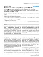
Figure 1
The mammalian families of NF-κB and IκB polypeptides. Conserved domains and their primary functions are indicated. Ankyrins, ankyrin repeat
domain (functions by binding and inhibiting RHDs; Bcl-3 and IκBζ are exceptions because they do not function as classical inhibitors of the NF-κB
activity); dimeriz., dimerization domain; DNA, DNA binding; NF-κB, nuclear factor-κB; IκB, inhibitor of NF-κB; RHD, Rel homology domain; NLS,
nuclear localization sequence; Transactivation, transactivating domain (functions at nuclear target sites).
Page 3 of 14
(page number not for citation purposes)
of the classical and alternative pathways of NF-κB activation
to the onset and maintenance of autoimmune reactivity, and
the subsequent inflammation that characterizes autoimmune
diseases. Examples will be drawn from several well studied
disease models, with particular attention given to RA.
Nuclear factor-
κκ
B in autoimmunity
NF-κB plays a central role in the differentiation, activation,
survival, and defense of mammalian cells. It contributes to
autoimmune diseases such as RA in multiple ways. First,
NF-κB is essential for normal lymphocyte and DC survival, for
their activation and development (including negative and
positive selection of B and T cells), and for lymphoid organ
morphogenesis [21,22]. Defects in NF-κB function or control
permit the survival and release into the periphery of auto-
reactive T cells from the thymus, where subsequent antigenic
stimuli may trigger autoimmune disease. Second, numerous
investigations into autoimmune disease have provided evi-
dence of NF-κB involvement in the induction of inflammatory
cytokines and other mediators of inflammation that drive the
pathology.
Nuclear factor-
κκ
B in lymphoid development
Signaling through NF-κB is essential for survival and activation
of most if not all mammalian cells, including lymphoid cells of
Available online />Figure 2
Classical pathway of NF-κB activation via IκB degradation. Ligand
engagement of specific membrane receptors triggers K63
polyubiquitination of TRAF2, TRAF6, RIP, MALT1, and NEMO. The
TAK kinase complex is recruited through association of the
polyubiquitin chains with TAB2 and TAB3. Activated TAK1 may
phosphorylate and activate IKKβ, which then phosphorylates IκB
bound to cytosolic NF-κB, triggering its βTrCP E3 ubiquitin ligase-
mediated K48 polyubiquitination and proteasomal degradation. Free
NF-κB then translocates to the nucleus and transactivates target
genes. CYLD and A20 are deubiquitinating enzymes that may block
NF-κB activation by removal of K63 ubiquitinated chains from activated
TRAFs, RIP, and NEMO. A20 may also terminate TNF-α induced NF-
κB activation by catalyzing the K48 ubiquitination of RIP, leading to its
proteasomal degradation. In addition to promoting survival via NF-κB
target genes, the TNF receptor (TNFR1) also stimulates competing
apoptotic pathways. T cell (and B cell) antigen receptors (TCR and
BCR, respectively [not shown]) may in some contexts enhance
apoptotic pathways but usually they contribute to survival (see text).
IκB, inhibitor of NF-κB; IKK, IκB kinase; MALT, mucosa-associated
lymphoid tissue lymphoma translocation gene; NEMO, NF-κB essential
modulator; NF-κB, nuclear factor-κB; RIP, receptor interacting protein;
TAB, TAK1-binding protein; TAK, transforming growth factor β-
activated kinase; TRAF, TNF receptor-associated factor.
Figure 3
Alternative pathway of NF-κB activation. In unstimulated cells, NIK is
destabilized by bound TRAF3. Activation through a subset of receptors
of the TNFR superfamily including the BAFFR, CD40, RANK and
lymphotoxin-βR leads to the recruitment of TRAF proteins (including
TRAF3) to the receptor. TRAF3 is inactivated (possibly by degradation
or sequestration) and active NIK is thus released. NIK then
phosphorylates and activates IKK; it also recruits NF-κB2/p100
(probably bound to RelB), which is phosphorylated by IKKα. This
triggers K48 polyubiquitination of p100 mediated by βTrCP E3
ubiquitin ligase and subsequent proteasomal processing to yield the
mature subunit p52. Predominantly RelB/p52 heterodimers are
generated, which migrate to the nucleus. The classical pathway is also
activated through these receptors with some receptors (BAFFR)
activating less strongly than others. Unlike TNFR (Figure 2), BAFFR
signaling is associated only with survival functions. BAFFR, B-cell
activating factor receptor; IKK, IκB kinase; LT, lymphotoxin; NF-κB,
nuclear factor-κB; NIK, NF-κB-inducing kinase; RANK, receptor
activator of NF-κB; TNFR, tumor necrosis factor receptor; TRAF, TNF
receptor-associated factor.
the immune system, both in the periphery and in the bone
marrow (B cells) and thymus (T cells). In autoimmune diseases
such as RA, defects in selection against autoreactive B cells
or in thymic selection of T cells may initiate the pathogenic
process. It is ultimately in the negative selection of self-
reactive B or T cells, in which a somewhat unusual pro-
apoptotic activity of NF-κB plays a role (or possibly its other
activities; see below), that defects in this activity can initiate
RA or other autoimmune disorders. Once B or T cells auto-
reactive for antigens present at the sites of RA (or reactive to
antigens arising from the environment, such as pathogen-
derived antigens) are released into the periphery and migrate
to those sites, further proinflammatory effects of NF-κB come
into play that aggravate and perpetuate the disease.
We recently reviewed the roles played by NF-κB in guiding
the survival and differentiation of developing B and T lympho-
cytes [21,22]. These are summarized in Figures 4 and 5. Brief
summaries of positive and negative selection of B and T cells
follow.
B-cell development
During B-cell development, immature B cells in the bone
marrow begin to express a BCR. If a given B cell’s BCR is
autoreactive, then that cell is either eliminated by apoptosis or
the BCR is ‘edited’ by RAG (recombinase-activating gene)
recombinase to generate a different BCR. RAG is negatively
regulated by NF-κB1 and positively regulated by NF-κB
dimers containing RelA and c-Rel [23]. It was suggested that
weak tonic signaling of the BCR may provide a positive
selection signal that represses RAG, possibly via NF-
κB1/p50 homodimers [24,25], thus blocking BCR editing. A
strong autoreactive signal may induce RAG expression (thus
facilitating editing) via activation of RelA-containing and c-
Rel-containing dimers. Failure to edit would trigger apoptosis
and negative selection. Survival of autoreactive cells (for at
least some time) may depend on survival factors including
BAFF, hemokinin-1, and thymic stromal lymphopoietin
[8,26,27]. Defects in NF-κB regulation both in bone marrow
and in spleen may allow autoreactive B cells to escape
negative selection, either directly via the above process or
indirectly because of defects in antigen-presenting cells
(DCs) or in bone marrow and splenic microarchitecture and
functions including those of stromal cells (see below). B-cell
selection can also occur in the periphery, where NF-κB is
essential for the maintenance of B-cell homeostasis. If this is
impaired, then survival of B cells may be prolonged and
autoimmune reactivity result [28] (see below).
T-cell development
During T-cell development in the thymus, positive and negative
selection occurs at the double-positive stage (Figure 5).
Autoreactive thymocytes are eliminated by apoptosis, whereas
those that weakly recognize self-antigens are positively
selected. The roles played by NF-κB in the process of T-cell
selection are complex and not fully elucidated. Apparently
contradictory results have been reported. First, negative
selection was found to be blocked by inhibition of NF-κB,
suggesting that NF-κB promotes apoptosis [29-31] (in
contrast to its well known anti-apoptotic activity). However,
Arthritis Research & Therapy Vol 10 No 4 Brown et al.
Page 4 of 14
(page number not for citation purposes)
Figure 4
NF-κB in B-lymphocyte development. A simplified schematic representation of B-lymphocyte development, highlighting some of the contributions
of NF-κB at various developmental checkpoints. See text for details. BAFFR, B-cell activating factor receptor; BCR, B-cell receptor; IKK, IκB
kinase; NF-κB, nuclear factor-κB; RAG, recombinase-activating gene; T1, transitional 1; T2, transitional 2; TNF, tumor necrosis factor.
negative selection was also reported to be due to repression
of NF-κB by IκB
NS
, an antigen-induced superrepressor
homologue of IκBα, suggesting a positive, anti-apoptotic role
for NF-κB in survival [32].
Positive selection of T cells that weakly recognized self-
antigens appeared to rely on the conventional anti-apoptotic
activity of NF-κB [31]. It is possible that NF-κB activity allows
the cell to assess TCR signal strength. Impairment of NF-κB
might be sensed by autoreactive cells as a weak TCR signal,
resulting in positive selection rather than correct negative
selection, thus promoting an autoimmune outcome. Similarly,
impairment of NF-κB under positive selection circumstances
might be sensed as a null signal, triggering death by neglect
[22]. Natural killer T cells and regulatory T cells (T
reg
s) are
positively selected by recognition of self-antigens at the
double-positive stage [33-36], or they are simply not
negatively selected [37] (Figure 5). Both are dependent on
NF-κB in their development [22], and the former at least
require NF-κB both in a cell-intrinsic role and in thymic
stromal cells in the form of RelB [33].
Nuclear factor-
κκ
B and immune tolerance
Both classical and alternative pathways of NF-κB activation
are involved in the control of autoimmune reactions exercised
by the thymic stroma. mTECs, which provide the thymic
microenvironment for developing T lymphocytes and myeloid
lineage DCs, play a critical role in preventing autoimmunity in
RA through their capacity to present self-antigen to T cells in
the thymus and (for DCs) in the periphery (draining lymph
nodes and spleen).
Several authors have shown that NF-κB is required for the
development of mTECs and organization of the thymic
stroma, and the development and differentiation of DCs
[38-43]. Genetic ablation of NF-κB family members in mice
and interference with or partial loss of NF-κB activation result
in defects in the thymic stromal development, absence of
mature mTECs and at least some subclasses of DCs, and
defects in the function of DCs. The phenotype of these mice
is characterized by severe autoimmunity with autoreactive
T cells, multiple organ lymphocytic infiltrates, and - in some
cases - early mortality. Both the classical and alternative
pathways of NF-κB activation appear to be essential for
correct thymic development and regulation of immune self-
tolerance. RelB, NF-κB-inducing kinase (NIK), and IKKα are
all components of the alternative pathway (leading to NF-κB2
activation and formation of p52/RelB heterodimers; Figure 3),
and defects in any one leads to impaired stromal cell
functions and autoimmune reactivity [38-42]. Deficiency of
NF-κB2 itself leads to a milder phenotype, possibly because
of compensation by NF-κB1, which can form heterodimers
with RelB (p50/RelB) in the absence of NF-κB2/p100 and
thus may be able to functionally replace p52/RelB in the NF-
κB2 knockout. Combined deficiency of NF-κB2 and the IκB
family member Bcl-3 leads to a full-blown autoimmune
phenotype, with complete loss of mTECs and consequent
loss of negative selection of autoreactive T cells [43].
Available online />Page 5 of 14
(page number not for citation purposes)
Figure 5
NF-κB in T lymphocyte development. A simplified schematic representation of T-lymphocyte development, highlighting some of the contributions of
NF-κB at various developmental checkpoints. T
Reg
and NKT cells branch off at some point after TCR expression on thymocytes. See text for details.
DP, double-positive stage; DN3/DN4, double-negative stages; IKK, IκB kinase; NF-κB, nuclear factor-κB; NKT, natural killer T cell; SP, single-
positive stage; TCR, T-cell receptor; T
Reg
, T-regulatory cell.
Intact upstream activators of the classical and alternative
pathways of NF-κB are also essential for normal lymphoid
organization and establishment of self-tolerance. TNFR-asso-
ciated factor (TRAF)6 is an essential component of many
signaling paths that activate the classic pathway, and TRAF6
deficiency in mice results in thymic atrophy: a disorganized
distribution of medullary epithelial cells, reduced T
reg
produc-
tion, absence of mature mTECs, and induction of auto-
immunity [39,41] (for review [44,45]). TRAF6 activates the
classical pathway (and activation of AP1 transcription
factors) after stimulation of members of the TNFR superfamily
and the Toll-like receptor/IL-1 receptor family (Figure 2). It
may indirectly activate the alternative pathway as a
consequence of activating the classic pathway [41,44,45].
This is because classically activated NF-κB regulates the
transcription of most NF-κB family members, including
NF-κB2 and RelB, the principal targets for activation by the
alternative pathway [46,47]. TRAF6 deficiency resulted in a
lack of RelB expression in mTECs and fetal thymic stroma
[41]. It was concluded that reduced T
reg
development and
reduced negative selection caused by absence of selecting
mTECs were two possible causes of the autoimmunity seen
in TRAF6 knockout mice. Others have also shown that
TRAF6 and RelB are critical for DC development and
maturation, and are essential for proper DC interaction with
T cells [38,39].
LTβ receptors, as well as RANK and CD40 receptors, are
expressed on stromal cells and, when stimulated, activate the
alternative NF-κB pathway [48-50]. Consistent with a role for
LTβR-, RANK-, and CD40-mediated activation of the
alternative pathway in stromal cells during thymic organo-
genesis, mutant mouse models deficient in signaling via the
LTβR, RANK, or CD40 have defects similar to those des-
cribed above for mice lacking components of the alternative
pathway. These include thymic defects and multiple organ
lymphocytic infiltrations characteristic of self-autoreactivity
[51-54]. However, loss of any one of the receptors and/or
their ligands results in relatively mild defects compared with
loss of the alternative pathway, most likely because the three
receptors are partially redundant.
Autoimmune mouse models associated with defective
central or peripheral tolerance
Several mouse models of autoimmune arthritis and lupus
implicate thymic selection defects in the pathogenesis. In the
SKG ζ-associated protein of 70 kDa (ZAP-70) model, spon-
taneous mutation in ZAP-70 (a key transduction molecule in
T cells that is responsible for transducing signals from the
T-cell antigen receptor to the classical pathway of NF-κB
activation and to other transcription factors) causes chronic
autoimmune arthritis in mice, which develops after encounter
with environmental stimuli (in particular, fungal β-glucans and
viruses) [55,56]. The disease closely resembles human RA.
Thus, although genetic predisposition plays an important role
in pathogenesis of this autoimmune disorder, like other
examples of autoimmune disease, exposure to infectious
agents also has an important part in the development of this
disorder (for review [57]). Altered signal transduction through
the mutant ZAP-70 protein changes the sensitivity of
developing T cells to both positive and negative selection of
thymocytes, thereby leading to the positive selection of
otherwise negatively selected self-reactive T cells. These self-
reactive T cells apparently overcome the mechanisms of
peripheral self-tolerance mediated by T
reg
s. Such potentially
arthritogenic T cells might also arise in a subset of humans
who go on to develop RA as a result of an SKG-like mutation,
driving a selection shift of the T-cell repertoire in the thymus
that could lead to the development of RA after exogenous
stimulation in the periphery by microbes [55,56].
Sakaguchi and coworkers [55] raised the interesting ques-
tion of why the general change in the T-cell repertoire in the
SKG mice should lead to autoimmune arthritis but not other
autoimmune diseases. They suggested that unlike other
organ-specific autoimmune diseases, in which self-reactive
T cells destroy the target cells (for example, in type 1
diabetes pancreatic β cells are destroyed), in autoimmune
arthritis in SKG mice (and in RA in humans) the self-reactive
T cells do not destroy synoviocytes but stimulate them to
proliferate [55,58-60]. They also secrete proinflammatory
cytokines (IL-1, IL-6, and tumor necrosis factor [TNF]-α) and
mediators that destroy the surrounding cartilage and bone.
In the New Zealand Black lupus-prone mouse model a
defective NF-κB/RelB pathway leads to disorganization of the
thymus and associated thymocyte selection defects [61].
Breakdown of self-tolerance in the periphery (after exit from
the bone marrow) during B-cell development and survival has
also been reported to lead to autoimmunity. BAFF is a crucial
B-lymphocyte survival factor [8,62,63], and one of its
receptors - BAFFR - appears to be the only mediator of
BAFF-mediated survival signals. BAFFR signals primarily
through the alternative NF-κB pathway and interacts directly
with TRAF3 (this is essential for its signal transduction).
Specific knockout of the gene encoding TRAF3 in mouse B
cells led to increased, constitutive activation of NF-κB2,
prolonged B-cell survival, and greatly expanded B-cell
compartments in secondary lymphoid organs. Splenomegaly,
lymphadenopathy, hyperimmunoglobulinemia, and autoimmune
reactivity resulted. This implicates TRAF3 and the alternative
NF-κB pathway in regulation of B-cell homeostasis and
peripheral self-tolerance [28].
Inflammatory effects of nuclear factor-
κκ
Bin
rheumatoid arthritis
Involvement of the alternative pathway at the site of
inflammation
RA is a chronic inflammatory disease of the joints in which
infiltration of immunocompetent cells and the proliferation of
synovial fibroblasts of the joint lining leads to formation of a
tumor-like tissue called the pannus, which invades and
Arthritis Research & Therapy Vol 10 No 4 Brown et al.
Page 6 of 14
(page number not for citation purposes)
destroys the joint cartilage and bone [64]. In the inflammatory
microenvironment of the synovium, lymphoid neogenesis
occurs, generating organized lymphocytic aggregates or
tertiary lymphoid organs (TLOs) with B-cell and T-cell areas
[65,66]. TLOs are also seen in some other chronic inflam-
matory diseases and in mouse models of such diseases,
including collagen-induced arthritis (CIA) [64]. The identity of
stromal cells initiating their development is unknown. The
alternative pathway of NF-κB activation may be implicated in
TLO generation, because constitutive expression of LTβ in
target tissues has been shown to cause TLO formation [67].
Decoy receptors for LTβ reduce inflammation in disease
models of CIA [68].
A further characteristic of most autoimmune diseases,
including RA, is the elevated level in target tissue fluids (in
RA, the synovial fluid) of the cytokine BAFF. This correlates
with the survival of B lymphocytes, which produce auto-
antibodies [69]. BAFF is an activator, principally of the alter-
native NF-κB pathway [8], and is needed for B-cell matura-
tion and for protection of otherwise negatively selected B
cells. It is also needed for plasma cell differentiation and
survival, and it is these cells that are responsible for antibody
production [70]. Antagonists of BAFF, including BAFF
antibody (belimumab) and decoy receptors, have been
developed and are under examination for targeting B cells in
RA and other autoimmune diseases [71,72].
NIK, a key mediator of the alternative pathway (Figure 3), has
also been shown in mouse models to be necessary for
antigen-mediated induction of the bone erosion caused by
inflammation-induced osteoclastogenesis. NIK-deficient mice
were largely resistant to RA, exhibiting less periarticular
osteoclastogenesis and less bone erosion [73].
Involvement of the classical pathway at sites of
inflammation
The classical pathway of NF-κB is also strongly implicated in
the inflammatory stages of RA. Inflammatory cells infiltrate
the synovial sublining and produce proinflammatory cyto-
kines, chemokines, and growth factors that stimulate synovial
lining hyperplasia. This results in increased numbers and
activation of macrophage-like synoviocytes and fibroblast-
like synoviocytes. In turn, synoviocytes release additional
cytokines, chemokines, and growth factors that help to
sustain inflammation and produce enzymes that degrade the
organized extracellular matrix, destroying cartilage and bone
[74-76]. Ectopic expression of IκBα (a principal inhibitor of
classical NF-κB activation; Figure 2) in human macrophages
and primary RA synoviocytes inhibited the production of
destructive enzymes (matrix metalloproteinases and aggre-
canases) and inflammatory cytokines (IL-1β, IL-6, IL-8, and
TNF-α) while sparing anti-inflammatory mediators, indicating
that the classical NF-κB pathway is essential for synthesis of
matrix-destructive enzymes and inflammatory cytokines
[74,75,77,78].
Evidence reviewed by Makarov [79] suggests that NF-κB
activation facilitates synovial hyperplasia by promoting pro-
liferation and inhibiting apoptosis of RA fibroblast-like
synoviocytes (FLSs). Briefly, NF-κB is a positive regulator of
cell growth in FLSs primarily via the induction of c-Myc and
cyclin D
1
, proteins required for cell cycle progression, but
also via inhibition of the pro-apoptotic effects of c-Myc.
Because c-Myc is highly expressed in RA synovium NF-κB
may thus contribute to hyperplasia by both inhibiting c-Myc-
induced apoptosis and promoting proliferation. NF-κB also
delivers an anti-apoptotic signal that counteracts other pro-
apoptotic stimuli such as TNF-α (which induces classical
NF-κB activation). Activation of NF-κB protected human RA
FLSs from the cytotoxic effects of TNF [80], whereas its
inhibition in arthritic rat joints by proteasome inhibitors (which
blocked IκB degradation) or by genetic introduction of IκB
NS
resulted in increased FLS apoptosis. These results suggest
an important role for NF-κB in protecting FLSs against
apoptosis in RA synovium, possibly by countering the
cytotoxicity of TNF-α and Fas ligand [81]. Because TNF is
also a potent mitogen in RA FLSs, NF-κB appears to be
critical in determining whether it exerts mitogenic or pro-
apoptotic effects.
The foregoing discussion implies that blocking NF-κB
activation by either the classical and/or the alternative path-
way may be therapeutically beneficial for human RA inflam-
mation. A major consideration, however, is the safety of this
approach, given the major roles played by this transcription
factor family in a host of essential functions, including
immunity and cell development [82,83].
The T-helper-17/IL-17/nuclear factor-
κκ
B axis in
rheumatoid arthritis
Continued inflammation and the resulting destruction of bone
and cartilage in joints of patients with RA depend on a
complex network of cells and cytokines [84]. Cells that are
critically involved in RA include synovial fibroblasts, chondro-
cytes, DCs, macrophages, monocytes, osteoclasts, neutro-
phils, and B and T cells. T cells may account for up to 40% of
the synovial cellular infiltrate [85]. Self-antigen specific T cells
play a role in the production of autoantibodies by providing
help to B cells, probably both locally and in draining lymph
nodes. However, the infiltrating T cells also play a more direct
role in RA. A critical T-helper (Th) cell type in RA is the Th17
subset, and these cells produce IL-17, which is emerging as
a primary effector of RA pathology [86]. IL-17 induces many
chemokines and cytokines, in part by activating NF-κB via the
classical pathway; it potently synergizes with TNF-α, which is
another cytokine that is critical in RA pathogenesis (see
below). Blocking TNF-α signaling with etanercept (a soluble
form of the TNFR α) has proven to be beneficial to many RA
patients [87]. In the following discussion, we first provide
some background on the generation of Th17 cells, which are
the main producers of IL-17. We then discuss the biologic
effects of Th17 and IL-17 in the context of RA, and the direct
Available online />Page 7 of 14
(page number not for citation purposes)
and indirect mechanisms by which IL-17 leads to activation of
NF-κB.
Th17 cell development
During the past few years there has been a shift in the
paradigm of T-cell help, which was thought to occur exclu-
sively through either Th type 1 (Th1) or type 2 (Th2) cells, but
now also includes Th17 cells (for review [88]). Th1 cells are
primarily responsible for cell-mediated immunity and Th2 cells
for humoral immunity. The exclusive division of T-cell help into
these two classes underwent a major correction when an
additional helper T-cell type was identified, named Th17 after
its signature cytokine IL-17. In mice, Th17 cells require
transforming growth factor-β and IL-6 for their differentiation
from naïve T cells, and their maintenance and expansion is
controlled by IL-23, a cytokine that is produced by DCs. Both
IFN-γ and IL-4 can suppress the differentiation of Th17 cells,
and there is some evidence that IL-17 can suppress Th2
responses [89]. Interestingly, transforming growth factor-β is
not only required for generation of Th17 cells but also for the
generation of T
reg
s, at least in the periphery, and so it is the
presence or absence of IL-6 that decides between the two
T-cell fates. It may be the particularly high levels of IL-6 present
in inflamed joints (see below) that shifts the balance from T
reg
s
to Th17, thus preventing resolution of the inflammation. The
division between Th1 and Th17 cells may not always be
absolute, especially at the site of inflammation in vivo,
because T cells producing IFN-γ and IL-17 can coexist, and
there is even some evidence that a single T-cell type can
coexpress both cytokines, especially in humans [90].
The initial development of Th17 in humans looks to be some-
what different from that in mouse; recent evidence suggests
that IL-6 and IL-1 may be the main initiators [91]. Thereafter,
IL-23 functions prominently in both human and mice.
Interestingly, bacterial peptidoglycan-derived muramyl dipep-
tide is a particularly potent inducer of IL-23 and IL-1 in DCs,
which in turn elicit strong IL-17 responses from the human
memory T-cell pool [92]. Muramyl dipeptide signals via the
NOD2 adaptor protein to induce transcription of IL-23 (and
probably IL-1) via the classical NF-κB pathway and it also
activates caspase-1 to process pro-IL-1β.
Th17/IL-17 in autoimmune diseases
Once the existence of Th17 cells was recognized, it soon
became evident that many inflammatory conditions may be
partly or largely driven by Th17 and not by Th1, as was
erroneously concluded previously [88,93-96]. Th17 and/or
IL-17 have been reported to be centrally involved in multiple
sclerosis (and its mouse model experimental autoimmune
encephalomyelitis) and RA (and its mouse model CIA). In
addition, evidence is accumulating for a role of the Th17/
IL-17 axis in many other inflammatory conditions and auto-
immune diseases, including inflammatory bowel disease,
psoriasis, periodontal disease, inflammatory airways diseases,
and possibly even systemic lupus erythematosus (see above).
Although there is considerable support for the involvement of
Th17/IL-17 in multiple sclerosis and RA (see below), evi-
dence for its roles in the other human diseases is more
circumstantial and often rests on the detection of high
expression levels of IL-17 at sites of inflammation. Th17 and
IL-17 are generally thought to be critical in defense against
extracellular bacteria and some fungi, especially at mucosal
and epithelial surfaces [88,95,97,98]. IL-17 is particularly
potent in inducing chemokines that recruit neutrophils to fight
these pathogens. The Th17/IL-17 axis thus represents
another instance in which the lines between innate and
adaptive immunity become blurred, because the antigen-
specific T cells elicit innate responses via IL-17 in this case.
Th17/IL-17 in rheumatoid arthritis
Regarding RA, multiple lines of investigation support the
critical involvement of Th17 and IL-17. For example, synovial
fluid from joints of RA patients contains high levels of IL-17,
and the T cells present in synovial cultures from RA patients
spontaneously secrete IL-17 [96]. Nevertheless, the impor-
tance of Th17 cells to the pathogenesis of RA remains to be
definitively proven; for example, one publication reports a
predominance of Th1 rather than Th17 in RA joints, although
it must be kept in mind that the presence of a mixed Th1/
Th17 type of helper might have been present (see above)
[99,100].
The importance of Th17/IL-17 in mouse RA models, however,
has been clearly established. CIA is markedly suppressed in
IL-17 deficient mice [101], and treatment of mice with a
neutralizing anti-IL-17 antibody in early and later phases of
CIA reduces joint inflammation, cartilage destruction, and
bone erosion [102]. Furthermore, IL-17 receptor deficient
mice are substantially blocked in development of strepto-
coccal cell wall induced arthritis [103]. It is worth noting that
IL-17 is produced not only by Th-17 cells, but also by some
other cells, including - in particular - oligoclonal γ/δ T cells;
these cells may also contribute to RA/CIA [104]. In the
naturally mutated SKG strain of mice discussed above
(recessive mutation in ZAP-70), the spontaneously arising
self-reactive T cells develop a T-cell mediated autoimmune
arthritis, resembling RA [105]. The self-reactive T cells are
able to induce expression of IL-6 in antigen-presenting cells,
and IL-6 in turn mediates differentiation of self-reactive T cells
into arthritogenic Th17 cells. Loss of either IL-6 or IL-17
completely blocks arthritis development in this model.
Interestingly, pathologic arthritis does require a trigger, which
can be supplied by stimulation of innate immunity or by IFN-γ
deficiency or any other stimulus that leads to expansion of the
Th17 cells [86,106-108]. Toll-like receptors are likely to be
involved in pathogen-derived triggers, and a significant part of
their intracellular effects is mediated by activation of the
classical pathway of NF-κB [109].
Experimentally induced over-expression of IL-17 in naïve
mouse joints leads to many of the signs of RA, including
Arthritis Research & Therapy Vol 10 No 4 Brown et al.
Page 8 of 14
(page number not for citation purposes)
chronic inflammation and bone erosion, and it exacerbates
existing pathology in acute arthritis models [109]. Further
evidence for a critical role for Th17 cells also comes from
investigations into IL-23. Synovial fluid from RA patients
contains elevated levels of IL-23 p19 protein, and the degree
of elevation was directly correlated with the levels of IL-17,
IL-1, and TNF-α; furthermore, levels were highest in patients
with bony erosions [108]. Finally, anti-IL-23 antibodies were
reported to attenuate CIA [110].
These findings clearly implicate Th17 and IL-17 in the patho-
genesis of RA, but why should this be so? IL-17 receptors
are fairly ubiquitously expressed, and IL-17 induces many
cytokines in various cells, including synovial fibroblasts, such
as IL-6, TNF-α, and IL-1, as well as chemokines, especially
CXC chemokines that can recruit neutrophils [84,95]. The
effect of IL-17 is greatly enhanced by synergy with TNF-α,
which is produced by T cells and activated macrophages,
among other cells (more details is provided on the synergy
between IL-17 and TNF-α below) [94,95]. Activated macro-
phages also produce IL-6 and IL-1. IL-6 (and by some
accounts IL-1, TNF-α and IL-17), in addition to Toll-like
receptor-2 and -4 ligands, directly or indirectly lead to
expression of RANK ligand (RANKL) on osteoblastic stromal
cells and synoviocytes [102,103,107,108,110-113]. RANKL
is the primary mediator of osteoclastogenesis and is essential
also for the maintenance and function of mature osteoclasts
(Figure 6). Th17 cells can directly stimulate this process as
well, because only this T-helper class preferentially expresses
RANKL [114]. IL-17 in addition leads to downregulation of
osteoprotegerin, the natural antagonist of RANKL [111,112].
The increased ratio of RANKL over osteoprotegerin assures
generation of osteoclasts from monocyte precursors and
continued activation and maintenance of mature osteoclasts;
activated osteoclasts erode bone and thus are critically
involved in RA pathology (Figure 6). IL-1 and TNF-α also
directly contribute to the differentiation of osteoclasts and
their activation after maturation [115,116].
IL-17 has additional pathogenic effects in RA. Activated
synoviocytes, chondrocytes, and infiltrating mononuclear cells
produce a variety of metalloproteases, cathepsin G and
elastase, leading to destruction of the extracelluar matrix and
cartilage, and further bone erosion [113]. IL-17 and IL-6
block matrix synthesis by articular chondrocytes; nitric oxide
produced via induction of inducible nitric oxide synthetase in
synoviocytes and macrophages leads to further degeneration
of chondrocytes; and IL-17-induced cyclo-oxygenase-2 leads
to production of prostaglandin E
2
and thus further inflam-
mation, cartilage damage, and bone erosion. Finally, neutro-
phils recruited via IL-17 induced chemokines further contribute
to tissue destruction [86,94,95,103,112,113] (Figure 6).
IL-17 and activation of the classical pathway
The interdependent network of cytokines in RA involves
various positive feedback loops. For example, optimal
differentiation and expansion of Th17 cells and production of
IL-17 requires IL-6, as well as IL-23 and IL-1, but these same
cytokines are also induced downstream of IL-17
[112,113,117]. The proinflammatory cytokines discussed
here, including TNF-α and IL-1 as well as IL-17, all induce the
classical pathway of NF-κB activation (see below), whereas
RANKL induces both the classical and the alternative
pathway. A number of studies have shown the importance of
both pathways in osteoclastogenesis and in subsequent
function of matured osteoclasts in response to RANKL
stimulation [115,116,118]. Given the central role of cytokines
in RA and their interdependence, it may not be too surprising
that therapeutic approaches aimed at disrupting this network
have shown great promise in patients with RA and in mouse
models. Treatments targeting the signaling via IL-6, TNF-α,
IL-1, IL-17, and RANKL were all quite effective in attenuating
pathogenesis [86,112].
Th17 cells produce IL-17A (also known as IL-17), as well as
IL-17F, which thus far appears to have same biologic activity
Available online />Page 9 of 14
(page number not for citation purposes)
Figure 6
The immune system regulates bone resorption through enhanced
osteoclastogenesis. Cells of the adaptive and innate immune systems
contribute to regulation of bone turnover through production of
cytokines and direct cell-cell interactions. Proinflammatory cytokines
such as IL-6, IL-1β, and TNF-α are secreted by macrophages and
fibroblasts secrete IL-6. Th17 lymphocytes produce IL-17, IL-6, and
TNF-α. In RA these cytokines drive bone erosion by induction of
RANKL expression by osteoblast stromal cells. Th17 lymphocytes also
secrete RANKL, which binds to RANK receptor on osteoclast
precursors triggering osteoclast maturation and activation, thus
enhancing bone loss. Osteoprotegerin (OPG) is a soluble decoy
receptor that inhibits RANKL binding to RANK thus limiting bone
resorption. IL-17 increases RANKL expression and concomitantly
decreases OPG expression in osteoblasts, causing enhanced
formation of osteoclasts and bone erosion. Neutrophils also contribute
to bone and cartilage degradation by secretion of degradative factors.
IL, interleukin; RANK, receptor activator of NF-κB; Th, T-helper; TNF,
tumor necrosis factor.
as IL-17, although it has a weaker affinity for the IL-17
receptor [95]. The receptor may be a heteromeric complex
containing the IL-17RA (also known as IL-17R) and RC
chains. The ligand family consists of six members (IL-17A-F),
whereas the receptor family has five members (IL-17RA-RE)
[88,94]. IL-17E (also known as IL-25) and its receptor
IL-17RB have been shown to play a role in Th2-type
responses [119], whereas relatively little is known about the
remaining members of the ligand and receptor families.
IL-17 stimulation induces the recruitment of the adaptor
protein CIKS (connection to IκB kinase and stress-activated
protein kinases; also known as Act1) to the IL-17R to trans-
duce signals [120,121]. This adaptor has been shown to be
essential for the development of experimental autoimmune
encephalomyelitis, complementing previous data implicating
Th17 and IL-17 in this disease [122]. Both CIKS and the
receptor chains contain a so-called SEFIR domain (similar
expression to fibroblast growth factor genes and IL-17Rs and
Toll and IL-1R), which is distantly related to the Toll and IL-1R
(TIR) domain. The recruitment of CIKS to the IL-17R occurs
via heterotypic SEFIR domain interactions, similar to the way
that Toll-like receptors recruit the adaptor MyD88 via TIR
domain interactions. IL-17 activates NF-κB and mitogen-
activated protein kinases via CIKS/Act1, although the
molecular mechanisms are not well understood at this point
[120,121]. CIKS is known to interact with NEMO/IKKγ, the
regulatory subunit of the IKK complex [123]. CIKS/Act1 can
also bind to TRAF3 and may bind to TRAF6 in response to
signals; furthermore, activation of NF-κB has been suggested
to proceed via TAK1 activation [120-122]. Signaling via the
IL-17Rs also activates CCAAT/enhancer binding protein
(c/EBP)β and c/EBPδ, which requires not only the SEFIR
domain (and CIKS) but also additional receptor domains
[124]. Many IL-17 target genes contain both c/EBP and NF-
κB binding sites and these appear to function cooperatively
on DNA to promote transcription, and IL-17 has been shown
to act synergistically with TNF-α in inducing many of its target
genes in fibroblasts in vitro [94,95].
The synergy between TNF-α and IL-17 may be due in part to
the ability of IL-17 to stabilize short-lived mRNAs that are only
transiently induced by TNF-α alone [125], although nothing is
known about how IL-17 may stabilize such mRNAs. Never-
theless, the synergy is profound because many target genes
are affected. Cumulative evidence also suggests that IL-17
can directly and immediately activate a modest level of NF-κB
activity, which is probably critical for its functions in the
absence of TNF-α or other signals that activate NF-κB. In
addition, IL-17, but not TNF-α, induces IκBζ, a member of the
IκB family that is able to promote NF-κB activity, in contrast
to the classic IκBs, which act as cytoplasmic inhibitors. It has
been suggested that IκBζ facilitates the synergy between
NF-κB and c/EBP transcription factors [126]. This may
provide an additional mechanism by which IL-17 synergizes
with TNF-α. As discussed above, IL-17 also activates NF-κB
indirectly in other cells through induction of various cytokines,
such as RANKL.
Conclusion
Both classical and alternative pathways of NF-κB activation
regulate survival and activation of T and B lymphocytes at
their sites of development in thymus, bone marrow and
spleen, and in the periphery. In normal conditions of health
the immune system balances antigen presentation and pro-
inflammatory activity in the periphery in response to patho-
gens and other environmental challenges to prevent excessive
autoreactivity of the T-cell and B-cell complement. Improperly
regulated NF-κB function leading to its constitutive activation
causes autoimmunity, engendering chronic inflammation, for
example in the articular joints in RA. Autoimmune diseases
may be initiated by malfunctioning lymphocytes whose
apoptotic pathways, normally activated by self-antigens, are
blocked by abnormal activation of NF-κB, enabling the
survival of self-reactive cells [21,127-130].
The multiple roles of NF-κB in autoimmune diseases make it
an important pharmaceutical target. Given its many crucial
roles in maintaining health, including roles in acute host
defense and lymphocyte development, systemic NF-κB
inhibitors are likely to have deleterious side effects, particu-
larly if used for long periods. Such inhibitors, however, might
be useful in doses that interfere with disease progression
while sparing normal processes. More promising are inhibi-
tors that target a specific subunit of NF-κB or the pathway(s)
that leads to its activation in a particular disease. To discover
such targets and inhibitors, we need to advance our under-
standing of the roles of NF-κB and its pathways of activation
in healthy and diseased cells. Furthermore, the unwanted
effects of blocking NF-κB activity might be reduced by
targeting inhibitors to specific tissues or cell types. Genetic
delivery of NF-κB inhibitors may be useful in this regard, and
local tissue delivery may avoid deleterious side effects of
systemic exposure and minimize broader immunosuppression
[104]. Recent reviews have outlined the advantages and
disadvantages of anti-inflammatory and anti-rheumatic NF-κB
inhibitors, and the effects (in animal models of RA and other
autoimmune diseases) of genetically inactivated NF-κB
subunits and ectopic IκBα. Together, the results support the
feasibility of using NF-κB inhibitors in therapeutic strategies
for RA and other autoimmune disorders [82,83,131-133].
Competing interests
The authors declare that they have no competing interests.
References
1. Karin M, Ben-Neriah Y: Phosphorylation meets ubiquitination:
the control of NF-kappa B activity. Annu Rev Immunol 2000,
18:621-663.
2. Li Q, Verma IM: NF-kappa B regulation in the immune system.
Nat Rev Immunol 2002, 2:725-734.
3. Brown K, Claudio E, Siebenlist U: New developments in NF-
kappa B. In Contemporary Targeted Therapies in Rheumatology.
Edited by Smolen JS, Lipsky PE. London: Informa; 2007:285-296.
Arthritis Research & Therapy Vol 10 No 4 Brown et al.
Page 10 of 14
(page number not for citation purposes)
4. Hayden MS, Ghosh S: Signaling to NF-kappaB. Genes Dev
2004, 18:2195-2224.
5. Bonizzi G, Karin M: The two NF-kappaB activation pathways
and their role in innate and adaptive immunity. Trends
Immunol 2004, 25:280-288.
6. Senftleben U, Cao Y, Xiao G, Greten FR, Krahn G, Bonizzi G,
Chen Y, Hu Y, Fong A, Sun SC, Karin M: Activation by IKKalpha
of a second, evolutionary conserved, NF-kappa B signaling
pathway. Science 2001, 293:1495-1499.
7. Dejardin E, Droin NM, Delhase M, Haas E, Cao Y, Makris C, Li
ZW, Karin M, Ware CF, Green DR: The lymphotoxin-beta
receptor induces different patterns of gene expression via
two NF-kappaB pathways. Immunity 2002, 17:525-535.
8. Claudio E, Brown K, Park S, Wang H, Siebenlist U: BAFF-
induced NEMO-independent processing of NF-kappa B2 in
maturing B cells. Nat Immunol 2002, 3:958-965.
9. Bonizzi G, Bebien M, Otero DC, Johnson-Vroom KE, Cao Y, Vu D,
Jegga AG, Aronow BJ, Ghosh G, Rickert RC, Karin M: Activation
of IKKalpha target genes depends on recognition of specific
kappaB binding sites by RelB:p52 dimers. Embo J 2004, 23:
4202-4210.
10. Cao Y, Bonizzi G, Seagroves TN, Greten FR, Johnson R, Schmidt
EV, Karin M: IKKalpha provides an essential link between
RANK signaling and cyclin D1 expression during mammary
gland development. Cell 2001, 107:763-775.
11. Novack DV, Yin L, Hagen-Stapleton A, Schreiber RD, Goeddel
DV, Ross FP, Teitelbaum SL: The IkappaB function of NF-
kappaB2 p100 controls stimulated osteoclastogenesis. J Exp
Med 2003, 198:771-781.
12. Silverstein AM: Autoimmunity versus horror autotoxicus: the
struggle for recognition. Nat Immunol 2001, 2:279-281.
13. Stefanova I, Dorfman JR, Germain RN: Self-recognition pro-
motes the foreign antigen sensitivity of naive T lymphocytes.
Nature 2002, 420:429-434.
14. Dale E, Davis M, Faustman DL: A role for transcription factor
NF-kappa B in autoimmunity: possible interactions of genes,
sex, and the immune response. Adv Physiol Educ 2006, 30:
152-158.
15. Lipsky PE: Systemic lupus erythematosus: an autoimmune
disease of B cell hyperactivity. Nat Immunol 2001, 2:764-766.
16. Anolik J, Sanz I: B cells in human and murine systemic lupus
erythematosus. Curr Opin Rheumatol 2004, 16:505-512.
17. Goodnow CC, Sprent J, Fazekas de St Groth, Vinuesa CG: Cel-
lular and genetic mechanisms of self tolerance and autoim-
munity. Nature 2005, 435:590-597.
18. Schulze-Koops H, Kalden JR: T cells-overview-update. In Con-
temporary Targeted Therapies in Rheumatology. Edited Smolen
JS, Lipsky PE. London: Informa; 2007:1-6.
19. Nutku E, Pugh-Bernard AE, Gauld S, Merrell K, Cambier JC: B-
cell antigen receptor signaling and autoimmunity. In Contem-
porary Targeted Therapies in Rheumatology. Edited by Smolen
JS, Lipsky PE. London: Informa; 2007:31-44.
20. Lutzky V, Thomas R: Dendritic cells. In Contemporary Targeted
Therapies in Rheumatology. Edited Smolen JS, Lipsky PE. UK:
Informa; 2007:63-78.
21. Siebenlist U, Brown K, Claudio E: Control of lymphocyte devel-
opment by nuclear factor-kappaB. Nat Rev Immunol 2005, 5:
435-445.
22. Claudio E, Brown K, Siebenlist U: NF-kappaB guides the sur-
vival and differentiation of developing lymphocytes. Cell Death
Differ 2006, 13:697-701.
23. Verkoczy L, Ait-Azzouzene D, Skog P, Martensson A, Lang J,
Duong B, Nemazee D: A role for nuclear factor kappa B/rel
transcription factors in the regulation of the recombinase acti-
vator genes. Immunity 2005, 22:519-531.
24. Wessells J, Baer M, Young HA, Claudio E, Brown K, Siebenlist U,
Johnson PF: BCL-3 and NF-kappaB p50 attenuate lipopolysac-
charide-induced inflammatory responses in macrophages. J
Biol Chem 2004, 279:49995-50003.
25. Driessler F, Venstrom K, Sabat R, Asadullah K, Schottelius AJ:
Molecular mechanisms of interleukin-10-mediated inhibition
of NF-kappaB activity: a role for p50. Clin Exp Immunol 2004,
135:64-73.
26. Thomas MD, Kremer CS, Ravichandran KS, Rajewsky K, Bender
TP: c-Myb is critical for B cell development and maintenance
of follicular B cells. Immunity 2005, 23:275-286.
27. Milne CD, Fleming HE, Zhang Y, Paige CJ: Mechanisms of
selection mediated by interleukin-7, the preBCR, and hemo-
kinin-1 during B-cell development. Immunol Rev 2004, 197:75-
88.
28. Xie P, Stunz LL, Larison KD, Yang B, Bishop GA: Tumor necro-
sis factor receptor-associated factor 3 is a critical regulator of
B cell homeostasis in secondary lymphoid organs. Immunity
2007, 27:253-267.
29. Hettmann T, DiDonato J, Karin M, Leiden JM: An essential role
for nuclear factor kappaB in promoting double positive thy-
mocyte apoptosis. J Exp Med 1999, 189:145-158.
30. Ren H, Schmalstieg A, van Oers NS, Gaynor RB: I-kappa B
kinases alpha and beta have distinct roles in regulating
murine T cell function. J Immunol 2002, 168:3721-3731.
31. Mora AL, Stanley S, Armistead W, Chan AC, Boothby M: Ineffi-
cient ZAP-70 phosphorylation and decreased thymic selection
in vivo result from inhibition of NF-kappaB/Rel. J Immunol
2001, 167:5628-5635.
32. Fiorini E, Schmitz I, Marissen WE, Osborn SL, Touma M, Sasada
T, Reche PA, Tibaldi EV, Hussey RE, Kruisbeek AM, Reinherz EL,
Clayton LK: Peptide-induced negative selection of thymocytes
activates transcription of an NF-kappa B inhibitor. Mol Cell
2002, 9:637-648.
33. Sivakumar V, Hammond KJ, Howells N, Pfeffer K, Weih F: Differ-
ential requirement for Rel/nuclear factor kappa B family
members in natural killer T cell development. J Exp Med 2003,
197:1613-1621.
34. Schmidt-Supprian M, Courtois G, Tian J, Coyle AJ, Israel A,
Rajewsky K, Pasparakis M: Mature T cells depend on signaling
through the IKK complex. Immunity 2003, 19:377-389.
35. Schmidt-Supprian M, Tian J, Grant EP, Pasparakis M, Maehr R,
Ovaa H, Ploegh HL, Coyle AJ, Rajewsky K: Differential depen-
dence of CD4
+
CD25
+
regulatory and natural killer-like T cells
on signals leading to NF-kappaB activation. Proc Natl Acad Sci
USA 2004, 101:4566-4571.
36. Zheng Y, Vig M, Lyons J, Van Parijs L, Beg AA: Combined defi-
ciency of p50 and cRel in CD4
+
T cells reveals an essential
requirement for nuclear factor kappaB in regulating mature T
cell survival and in vivo function. J Exp Med 2003, 197:861-
874.
37. van Santen HM, Benoist C, Mathis D: Number of T reg cells that
differentiate does not increase upon encounter of agonist
ligand on thymic epithelial cells. J Exp Med 2004, 200:1221-
1230.
38. Burkly L, Hession C, Ogata L, Reilly C, Marconi LA, Olson D,
Tizard R, Cate R, Lo D: Expression of relB is required for the
development of thymic medulla and dendritic cells. Nature
1995, 373:531-536.
39. Kobayashi T, Walsh PT, Walsh MC, Speirs KM, Chiffoleau E, King
CG, Hancock WW, Caamano JH, Hunter CA, Scott P, Turka LA,
Choi Y: TRAF6 is a critical factor for dendritic cell maturation
and development. Immunity 2003, 19:353-363.
40. Kajiura F, Sun S, Nomura T, Izumi K, Ueno T, Bando Y, Kuroda N,
Han H, Li Y, Matsushima A, Takahama Y, Sakaguchi S, Mitani T,
Matsumoto M: NF-kappa B-inducing kinase establishes self-
tolerance in a thymic stroma-dependent manner. J Immunol
2004, 172:2067-2075.
41. Akiyama T, Maeda S, Yamane S, Ogino K, Kasai M, Kajiura F, Mat-
sumoto M, Inoue J: Dependence of self-tolerance on TRAF6-
directed development of thymic stroma. Science 2005, 308:
248-251.
42. Zhang B, Wang Z, Ding J, Peterson P, Gunning WT, Ding HF:
NF-kappaB2 is required for the control of autoimmunity by
regulating the development of medullary thymic epithelial
cells. J Biol Chem 2006, 281:38617-38624.
43. Zhang X, Wang H, Claudio E, Brown K, Siebenlist U: A role for
the IkappaB family member Bcl-3 in the control of central
immunologic tolerance. Immunity 2007, 27:438-452.
44. Thomas R: The TRAF6-NF-kappa B signaling pathway in
autoimmunity: not just inflammation. Arthritis Res 2005, 7:170-
173.
45. Derbinski J, Kyewski B: Linking signalling pathways, thymic
stroma integrity and autoimmunity. Trends Immunol 2005,
26:503-506.
46. Pahl HL: Activators and target genes of Rel/NF-kappa B tran-
scription factors. Oncogene 1999, 18:6853-6866.
47. Bren GD, Solan NJ, Miyoshi H, Pennington KN, Pobst LJ, Paya
CV: Transcription of the RelB gene is regulated by NF-kappa
Available online />Page 11 of 14
(page number not for citation purposes)
B. Oncogene 2001, 20:7722-7733.
48. Mueller JR, Siebenlist U: Lymphotoxin beta receptor induces
sequential activation of distinct NF-kappa B factors via sepa-
rate signaling pathways. J Biol Chem 2003, 278:12006-12012.
49. Basak S, Kim H, Kearns JD, Tergaonkar V, O’Dea E, Werner SL,
Benedict CA, Ware CF, Ghosh G, Verma IM, Hoffmann A: A
fourth IkappaB protein within the NF-kappaB signaling
module. Cell 2007, 128:369-381.
50. Dejardin E: The alternative NF-kappaB pathway from biochem-
istry to biology: pitfalls and promises for future drug develop-
ment. Biochem Pharmacol 2006, 72:1161-1179.
51. Matsushima A, Kaisho T, Rennert PD, Nakano H, Kurosawa K,
Uchida D, Takeda K, Akira S, Matsumoto M: Essential role of
nuclear factor (NF)-kappaB-inducing kinase and inhibitor of
kappaB (IkappaB) kinase alpha in NF-kappaB activation
through lymphotoxin beta receptor, but not through tumor
necrosis factor receptor I. J Exp Med 2001, 193:631-636.
52. Hehlgans T, Pfeffer K: The intriguing biology of the tumour
necrosis factor/tumour necrosis factor receptor superfamily:
players, rules and the games. Immunology 2005, 115:1-20.
53. Gray DH, Seach N, Ueno T, Milton MK, Liston A, Lew AM,
Goodnow CC, Boyd RL: Developmental kinetics, turnover, and
stimulatory capacity of thymic epithelial cells. Blood 2006,
108:3777-3785.
54.Rossi SW, Kim MY, Leibbrandt A, Parnell SM, Jenkinson WE,
Glanville SH, McConnell FM, Scott HS, Penninger JM, Jenkinson
EJ, Lane PJ, Anderson G: RANK signals from CD4
+
3
-
inducer
cells regulate development of Aire-expressing epithelial cells
in the thymic medulla. J Exp Med 2007, 204:1267-1272.
55. Sakaguchi N, Takahashi T, Hata H, Nomura T, Tagami T, Yamazaki
S, Sakihama T, Matsutani T, Negishi I, Nakatsuru S, Sakaguchi S:
Altered thymic T-cell selection due to a mutation of the ZAP-
70 gene causes autoimmune arthritis in mice. Nature 2003,
426:454-460.
56. Yoshitomi H, Sakaguchi N, Kobayashi K, Brown GD, Tagami T,
Sakihama T, Hirota K, Tanaka S, Nomura T, Miki I, Gordon S, Akira
S, Nakamura T, Sakaguchi S: A role for fungal
ββ
-glucans and
their receptor Dectin-1 in the induction of autoimmune arthritis
in genetically susceptible mice. J Exp Med 2005, 201:949-960.
57. Karin M, Lawrence T, Nizet V: Innate immunity gone awry:
linking microbial infections to chronic inflammation and
cancer. Cell 2006, 124:823-835.
58. Harris ED: Rheumatoid Arthritis. Philadelphia, PA: WB Saunders;
1997.
59. Feldmann M, Brennan FM, Maini RN: Rheumatoid arthritis. Cell
1996, 85:307-310.
60. Firestein GS: Etiology and pathogenesis of rheumatoid arthritis.
In Textbook of Rheumatology. Edited by Kelley WN, Harris ED Jr,
Ruddy S, Sledge C. Philadelphia, PA: WB Saunders; 1997:851-
897.
61. Valero R, Baron ML, Guerin S, Beliard S, Lelouard H, Kahn-Perles
B, Vialettes B, Nguyen C, Imbert J, Naquet P: A defective NF-
kappa B/RelB pathway in autoimmune-prone New Zealand
black mice is associated with inefficient expansion of thymo-
cyte and dendritic cells. J Immunol 2002, 169:185-192.
62. Mackay F, Schneider P, Rennert P, Browning J: BAFF AND
APRIL: a tutorial on B cell survival. Annu Rev Immunol 2003,
21:231-264.
63. Miller JP, Stadanlick JE, Cancro MP: Space, selection, and sur-
veillance: setting boundaries with BLyS. J Immunol 2006,
176:6405-6410.
64. Calzado MA, Bacher S, Schmitz ML: NF-kappaB inhibitors for
the treatment of inflammatory diseases and cancer. Curr Med
Chem 2007, 14:367-376.
65. Dejardin E: The alternative NF-kappaB pathway from biochem-
istry to biology: pitfalls and promises for future drug develop-
ment. Biochem Pharmacol 2006, 72:1161-1179.
66. Wengner AM, Hopken UE, Petrow PK, Hartmann S, Schurigt U,
Brauer R, Lipp M: CXCR5- and CCR7-dependent lymphoid
neogenesis in a murine model of chronic antigen-induced
arthritis. Arthritis Rheum 2007, 56:3271-3283.
67. Drayton DL, Liao S, Mounzer RH, Ruddle NH: Lymphoid organ
development: from ontogeny to neogenesis. Nat Immunol
2006, 7:344-353.
68. Gommerman JL, Browning JL: Lymphotoxin/light, lymphoid
microenvironments and autoimmune disease. Nat Rev
Immunol 2003, 3:642-655.
69. Ng LG, Mackay CR, Mackay F: The BAFF/APRIL system: life
beyond B lymphocytes. Mol Immunol 2005, 42:763-772.
70. Ettinger R, Sims GP, Robbins R, Withers D, Fischer RT, Grammer
AC, Kuchen S, Lipsky PE: IL-21 and BAFF/BLyS synergize in
stimulating plasma cell differentiation from a unique popula-
tion of human splenic memory B cells. J Immunol 2007 178:
2872-2882.
71. Edwards JC, Cambridge G: B-cell targeting in rheumatoid
arthritis and other autoimmune diseases. Nat Rev Immunol
2006, 6:394-403.
72. Stohl W: Targeting B-lymphocyte stimulator (BLyS) in
immune-based rheumatic diseases: a therapeutic promise
waiting to be fulfilled. In Contemporary Targeted Therapies in
Rheumatology. Edited by Smolen JS, Lipsky PE. London: Informa;
2007:527-542.
73. Aya K, Alhawagri M, Hagen-Stapleton A, Kitaura H, Kanagawa O,
Novack DV: NF-(kappa)B-inducing kinase controls lymphocyte
and osteoclast activities in inflammatory arthritis. J Clin Invest
2005, 115:1848-1854.
74. Bondeson J, Foxwell B, Brennan F, Feldmann M: Defining thera-
peutic targets by using adenovirus: blocking NF-kappa B
inhibits both inflammatory and destructive mechanisms in
rheumatoid synovium but spares anti-inflammatory media-
tors. Proc Natl Acad Sci USA 1999, 96:5668-5673.
75. Amos N, Lauder S, Evans A, Feldmann M, Bondeson J: Adenovi-
ral gene transfer into osteoarthritis synovial cells using the
endogenous inhibitor Ikappa B alpha reveals that most, but
not all, inflammatory and destructive mediators are NF-kappa
B dependent. Rheumatology (Oxford) 2006, 45:1201-1209.
76. Noss EH, Brenner MB: Cadherin-11 mediates synovial lining
organization: a new therapeutic target in inflammatory arthri-
tis. In Contemporary Targeted Therapies in Rheumatology. Edited
by Smolen JS, Lipsky PE. London: Informa; 2007:121-131.
77. Foxwell B, Browne K, Bondeson J, Clarke C, de Martin R, Brennan
F, Feldmann M: Efficient adenoviral infection with Ikappa B
alpha reveals that macrophage tumor necrosis factor alpha
production in rheumatoid arthritis is NF-kappa B dependent.
Proc Natl Acad Sci USA 1998, 95:8211-8215.
78. Bondeson J, Lauder S, Wainwright S, Amos N, Evans A. Hughes
C, Feldmann M, Caterson B: Adenoviral gene transfer of the
endogenous inhibitor Ikappa B alpha into human osteoarthri-
tis synovial fibroblasts demonstrates that several matrix
metalloproteinases and aggrecanases are nuclear factor-
kappaB-dependent. J Rheumatol 2007, 34:523-533.
79. Makarov SS: NF-kappa B in rheumatoid arthritis: a pivotal reg-
ulator of inflammation, hyperplasia, and tissue destruction.
Arthritis Res 2001, 3:200-206.
80. Zhang HG, Huang N, Liu D, Bilbao L, Zhang X, Yang P, Zhou T,
Curiel DT, Mountz JD: Gene therapy that inhibits nuclear
translocation of nuclear factor kappaB results in tumor necro-
sis factor alpha-induced apoptosis of human synovial fibrob-
lasts. Arthritis Rheum 2000, 43:1094-1105.
81. Miagkov AV, Kovalenko DV, Brown CE, Didsbury JR, Cogswell JP,
Stimpson SA, Baldwin AS, Makarov SS: NF-kappaB activation
provides the potential link between inflammation and hyper-
plasia in the arthritic joint. Proc Natl Acad Sci USA 1998, 95:
13859-13864.
82. Feldmann M, Andreakos E, Smith C, Bondeson J, Yoshimura S,
Kiriakidis S, Monaco C, Gasparini C, Sacre S, Lundberg A, Pale-
olog E, Horwood NJ, Brennan FM. Foxwell BMJ: Is NF-kappa B a
useful therapeutic target in rheumatoid arthritis? Ann Rheum
Dis 2002, 61(suppl II):ii13-ii18.
83. Drexler SK, Turner JJO, Foxwell BM: Clinical prospects of NF-
kappa B inhibitors to further targeted therapies in rheumatol-
ogy. In Contemporary Targeted Therapies in Rheumatology.
Edited by Smolen JS, Lipsky PE. London: Informa; 2007:581-600.
84. McInnes IB, Schett G: Cytokines in the pathogenesis of
rheumatoid arthritis. Nat Rev Immunol 2007, 7:429-442.
85. Toh ML, Miossec P: The role of T cells in rheumatoid arthritis:
new subsets and new targets. Curr Opin Rheumatol 2007, 19:
284-288.
86. Koenders MI, Lubberts E, van de Loo FA, Oppers-Walgreen B,
van den Bersselaar L, Helsen MM, Kolls JK, Di Padova FE,
Joosten LA, van den Berg WB: Interleukin-17 acts indepen-
dently of TNF-alpha under arthritic conditions. J Immunol
2006, 176:6262-6269.
87. Kageyama Y, Ichikawa T, Nagafusa T, Torikai E, Shimazu M,
Arthritis Research & Therapy Vol 10 No 4 Brown et al.
Page 12 of 14
(page number not for citation purposes)
Nagano A: Etanercept reduces the serum levels of interleukin-
23 and macrophage inflammatory protein-3 alpha in patients
with rheumatoid arthritis. Rheumatol Int 2007, 28:137-143.
88. Weaver CT, Hatton RD, Mangan PR, Harrington LE: IL-17 family
cytokines and the expanding diversity of effector T cell lin-
eages. Annu Rev Immunol 2007, 25:821-852.
89. Schnyder-Candrian S, Togbe D, Couillin I, Mercier I, Brombacher
F, Quesniaux V, Fossiez F, Ryffel B, Schnyder B: Interleukin-17
is a negative regulator of established allergic asthma. J Exp
Med 2006, 203:2715-2725.
90. Muller A, Lamprecht P: Interleukin-17 in chronic inflammatory
and autoimmune diseases: rheumatoid arthritis, Crohn’s
disease, and Wegener’s granulomatosis [in German]. Z
Rheumatol 2008, 67:72-74.
91. Acosta-Rodriguez EV, Napolitani G, Lanzavecchia A, Sallusto F:
Interleukins 1beta and 6 but not transforming growth factor-
beta are essential for the differentiation of interleukin 17-pro-
ducing human T helper cells. Nat Immunol 2007, 8:942-949.
92. van Beelen AJ, Zelinkova Z, Taanman-Kueter EW, Muller FJ,
Hommes DW, Zaat SA, Kapsenberg ML, de Jong EC: Stimula-
tion of the intracellular bacterial sensor NOD2 programs den-
dritic cells to promote interleukin-17 production in human
memory T cells. Immunity 2007, 27:660-669.
93. Afzali B, Lombardi G, Lechler RI, Lord GM: The role of T helper
17 (Th17) and regulatory T cells (Treg) in human organ trans-
plantation and autoimmune disease. Clin Exp Immunol 2007,
148:32-46.
94. Kramer JM, Gaffen SL: Interleukin-17: a new paradigm in
inflammation, autoimmunity, and therapy. J Periodontol 2007,
78:1083-1093.
95. Yu JJ, Gaffen SL: Interleukin-17: a novel inflammatory cytokine
that bridges innate and adaptive immunity. Front Biosci 2008,
13:170-177.
96. Paradowska A, Masliniski W, Grzybowska-Kowalczyk A, Lacki J:
The function of interleukin 17 in the pathogenesis of rheuma-
toid arthritis. Arch Immunol Ther Exp (Warsz) 2007, 55:329-
334.
97. Stockinger B, Veldhoen M, Martin B: Th17 T cells: linking innate
and adaptive immunity. Semin Immunol 2007, 19:353-361.
98. Schmidt-Weber CB, Akdis M, Akdis CA: TH17 cells in the big
picture of immunology. J Allergy Clin Immunol 2007, 120:247-
254.
99. Romagnani S: Human Th17 cells. Arthritis Res Ther 2008, 10:
206-213.
100. Yamada H, Nakashima Y, Okazaki K, Mawatari T, Fukushi JI,
Kaibara N, Hori A, Iwamoto Y, Yoshikai Y: Th1 but not Th17 cells
predominate in the joints of patients with rheumatoid arthritis.
Ann Rheum Dis 2007 [Epub ahead of print].
101. Nakae S, Nambu A, Sudo K, Iwakura Y: Suppression of immune
induction of collagen-induced arthritis in IL-17-deficient mice.
J Immunol 2003, 171:6173-6177.
102. Lubberts E, Koenders MI, Oppers-Walgreen B, van den Bersse-
laar L, Coenen-de Roo CJ, Joosten LA, van den Berg WB: Treat-
ment with a neutralizing anti-murine interleukin-17 antibody
after the onset of collagen-induced arthritis reduces joint
inflammation, cartilage destruction, and bone erosion. Arthritis
Rheum 2004, 50:650-659.
103. Koenders MI, Kolls JK, Oppers-Walgreen B, van den Bersselaar L,
Joosten LA, Schurr JR, Schwarzenberger P, van den Berg WB,
Lubberts E: Interleukin-17 receptor deficiency results in
impaired synovial expression of interleukin-1 and matrix met-
alloproteinases 3, 9, and 13 and prevents cartilage destruc-
tion during chronic reactivated streptococcal cell wall-induced
arthritis. Arthritis Rheum 2005, 52:3239-3247.
104. Roark CL, French JD, Taylor MA, Bendele AM, Born WK, O’Brien
RL: Exacerbation of collagen-induced arthritis by oligoclonal,
IL-17-producing gamma delta T cells. J Immunol 2007, 179:
5576-5583.
105. Hirota K, Hashimoto M, Yoshitomi H, Tanaka S, Nomura T, Yam-
aguchi T, Iwakura Y, Sakaguchi N, Sakaguchi S: T cell self-reac-
tivity forms a cytokine milieu for spontaneous development of
IL-17
+
Th cells that cause autoimmune arthritis. J Exp Med
2007, 204:41-47.
106. Abdollahi-Roodsaz S, Joosten LA, Koenders MI, Devesa I, Roelofs
MF, Radstake TR, Heuvelmans-Jacobs M, Akira S, Nicklin MJ,
Ribeiro-Dias F, van den Berg WB: Stimulation of TLR2 and
TLR4 differentially skews the balance of T cells in a mouse
model of arthritis. J Clin Invest 2008, 118:205-216.
107. van den Berg WB, van Lent PL, Joosten LA, Abdollahi-Roodsaz S,
Koenders MI: Amplifying elements of arthritis and joint
destruction. Ann Rheum Dis 2007, 66(suppl 3):iii45-iii48.
108. Kim KW, Cho ML, Lee SH, Oh HJ, Kang CM, Ju JH, Min SY, Cho
YG, Park SH, Kim HY: Human rheumatoid synovial fibroblasts
promote osteoclastogenic activity by activating RANKL via
TLR-2 and TLR-4 activation. Immunol Lett 2007, 110:54-64.
109. Kawai T, Akira S: Signaling to NF-kappaB by Toll-like recep-
tors. Trends Mol Med 2007, 13:460-469.
110. Yago T, Nanke Y, Kawamoto M, Furuya T, Kobashigawa T,
Kamatani N, Kotake S: IL-23 induces human osteoclastogene-
sis via IL-17 in vitro, and anti-IL-23 antibody attenuates colla-
gen-induced arthritis in rats. Arthritis Res Ther 2007, 9:R96.
111. Schett G: Joint remodelling in inflammatory disease. Ann
Rheum Dis 2007, 66(suppl 3):iii42-iii44.
112. Tarner IH, Muller-Ladner U, Gay S: Emerging targets of biologic
therapies for rheumatoid arthritis. Nat Clin Pract Rheumatol
2007, 3:336-345.
113. Cho YG, Cho ML, Min SY, Kim HY: Type II collagen autoimmu-
nity in a mouse model of human rheumatoid arthritis. Autoim-
mun Rev 2007, 7:65-70.
114. Sato K, Suematsu A, Okamoto K, Yamaguchi A, Morishita Y,
Kadono Y, Tanaka S, Kodama T, Akira S, Iwakura Y, Cua DJ,
Takayanagi H: Th17 functions as an osteoclastogenic helper T
cell subset that links T cell activation and bone destruction. J
Exp Med 2006, 203:2673-2682.
115. Boyce BF, Schwarz EM, Xing L: Osteoclast precursors:
cytokine-stimulated immunomodulators of inflammatory
bone disease. Curr Opin Rheumatol 2006, 18:427-432.
116. Boyce BF, Xing L: Biology of RANK, RANKL, and osteoprote-
gerin. Arthritis Res Ther 2007, 9(suppl 1):S1.
117. Sheibanie AF, Khayrullina T, Safadi FF, Ganea D: Prostaglandin
E2 exacerbates collagen-induced arthritis in mice through the
inflammatory interleukin-23/interleukin-17 axis. Arthritis
Rheum 2007, 56:2608-2619.
118. Xing L, Schwarz EM, Boyce BF: Osteoclast precursors, RANKL/
RANK, and immunology. Immunol Rev 2005, 208:19-29.
119. Nakajima H, Takatsu K: Role of cytokines in allergic airway
inflammation. Int Arch Allergy Immunol 2007, 142:265-273.
120. Linden A: A role for the cytoplasmic adaptor protein Act1 in
mediating IL-17 signaling. Sci STKE 2007, 398:re4.
121. Li X: Act1 modulates autoimmunity through its dual functions
in CD40L/BAFF and IL-17 signaling. Cytokine 2008, 41:105-
113.
122. Qian Y, Liu C, Hartupee J, Altuntas CZ, Gulen MF, Jane-Wit D,
Xiao J, Lu Y, Giltiay N, Liu J, Kordula T, Zhang QW, Vallance B,
Swaidani S, Aronica M, Tuohy VK, Hamilton T, Li X: The adaptor
Act1 is required for interleukin 17-dependent signaling asso-
ciated with autoimmune and inflammatory disease. Nat
Immunol 2007, 8:247-256.
123. Leonardi A, Chariot A, Claudio E, Cunningham K, Siebenlist U:
CIKS, a connection to Ikappa B kinase and stress-activated
protein kinase. Proc Natl Acad Sci USA 2000, 97:10494-
10499.
124. Maitra A, Shen F, Hanel W, Mossman K, Tocker J, Swart D,
Gaffen SL: Distinct functional motifs within the IL-17 receptor
regulate signal transduction and target gene expression. Proc
Natl Acad Sci USA 2007, 104:7506-7511.
125. Hartupee J, Liu C, Novotny M, Li X, Hamilton T: IL-17 enhances
chemokine gene expression through mRNA stabilization. J
Immunol 2007, 179:4135-4141.
126. Matsuo S, Yamazaki S, Takeshige K, Muta T: Crucial roles of
binding sites for NF-kappaB and C/EBPs in IkappaB-zeta-
mediated transcriptional activation. Biochem J 2007, 405:605-
615.
127. Kayagaki N, Yan M, Seshasayee D, Wang H, Lee W, French DM,
Grewal IS, Cochran AG, Gordon NC, Yin J, Starovasnik MA, Dixit
VM: BAFF/BLyS receptor 3 binds the B cell survival factor
BAFF ligand through a discrete surface loop and promotes
processing of NF-kappaB2. Immunity 2002, 17:515-524.
128. Mackay F, Woodcock SA, Lawton P, Ambrose C, Baetscher M,
Schneider P, Tschopp J, Browning JL: Mice transgenic for BAFF
develop lymphocytic disorders along with autoimmune mani-
festations. J Exp Med 1999, 190:1697-1710.
129. Khare SD, Sarosi I, Xia XZ, McCabe S, Miner K, Solovyev I,
Hawkins N, Kelley M, Chang D, Van G, Ross L, Delaney J, Wang
Available online />Page 13 of 14
(page number not for citation purposes)
L, Lacey D, Boyle WJ, Hsu H: Severe B cell hyperplasia and
autoimmune disease in TALL-1 transgenic mice. Proc Natl
Acad Sci USA 2000, 97:3370-3375.
130. Gross JA, Johnston J, Mudri S, Enselman R, Dillon SR, Madden K,
Xu W, Parrish-Novak J, Foster D, Lofton-Day C, Moore M, Littau A,
Grossman A, Haugen H, Foley K, Blumberg H, Harrison K,
Kindsvogel W, Clegg CH: TACI and BCMA are receptors for a
TNF homologue implicated in B-cell autoimmune disease.
Nature 2000, 404:995-999.
131. Makarov SS: Gene therapy for rheumatoid arthritis: preclinical
studies. In Gene Therapy in Inflammatory Diseases. Edited by
Evans CH, Robbins PD. Basel, Switzerland: Verlag; 2000:13-35.
132. Bacher S, Schmitz ML: The NF-kappaB pathway as a potential
target for autoimmune disease therapy. Curr Pharm Des 2004,
10:2827-2837.
133. Okamoto T: NF-kappaB and rheumatic diseases. Endocr Metab
Immune Disord Drug Targets 2006, 6:359-372.
Arthritis Research & Therapy Vol 10 No 4 Brown et al.
Page 14 of 14
(page number not for citation purposes)