Báo cáo y học: "Synergistic role of c-Myc and ERK1/2 in the mitogenic response to TGF-1 in cultured rat nucleus pulposus cells" ppsx
Bạn đang xem bản rút gọn của tài liệu. Xem và tải ngay bản đầy đủ của tài liệu tại đây (830.19 KB, 12 trang )
Open Access
Available online />Page 1 of 12
(page number not for citation purposes)
Vol 10 No 6
Research article
Synergistic role of c-Myc and ERK1/2 in the mitogenic response to
TGF-1 in cultured rat nucleus pulposus cells
Tomoko Nakai
1
, Joji Mochida
1,2
and Daisuke Sakai
1,2
1
Division of Organogenesis, Research Center for Regenerative Medicine, Tokai University School of Medicine, Shimokasuya 143, Isehara, Kanagawa,
259-1193, Japan
2
Department of Orthopaedic Surgery, Surgical Science, Tokai University School of Medicine, Shimokasuya 143, Isehara, Kanagawa, 259-1193,
Japan
Corresponding author: Daisuke Sakai,
Received: 21 May 2008 Revisions requested: 1 Aug 2008 Revisions received: 29 Nov 2008 Accepted: 5 Dec 2008 Published: 5 Dec 2008
Arthritis Research & Therapy 2008, 10:R140 (doi:10.1186/ar2567)
This article is online at: />© 2008 Nakai et al.; licensee BioMed Central Ltd.
This is an open access article distributed under the terms of the Creative Commons Attribution License ( />),
which permits unrestricted use, distribution, and reproduction in any medium, provided the original work is properly cited.
Abstract
Introduction Although transforming growth factor 1 (TGF1)
is known to be a potent inhibitor of proliferation in most cell
types, it accelerates proliferation in certain mesenchymal cells,
such as articular chondrocytes and nucleus pulposus cells. The
low ability for self-renewal of nucleus pulposus cells is one
obstacle in developing new therapeutic options for
intervertebral disc diseases, and utilizing cytokines is one of the
strategies to regulate nucleus pulposus cell proliferation.
However, the precise cell cycle progression and molecular
mechanisms by which TGF1 stimulates cell growth remain
unclear. The aim of this study was to elucidate a mechanism that
enables cell proliferation with TGF1 stimulation.
Methods We tested cultured rat nucleus pulposus cells for
proliferation and cell cycle distribution under exogenous TGF1
stimulation with and without putative pharmaceutical inhibitors.
To understand the molecular mechanism, we evaluated the
expression levels of key regulatory G
1
phase proteins, c-Myc
and the cyclin-dependent kinase inhibitors.
Results We found that TGF1 promoted proliferation and cell
cycle progression while reducing expression of the cyclin-
dependent kinase inhibitors p21 and p27, which are
downregulators of the cell cycle. Robust c-Myc expression for 2
h and immediate phosphorylation of extra cellular signal
regulated kinase (ERK1/2) were detected in cultures when
TGF1 was added. However, pretreatment with 10058-F4 (an
inhibitor of c-Myc transcriptional activity) or PD98059 (an
inhibitor of ERK1/2) suppressed c-Myc expression and ERK1/2
phosphorylation, and inhibited cell cycle promotion by TGF1.
Conclusions Our experimental results indicate that TGF1
promotes cell proliferation and cell cycle progression in rat
nucleus pulposus cells and that c-Myc and phosphorylated
ERK1/2 play important roles in this mechanism. While the
difference between rat and human disc tissues requires future
studies using different species, investigation of distinct
response in the rat model provides fundamental information to
elucidate a specific regulatory pathway of TGF1.
Introduction
Transforming growth factor 1 (TGF1) is known to be a
potent inhibitor of proliferation in most cell types, including
keratinocytes [1], endothelial cells [2-4] lymphoid cells [5-7]
and mesangial cells [8]. Conversely, TGF1 stimulates prolif-
eration in certain mesenchymal cells such as bone marrow
derived mesenchymal stem cells (BM-MSCs) [9], chondro-
cytes [10-12] and cells with osteoblastic phenotypes [13].
However, the exact mechanism of stimulation of cell prolifera-
tion by TGF1 has not been elucidated.
Previous studies suggested that endogenous c-Myc mRNA
and protein decrease rapidly when TGF1 inhibits cell growth
[14-17]. c-Myc is a helix-loop-helix-leucine zipper oncoprotein
AC: articular chondrocytes; BM-MSCs: bone marrow derived mesenchymal stem cells; BSA: bovine serum albumin; CDK: cyclin dependent kinase;
CKIs: cyclin dependent kinase inhibitors; DMEM: Dulbecco's modified Eagle medium; DPBS: Dulbecco's phosphate-buffered saline; ERK1/2: extra-
cellular signal regulated kinase 1/2; FACS: fluorescence-activated cell sorting; FBS: fetal bovine serum; GSK-3: glycogen synthase kinase-3; KT:
keratinocytes; MAPK: mitogen activated protein kinase; Max: Myc-associated factor X; MEK: MAP/ERK kinase; MEM: minimum essential medium;
MKK: MAP kinase kinase; NP: nucleus pulposus; PVDF: polyvinylidene difluoride; RT-PCR: reverse transcriptase-polymerase chain reaction; TBST:
Tris-buffered saline/Tween; TGF1: transforming growth factor 1; SDS-PAGE: sodium dodecyl sulfate polyacrylamide gel electrophoresis; SEM:
standard error of the mean.
Arthritis Research & Therapy Vol 10 No 6 Nakai et al.
Page 2 of 12
(page number not for citation purposes)
that plays an important role in cell cycle regulation [18]. It has
been also shown that elevated c-Myc activity is able to abro-
gate the cell cycle suppressing effect of TGF1; the mouse
keratinocyte cell line (BALB/MK) constitutively expresses
endogenous c-myc, and showed resistance to the arrest of
growth by TGF1 [19]. Similarly, c-myc-transfected Fisher rat
3T3 fibroblasts showed upregulation in colony formation in
soft agar with TGF1 treatment [20]. At the same time, these
investigators suggested that TGF is a bifunctional regulator
of cellular growth [19,20].
Considering these findings, we hypothesized that the cells
that show mitogenic response to TGF1 have a unique mech-
anism dependent on endogenous c-Myc. We determined the
mitogenic effect of TGF1 on cultured rat nucleus pulposus
cells and whether the small-molecule c-Myc inhibitor, 10058-
F4, obstructed cell proliferation caused by exogenous TGF1.
This inhibitor is a recently identified compound that inhibits the
association between c-Myc and Myc-associated factor X
(Max). Because c-Myc/Max heterodimers are necessary for
binding E-box DNA in the target gene, the interruption of their
association inhibits the transcriptional function of c-Myc [21].
Secondly, to suppress expression of c-Myc in protein level, we
tested an inhibitor of extracellular signal regulated kinase
(ERK)1/2, PD98059 [22]. This was investigated since, it has
been reported that mitogen activated protein kinase (MAPK)
subtype ERK1/2 mediates TGF1 signaling in rat articular
chondrocytes [23] and stabilizes c-Myc protein expression
[24].
To understand the molecular mechanism of cell cycle regula-
tion by TGF1, we utilized western blot analysis. The cell cycle
is known to be controlled by positive and negative regulators.
The positive regulators are cyclin and cyclin-dependent kinase
(CDK) complexes [25]. Cell cycle progression through G
1
into
S phase requires cyclin D-CDK4/6 and cyclin E-CDK2, which
phosphorylate the retinoblastoma protein [26]. CDK inhibitors
(CKIs) are the negative regulators and are grouped into two
families [27]. The INK4 family (p15, p16, p18, p19 and p20)
only bind and inactivate cyclin D-CDK4/6 complex, while the
Cip/Kip family (p21, p27, and p57) show broader substrate
specificity inactivating both cyclin D-CDK4/6 and cyclin E-
CDK2 kinase complexes [28]. We examined the expression of
p15
INK4
, p21
WAF1/Cip1
and p27
Kip1
, which are known to prevent
cell cycle progression under the growth inhibitory effect of
TGF1 [29-32].
The aim of the present study was therefore to reveal the role
of c-Myc in mitogenic response to TGF1 in nucleus pulposus
cells. The study was designed to (1) analyze the effect of
TGF1 on cell proliferation and the cell cycle progression in
nucleus pulposus cells, (2) determine if c-Myc transcription
inhibitor obstructed the effect of TGF1, and (3) determine the
role of ERK1/2 in stabilizing the expression of c-Myc.
Materials and methods
Antibodies and reagents
Recombinant human TGF1 was obtained from PeproTech
Pharmacological (London, UK). Pharmacological c-Myc inhib-
itor, 10058-F4, ((Z, E)-5-(4-Ethylbenzylidine)-2-thioxothiazoli-
din-4-one), which inhibits c-Myc transcriptional activity was
supplied by Calbiochem (Darmstadt, Germany). Pharmaco-
logical MAPK/ERK kinase inhibitor PD98059 was from
Upstate (Lake Placid, NY, USA). Polyclonal rabbit antibodies
against rat phospho-MAPK (ERK1/2) (Thr202/Tyr204), p44/
42 MAPkinase (ERK1/2), and p27 Kip1 were from Cell Sign-
aling Technology (Beverly, MA, USA). Polyclonal rabbit anti-
bodies against rat p15 INK4b, p21 WAF1/Cip1 and c-Myc
were from Abcam (Cambridge, UK) and monoclonal mouse
antibody for beta-Actin was from Sigma-Adrich Corp. (St
Louis, MO, USA).
Cell culture
All animal experiments were performed with approval from the
Tokai University animal study institutional review board
(No.073008). A total of 14 female Sprague-Dawley rats (12
months old; CLEA Japan Inc., Tokyo, Japan) were utilized for
the entire study and the cells from at least 3 animals were
applied to each experiment. Cryopreserved primary passage
rat epidermal keratinocytes were obtained from Cell Applica-
tions Inc. (San Diego, CA, USA) and maintained in growth
medium (Cell Applications Inc.). Cells from rat intervertebral
disc tissues were isolated and processed as previously
described [33]. Briefly, the nucleus pulposus was harvested
from coccygeal discs of rats and suspended in Dulbecco's
phosphate-buffered saline (DPBS; DS Pharma Biomedical,
Osaka, Japan) with 0.05% trypsin/0.53 mM Ethylenediamine-
tetraacetic acid (EDTA; Gibco Invitrogen Corp., Carlsbad, CA,
USA) added to achieve final concentrations of 0.01% trypsin
and 0.1 mM EDTA and allowed to digest at 37°C for 15 min.
Chondrocytes from articular cartilage were prepared following
the method of Tukazaki et al. [10]. Cartilage slices from knee
joints of rats were digested with 0.05% trypsin and 0.53 mM
EDTA (Gibco Invitrogen) at 37°C for 30 min, followed by 0.3
mg/mL collagenase P (Roche Diagnostics GmbH, Mannheim,
Germany) at 37°C for 4 h. The isolated nucleus pulposus cells
and articular chondrocytes were cultured in Dulbecco's modi-
fied Eagle medium: Nutrient Mixture F-12, 1:1 Mixture (DMEM/
F-12) (Wako Pure Chemical Industries Ltd., Osaka, Japan),
containing 10% fetal bovine serum (FBS; Gibco Invitrogen),
100 U/mL penicillin (Gibco Invitrogen) and 100 g/mL strep-
tomycin (Gibco Invitrogen), at 37°C in 5% CO
2
humidified
atmosphere. The medium was replaced twice a week and the
cells were trypsinized and subcultured before the cultured
cells reached confluency. The nucleus pulposus has been
reported to consist of at least two major cell populations, noto-
chordal cells and chondrocyte-like cells [34,35]. Because
cells obtained from the rat disc tissues were variable in mor-
phology until the second passage, we expanded the culture to
the third or fourth passage to prepare enough number of the
Available online />Page 3 of 12
(page number not for citation purposes)
morphologically uniformed cells from each animal. Conversely,
because articular chondrocytes were morphologically uniform
since primary culture, the second passage was used for the
experiments. With regard to keratinocytes, they will not prolif-
erate if keratinization is triggered by passage. Therefore, the
primary culture was applied for the experiment in the medium
specified by the supplier. Nucleus pulposus and articular
chondrocytes were subjected to the experiments using Opti
Minimum Essential Medium (Opti-MEM, Gibco Invitrogen).
Serum deprivation was performed with 24 h incubation with
medium containing 2% FBS followed by 2 h incubation with
medium containing 0.5% FBS; 0.5% FBS was fed to maintain
cell adhesion throughout every experimental period. All exper-
iments were performed at least three times to confirm consist-
ency.
Reverse transcriptase-polymerase chain reaction (RT-
PCR)
Cells cultured in serum-deprived medium were treated with
and without 5 ng/mL TGF1 for 24 h. The cells were then har-
vested and total RNA was isolated using the SV Total RNA
Isolation System (Promega, Madison, WI, USA), which
included DNase digestion and spin column purification. Prim-
ers for rat c-myc, p15, p21, p27 and
-actin were designed
based on the coding sequences from GenBank ([Gen-
bank:BC091699
, AF474979, BC100620, NM_031762,
NM_031144
] respectively), and synthesized by Invitrogen. For
c-myc the primers used were CAACGTCTTGGAACGT-
CAGA (forward) and CTCGCCGTTTCCTCAGTAAG
(reverse). For p15 the primers used were CAGAGCTGTT-
GCTCCTCCAC (forward) and CGTGCAGATACCTCG-
CAATA (reverse). For p21 the primers used were
AGCAAAGTATGCCGTCGTCT (forward) and ACACGCTC-
CCAGACGTAGTT (reverse). For p27 the primers used were
ATAATCGCCACAGGGAGTTG (forward) and CCA-
GAGTTTTGCCCAGTGTT (reverse). For
-actin, the primers
were AGCCATGTACGTAGCCATCC (forward) and CTCT-
CAGCTGTGGTGGTGAA (reverse). For each sample, 2 g of
total RNA was reverse transcribed into cDNA using Multi-
Scribe Reverse Transcriptase (Applied Biosystems, Foster
City, CA, USA) and oligo(dT) primers (Applied Biosystems).
For PCR 5 L of cDNA template was amplified in a 25-L
reaction volume of GeneAmp PCR buffer (Applied Biosys-
tems), containing 5.5 mM MgCl
2
, 200 M of each dNTP, 0.5
M of appropriate primer pairs and 1 unit of AmpliTaq Gold
DNA polymerase (Applied Biosystems). The reaction mixture
was kept at 95°C for 10 min for a 'hot-start', followed by PCR
of 31 cycles for p15, 28 cycles for p21, 27 cycles for p27, 30
cycles for c-myc and 26 cycles for
-actin. Each cycle
included denaturation at 95°C for 15 s, followed by annealing
and extension at 61°C for 1 min. A total of 10 L of each PCR
product was applied to 3% agarose gel for electrophoresis.
Resolved bands on the gels were visualized with ethidium bro-
mide on a densitograph system (ATTO Biotechnologies Inc.,
Tokyo, Japan).
Cell proliferation assay
To determine cell proliferation, nucleus pulposus cells were
plated in 96-well plates at a density of 3,000 cells/well. The
cells were allowed to adhere for 24 h in OptiMEM containing
2% FBS. The medium was replaced with OptiMEM containing
0.5% FBS and recombinant human TGF1 in final concentra-
tions of 0 (control), 5, or 20 ng/mL. For experiments using
pathway specific inhibitors, appropriate concentrations of
10058-F4 or PD98059 were added to the medium as concen-
trated stock solutions dissolved in dimethyl sulfoxide (DMSO,
Wako). The solvent alone was added at 0.08% to serve as the
vehicle control. During the 6 days of culture, the culture media
were replaced on day 3 with the appropriate medium. After
cultivation for the scheduled period, cell numbers were deter-
mined using the 3-(4,5-dimethyl-2-thiazolyl)-2,5-diphenyl-2H-
tetrazolium bromide (MTT; Wako) assay [36]. Briefly, the cul-
ture medium was replaced with 0.1 mL of MTT solution (0.5
mg/mL MTT) in serum-free DMEM without phenol red (Gibco
Invitrogen). The cells were incubated at 37°C for 2 h, and then
the MTT solution was replaced by 0.2 mL of solubilizer solution
(80% isopropanol; 20% DMSO; 4% Tween 20) and mixed.
The absorbance at 562 nm was determined using a microplate
reader (SPECTRA MAX 250, Molecular Devices, Sunnyvale,
CA, USA). The cell number was calculated based on the
absorbance according to a standard curve of rat nucleus pul-
posus cells prepared prior to the experiments. The wells for
each experimental condition were replicated five times and the
representative results from three individual experiments were
shown.
Cell cycle analysis by fluorescence-activated cell sorting
(FACS)
The cells were trypsinized, washed and seeded in 25 cm
2
flasks at 1 × 10
5
cells/flask. The cells were allowed to adhere
for 24 h in medium containing 2% FBS. The culture medium of
each flask was then replaced with medium containing 0.5%
FBS. The appropriate concentrations of 10058-F4 or
PD98059 were then added to this medium as concentrated
stock solutions dissolved in DMSO. After incubation for 2 h,
TGF1 (5 or 20 ng/mL) was added to the cultures. After an
additional incubation period of 24 h, cell cycle distribution of
the nucleus pulposus cells was analyzed by FACS after DNA
staining with propidium iodide using the CycleTEST™ PLUS
(BD PharMingen, San Diego, CA, USA) kit. CELLQuest (BD
PharMingen) and ModiFit LT (BD PharMingen) software was
used for calculations of cell acquisition and analysis. Each
experiment was duplicated and the results from three individ-
ual experiments were shown.
Western blot
The cells were lysed in ice-cold cell lysis buffer (50 mM Tris/
HCl, pH7.5; 2 mM CaCl
2
; 1% TritonX-100) containing pro-
tease and phosphatase inhibitors (0.5 mM phenylmethylsulfo-
nyl fluoride (PMSF); 1/50 Complate, a protease inhibitor
cocktail (Roche Molecular Biochemicals, Mannheim, Ger-
Arthritis Research & Therapy Vol 10 No 6 Nakai et al.
Page 4 of 12
(page number not for citation purposes)
many); 1 mM Na
3
VO
4
and 1 mM NaF). Cell lysates were soni-
cated for 10 s to shear the DNA, then centrifuged at 10,000 g
for 10 min at 4°C. The supernatant was collected and its total
protein concentration was determined using the DC Protein
Assay Reagent (Bio-Rad, Hercules, CA, USA). Equal amounts
of protein were diluted with sodium dodecyl sulfate (SDS)
sample buffer, (reducing conditions were used only for p21)
boiled for 5 min, and electrophoresis performed using SDS-
polyacrylamide gel electrophoresis (SDS-PAGE). The protein
bands separated in the gel were electrotransferred by elec-
troblotting to a polyvinylidene difluoride (PVDF) membrane fil-
ter (Bio-Rad). The membrane was then blocked with 3% w/v
bovine serum albumin (BSA, Serologicals, Kankakee, IL, USA)
in Tris-buffered saline/Tween (TBST: 50 mM Tris, pH 7.6; 150
mM NaCl; 0.1% Tween-20) for 1 h at room temperature. Incu-
bation with the indicated primary antibodies overnight at 4°C
in 1% BSA in TBST followed this step. After washing in TBST,
the membrane was incubated with secondary anti-IgG anti-
body conjugated with horseradish peroxidase (Amersham Life
Science, Arlington Heights, IL, USA) for 1 h at room tempera-
ture. The signals were detected using enhanced chemilumi-
nescence reagent (ECL Plus, Amersham Pharmacia Biotech,
Bjorkgatan, Sweden).
Statistical analysis
The data are presented as the mean and standard error of the
mean (SEM). Statistical analysis was performed basically by
non-repeated measures analysis of variance (ANOVA) except
for the cell cycle experiment, where repeated measures
ANOVA was used. When a p-value of < 0.05 was found, the
Student-Newman-Keuls test for multiple pair comparisons
was used. **Indicates highly significant differences (p < 0.01),
* indicates significant differences (p < 0.05) throughout.
Results
Different response to TGF1 treatment in c-Myc mRNA
expression dependent on cell type
To investigate endogenous c-Myc mRNA expression and the
influence of TGF1 treatment on cells derived from different
organs, we analyzed gene expression in rat keratinocytes,
nucleus pulposus cells, and articular chondrocytes. As shown
in Figure 1a, c-Myc mRNA decreased in rat keratinocytes with
TGF1 treatment, while it was unchanged in nucleus pulposus
cells and articular chondrocytes. Further analyses of nucleus
pulposus cells indicated that levels of p21 mRNA decreased
with TGF1 treatment and that levels of c-Myc mRNA were
downregulated at the 60 and 120 min time points (Figure 1b).
Differences in concentration of FBS in the medium did not
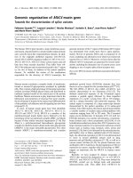
Figure 1
Effect of transforming growth factor 1 (TGF1) treatment on mRNA expression in different cell types (a), Cells were treated with or without 5 ng/mL TGF1 for 24 hEffect of transforming growth factor 1 (TGF1) treatment on mRNA expression in different cell types (a), Cells were treated with or with-
out 5 ng/mL TGF1 for 24 h. The expression of c-myc in nucleus pulposus cells (NP), in articular chondrocytes (AC) and keratinocytes (KT) are
presented. The expression of p15, p21 and p27 in NP was also determined. Time course of c-myc expression in NP treated with 5 ng/mL TGF1
(b). The graph shows the relative intensities of c-myc bands normalized for
-actin levels by densitographic analysis. Incubation for 24 h with medium
containing various concentrations of fetal bovine serum (FBS) did not alter the level of c-myc expression in NP (c). The reverse transcription-polymer-
ase chain reaction (RT-PCR) was performed on total RNA extracted from the cells.
-actin was used as an internal control.
Available online />Page 5 of 12
(page number not for citation purposes)
alter the expression of c-Myc mRNA in nucleus pulposus cells
(Figure 1c).
TGF1 treatment enhanced the proliferation of nucleus
pulposus cells
To determine the effect of TGF1 on cell proliferation, cell
number was measured at the given time intervals. Treatment
was with either 5 or 20 ng/mL TGF1 upregulated cell prolif-
eration on days 3 and 6 (up to 160% compared to the day 3
control (Figure 2)). The statistical significance among the
groups in this proliferation assay by ANOVA was p = 4.408E-
7. The significances of individual differences by the multiple
pair comparisons are shown in Figure 2 (**p < 0.01, *p <
0.05).
Influence of pathway inhibitors blocked cell growth
under TGF1 stimulation
As nucleus pulposus cells maintained c-Myc mRNA expres-
sion under TGF1 stimulation (Figure 1a,b), we hypothesized
that c-Myc plays a central role in TGF1 signaling for cell
growth stimulation. Additionally, to examine the possibility of
involvement of the MAPK pathway in regulation of c-Myc sta-
bility, we devised serial experiments using the pathway spe-
cific inhibitors 10058-F4, an inhibitor of c-Myc transcriptional
activity, and PD98059, an inhibitor of extracellular signal reg-
ulated kinase (ERK1/2). As shown in Figure 3, 5 or 20 ng/mL
TGF1 treatment increased the nucleus pulposus cell number
(up to 160%, p < 0.01) compared with control. Pretreatment
with the c-Myc inhibitor, 10058-F4, caused a dose-dependent
significant decrease in cell number (from 32% to 79%, com-
pared with the TGF1-treated group, p < 0.01). The 20-ng/mL
TGF1-treated cultures showed higher resistance to the inhib-
itory effect of 10058-F4 (8 and 12 M) than 5 ng/mL TGF1.
The statistical significance of this experiment using 10058-F4
was p = 1.116E-18.
Similar results from the cell proliferation assay using the
ERK1/2 inhibitor (Figure 4), demonstrated that while treatment
with 5 or 20 ng/mL TGF1 increased the nucleus pulposus
cell number (up to 130% compared with control, p < 0.05),
pretreatment with the ERK1/2 inhibitor, PD98059, caused a
significant decrease in cell number (from 66% to 76% com-
pared with TGF1-treated group, p < 0.01). In contrast to the
10058-F4 results, the differences were not clearly dose-
dependent. The statistical significance of this experiment
using PD98059 was p = 1.334E-8.
Effects of TGF1 and pathway inhibitors on cell cycle
distribution in nucleus pulposus cells
We then used flow cytometry to determine cell cycle progres-
sion by quantifying DNA. Effects of inhibition of c-Myc tran-
scriptional activity and inhibition of ERK1/2 activity in the
presence of 5 ng/mL TGF1 were determined. After serum
deprivation, 79.0% of nucleus pulposus cells were in the G
0
/
G
1
phase, 10.9% in the S phase, and 10.1% in the G
2
/M
phase (Figure 5a). Treatment with TGF1 for 24 h (Figure 5b)
significantly increased the percentage of cells in the S phase
to 26.4%, indicating that TGF1 did not cause cell cycle
Figure 2
Nucleus pulposus cell proliferation is upregulated by TGF1 treatmentNucleus pulposus cell proliferation is upregulated by TGF1 treat-
ment. Cells were plated in 96-well plates in medium containing 2%
fetal bovine serum (FBS) for 24 h. This medium was replaced with
medium containing 0.5% FBS and cells were treated with 5 or 20 ng/
mL transforming growth factor 1 (TGF1). Cell proliferation was evalu-
ated by the 3-(4,5-dimethyl-2-thiazolyl)-2,5-diphenyl-2H-tetrazolium
bromide (MTT) assay on days 3 and 6 after treatment. Five replicates
per experimental condition were made. Data are normalized to values
obtained for cells cultured for 3 days in 0.5% FBS containing medium
and shown as mean ± standard error of the mean (SEM) (*p < 0.05,
**p < 0.01).
Figure 3
c-Myc transcription inhibition prevents transforming growth factor 1 (TGF1)-stimulated cell proliferationc-Myc transcription inhibition prevents transforming growth factor
1 (TGF1)-stimulated cell proliferation. Serum-deprived cells in 96-
well plates were treated with 5 or 20 ng/mL TGF1 (abbreviated to T)
with or without 8, 12, 16 M 10058-F4. Cell proliferation was evalu-
ated by the 3-(4,5-dimethyl-2-thiazolyl)-2,5-diphenyl-2H-tetrazolium
bromide (MTT) assay on day 3 after treatment. Five replicates per
experimental condition were made. Data are normalized to values
obtained for untreated cells cultured in 0.5% serum containing medium
and represented as mean ± standard error of the mean (SEM) (**p <
0.01 when compared with control, #p < 0.01 when compared with the
TGF1-treated group).
Arthritis Research & Therapy Vol 10 No 6 Nakai et al.
Page 6 of 12
(page number not for citation purposes)
arrest but acted as a mitogen, unlike its action in some other
cell types. In contrast, marked decrease in the percentage of
cells in the S phase were observed in the presence of 10058-
F4, 4.5% (Figure 5c) or PD98059, 8.4% (Figure 5d). In addi-
tion, increase in the G
0
/G
1
phase were found when cells were
treated with these inhibitors (87.7% (Figure 5c) and 85.6%
(Figure 5d), respectively), compared to control (79.0% (Figure
5a)). This indicates that these inhibitors have caused cell cycle
arrest in the G
0
/G
1
phase even with treatment with TGF1.
The results obtained from three different rats are shown in Fig-
ure 6. Although the percentages of cells in the S phase differ
among individuals, these inhibitors both seem to block the
mitogenic effect of TGF1 completely. The statistical signifi-
cance by the repeated measures ANOVA of the cell cycle
experiment was p = 3.213E-3.
TGF1 did not abolish c-Myc expression but decreased
CDKIs p21 and p27
In parallel experiments, we evaluated the expression levels of
key regulatory G1 phase proteins c-Myc, p15, p21 and p27
utilizing western blotting. As seen in Figure 7, TGF1 treat-
ment (b) did not abolish c-Myc expression, but pretreatment
with either 10058-F4 (c) or PD98059 (d) diminished the level
of expression. In contrast, TGF1 treatment showed the low-
est levels of p21 and p27 when compared with other experi-
mental conditions. Note that pretreatment with either 10058-
F4 or PD98059 upregulated the levels of p21 and p27 com-
pared to TGF1 treatment. However, no distinguishable
change was observed in p15 expression.
Mitogenic effect of TGF1 is supported by coexpression
of c-Myc and phospho-ERK1/2
To understand the molecular mechanism underlying TGF1-
mediated cell cycle modulation, we performed a time-course
Figure 4
The inhibition of extracellular signal regulated kinase (ERK)1/2 phos-phorylation prevents transforming growth factor 1 (TGF1)-stimulated cell proliferationThe inhibition of extracellular signal regulated kinase (ERK)1/2
phosphorylation prevents transforming growth factor 1 (TGF1)-
stimulated cell proliferation. Serum-deprived cells in 96-well plates
were treated with 5 or 20 ng/mL TGF1 (abbreviated to T) with or with-
out 10, 20, 30 M PD98059. Cell proliferation was evaluated by the 3-
(4,5-dimethyl-2-thiazolyl)-2,5-diphenyl-2H-tetrazolium bromide (MTT)
assay on day 3 after treatment. Five replicates per experimental condi-
tion were made. Data are normalized to values obtained for untreated
cells cultured in 0.5% serum containing medium and represented as
mean ± standard error of the mean (SEM) (*p < 0.05, **p < 0.01 when
compared with control, #p < 0.01 when as compared with TGF1-
treated group).
Figure 5
Cell cycle distribution of nucleus pulposus cellsCell cycle distribution of nucleus pulposus cells. Serum-deprived nucleus pulposus cells were cultured with no supplements for 24 h (a). The
cells were treated with 5 ng/mL transforming growth factor 1 (TGF1) for 24 h (b). At 2 h before the addition of TGF1, the cells were treated with
16 M 10058-F4 (c), or with 30 M PD98059 (d). The cells were harvested 24 h after the addition of TGF1 and the nuclei were stained with pro-
pidium iodide. DNA histograms were generated using flow cytometry. Each plot represents the analysis of 10,000 events. The histograms present
typical results and the percentage of cells in G
0
/G
1
, S and G
2
/M phases are shown as the average of duplicated measurements.
Available online />Page 7 of 12
(page number not for citation purposes)
study on c-Myc and phospho-ERK1/2. Serum-deprived cells
were pretreated with or without 10058-F4 or PD98059 then
treated with TGF1 for different time periods. The cells were
harvested and whole cell lysates were analyzed for the expres-
sion of c-Myc, phospho-ERK1/2, and total ERK1/2 by western
blot. Robust c-Myc expression from the beginning was sup-
pressed at 6 h and ERK1/2 was immediately phosphorylated
(activated) by 0.5 to 2 h in TGF1-treated preparations (Figure
8a). Both c-Myc and phospho-ERK1/2 were detected
throughout the experimental period. The lane on the far right
indicates the result of 24 h treatment with 10% FBS in which
c-Myc and phospho-ERK1/2 appear distinctly (Figure 8a).
These data indicate that coexpression of c-Myc and phospho-
ERK1/2 correlates with vigorous cell proliferation.
By contrast, pretreatment with the ERK inhibitor PD98059
diminished the expression of c-Myc and mainly blocked the
phosphorylation of ERK1 induced by TGF1 treatment (Figure
8b). A single isoform corresponding to phospho-ERK2 was
detected at all time points; this suggests that c-Myc expres-
sion under TGF1 stimulation requires activated ERK1/2,
especially ERK1. Similarly, pretreatment with the c-Myc inhib-
itor 10058-F4 unexpectedly decreased c-Myc expression and
interrupted the phosphorylation of ERK1/2 induced by TGF1
(Figure 8c). The expression of phospho-ERK1/2 was delayed
until the 2-h time point and disappeared after 12 h in spite of
coexistent TGF1. These data indicate that the inhibition of c-
Myc transcriptional activity diminished the level of c-Myc pro-
tein itself and also downregulated the phosphorylation (activa-
tion) of ERK1/2.
The results of these blot analyses reveal that the effect of the
TGF1 signal can be mitogenic when c-Myc and phospho-
ERK1/2 are both expressed in nucleus pulposus cells.
Discussion
Although TGF1 is a potent inhibitor of growth in most cell
types, it has been shown to stimulate growth of certain mes-
enchymal cells in culture, such as mouse BM-MSCs [9], rat
and avian articular chondrocytes [10,11,23,37], human nasal
septal chondrocytes [12], and cells with an osteoblastic phe-
notype from rat parietal bone [38] and from calvariae of 1-day-
old mice [13]. In these previous investigations, growth stimu-
lation was shown by upregulation in proliferation or in [
3
H]-thy-
midine uptake. With regard to intervertebral disc cells, the
enhancement of colony formation of human annulus fibrosus
cells and increase in density of nucleus pulposus cells in three-
dimensional scaffold cultures have been reported [39,40].
In the present study, we found that TGF1 significantly stimu-
lated growth of nucleus pulposus cells (Figure 2). To ascertain
the effects of TGF1, we examined the cell cycle regulatory
effect of TGF1 in rat nucleus pulposus cells in vitro.
TGF1 regulates gene expression through Smad transcription
factors [41-43]. When TGF1 inhibits cell growth, a rapid
decrease in endogenous c-Myc mRNA and protein has been
observed [14-17]. c-Myc is a transcription factor that pro-
motes cell growth and proliferation, and under certain condi-
tions, apoptosis, and tumor cell immortalization [44]. Levels of
c-Myc are increased or decreased in response to mitogenic or
growth inhibitory stimuli, respectively [17]. It is notable that c-
myc transfected Fisher rat 3T3 fibroblast have a proliferative
Figure 6
Effects of inhibitors and transforming growth factor 1 (TGF1) on cell cycle progressionEffects of inhibitors and transforming growth factor 1 (TGF1) on cell cycle progression. Serum-deprived nucleus pulposus cells were
treated with or without inhibitors (16 M 10058-F4, or 30 M PD98059) then treated with 5 or 20 ng/mL TGF1 for 24 h. The percentage of cells
in S-phase was determined with fluorescence-activated cell sorting (FACS). Black bar, white bar and gray bar indicate the results obtained for three
rats respectively. (*p < 0.05)
Arthritis Research & Therapy Vol 10 No 6 Nakai et al.
Page 8 of 12
(page number not for citation purposes)
response to TGF1 [20], and that the mouse keratinocyte cell
line (BALB/MK) expressing the chimeric estrogen-inducible
form of c-myc-encoded protein (mycER) suppresses the
growth-inhibitory effect of TGF1 [19].
As shown in Figure 1, TGF1 treatment decreased c-Myc
mRNA after 24 h in keratinocytes, while nucleus pulposus
cells and articular chondrocytes showed a constant level of c-
Myc mRNA. In keratinocytes, we confirmed earlier findings
[14,15]. In contrast, nucleus pulposus cells and articular
chondrocytes respond differently to TGF1 treatment.
Although the passage numbers of these cultures are different,
we used all of the cultures at the constantly proliferative stage.
Considering that keratinocytes has been reported to be
growth arrested by TGF1 [1], these results suggest that c-
Myc mRNA expression correlates with the mitogenic response
of the cells to TGF1 stimulation. To investigate the effects of
c-Myc on cell growth under TGF1 stimulation, we inhibited c-
Myc function in nucleus pulposus cells using specific inhibi-
tors.
The mitogenic response to TGF1 suppressed by
pathway inhibitors
Figure 7a,b indicate that the same levels of endogenous c-Myc
protein were detected in nucleus pulposus cells, independent
of TGF1 treatment. The cell cycle distribution in TGF1-
treated cells (Figure 5b) indicates a large increase in cells in
the S phase, associated with the suppression of p21 and p27
which belong to the Cip/Kip family of cyclin-dependent kinase
inhibitors (CKIs) (Figure 7a,b). By contrast, pretreatment with
either 10058-F4, a c-Myc, inhibitor or PD98059, an ERK1/2
inhibitor, arrested cell proliferation and cell cycle progression
when coexistent with TGF1 (Figures 3, 4, 5, 6). Additionally,
both inhibitors suppressed c-Myc expression while upregulat-
ing p21 and p27 expression (Figure 7c,d) compared to
TGF1-treated cells (Figure 7b). The elevation of p15, p21
and p27 has been reported to be the main cause of cell cycle
arrest by TGF1 [29-32]. We therefore analyzed the expres-
sion of these three CKIs, but found that p21 and p27 were
decreased by TGF1, while there was no change in p15
expression (Figure 7). The findings that TGF1 did not cause
cell cycle arrest in nucleus pulposus cells and that it
decreased p21 and p27 expression can be attributed to the
sustained c-Myc expression. Previous investigations have sug-
gested the special regulation of CKIs under TGF1, mediated
by an elevated level of c-Myc [45-47].
The immediate phosphorylation of ERK1/2 with robust c-
Myc expression for 2 h after TGF1 treatment
In the time course study, the top panel shows TGF1 treat-
ment kept the robust c-Myc expression for 2 h but downregu-
lated it after 6 h (Figure 8a). The downregulation of c-Myc was
considered to result from the downregulation of c-Myc mRNA
transcription by TGF1 through the Smad pathway [16]. As
shown in Figure 1b, the level of c-Myc mRNA was downregu-
lated at 60 min and recovered after 240 min. In the protein lev-
els, distinct recovery of c-Myc expression was not detected;
nonetheless it was sustained for 24 h. The second panel in
Figure 8a shows that TGF1 induces the immediate phospho-
rylation (activation) of ERK1/2; this observation agrees with an
earlier study using rat articular chondrocytes by Hirota et al.
[48]. ERK1 and ERK2 are subtypes of MAPKs activated by a
diverse array of extracellular stimuli [49]. The phosphorylation
of ERK1/2 in nucleus pulposus cells has been reported to be
critical for survival in a hypoxic environment [50]. We also
detected marked phosphorylation of ERK1/2 and c-Myc
expression in 10% FBS-added cultures. Therefore, growth
factors can be considered to drive c-Myc expression and
phosphorylation of ERK1/2 in nucleus pulposus cells. How-
ever, serum-deprived cells with no supplements (time 0 in Fig-
ure 8a) expressed c-Myc, but no phosphorylated ERK1/2.
These results suggest that c-Myc itself does not enhance cell
growth, but acts as a mediator of exogenous growth stimuli.
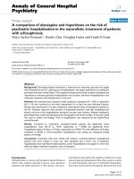
Figure 7
Western blot analysis of cell cycle regulatorsWestern blot analysis of cell cycle regulators. After 24 h incubation
in a medium containing 2% fetal bovine serum (FBS), this medium was
replaced with medium containing 0.5% FBS. Nucleus pulposus cells
were cultured with no supplements for an additional 24 h (a). The cells
were treated with 5 ng/mL transforming growth factor 1 (TGF1) for
24 h (b). At 2 h before the addition of TGF1, the cells were treated
with 16 M 10058-F4 (c), or with 30 M PD98059 (d). The cells were
harvested 24 h after the TGF1 treatment and lysed. Aliquots of the
lysates were electrophoresed on 12.5% sodium dodecyl sulfate poly-
acrylamide gel electrophoresis (SDS-PAGE). The protein bands were
blotted to a polyvinylidene diflouride (PVDF) membrane and probed
with antibodies against c-Myc, p15, p21, and p27. -Actin was used as
a quantity loading control. Treatment with TGF1 without inhibitors (b)
did not abolish c-Myc expression but decreased the level of cyclin-
dependent kinase inhibitors (CKIs) (p21, p27) compared to the control,
while treatments with inhibitors (c, d) diminished c-Myc and upregu-
lated p21 and p27. In contrast, p15 levels were unchanged by any of
these treatments.
Available online />Page 9 of 12
(page number not for citation purposes)
Figure 8
Time course study of c-Myc and phospho-extracellular signal regulated kinase (ERK)1/2 expression by western blot analysisTime course study of c-Myc and phospho-extracellular signal regulated kinase (ERK)1/2 expression by western blot analysis. Serum-
deprived nucleus pulposus cells were treated with or without 16 M 10058-F4 or 30 M PD98059 before the addition of 5 ng/mL transforming
growth factor 1 (TGF1). The cells were harvested at the times indicated and lysed. Aliquots of the lysates were electrophoresed on 5% to 20%
gradient sodium dodecyl sulfate polyacrylamide gel electrophoresis SDS-PAGE). The protein bands were blotted to a polyvinylidene diflouride
(PVDF) membrane and probed with antibodies against c-Myc, total ERK1/2 and phospho-ERK1/2. -Actin was used as a quantity loading control.
(a) TGF1 treatment induced immediate phosphorylation of ERK1/2 with robust c-Myc expression for 2 h. The expression of c-Myc, phospho-ERK1/
2, and total ERK1/2 were detected throughout the experimental period. The right lane indicates the result of 24 h treatment with 10% FBS; c-Myc
and phospho-ERK1/2 appear distinctly. (b) Pretreatment with ERK1/2 inhibitor 30 M PD98059 diminished the expression of c-Myc and interrupted
the phosphorylation of ERK1/2. Note that a single isoform corresponding to phospho-ERK2 was detected at all times. (c) Pretreatment with c-Myc
inhibitor 16 M 10058-F4 diminished c-Myc expression and limited ERK1/2 phosphorylation for a short time under TGF1 stimulation. Graphs show
relative intensities in expression of c-Myc normalized to
-actin levels and in expression of phospho-ERK1/2 normalized to total ERK1/2 levels,
respectively.
Arthritis Research & Therapy Vol 10 No 6 Nakai et al.
Page 10 of 12
(page number not for citation purposes)
10058-F4 downregulates c-Myc expression and ERK1/2
phosophorylation
The c-Myc inhibitor 10058-F4 we used was isolated by Yin et
al. [21] using a yeast two-hybrid system. In order to bind DNA,
c-Myc must dimerize with Max. 10058-F4 inhibits c-Myc tran-
scriptional activity by disrupting the c-Myc/Max association.
The half-life of Myc is known to be less than 30 min [51]; it has
also been reported that the instability of oncoprotein Myc is
important to avoid its accumulation in normal cells and that
Myc is destroyed by ubiquitin-mediated proteolysis [52]. In this
study, we showed almost constant levels of c-Myc mRNA
expression in nucleus pulposus cells independent of serum
concentrations (Figure 1c) and sustained c-Myc protein
expression during treatment with TGF1 (Figures 7a,b and
8a). However, inhibition of c-Myc transcriptional activity by
10058-F4 in the presence TGF1 resulted in suppression of
the mitogenic effect of TGF1 (MTT assay (Figure 3) and the
cell cycle distribution (Figures 5c, 6)). These results suggest
that c-Myc implicates in the effect of TGF1. We also
observed that 10058-F4 unexpectedly interrupted phosphor-
ylation of ERK1/2 as well as downregulating c-Myc expression
(Figure 8c). Because Myc is associated with an extraordinarily
large number of genomic sites, it can regulate complex
genomic pathways [53-55]. It was also reported that transcrip-
tional response to Myc binding differs markedly according to
context and cell type [55]. The elucidation of the role of c-Myc
in ERK1/2 phosphorylation in nucleus pulposus cells requires
further investigation.
Recent studies investigating 10058-F4 report cell cycle arrest
accompanied by suppression of c-Myc mRNA in lymphoma
[56] and the suppression of c-Myc with upregulation of levels
of p21 and p27 in myeloid leukemia [57,58]. These reports
correspond with our observations (Figure 7).
PD98059 downregulates ERK1 phosphorylation and c-
Myc expression
We show that pretreatment with PD98059 significantly
blocked the mitogenic and cell cycle promotive effects of
TGF1 (MTT assay (Figure 4) and cell cycle distribution (Fig-
ures 5d, 6)) associated with suppression of c-Myc expression
(Figure 7d). In the preliminarily experiments we also examined
a protein kinase C inhibitor peptide (19–36) obtained from
Calbiochem (Darmstadt, Germany), because inhibition of pro-
tein kinase C had been reported to cause abolition of TGF1
induced cell growth in rat articular chondrocytes [37], but it
did not exert the abolition in nucleus pulposus cells (data not
shown). By contrast, PD98059 showed a significant inhibitory
effect. PD98059 is an inhibitor for MAP kinase kinases 1 and
2 (MKK), also called MAP/ERK kinases (MEK), the upstream
activator of ERK1/2. In the time course study (Figure 8b), the
second panel shows only phospho-ERK2 protein bands with
the complete absence of phospho-ERK1 for 24 h. The inhibi-
tory effect of PD98059 on MEK2 is known to be less potent
than MEK1. The concentration of PD98059 required to give
50% inhibition (IC50) of MEK1 is 4 M and of MEK2 is 50 M
[22]. Because we used a maximum dose of 30 M of
PD98059, MEK1 was considered to be strongly inhibited.
These results suggest that phosphorylated ERK1 is necessary
to maintain c-Myc expression and promote cell cycle progres-
sion under TGF1 stimulation. Our results agree with earlier
reports showing that ERK1/2 plays a crucial mediating role in
mitogenic signaling of TGF1 in mouse BM-MSCs cultured in
chondrogenic condition [9] and in rat articular chondrocytes
[23].
Possibility of c-Myc stability supported by phospho-
ERK1/2
We showed the persistent expression of c-Myc in nucleus pul-
posus cells, which are not tumor cells or immortalized cells. As
described above, c-Myc appears to be supported by phospho-
ERK1/2. Lefevre et al. [59] showed that treatment with Raf-1
kinase inhibitor or ERK1/2 inhibitor PD98059 decreased c-
Myc production in cultured ocular choroidal melanoma which
had a high and constant level of c-Myc. Also, the contribution
of Ras/Raf/ERK prevented the rapid degradation of c-Myc by
phosphorylation of the serine 62 residue in the N-terminal of c-
Myc [24]. They also found that the suppression of glycogen
synthase kinase 3 beta (GSK-3) activity, which phosphor-
ylates threonine 58, a phosphorylation site adjacent to serine
62, enhances c-Myc stability. Although we did not analyze the
phosphorylation of c-Myc, these proposed kinetics should be
investigated to explain the enhanced stability of c-Myc in
nucleus pulposus cells.
Recent investigations have revealed that Myc stability is
required in self-renewal and maintenance of murine ES cell
pluripotency [44]. These authors evaluated Myc protein levels
in ES cells and concluded that elevated Myc activity is able to
block the differentiation of multiple cell lineages and that this
blocking of differentiation promotes self-renewal. Similarly, c-
Myc has been reported to inhibit the terminal stages of adi-
pocyte differentiation [60].
We used cells derived from rat nucleus pulposus of the
intervertebral disc to examine how they respond to TGF-1
stimulation. Cells constituting the nucleus pulposus are known
to be sparse and have a low ability for self-renewal [61].
Although efforts to regenerate disc tissue using cell therapy
have accelerated their profiling [62], the precise phenotype of
nucleus pulposus cells and their response to various cytokines
are still under investigation. In this study, we suggested a spe-
cific regulatory pathway of TGF1 in which c-Myc and phos-
pho-ERK1/2 play important roles. However, we used the third
or fourth passaged culture, which did not contain large noto-
chordal cells. Therefore, some phenotypic change (that is ded-
ifferentiation) may have been induced, as is known to occur for
articular chondrocytes. Inevitably, the correlation between dif-
ferentiation level in the cells and responsiveness to TGF1
remains to be elucidated. Moreover, in view of the therapeutic
Available online />Page 11 of 12
(page number not for citation purposes)
use of TGF1 for nucleus pulposus regeneration, the limitation
in the use of the rat model needs to be carefully considered.
This is because the presence of notochordal cells in the rat
coccygeal disc is different from the human situation, in which
notochordal cells have been known to disappear after birth.
Therefore, future studies using different animal models are
necessary to confirm whether the implication of c-Myc and
ERK1/2 can generally be attributed to nucleus pulposus cells
or it depends on the species of the donor.
Conclusion
Because our results indicate that both c-Myc and phospho-
ERK1/2 are required for proliferation and cell cycle progres-
sion, we conclude that the synergistic effect between c-Myc
and phospho-ERK1/2 plays a key role in nucleus pulposus cell
growth under TGF1 stimulation. Therefore, treatment with
TGF1 should yield different effects depending on the status
of these mediators in the target cells.
Competing interests
The authors declare that they have no competing interests.
Authors' contributions
TN and DS conceived of the study and performed the experi-
mental work. DS and JM participate in its design and coordi-
nation. TN, DS and JM helped to draft the manuscript. TN and
DS performed the statistical analysis. All authors read and
approved the final manuscript.
Acknowledgements
We thank Dr Hideo Tsukamoto and Dr Yoshinori Okada, of Teaching
and Research Support Center of Tokai University, for sharing their
sophisticated understanding of techniques. This work is supported by a
grant from the Academic Frontier Project of the Ministry of Education,
Culture, Sports, Science and Technology (MEXT) of Japan and a grant
from AO Spine International to JM and DS.
References
1. Shipley GD, Pittelkow MR, Wille JJ Jr, Scott RE, Moses HL:
Reversible inhibition of normal human prokerationocyte prolif-
eration by type transforming growth factor-growth inhibitor
in serum free medium. Cancer Res 1986, 46:2068-2071.
2. Heimark RL, Twardzik DR, Schwartz SM: Inhibition of endothelial
cell regeneration by type-beta transforming growth factor
from platelets. Science 1986, 233:1078-1080.
3. Takehara K, LeRoy EC, Grotendorst GR: TGF-beta inhibition of
endothelial cell proliferation: alteration of EGF binding and
EGF-induced growth-regulatory (competence) gene expres-
sion. Cell 1987, 49:415-422.
4. Merwin JR, Newman W, Beall LD, Tucker A, Madri J: Vascular
cells respond differentially to transforming growth factors-1
and 2 in vitro. Am J Pathol 1991, 138:37-51.
5. Kehrl JH, Wakefield LM, Roberts AB, Jakowlew S, Alvarez-Mon M,
Derynck R, Sporn MB: Production of transforming growth fac-
tor- by human T lymphocytes and its potential role in the reg-
ulation of T cell growth. J Exp Med 1986, 163:1037-1050.
6. Shull MM, Ormsby I, Kier AB, Pawlowski S, Diebold RJ, Yin M,
Allen R, Sidman C, Proetzel G, Calvin D: Targeted disruption of
the mouse transforming growth factor-1 gene results in
multifocal inflammatory disease. Nature 1992, 359:693-699.
7. Kulkarni AB, Huh CG, Becker D, Geiser A, Lyght M, Flanders KC,
Roberts AB, Sporn MB, Ward JM, Karlsson S: Transforming
growth factor beta 1 null mutation in mice causes excessive
inflammatory response and early death. Proc Natl Acad Sci
USA 1993, 90:770-774.
8. Abdel-Wahab N, Weston BS, Roberts T, Mason RM: Connective
tissue growth factor and regulation of the mesangial cell cycle:
role in cellular hypertrophy. J Am Soc Nephrol 2002,
13:2437-2445.
9. Longobardi L, O'Rear L, Aakula S, Jhonstone B, Shimer K, Chtil A,
Horton WA, Moses HL, Spagnoli A: Effect of IGF-I in the chon-
dorogenesis of bone marrow mesenchymal stem cells in the
presence or absence of TGF- signaling. J Bone Miner Res
2006, 21:626-636.
10. Tsukazaki T, Usa T, Matsumoto T, Enomoto H, Ohtsuru A, Namba
H, Iwasaki K, Yamashita S: Effect of transforming growth factor-
on the insulin-like growth factor-I autocrine/paracirine axis
in cultured rat articular chondrocytes. Exp Cell Res 1994,
215:9-16.
11. Rousche KT, Ford BC, Praul CA, Leach RM: The use of growth
factors in the proliferation of avian articular chondrocytes in a
serum-free culture system. Connect Tissue Res 2001,
42:165-174.
12. Richmon JD, Sage AB, Wong VW, Chen AC, Pan C, Sah RL,
Watson D: Tensile biomechanical properties of human nasal
septal cartilage. Am J Rhinol 2005, 19:617-622.
13. Urano T, Yashiroda H, Muraoka M, Tanaka K, Hosoi T, Inoue S,
Ouchi Y, Toyoshima H: p57(Kip2) is degraded through the pro-
teasome in osteoblasts stimulated to proliferation by trans-
forming growth factor beta1. J Biol Chem 1999,
274:12197-12200.
14. Pietenpol JA, Holt JT, Stein RW, Moses HL: Transforming growth
factor beta 1 suppression of c-myc gene transcription: role in
inhibition of keratinocyte proliferation. Proc Natl Acad Sci USA
1990, 87:3758-3762.
15. Münger K, Pietenpol JA, Pittelkow MR, Holt JT, Moses HL: Trans-
forming growth factor beta 1 regulation of c-myc expression,
pRB phosphorylation, and cell cycle progression in keratinoc-
ytes. Cell Growth Differ 1992, 3:291-298.
16. Yagi K, Furuhashi M, Aoki H, Goto D, Kuwano H, Sugamura K,
Miyazono K, Kato M: c-myc is a downstream target of the Smad
pathway. J Biol Chem 2002, 277:854-861.
17. Seoane J, Le HV, Massagué J: Myc suppression of the p21(Cip1)
Cdk inhibitor influences the outcome of the p53 response to
DNA damage. Nature 2002, 419:729-734.
18. Lüscher B, Larsson LG: The basic region/helix-loop-helix/leu-
cine zipper domain of Myc proto-oncoproteins: function and
regulation. Oncogene 1999, 18:
2955-2966.
19. Alexandrow MG, Kawabata M, Aakre M, Moses HL: Overexpres-
sion of the c-Myc oncoprotein blocks the growth-inhibitory
response but is required for the mitogenic effects of trans-
forming growth factor 1. Proc Natl Acad Sci USA 1995,
92:3239-3243.
20. Roberts AB, Anzano MA, Wakefield LM, Roche NS, Stern DF,
Sporn MB: Type beta transforming growth factor: a bifunc-
tional regulator of cellular growth. Proc Natl Acad Sci U S A
1985, 82:119-123.
21. Yin X, Giap C, Lazo JS, Prochowink EV: Low molecular weight
inhibitors of Myc-Max interaction and function. Oncogene
2003, 22:6151-6159.
22. Alessi DR, Cuenda A, Cohen P, Dudley DT, Saltiel AR: PD098059
is a specific inhibitor of the activation of mitogen-activated
protein kinase kinase in vitro and in vivo. J Biol Chem 1995,
270:27489-27494.
23. Yonekura A, Osaki M, Hirota Y, Tsukazaki T, Miyazaki Y, Mat-
sumoto T, Ohtsuru A, Namba H, Shindo H, Yamashita S: Trans-
forming growth factor-beta stimulates articular chondrocyte
cell growth through p44/42 MAP kinase (ERK) activation.
Endocr J 1999, 46:545-553.
24. Sears R, Nuckolls F, Haura E, Taya Y, Tamai K, Nevins JR: Multiple
Ras-dependent phosphorylation pathways regulate Myc pro-
tein stability. Genes Dev 2000, 14:2501-2514.
25. Morgan DO: Principles of CDK regulation. Nature 1995,
374:131-134.
26. Sherr CJ: G1 phase progression: cycling on cue. Cell 1994,
79:551-555.
27. Sherr CJ, Roberts JM: CDK inhibitors: positive and negative
regulators of G1-phase progression. Genes Dev 1999,
13:1501-1512.
Arthritis Research & Therapy Vol 10 No 6 Nakai et al.
Page 12 of 12
(page number not for citation purposes)
28. Hunter T, Pines J: Cyclins and cancer. II: cyclin D and CDK inhib-
itors come of age. Cell 1994, 79:573-582.
29. Hannon GJ, Beach D: p15INK4B is a potential effector of TGF-
beta-induced cell cycle arrest. Nature 1994, 371:257-261.
30. Polyak K, Kato JY, Solomon MJ, Sherr CJ, Massague J, Roberts JM,
Koff A: p27Kip1, a cyclin-Cdk inhibitor, links transforming
growth factor-beta and contact inhibition to cell cycle arrest.
Genes Dev 1994, 8:9-22.
31. Li JM, Nichols MA, Chandrasekharan S, Xiong Y, Wang XF: Trans-
forming growth factor beta activates the promoter of cyclin-
dependent kinase inhibitor p15INK4B through an Sp1 consen-
sus site. J Biol Chem 1995, 270:26750-26753.
32. Li CY, Suardet L, Little JB: Potential role of WAF1/Cip1/p21 as
a mediator of TGF-beta cytoinhibitory effect. J Biol Chem
1995, 270:4971-4974.
33. Iwabuchi S, Sakai D: Low-intensity pulsed ultrasound stimula-
tion enhances TIMP-1 in nucleus pulposus cells and MCP-1 in
macrophages in the rat. J Orthop Res 2008, 26:865-871.
34. Trout JJ, Buckwalter JA, Moore KC, Landas SK: Ultrastructure of
the human intervertebral disc. I. Changes in notochordal cells
with age. Tissue Cell 1982, 14:359-369.
35. Trout JJ, Buckwalter JA, Moore KC: Ultrastructure of the human
intervertebral disc: II. Cells of the nucleus pulposus. Anat Rec
1982, 204:307-314.
36. Ambs S, Dennis S, Fairman J, Wright M, Papkoff J: Inhibition of
tumor growth correlates with the expression level of a human
angiostatin transgene in transfected B16F10 melanoma cells.
Cancer Res 1999, 59:5773-5777.
37. Osaki M, Tsukazaki T, Yonekura A, Miyazaki Y, Iwasaki K, Shindo
H, Yamashita S: Regulation of c-fos gene induction and
mitogenic effect of transforming growth factor-1 in rat articu-
lar chondrocyte. Endocr J 1999, 46:253-261.
38. Centrella M, McCarthy TL, Canalis E: Transforming growth factor
beta is a bifunctional regulator of replication and collagen syn-
thesis in osteoblast-enriched cell cultures from fetal rat bone.
J Biol Chem 1987,
262:2869-2874.
39. Gruber HE, Fisher EC Jr, Desai B, Stasky AA, Hoelscher G, Hanley
EN Jr: Human intervertebral disc cells from the annulus: three-
dimensional culture in agarose or alginate and responsive-
ness to TGF-beta1. Exp Cell Res 1997, 235:13-21.
40. Alini M, Li W, Markovic P, Aebi M, Spiro RC, Roughley PJ: The
potential and limitations of a cell-seeded collagen/hyaluronan
scaffold to engineer an intervertebral disc-like matrix. Spine
2003, 28:446-454.
41. Heldin CH, Miyazono K, ten Dijke P: TGF- signalling from cell
membrane to nucleus through SMAD proteins. Nature 1997,
390:465-471.
42. Massaous J, Hata A: TGF- signalling through the Smad path-
way. Trends Cell Biol 1997, 7:187-192.
43. Feng XH, Lin X, Derynck R: Smad2, Smad3 and Smad4 cooper-
ate with Sp1 to induce p15 (Ink4B) transcription in response
to TGF-. EMBO J 2000, 19:5178-5193.
44. Cartwright P, McLean C, Sheppard A, Rivett D, Jones K, Dalton S:
LIF/STAT3 controls ES cell self-renewal and pluripotency by a
Myc-dependent mechanism. Development 2005, 132:885-896.
45. Vlach J, Hennecke S, Alevizopoulos K, Conti D, Amati B: Growth
arrest by the cyclin-dependent kinase inhibitor p27Kip1 is
abrogated by c-Myc. EMBO J 1996, 15:6595-6604.
46. Mitchell KO, El-Deiry WS: Overexpression of c-Myc inhibits
p21WAF1/CIP1 expression and induces S-phase entry in 12-
O-tetradecanoylphorbol-13-acetate (TPA)-sensitive human
cancer cells. Cell Growth Differ 1999, 10:223-230.
47. Claassen GF, Hann SR: A role for transcriptional repression of
p21CIP1 by c-Myc in overcoming transforming growth factor
beta-induced cell-cycle arrest. Proc Natl Acad Sci USA 2000,
97:9498-9503.
48. Hirota Y, Tsukazaki T, Yonekura A, Miyazaki Y, Osaki M, Shindo H,
Yamashita S: Activation of specific MEK-ERK cascade is neces-
sary for TGF
signaling and crosstalk with PKA and PKC path-
ways in cultured rat articular chondrocytes. Osteoarthritis
Cartilage 2000, 8:241-247.
49. Cobb MH, Goldsmith EJ: How MAP kinases are regulated. J
Biol Chem 1995, 270:14843-14846.
50. Risbud MV, Guttapalli A, Albert TJ, Shapiro IM: Hypoxia activates
MAPK activity in rat nucleus pulposus cells: regulation of
integrin expression and cell survival. Spine 2005,
30:2503-2509.
51. Hann SR, Eisenman RN: Proteins encoded by the human c-myc
oncogene: differential expression in neoplastic cells. Mol Cell
Biol 1984, 11:2486-2497.
52. Salghetti SE, Kim SY, Tansey WP: Destruction of Myc by ubiqui-
tin-mediated proteolysis: cancer-associated and transforming
mutations stabilize Myc. EMBO J 1999, 18:717-726.
53. Fernandez PC, Frank SR, Wang L, Schroeder M, Liu S, Greene J,
Cocito A, Amati B: Genomic targets of the human c-Myc pro-
tein. Genes Dev 2003, 17:1115-1129.
54. Orian A, van Steensel B, Delrow J, Bussemaker HJ, Li L, Sawado
T, Williams E, Loo LW, Cowley SM, Yost C, Pierce S, Edgar BA,
Parkhurst SM, Eisenman RN: Genomic binding by the dro-
sophila Myc, Max, Mad/Mnt transcription factor network.
Genes Dev 2003, 17:1101-1114.
55. Ellwood-Yen K, Graeber TG, Wongvipat J, Iruela-Arispe ML, Zhang
J, Matusik R, Thomas GV, Sawyers CL: Myc-driven murine pros-
tate cancer shares molecular features with human prostate
tumors. Cancer Cell 2003, 4:223-238.
56. Gomez-Curet I, Perkins RS, Bennett R, Feidler KL, Dunn SP,
Krueger LJ: c-Myc inhibition negatively impacts lymphoma
growth. J Pediatr Surg 2006, 41:207-211.
57. Huang MJ, Cheng YC, Liu CR, Lin S, Liu HE: A small-molecule c-
Myc inhibitor, 10058-F4, induces cell-cycle arrest, apoptosis,
and myeloid differentiation of human acute myeloid leukemia.
Exp Hematol 2006, 34:1480-1489.
58. Lin CP, Liu JD, Chow JM, Liu CR, Liu HE: Small-molecule c-Myc
inhibitor, 10058-F4, inhibits proliferation, downregulates
human telomerase reverse transcriptase and enhances chem-
osensitivity in human hepatocellular carcinoma cells. Antican-
cer Drugs 2007, 18:161-170.
59. Lefevre G, Calipel A, Mouriaux F, Hecquet C, Malecaze F, Mas-
carelli F: Opposite long-term regulation of c-Myc and p27Kip1
through overactivation of Raf-1 and the MEK/ERK module in
proliferating human choroidal melanoma cells. Oncogene
2003, 22:8813-8822.
60. Heath VJ, Gillespie DA, Crouch DH: Inhibition of the terminal
stages of adipocyte differentiation by cMyc. Exp Cell Res
2000, 254:91-8.
61. Ichimura K, Tsuji H, Matsui H, Makiyama N: Cell culture of the
intervertebral disc of rats: factors influencing culture, prote-
oglycan, collagen, and deoxyribonucleic acid synthesis. J Spi-
nal Disord 1991, 4:428-436.
62. Sakai D, Mochida J, Iwashina T, Watanabe T, Nakai T, Ando K,
Hotta T: Differentiation of mesenchymal stem cells trans-
planted to a rabbit degenerative disc model: potential and lim-
itations for stem cell therapy in disc regeneration. Spine 2005,
30:2379-2387.