Báo cáo y học: "Caveolin-1 expression and stress-induced premature senescence in human intervertebral disc degeneration" docx
Bạn đang xem bản rút gọn của tài liệu. Xem và tải ngay bản đầy đủ của tài liệu tại đây (569.49 KB, 9 trang )
Open Access
Available online />Page 1 of 9
(page number not for citation purposes)
Vol 10 No 4
Research article
Caveolin-1 expression and stress-induced premature senescence
in human intervertebral disc degeneration
Sarah Kathleen Heathfield
1
, Christine Lyn Le Maitre
2
and Judith Alison Hoyland
1
1
Tissue Injury and Repair Group, Research School of Clinical and Laboratory Sciences, Faculty of Medical and Human Sciences, Stopford Building,
The University of Manchester, Oxford Road, Manchester, M13 9PT, UK
2
Biomedical Research Centre, Biosciences, Faculty of Health and Wellbeing, Sheffield Hallam University, City Campus, Howard Street, Sheffield, S1
1WB, UK
Corresponding author: Judith Alison Hoyland,
Received: 20 May 2008 Revisions requested: 12 Jun 2008 Revisions received: 9 Jul 2008 Accepted: 5 Aug 2008 Published: 5 Aug 2008
Arthritis Research & Therapy 2008, 10:R87 (doi:10.1186/ar2468)
This article is online at: />© 2008 Heathfield et al.; licensee BioMed Central Ltd.
This is an open access article distributed under the terms of the Creative Commons Attribution License ( />),
which permits unrestricted use, distribution, and reproduction in any medium, provided the original work is properly cited.
Abstract
Introduction Chronic and debilitating low back pain is a
common condition and a huge economic burden. Many cases
are attributed to age-related degeneration of the intervertebral
disc (IVD); however, age-related degeneration appears to occur
at an accelerated rate in some individuals. We have previously
demonstrated biomarkers of cellular senescence within the
human IVD and suggested a role for senescence in IVD
degeneration. Senescence occurs with ageing but can also
occur prematurely in response to stress. We hypothesised that
stress-induced premature senescence (SIPS) occurs within the
IVD and here we have investigated the expression and
production of caveolin-1, a protein that has been shown
previously to be upregulated in SIPS.
Methods Caveolin-1 gene expression in human nucleus
pulposus (NP) cells was assessed by conventional and
quantitative real-time polymerase chain reaction (PCR), and
caveolin-1 protein expression was examined within human IVDs
using immunohistochemistry. The correlation between caveolin-
1 and p16
INK4a
(biomarker of cellular senescence) gene
expression was investigated using quantitative real-time PCR.
Results Caveolin-1 gene expression and protein expression
were demonstrated within the human IVD for the first time. NP
cells from degenerate discs exhibited elevated levels of
caveolin-1 which did not relate to increasing chronological age.
A negative correlation was observed between gene expression
for caveolin-1 and donor age, and no correlation was found
between caveolin-1 protein expression and age. A positive
correlation was identified between gene expression of caveolin-
1 and p16
INK4a
.
Conclusion Our findings are consistent with a role for caveolin-
1 in degenerative rather than age-induced changes in the NP. Its
expression in IVD tissue and its association with the senescent
phenotype suggest that caveolin-1 and SIPS may play a
prominent role in the pathogenesis of IVD degeneration.
Introduction
Low back pain (LBP) is a condition that affects a significant
proportion of the population, with a lifetime incidence rate in
excess of 70% in industrialised nations [1]. It not only impacts
on quality of life, but also places a substantial financial burden
on the National Health Service and the economy in general
due to loss of working days [1,2]. Many cases of LBP are
attributed to degeneration of the intervertebral disc (IVD) and
imaging studies have indicated a link between IVD degenera-
tion and LBP [3,4].
To date, no clear mechanism for IVD degeneration has been
identified, although the involvement of both environmental and
genetic factors has been proposed [5-8]. The occurrence of
ABI = Applied Biosystems (Warrington, UK); ADAMTS = a disintegrin and metalloprotease with thrombospondin motifs; AF = annulus fibrosus; AGE
= advanced glycation endproduct; CML = carboxymethyl-lysine; Ct = cycle threshold; DMEM + F-12 = Dulbecco's modified Eagle's medium and
Ham's F-12 nutrient medium; gDNA = genomic DNA; IHC = immunohistochemistry; IL = interleukin; IVD = intervertebral disc; LBP = low back pain;
MMP = matrix metalloproteinase; NP = nucleus pulposus; PCR = polymerase chain reaction; PDAR = pre-developed assay reagent; PM = post mor-
tem; qRT-PCR = quantitative real-time reverse transcription-polymerase chain reaction; RAGE = receptor for advanced glycation endproducts; RS
= replicative senescence; SA-β-gal = senescence-associated β-galactosidase; SD = standard deviation; SEM = standard error of the mean; SIPS =
stress-induced premature senescence; TBS = Tris-buffered saline; uPAR = urokinase plasminogen activator receptor.
Arthritis Research & Therapy Vol 10 No 4 Heathfield et al.
Page 2 of 9
(page number not for citation purposes)
IVD degeneration increases with age [9,10]; however, a sub-
set of individuals appear to exhibit accelerated degeneration
that is independent of age [5,6]. This has led to speculation
that additional factors could play a key role in the development
of degeneration in some individuals.
There is increasing evidence that many features of IVD degen-
eration, including altered matrix synthesis and enhanced matrix
degradation, originate at a cellular level [6,11,12]. Cellular
senescence is a strong candidate for the prolonged alteration
in cellular activity observed during degeneration. Senescence
and accompanying alterations in cell function have been impli-
cated in ageing-related, degenerative, and pathological
changes in a variety of tissues, including atherosclerotic
plaque development within blood vessels and osteoarthritic
alterations to cartilage [13-15]. Two groups have shown
increased staining for senescence-associated β-galactosi-
dase (SA-β-gal) in cells from prolapsed and degenerate IVD
cells, respectively, when compared with non-degenerate discs
[16,17]. More recently, our group has presented more com-
prehensive evidence of senescence biomarkers in human IVD
samples, demonstrating increased cellular senescence during
IVD degeneration [18]. In particular, cells from degenerate
discs exhibited increased SA-β-gal activity, elevated expres-
sion of the cell cycle inhibitor p16
INK4a
, telomere erosion, and
a decrease in replicative potential. Furthermore, a correlation
was observed between p16
INK4a
expression and the expres-
sion of matrix-degrading enzymes matrix metalloproteinase
(MMP)-13 and a disintegrin and metalloproteinase with throm-
bospondin motifs (ADAMTS)-5, suggesting a role for cell
senescence in the molecular processes observed during IVD
degeneration [18].
Senescence occurs naturally with ageing but can also occur
prematurely in response to stresses (such as exposure to
cytokines or oxidative stress) in a number of cell types [19-24].
Since telomeric erosion and p16
INK4a
protein expression are
increased in degenerate discs compared to non-degenerate
age-matched samples [18], we hypothesised that stress-
induced premature senescence (SIPS) occurs within the IVD
and may be responsible for the accelerated degeneration
observed in some individuals.
Caveolae are plasma membrane compartments found abun-
dantly in terminally differentiated cells such as fibroblasts and
endothelial and muscle cells [25]. The mammalian caveolin
gene family codes for three 21 to 25 kDa caveolin proteins,
which are integral membrane proteins essential for the struc-
tural integrity and function of caveolae [26]. Expression of
caveolin-3 is muscle-specific, whereas caveolin-1 and caveo-
lin-2 are coexpressed in many cell types [26]. Proposed func-
tions include lipid transport, membrane trafficking, and a role
in intracellular signalling pathways which stems from the colo-
calisation of caveolins with a variety of signal transduction mol-
ecules [25-28]. Interestingly, caveolin-1 has been implicated
in the senescent phenotype of several cell types, including
human fibroblasts, lung adenocarcinoma cells, endothelial
cells, and articular chondrocytes [19,29-33]. Moreover, cave-
olin-1 has been proposed to mediate SIPS in murine fibrob-
lasts and human articular chondrocytes in response to
oxidative stress and the inflammatory cytokine interleukin-1β
(IL-1β) (both of which are known to be increased during IVD
degeneration) [19,31,34-38]. Here, we have investigated the
expression of caveolin-1 in human IVDs and correlated its
expression with the cell cycle inhibitor and the biomarker of
senescence p16
INK4a
, focusing on the nucleus pulposus (NP)
as this area shows the most evidence of cell senescence in
human IVDs [18].
Materials and methods
Tissue samples
Human IVD tissue was obtained either at post mortem (PM)
examination or from patients undergoing surgery, where
patients were selected on the basis of magnetic resonance
imaging-diagnosed degeneration and progression to anterior
resection either for spinal fusion or disc replacement surgery
for chronic LBP. Local research ethics committee approval
was obtained together with informed consent from the patient
or relatives. Disc tissue was removed as detailed previously
[37].
General procedure for tissue specimens
A block of tissue (incorporating annulus fibrosus [AF] and NP
in continuity) was fixed in 10% vol/vol neutral buffered formalin
and embedded in paraffin wax. Four micron sections were
stained with haematoxylin and eosin to grade the degree of
morphological degeneration according to previously pub-
lished criteria that assess the demarcation between NP and
AF, proteoglycan content of the NP, presence and extent of
structural fissures, and cell cluster formation [39]. Potential
grades range between 0 and 12. A grade of 0 to 3 indicates a
histologically non-degenerate IVD, 4 to 7 indicates evidence of
intermediate (or moderate) degeneration, and 8 to 12 indi-
cates severe degeneration. Further tissue sections were taken
for immunohistochemical analysis of caveolin-1.
Isolation of nucleus pulposus cells
To obtain NP cells from human IVD tissue, NP tissue was iden-
tified and dissected from AF. NP tissue was finely chopped
and digested in a solution of 2 U/mL protease (Sigma-Aldrich,
Gillingham, UK) in Dulbecco's modified Eagle's medium plus
Ham's F-12 nutrient medium (DMEM + F-12) (Gibco BRL,
now part of Invitrogen, Paisley, UK) for 30 minutes at 37°C. NP
cells were washed twice with DMEM + F-12 prior to cell iso-
lation with collagenase type I treatment (0.4 mg/mL;
Invitrogen).
Available online />Page 3 of 9
(page number not for citation purposes)
Conventional reverse transcription-polymerase chain
reaction
To investigate gene expression of caveolin-1 in human NP
cells, RNA was extracted from isolated cells following the
standard procedure for TRIzol
®
reagent (Invitrogen). cDNA
was then synthesised using Superscript II in accordance with
the instructions of the manufacturer (Invitrogen). A standard
Platinum Taq (Invitrogen) method was used for conventional
polymerase chain reaction (PCR), using a concentration of 1.5
mM MgCl
2
. Primers specific for caveolin-1 [19] and the house-
keeping gene 18S (Invitrogen) are detailed in Table 1. All prim-
ers were confirmed for gene specificity using BLAST (Basic
Local Alignment Search Tool) (Genbank database
sequences). Reactions, including non-template controls, were
conducted for 35 cycles, including the annealing temperature
of 58°C on a thermal cycler (MJ Research, now part of Bio-
Rad Laboratories, Hercules, CA, USA), and products were
analysed alongside a 100-base pair DNA ladder (Hyperladder
IV; Bioline, London, UK) by electrophoresis on a 1.5% wt/vol
agarose gel containing 0.2 μg/mL ethidium bromide (Sigma-
Aldrich). Product bands were visualised by UV transillumina-
tion and images were captured using Gene Snap software
(Syngene, Cambridge, UK).
Quantitative real-time polymerase chain reaction
Quantitative real-time reverse transcription-PCR (qRT-PCR)
was performed to further examine caveolin-1 gene expression
in human NP cells and to investigate any correlation between
caveolin-1 and p16
INK4a
gene expression in isolated NP cells
using the standard curve method of analysis as described pre-
viously [18].
Primers and probe design
Primers and FAM-MGB probe specific for human caveolin-1
were designed by Applied Biosystems (ABI) (Warrington, UK)
upon provision of caveolin-1-specific exon sequence (Gene
expression assays) (Table 1). p16
INK4a
primers and probe were
as described previously [18], and 18S primer/VIC-TAMRA
probe set was a pre-developed assay reagent (PDAR) pur-
chased from ABI.
Genomic curve standards
Genomic DNA (gDNA) was used to create standard curves for
absolute quantification of copy number per reaction. gDNA
(Promega Corporation, Southampton, UK) was homogenised,
diluted to 100 ng/μL, and sonicated on ice. Serial dilutions of
gDNA were prepared to generate standards with gene copy
numbers of 75,000, 7,500, 750, 75, and 0 copies per 25 μL
reaction.
Quantitative real-time reverse transcription-polymerase
chain reaction amplification
qRT-PCRs were carried out in triplicate in a 96-well plate.
Reactions contained 12.5 μL of mastermix (Taqman
®
Univer-
sal PCR mastermix; ABI) and 2.5 μL of template cDNA or
gDNA. Primers were added to a final concentration of 900 nM
and probe to a concentration of 250 nM, and molecular-grade
water was added to a total reaction volume of 25 μL. A gDNA
standard curve for each gene was included on each plate.
Real-time PCR was performed using an ABI Prism 7000
sequence detection system (ABI). Reactions consisted of an
initial Taq activation step of 95°C for 10 minutes to denature
DNA and activate Taq polymerase followed by 40 cycles of
95°C for 15 seconds and 60°C for 1 minute.
Quantitative real-time reverse transcription-polymerase
chain reaction analysis
Following amplification, an auto-baseline was set using the
ABI 7000 sequence detection software and a threshold was
set for each gene, above background levels and within the
exponential phase. From these, a cycle threshold (Ct) was
obtained for each well and data exported into Microsoft Excel
Table 1
Details of polymerase chain reaction (PCR) primers, probes, and amplicon sizes
Conventional PCR conditions
Target Forward primer 5' to 3' Reverse primer 5' to 3' Amplicon size, base pairs (bp)
18S GCC ATG CAT GTC TAA GTA CG GCT GGC ACC AGA CTT GCC 574 bp
Caveolin-1 AAG GAG ATC GAC CTG G GGA ATA GAC ACG GCT G 309 bp
Real-time PCR primers and probes
Target Forward primer 5' to 3' Probe 5' to 3' Reverse primer 5' to 3'
18S PDAR PDAR (VIC-TAMRA) PDAR
Caveolin-1 ACT TGC AAC CGT CTG TTA TGC T FAM – ACA TGG CCC CTC CCC – MGB GCA AAG GGA TGC TTG GAT TAG GT
p16
INK4a
GGC TCT ACA CAA GCT TCC TTT CC FAM – ACC CTG GCT CTG ACC A –
MGB
TCA TGA CCT GCC AGA GAG AAC A
PDAR, pre-developed assay reagent.
Arthritis Research & Therapy Vol 10 No 4 Heathfield et al.
Page 4 of 9
(page number not for citation purposes)
(Microsoft Corporation, Redmond, WA, USA), where the three
Ct values for each sample were averaged. Data were analysed
as described previously [18] and results were expressed as
copy number of target gene per 100 ng cDNA normalised to
18S.
Immunohistochemistry
Immunohistochemistry (IHC) was used to determine the
expression and localisation of caveolin-1 protein in the NP of
28 paraffin-embedded disc samples (Table 2). Normal human
skin tissue was used as a positive control. The protocol was
based upon previously published IHC [40]. Briefly, following
deparaffination, blocking of endogenous peroxidase activity,
and enzyme retrieval in 0.01% wt/vol chymotrypsin (Sigma-
Aldrich) solution at 37°C for 20 minutes, sections were
washed and incubated with 25% rabbit serum (Sigma-Aldrich)
to block non-specific binding sites. Sections were then incu-
bated at 4°C overnight with mouse monoclonal antibody
against human caveolin-1 (BD Transduction Laboratories cat-
alogue number 610406, clone 2297; BD Biosciences,
Oxford, UK) (1:10 dilution in 25% rabbit serum in 0.1% bovine
serum albumin; Sigma-Aldrich). Negative control sections
were incubated with an equivalent concentration of mouse
IgG1 (Dako UK Ltd., Ely, UK). Following washes in Tris-buff-
ered saline (TBS), sections were incubated with biotinylated
rabbit anti-mouse antiserum (1:400; Dako UK Ltd.) for 30 min-
utes at room temperature. After further washes in TBS, immu-
noreactivity was visualised using the streptavidin-biotin
complex (Dako UK Ltd.) technique with 3,3'-diaminobenzidine
tetrahydrochloride solution (Sigma-Aldrich). Sections were
subsequently rinsed in water, counterstained with Mayer's
haematoxylin, dehydrated, and mounted with Pertex (HistoLab,
Gothenburg, Sweden).
Sections were visualised using a Leica RMDB microscope
(Leica Camera Limited, Knowlhill, Milton Keynes, UK), and
images were captured using a digital camera and Bioquant
Nova image analysis system (Bioquant Image Analysis Corpo-
ration, Nashville, TN, USA). For analysis, the NP was identified
morphologically within each disc section. Within each section,
a minimum of 200 NP cells were analysed from at least five dif-
ferent fields of view and immunopositivity was calculated as a
percentage of the total cell population.
Statistical analysis
Data were non-parametric and thus Mann-Whitney U tests
were conducted to compare gene copy number and numbers
of caveolin-1-immunopositive cells in non-degenerate NP
(grades 0 to 3) and degenerate NP (grades 4 to 7 and 8 to
12). Non-parametric linear regression analysis was performed
to analyse the correlation between copy numbers of different
genes and between gene copy numbers and subject age or
number of caveolin-1-immunopositive cells and subject age.
Results
Caveolin-1 gene expression in human nucleus pulposus
cells
cDNAs derived from cells directly extracted from the NP of 19
different IVDs, from both PM and surgical sources, were ana-
lysed for expression of the caveolin-1 gene. Eight samples
were taken from non-degenerate IVD (grades 0 to 3; mean age
± standard deviation [SD] 45.4 ± 18.7 years) and 11 samples
from degenerate IVD (grades 4 to 9; 51.7 ± 24.3 years). Gene
expression for caveolin-1 was detected in the NP tissue of
every sample analysed (qRT-PCR analysis). Comparison of
Table 2
Details of human nucleus pulposus samples used to study
caveolin-1 protein expression by immunohistochemistry
Laboratory number Histological grade Age, years Source
1125Surgery
2130PM
3147PM
4247PM
5275PM
62UnknownPM
7330PM
8330PM
9337PM
10 3 74 PM
11 4 30 PM
12 4 37 PM
13 5 30 PM
14 5 74 PM
15 5 Unknown PM
16 5 Unknown PM
17 6 74 PM
18 6 75 PM
19 7 75 PM
20 7 78 PM
21 8 58 PM
22 8 75 PM
23 9 58 PM
24 9 74 PM
25 9 74 PM
26 10 58 PM
27 11 46 Surgery
28 12 Unknown PM
PM, post mortem tissue.
Available online />Page 5 of 9
(page number not for citation purposes)
caveolin-1 gene expression by non-degenerate and degener-
ate samples demonstrated higher gene expression in
degenerate samples (conventional RT-PCR analysis, Figure
1). This was supported by qRT-PCR analysis (Figure 2a) in
that non-degenerate samples demonstrated a median caveo-
lin-1 gene copy number of 35,220 with a range of 6,740 to
70,9222 copies per 100 ng cDNA compared with the ele-
vated degenerate median caveolin-1 gene copy number of
45,695 with a range of 7,589 to 105,626 copies per 100 ng
cDNA (Figure 2a). A negative correlation was observed
between gene expression for caveolin-1 and age of the donor
(P = 0.0472) (Figure 2b).
Immunohistochemical detection of caveolin-1 protein in
human nucleus pulposus
Caveolin-1 protein expression was investigated in 28 IVD sam-
ples (for sample details, see Table 2). Immunohistochemical
analysis for caveolin-1 demonstrated cytoplasmic/membrane
staining within the chondrocyte-like cells of the NP (Figure 3).
The percentage of immunopositive cells for caveolin-1
increased from 2.59% ± 1.01% (mean ± standard error of the
mean [SEM]) in non-degenerate discs to 13.62% ± 6.51% in
severely degenerate samples (Figure 4a). All IgG1 controls
were negative. It must be noted that the majority of patients
with severely degenerate discs were above 50 years of age;
however, in the 24 samples of all grades for which the chron-
ological age of individuals was known, no correlation was
observed between caveolin-1 immunopositivity and age of the
donors (P = 0.6609) (Figure 4b).
Correlation between caveolin-1 gene expression and
gene expression of the senescence biomarker p16
INK4a
Seventeen NP samples were analysed for both caveolin-1 and
p16
INK4a
gene expression using qRT-PCR. Analysis of
p16
INK4a
expression agreed with our previous study [18] in that
a higher proportion of degenerate than non-degenerate discs
expressed p16
INK4a
. Of the five non-degenerate samples (from
PM source, mean age ± SD 45.8 ± 18.4 years), only two sam-
ples expressed p16
INK4a
at copy numbers of 1.4 and 55.8 cop-
ies per 100 ng cDNA from individuals of 30 and 75 years of
age, respectively. Eleven of the 12 degenerate samples (from
both PM and surgical sources, 35.4 ± 12.7 years) expressed
p16
INK4a
with median and maximum copy numbers of 32.5 and
17,075 copies per 100 ng cDNA, respectively. qRT-PCR
analysis demonstrated a significant correlation between cave-
olin-1 and p16
INK4a
gene expression in the degenerate NP
samples (P = 0.02) (Figure 5).
Discussion
This study has demonstrated for the first time that cells from
the NP of human IVDs express caveolin-1 and furthermore that
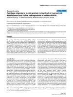
Figure 1
Conventional reverse transcription-polymerase chain reaction (RT-PCR) for caveolin-1 and housekeeping gene 18SConventional reverse transcription-polymerase chain reaction (RT-
PCR) for caveolin-1 and housekeeping gene 18S. Representative pho-
tographs following agarose gel electrophoresis of products from con-
ventional RT-PCR for caveolin-1 and 18S. cDNA samples displayed are
non-degenerate samples from a post mortem (PM) source (respective
grades [G] and ages of subjects: G3, 30 years; G1, 30 years; and G2,
75 years) and degenerate samples from surgical and PM sources (G5,
29 years; G6, 34 years; and G9, 74 years). Photographs are inverted
to improve visualisation of product bands. Cav-1, caveolin-1.
Figure 2
Quantitative real-time reverse transcription-polymerase chain reaction analysis of caveolin-1 gene expression levels in nucleus pulposus (NP) cells from human intervertebral discQuantitative real-time reverse transcription-polymerase chain reaction
analysis of caveolin-1 gene expression levels in nucleus pulposus (NP)
cells from human intervertebral disc. (a) Caveolin-1 gene expression
per 100 ng cDNA normalised to 18S in non-degenerate and degener-
ate NP presented as box-and-whisker plot (5–95 percentile). (b) Corre-
lation of caveolin-1 gene expression with age of subject. Non-
parametric linear regression analysis (P = 0.0472; R
2
= 0.2122).
Arthritis Research & Therapy Vol 10 No 4 Heathfield et al.
Page 6 of 9
(page number not for citation purposes)
caveolin-1 gene expression and protein expression are ele-
vated in degenerate IVDs, but that this rise in caveolin-1
expression does not correlate with increasing age. This is con-
sistent with a role for caveolin-1 in degenerative rather than
age-induced changes in the NP.
Changes associated with tissue ageing and degeneration
have been postulated to involve cellular senescence [41-43].
Two major categories of senescence are generally described
in the literature as replicative senescence (RS) and SIPS. RS
was first described by Hayflick in 1965 [44] and is widely
regarded as one of the main mechanisms underlying the nor-
mal ageing process via reduction of telomere length to critical
levels following cumulative population doublings. In addition,
there are a number of reports describing premature induction
of senescence as a result of cellular exposure to stress. Fac-
tors linked to the induction of SIPS vary widely, from DNA
damage – for example, radiation (bovine aortic endothelial
cells [45]), UV light (human fibroblasts [46] and human
melanocytes [47]), chemical treatment (nasopharyngeal carci-
noma cells [48] and human fibroblasts [49,50]), and oxidative
stress (human fibroblasts [20,22,24] and human articular
chondrocytes [19]) – to oncogenic protein overexpression (for
example, ras in human fibroblasts [51]) and exposure to
inflammatory cytokines such as IL-1 and tumour necrosis fac-
tor-α (human chondrocytes and fibroblasts [19,21,23]). Previ-
ous data from our laboratory described accelerated
senescence (characterised by a variety of biomarkers, includ-
ing reduced cell replication potential, elevated levels of the cell
cycle inhibitor p16
INK4a
, increased SA-β-gal activity, and
telomere erosion) in degenerate human IVDs compared with
age-matched non-degenerate discs [18], suggesting that
SIPS may be involved in IVD degeneration.
Figure 3
Caveolin-1 immunohistochemistryCaveolin-1 immunohistochemistry. (a) Photomicrograph demonstrating
staining for caveolin-1 protein in degenerate human nucleus pulposus
(sample 28). (b) Immunoglobulin G controls were negative.
Figure 4
Analysis of caveolin-1 immunohistochemistryAnalysis of caveolin-1 immunohistochemistry. (a) Percentage of cells
immunopositive for caveolin-1 protein in non-degenerate, moderately
degenerate, and severely degenerate intervertebral discs. Data are
shown as mean ± SEM. (b) Correlation of caveolin-1 protein expres-
sion with age of subject. Non-parametric linear regression analysis (P =
0.6609; R
2
= 0.0089).
Available online />Page 7 of 9
(page number not for citation purposes)
Caveolin-1 forms homodimers, or heterodimers with its family
member caveolin-2, that insert into the plasma membrane of
terminally differentiated cells [25]. The caveolin-1-rich areas
termed caveolae and the caveolin proteins themselves are pro-
posed to regulate cellular processes, including membrane
traffic, signal transduction, and cellular senescence [25-
28,52]. Caveolin-1 was investigated here due to its possible
role in cellular senescence, in particular SIPS [19,31,52].
Here, we show that caveolin-1 gene expression and protein
expression are increased during IVD degeneration, but not in
a manner that is associated with increasing chronological age.
Moreover, we demonstrate a correlation between caveolin-1
and p16
INK4a
gene expression. p16
INK4a
is a cyclin-dependent
kinase inhibitor that prevents retinoblastoma phosphorylation
and arrests the cell cycle in the G
0
/G
1
phase prior to entry into
the synthesis phase [53,54]. Many studies have shown
increased levels of p16
INK4a
alongside the occurrence and
maintenance of permanent growth arrest and senescence,
including a rodent model of ageing [55-57]. Previous studies
by our group and others strongly suggest a role for p16
INK4a
in
cellular senescence within degenerate tissue when compared
with age-matched controls [18,58]. Furthermore, elevated
p16
INK4a
expression has been described in the premature
senescence of human fibroblasts and leukaemic cells exposed
to oncogenic ras and DNA double-strand breaks [51,59,60],
strengthening the reports that p16
INK4a
is a biological marker
for senescence. The present study demonstrated that the
increased expression of caveolin-1 seen in the degenerate NP
positively correlated with gene expression for p16
INK4a
, sug-
gesting that caveolin-1 expression is linked to the senescent
phenotype observed in these cells.
The literature describes evidence linking cell exposure to
stressful stimuli to both caveolin-1 expression and cellular
senescence. In mouse NIH 3T3 fibroblasts, administration of
subcytotoxic levels of H
2
O
2
to experimentally mimic oxidative
stress induced cellular senescence and increased caveolin-1
expression. Treatment with H
2
O
2
in the presence of caveolin-
1 antisense oligonucleotides reduced expression of senes-
cence biomarkers, whereas transgenic overexpression of
caveolin-1 induced SIPS [31]. In human endothelial cells, iso-
lated from atherosclerotic patients and induced to senesce,
caveolin-1 expression was correlated with senescence
biomarkers and with expression of 4-hydroxynonenal expres-
sion (a marker of lipid peroxidation and thus oxidative stress)
independently of an effect on telomere length [31]. These
studies strongly support a role for caveolin-1 in SIPS induced
by oxidative stress and this is further strengthened by work
conducted on osteoarthritic articular chondrocytes. Adminis-
tration of H
2
O
2
to these chondrocytes induced cellular senes-
cence via expression of the caveolin-1 protein, a mechanism
reversed by antisense oligonucleotide-mediated downregula-
tion of the caveolin-1 gene [19]. The same study demon-
strated an identical role for the inflammatory cytokine IL-1β.
Articular chondrocytes and the degenerative process
observed during osteoarthritis share many characteristics with
IVD cells and IVD degeneration [12,43]. Interestingly, IVD
cells are subjected to both oxidative stress and catabolic
cytokines, which have been implicated in the induction of SIPS
[19-22,24]. Work published by our group suggests that IL-1β
not only is increased in degenerate discs but is an important
factor involved in catabolic events during IVD degeneration,
including decreased matrix production and increased MMP
and ADAMTS expression [37,38,61,62]. Moreover, advanced
glycation endproducts (AGEs) such as carboxymethyl-lysine
(CML) and the receptor for AGEs (RAGE) have been localised
to the NP of degenerate IVD [34-36]. CML is a tissue marker
for accumulated oxidative stress [35]; therefore, its presence
and that of its receptor RAGE are highly significant for both
mechanisms underlying IVD degeneration and the likelihood
that they could cause SIPS in human NP cells. Furthermore,
RAGE has been localised to caveolin-1-rich membranes in
endothelial cells [63]. This gives evidence, together with
studies involving IL-1, that there are factors in the degenerate
disc that may induce caveolin-1 expression and thus lead to
the senescent phenotype described in IVD cells [16-18].
Caveolin-1-rich regions of the plasma membrane have been
associated with several receptors and signalling molecules,
predominantly through isolation of caveolae and colocalisation
studies. These studies have highlighted a subset of proteins
that are relevant to IVD degeneration and to SIPS. First,
RAGE, described above, is known to regulate several
intracellular signalling pathways, including the nuclear factor-
kappa-B pathway, which is essential for the expression of
MMPs present in the degenerate IVD [34,64]. Second, there
Figure 5
Correlation between caveolin-1 and p16
INK4a
gene expression in degenerate nucleus pulposus samplesCorrelation between caveolin-1 and p16
INK4a
gene expression in
degenerate nucleus pulposus samples. Caveolin-1 and p16
INK4a
gene
expression (copy number per 100 ng cDNA normalised to 18S) ana-
lysed by quantitative real-time reverse transcription-polymerase chain
reaction. Non-parametric linear regression analysis (P = 0.02; R
2
=
0.4725).
Arthritis Research & Therapy Vol 10 No 4 Heathfield et al.
Page 8 of 9
(page number not for citation purposes)
is evidence suggesting that caveolin-1, β1 integrin, and uroki-
nase plasminogen activator receptor (uPAR) colocalise in
human articular chodrocytes [65]. uPAR has an integral role in
plasmin activation and thereby promotes catabolic events
through initiation of a proteolytic cascade through which
matrix-degrading enzymes described in IVD degeneration
such as MMPs are activated [66]. Both could conceivably be
pathways via which elevated caveolin-1 levels exert aspects of
the senescent cellular phenotype observed in IVD
degeneration.
Conclusion
This study has shown that caveolin-1 expression in human NP
cells is linked to IVD degeneration and is associated with the
senescent phenotype as depicted by increased expression of
p16
INK4a
. Caveolin-1 expression was not linked to increasing
chronological age, suggesting a role in accelerated degenera-
tion which could be due to SIPS, rather than RS. Further work
will elucidate the role of caveolin-1 in these related areas.
Competing interests
The authors declare that they have no competing interests.
Authors' contributions
SKH participated in the design of the study, performed the
majority of the laboratory work and analysis, and drafted the
manuscript. CLM helped to secure funding, participated in the
design of the study and the interpretation of data, and assisted
in the preparation of the final manuscript. JAH conceived the
study, secured funding, contributed to the design and coordi-
nation of the study, and participated in the interpretation of
data and extensive preparation of the final manuscript. All
authors read and approved the final manuscript.
Acknowledgements
This work was funded by a grant from DISCS (Diagnostic Investigation
of Spinal Conditions and Sciatica) and was undertaken in the Human
Tissue Profiling Laboratories of the Tissue Injury and Repair research
group.
References
1. Burton AK, Balague F, Cardon G, Eriksen HR, Henrotin Y, Lahad
A, Leclerc A, Muller G, Beek AJ van der: Chapter 2. European
guidelines for prevention in low back pain: November 2004.
Eur Spine J 2006, 15(Suppl 2):S136-168.
2. Maniadakis N, Gray A: The economic burden of back pain in the
UK. Pain 2000, 84:95-103.
3. Peterson CK, Bolton JE, Wood AR: A cross-sectional study cor-
relating lumbar spine degeneration with disability and pain.
Spine 2000, 25:218-223.
4. Luoma K, Riihimaki H, Luukkonen R, Raininko R, Viikari-Juntura E,
Lamminen A: Low back pain in relation to lumbar disc
degeneration. Spine 2000, 25:487-492.
5. Roughley PJ: Biology of intervertebral disc aging and degener-
ation: involvement of the extracellular matrix. Spine 2004,
29:2691-2699.
6. Adams MA, Roughley PJ: What is intervertebral disc degenera-
tion, and what causes it? Spine 2006, 31:2151-2161.
7. Sambrook PN, MacGregor AJ, Spector TD: Genetic influences
on cervical and lumbar disc degeneration: a magnetic reso-
nance imaging study in twins. Arthritis Rheum 1999,
42:366-372.
8. MacGregor AJ, Andrew T, Sambrook PN, Spector TD: Structural,
psychological, and genetic influences on low back and neck
pain: a study of adult female twins. Arthritis Rheum 2004,
51:160-167.
9. Miller JA, Schmatz C, Schultz AB: Lumbar disc degeneration:
correlation with age, sex, and spine level in 600 autopsy
specimens. Spine 1988, 13:173-178.
10. Boos N, Weissbach S, Rohrbach H, Weiler C, Spratt KF, Nerlich
AG: Classification of age-related changes in lumbar interver-
tebral discs: 2002 Volvo Award in basic science. Spine 2002,
27:2631-2644.
11. Anderson DG, Tannoury C: Molecular pathogenic factors in
symptomatic disc degeneration. Spine J 2005, 5(Suppl
6):260S-266S.
12. Freemont AJ, Watkins A, Le Maitre C, Jeziorska M, Hoyland JA:
Current understanding of cellular and molecular events in
intervertebral disc degeneration: implications for therapy.
J
Pathol 2002, 196:374-379.
13. Minamino T, Komuro I: Vascular cell senescence: contribution to
atherosclerosis. Circ Res 2007, 100:15-26.
14. Price JS, Waters JG, Darrah C, Pennington C, Edwards DR, Donell
ST, Clark IM: The role of chondrocyte senescence in
osteoarthritis. Aging Cell 2002, 1:57-65.
15. Martin JA, Buckwalter JA: Roles of articular cartilage aging and
chondrocyte senescence in the pathogenesis of osteoarthritis.
Iowa Orthop J 2001, 21:1-7.
16. Roberts S, Evans EH, Kletsas D, Jaffray DC, Eisenstein SM:
Senescence in human intervertebral discs. Eur Spine J 2006,
15(Suppl 15):312-316.
17. Gruber HE, Ingram JA, Norton HJ, Hanley EN Jr: Senescence in
cells of the aging and degenerating intervertebral disc: immu-
nolocalization of senescence-associated beta-galactosidase
in human and sand rat discs. Spine 2007, 32:321-327.
18. Le Maitre CL, Freemont AJ, Hoyland JA: Accelerated cellular
senescence in degenerate intervertebral discs: a possible role
in the pathogenesis of intervertebral disc degeneration. Arthri-
tis Res Ther 2007, 9:R45.
19. Dai SM, Shan ZZ, Nakamura H, Masuko-Hongo K, Kato T, Nishioka
K, Yudoh K: Catabolic stress induces features of chondrocyte
senescence through overexpression of caveolin 1: possible
involvement of caveolin 1-induced down-regulation of articu-
lar chondrocytes in the pathogenesis of osteoarthritis. Arthritis
Rheum 2006, 54:818-831.
20. Frippiat C, Chen QM, Zdanov S, Magalhaes JP, Remacle J, Tous-
saint O: Subcytotoxic H2O2 stress triggers a release of trans-
forming growth factor-beta 1, which induces biomarkers of
cellular senescence of human diploid fibroblasts. J Biol Chem
2001, 276:2531-2537.
21. Dumont P, Balbeur L, Remacle J, Toussaint O: Appearance of
biomarkers of in vitro ageing after successive stimulation of
WI-38 fibroblasts with IL-1alpha and TNF-alpha: senescence
associated beta-galactosidase activity and morphotype
transition. J Anat
2000, 197(Pt 4):529-537.
22. Dumont P, Burton M, Chen QM, Gonos ES, Frippiat C, Mazarati
JB, Eliaers F, Remacle J, Toussaint O: Induction of replicative
senescence biomarkers by sublethal oxidative stresses in
normal human fibroblast. Free Radic Biol Med 2000,
28:361-373.
23. Mendez MV, Raffetto JD, Phillips T, Menzoian JO, Park HY: The
proliferative capacity of neonatal skin fibroblasts is reduced
after exposure to venous ulcer wound fluid: a potential mech-
anism for senescence in venous ulcers. J Vasc Surg 1999,
30:734-743.
24. Chen Q, Ames BN: Senescence-like growth arrest induced by
hydrogen peroxide in human diploid fibroblast F65 cells. Proc
Natl Acad Sci USA 1994, 91:4130-4134.
25. Parton RG, Simons K: The multiple faces of caveolae. Nat Rev
Mol Cell Biol 2007, 8:185-194.
26. Smart EJ, Graf GA, McNiven MA, Sessa WC, Engelman JA,
Scherer PE, Okamoto T, Lisanti MP: Caveolins, liquid-ordered
domains, and signal transduction. Mol Cell Biol 1999,
19:7289-7304.
27. Liu P, Rudick M, Anderson RG: Multiple functions of caveolin-1.
J Biol Chem 2002, 277:41295-41298.
28. Okamoto T, Schlegel A, Scherer PE, Lisanti MP: Caveolins, a
family of scaffolding proteins for organizing "preassembled
Available online />Page 9 of 9
(page number not for citation purposes)
signaling complexes" at the plasma membrane. J Biol Chem
1998, 273:5419-5422.
29. Park WY, Park JS, Cho KA, Kim DI, Ko YG, Seo JS, Park SC: Up-
regulation of caveolin attenuates epidermal growth factor sig-
naling in senescent cells. J Biol Chem 2000,
275:20847-20852.
30. Wheaton K, Sampsel K, Boisvert FM, Davy A, Robbins S, Riabowol
K: Loss of functional caveolae during senescence of human
fibroblasts. J Cell Physiol 2001, 187:226-235.
31. Volonte D, Zhang K, Lisanti MP, Galbiati F: Expression of caveo-
lin-1 induces premature cellular senescence in primary cul-
tures of murine fibroblasts. Mol Biol Cell 2002, 13:2502-2517.
32. Linge A, Weinhold K, Blasche R, Kasper M, Barth K: Downregu-
lation of caveolin-1 affects bleomycin-induced growth arrest
and cellular senescence in A549 cells. Int J Biochem Cell Biol
2007, 39:1964-1974.
33. Voghel G, Thorin-Trescases N, Farhat N, Nguyen A, Villeneuve L,
Mamarbachi AM, Fortier A, Perrault LP, Carrier M, Thorin E: Cellu-
lar senescence in endothelial cells from atherosclerotic
patients is accelerated by oxidative stress associated with car-
diovascular risk factors. Mech Ageing Dev 2007, 128:662-671.
34. Nerlich AG, Bachmeier BE, Schleicher E, Rohrbach H, Paesold G,
Boos N: Immunomorphological analysis of RAGE receptor
expression and NF-kappaB activation in tissue samples from
normal and degenerated intervertebral discs of various ages.
Ann N Y Acad Sci 2007, 1096:239-248.
35. Nerlich AG, Schleicher ED, Boos N: 1997 Volvo Award winner in
basic science studies. Immunohistologic markers for age-
related changes of human lumbar intervertebral discs. Spine
1997, 22:2781-2795.
36. Schleicher ED, Wagner E, Nerlich AG: Increased accumulation
of the glycoxidation product N(epsilon)-(carboxymethyl)lysine
in human tissues in diabetes and aging. J Clin Invest 1997,
99:457-468.
37. Le Maitre CL, Freemont AJ, Hoyland JA: The role of interleukin-1
in the pathogenesis of human intervertebral disc
degeneration. Arthritis Res Ther 2005, 7:R732-745.
38. Le Maitre CL, Hoyland JA, Freemont AJ: Catabolic cytokine
expression in degenerate and herniated human intervertebral
discs: IL-1beta and TNFalpha expression profile. Arthritis Res
Ther 2007, 9:R77.
39. Sive JI, Baird P, Jeziorsk M, Watkins A, Hoyland JA, Freemont AJ:
Expression of chondrocyte markers by cells of normal and
degenerate intervertebral discs. Mol Pathol 2002, 55:91-97.
40. Le Maitre CL, Freemont AJ, Hoyland JA: Localization of degrada-
tive enzymes and their inhibitors in the degenerate human
intervertebral disc. J Pathol 2004, 204:47-54.
41. Toussaint O, Dumont P, Dierick JF, Pascal T, Frippiat C, Chainiaux
F, Sluse F, Eliaers F, Remacle J: Stress-induced premature
senescence. Essence of life, evolution, stress, and aging. Ann
N Y Acad Sci 2000, 908:85-98.
42. Campisi J, Kim SH, Lim CS, Rubio M: Cellular senescence, can-
cer and aging: the telomere connection. Exp Gerontol 2001,
36:1619-1637.
43. Martin JA, Buckwalter JA: Aging, articular cartilage chondrocyte
senescence and osteoarthritis. Biogerontology 2002,
3:257-264.
44. Hayflick L: The limited in vitro lifetime of human diploid cell
strains. Exp Cell Res 1965, 37:614-636.
45. Oh CW, Bump EA, Kim JS, Janigro D, Mayberg MR: Induction of
a senescence-like phenotype in bovine aortic endothelial cells
by ionizing radiation. Radiat Res 2001, 156:232-240.
46. Chainiaux F, Magalhaes JP, Eliaers F, Remacle J, Toussaint O:
UVB-induced premature senescence of human diploid skin
fibroblasts. Int J Biochem Cell Biol 2002, 34:1331-1339.
47. Medrano EE, Im S, Yang F, Abdel-Malek ZA: Ultraviolet B light
induces G1 arrest in human melanocytes by prolonged inhibi-
tion of retinoblastoma protein phosphorylation associated
with long-term expression of the p21Waf-1/SDI-1/Cip-1
protein. Cancer Res 1995, 55:4047-4052.
48. Wang X, Wong SC, Pan J, Tsao SW, Fung KH, Kwong DL, Sham
JS, Nicholls JM:
Evidence of cisplatin-induced senescent-like
growth arrest in nasopharyngeal carcinoma cells. Cancer Res
1998, 58:5019-5022.
49. Rodemann HP: Differential degradation of intracellular pro-
teins in human skin fibroblasts of mitotic and mitomycin-C
(MMC)-induced postmitotic differentiation states in vitro. Dif-
ferentiation 1989, 42:37-43.
50. Robles SJ, Buehler PW, Negrusz A, Adami GR: Permanent cell
cycle arrest in asynchronously proliferating normal human
fibroblasts treated with doxorubicin or etoposide but not
camptothecin. Biochem Pharmacol 1999, 58:675-685.
51. Serrano M, Lin AW, McCurrach ME, Beach D, Lowe SW: Onco-
genic ras provokes premature cell senescence associated
with accumulation of p53 and p16INK4a. Cell 1997,
88:593-602.
52. Cho KA, Park SC: Caveolin-1 as a prime modulator of aging: a
new modality for phenotypic restoration? Mech Ageing Dev
2005, 126:105-110.
53. Sherr CJ, Roberts JM: CDK inhibitors: positive and negative
regulators of G1-phase progression. Genes Dev 1999,
13:1501-1512.
54. Huschtscha LI, Reddel RR: p16(INK4a) and the control of cellu-
lar proliferative life span. Carcinogenesis 1999, 20:921-926.
55. Satyanarayana A, Rudolph KL: p16 and ARF: activation of teen-
age proteins in old age. J Clin Invest 2004, 114:1237-1240.
56. Beausejour CM, Krtolica A, Galimi F, Narita M, Lowe SW, Yaswen
P, Campisi J: Reversal of human cellular senescence: roles of
the p53 and p16 pathways. Embo J 2003, 22:4212-4222.
57. Krishnamurthy J, Torrice C, Ramsey MR, Kovalev GI, Al-Regaiey K,
Su L, Sharpless NE: Ink4a/Arf expression is a biomarker of
aging. J Clin Invest 2004, 114:1299-1307.
58. Zhou HW, Lou SQ, Zhang K: Recovery of function in osteoar-
thritic chondrocytes induced by p16INK4a-specific siRNA in
vitro.
Rheumatology (Oxford) 2004, 43:555-568.
59. Robles SJ, Adami GR: Agents that cause DNA double strand
breaks lead to p16INK4a enrichment and the premature
senescence of normal fibroblasts. Oncogene 1998,
16:1113-1123.
60. Park JI, Jeong JS, Han JY, Kim DI, Gao YH, Park SC, Rodgers GP,
Kim IH: Hydroxyurea induces a senescence-like change of
K562 human erythroleukemia cell. J Cancer Res Clin Oncol
2000, 126:455-460.
61. Le Maitre CL, Hoyland JA, Freemont AJ: Interleukin-1 receptor
antagonist delivered directly and by gene therapy inhibits
matrix degradation in the intact degenerate human interverte-
bral disc: an in situ zymographic and gene therapy study.
Arthritis Res Ther 2007, 9:R83.
62. Le Maitre CL, Pockert A, Buttle DJ, Freemont AJ, Hoyland JA:
Matrix synthesis and degradation in human intervertebral disc
degeneration. Biochem Soc Trans 2007, 35:652-655.
63. Lisanti MP, Scherer PE, Vidugiriene J, Tang Z, Hermanowski-
Vosatka A, Tu YH, Cook RF, Sargiacomo M: Characterization of
caveolin-rich membrane domains isolated from an endothe-
lial-rich source: implications for human disease. J Cell Biol
1994, 126:111-126.
64. Kislinger T, Fu C, Huber B, Qu W, Taguchi A, Du Yan S, Hofmann
M, Yan SF, Pischetsrieder M, Stern D, Schmidt AM: N(epsilon)-
(carboxymethyl)lysine adducts of proteins are ligands for
receptor for advanced glycation end products that activate cell
signaling pathways and modulate gene expression. J Biol
Chem 1999, 274:31740-31749.
65. Schwab W, Gavlik JM, Beichler T, Funk RH, Albrecht S, Magdolen
V, Luther T, Kasper M, Shakibaei M: Expression of the uroki-
nase-type plasminogen activator receptor in human articular
chondrocytes: association with caveolin and beta 1-integrin.
Histochem Cell Biol 2001, 115:317-323.
66. Nicholl SM, Roztocil E, Davies MG: Plasminogen activator sys-
tem and vascular disease. Curr Vasc Pharmacol 2006,
4:101-116.