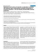
Báo cáo y học: "Raised intrathecal levels of APRIL and BAFF in patients with systemic lupus erythematosus: relationship to neuropsychiatric symptoms" docx
Bạn đang xem bản rút gọn của tài liệu. Xem và tải ngay bản đầy đủ của tài liệu tại đây (437.52 KB, 9 trang )
Open Access
Available online />Page 1 of 9
(page number not for citation purposes)
Vol 10 No 4
Research article
Raised intrathecal levels of APRIL and BAFF in patients with
systemic lupus erythematosus: relationship to neuropsychiatric
symptoms
Annie George-Chandy
1,2
, Estelle Trysberg
3
and Kristina Eriksson
1
1
Department of Rheumatology and Inflammation Research, Guldhedsgatan 10A, 413 46 Gothenburg, Sweden
2
Department of Infection Immunology, Statens Serum Institut, Artillerivej 5, 232 300 Copenhagen, Denmark
3
Rheumatogy Unit at Karolinska University Hospital/Huddinge, Hälsovägen 141, 141 52 Huddinge, Sweden
Corresponding author: Annie George-Chandy,
Received: 2 Apr 2008 Revisions requested: 29 Apr 2008 Revisions received: 28 Jul 2008 Accepted: 22 Aug 2008 Published: 22 Aug 2008
Arthritis Research & Therapy 2008, 10:R97 (doi:10.1186/ar2484)
This article is online at: />© 2008 George-Chandy et al.; licensee BioMed Central Ltd.
This is an open access article distributed under the terms of the Creative Commons Attribution License ( />),
which permits unrestricted use, distribution, and reproduction in any medium, provided the original work is properly cited.
Abstract
Introduction The tumour necrosis factor (TNF) family ligands
BAFF (B-cell activating factor of TNF family) and APRIL (a
proliferation-inducing ligand) are essential for B-cell survival and
function. Elevated serum levels of BAFF and APRIL have been
reported earlier in patients with systemic lupus erythematosus
(SLE). Since autoantibody formation in the central nervous
system (CNS) is a distinct feature of neuropsychiatric SLE
(NPSLE), we have investigated whether NPSLE is associated
with an enhanced intrathecal production of APRIL and BAFF.
Methods Levels of BAFF and APRIL in cerebrospinal fluid
(CSF) and serum from healthy controls, SLE patients without
CNS involvement, and patients with NPSLE were determined by
enzyme-linked immunosorbent assay. Interleukin-6 (IL-6) levels
were determined by an IL-6-specific bioassay.
Results SLE patients had levels of APRIL in CSF that were
more than 20-fold higher and levels of BAFF in CSF that were
more than 200-fold higher than those of healthy controls.
Separate analyses of SLE patients with and without CNS
involvement revealed that NPSLE patients had enhanced levels
of APRIL in CSF. BAFF and APRIL were likely produced locally
in the CNS as CSF and serum levels did not correlate.
Moreover, CSF levels of APRIL correlated with BAFF but not
with IL-6, suggesting that APRIL and BAFF in the CNS are
regulated together but that they are produced independently of
IL-6.
Conclusion To our knowledge this is the first study to show
elevated levels of BAFF and APRIL in CSF of SLE patients.
APRIL was augmented in NPSLE patients compared with SLE
patients without CNS involvement. APRIL and BAFF
antagonists breeching the blood-brain barrier therefore could
have beneficial effects on SLE patients, in particular patients
with NPSLE.
Introduction
Systemic lupus erythematosus (SLE) is a chronic, usually life-
long, potentially fatal autoimmune disease characterised by an
increased production of autoantibodies, impairment of B- and
T-cell functions, cytokine production, and immune complex
deposition. SLE is manifested, for example, in neurological,
dermal, haematological, musculoskeletal, and renal symptoms
[1]. Central nervous system (CNS) involvement has been
reported to occur in 14% to 75% of patients with SLE and is
a major factor contributing to morbidity and mortality in
patients [2]. The aetiology of neuropsychiatric SLE (NPSLE)
includes autoantibody production specific for brain structures,
immune complex depositions, microangiopathy, and intrathe-
cal production of proinflammatory cytokines. Seizures, stroke,
depression, psychoses, and disordered mentions are manifes-
tations of this disease [3]. Beneficial treatment in the form of
APRIL: a proliferation-inducing ligand; ARA: American Rheumatism Association; BAFF: B-cell activating factor of tumour necrosis factor family;
BCMA: B-cell maturation antigen; CNS: central nervous system; CSF: cerebrospinal fluid; DC: dendritic cell; ELISA: enzyme-linked immunosorbent
assay; GFP: glial fibrillary acidic protein; IFN: interferon; IL: interleukin; MRI: magnetic resonance imaging; MS: multiple sclerosis; NFL: neurofilament;
NPSLE: neuropsychiatric systemic lupus erythematosus; SD: standard deviation; SLE: systemic lupus erythematosus; TACI: transmembrane activator
and calcium-modulating cyclophilin ligand interactor; TNF: tumour necrosis factor.
Arthritis Research & Therapy Vol 10 No 4 George-Chandy et al.
Page 2 of 9
(page number not for citation purposes)
cytotoxic drugs is available [4] but requires early recognition of
CNS involvement. Due to the multiple pathogenic mecha-
nisms causing NPSLE, there is no single confirmatory diag-
nostic test for NPSLE. Several clinical, laboratory, and
radiographic test findings are reported to be abnormal in some
but not all patients. Magnetic resonance imaging (MRI) of the
brain has been shown to be valuable in detecting even minor
NPSLE-induced lesions [5]. Pleocytosis and elevated protein
levels are found in some but not all NPSLE patients. Elevated
concentrations of IgG in cerebrospinal fluid (CSF) IgG-albu-
min ratio, IgG index, and the presence of oligoclonal bands
have all been described with varying frequencies. Increased
levels of interleukin (IL)-1 [6], IL-6 [6,7], IL-8 [8], and inter-
feron-gamma (IFN-γ) [9] have been found in CSF of NPSLE
patients. We have reported earlier that patients with NPSLE
displayed elevated CSF levels of matrix metalloprotease-9 well
as intrathecal neurofilament (NFL) and glial fibrillary acidic pro-
tein (GFP) [10], which are markers for neuronal and astroglial
brain damage.
The tumour necrosis factor (TNF) family ligands BAFF (B-cell
activating factor of TNF family) and APRIL (a proliferation-
inducing ligand) are implicated in several immunological phe-
nomena such as peripheral B-cell survival, CD40L-independ-
ent antibody production and isotype switching, autoimmunity
as well as tumour cell growth [11,12]. BAFF is expressed on
the cell surface or cleaved and secreted [12], while APRIL is
cleaved from the Golgi and solely exists as a secreted soluble
ligand [13]. BAFF and APRIL share two receptors: B-cell mat-
uration antigen (BCMA) and transmembrane activator and cal-
cium-modulating cyclophilin ligand interactor (TACI), which
are found mainly on B cells and plasma cells [14]. In addition,
BAFF binds to BAFF receptor found mainly on B cells, plasma
cells, and some subsets of T cells [15,16], while APRIL inter-
acts with heparin sulfate proteoglycans, which likely consti-
tutes a third receptor for APRIL [17].
In the context of autoimmunity, both BAFF and APRIL are
implicated in the establishment and/or maintenance of autoim-
mune disease. Abnormal serum levels of BAFF and APRIL
have been observed in patients with rheumatoid arthritis [18],
Sjögren syndrome [19], and SLE [20]. In SLE patients,
increased serum levels of BAFF, APRIL, and BAFF/APRIL het-
erotrimers correlate with anti-double-stranded DNA autoanti-
bodies and disease activity [21]. Gene polymorphism of
APRIL has been reported to be associated with SLE [22]. The
association between enhanced levels of BAFF and autoim-
mune disease in humans has been substantiated in mice ren-
dered transgenic or deficient for this cytokine. Mice
overexpressing BAFF develop a lupus-like phenotype charac-
terised by high titres of anti-DNA antibodies, hypergamma-
globulinaemia, and glomerulonephritis [23], while mice lacking
BAFF are deficient in mature B cells and marginal zone B cells
[24]. The association between elevated circulating levels of
BAFF and polyclonal hypergammaglobulinaemia extends to
humans as well. Increased serum and/or plasma levels of
BAFF have been documented in SLE, rheumatoid arthritis, and
Sjögren syndrome [25-27], all conditions associated with pol-
yclonal hypergammaglobulinaemia. Overexpression of APRIL,
in contrast, has not been associated with autoimmunity in mice
but leads to enhanced IgM production, T-cell-independent
type 2 humoral responses, and T-cell survival [28], while lack
of APRIL is associated with enhanced numbers of effector/
memory T cells and impaired IgA responses [29,30]. However,
APRIL and BAFF/APRIL heterotrimers have been found to be
elevated in sera and target organs of autoimmune disease
patients, including SLE, Sjögren syndrome, multiple sclerosis
(MS), and myasthenia gravis [31-34]. The purpose of this
study was to examine BAFF and APRIL levels in the CSF of
SLE patients with or without NPSLE in order to investigate
whether BAFF and/or APRIL could have a role in the pathology
of NPSLE.
Materials and methods
Participants
Seventy-nine patients who fulfilled at least four of the Ameri-
can Rheumatism Association (ARA) 1987 revised criteria for
the classification of SLE [35] and who ranged in age from 19
to 75 years (mean age ± standard deviation [SD]: 45 ± 15
years; 66 females and 13 males mostly of Caucasian origin)
were included in the study. All subjects were patients at the
Department of Rheumatolgy, Sahlgrenska University Hospital.
The patients were consecutively incorporated into the study.
The disease duration varied between 0 and 41 years with a
mean of 9 ± 9 years. The patients underwent a thorough clin-
ical examination by an experienced rheumatologist and a neu-
rologist. Examination of CNS signs and symptoms included
lumbar puncture, neuropsychological tests, and MRI of the
brain. The proposed definition of CNS lupus according to ARA
criteria for SLE [35] appears inadequate, given that only two
elements, psychosis and seizures, are included. We defined
NPSLE as the presence of at least two of the following seven
items occurring in association with clinical evidence of disease
progression: (a) recent-onset psychosis, (b) transverse myeli-
tis, (c) aseptic meningitis, (d) seizures, (e) pathological brain
MRI, (f) severely abnormal neuropsychiatric tests [36], and (g)
oligoclonal IgG bands in the CSF. The pathogenesis of anti-
phospholipid-antibody-mediated brain damage is thrombotic
rather than inflammatory, and it has previously been shown
that stroke gives rise to increased values of GFP and NFL.
Consequently, we decided to exclude this condition from the
definition of CNS lupus. Patients with non-SLE causes of neu-
rological events (for example, cerebral infections) were also
excluded. Based on the criteria above, the patients were
divided into two distinct groups: (a) patients with NPSLE (n =
37) and (b) patients with SLE but without any signs of NPSLE
(n = 45). Clinical CNS and peripheral nervous system symp-
toms of included SLE patients are specified in Table 1. CSF
from healthy blood donors with no previous history of neuro-
logical disorder and with a normal neurological status served
Available online />Page 3 of 9
(page number not for citation purposes)
as controls in the present study. The age of the control sub-
jects (11 males and 9 females) was 38 ± 11 years. The Med-
ical Ethics Committee at Göteborg University approved the
study, and informed consent was obtained from all patients
and participating blood donor volunteers after written and ver-
bal information had been given.
Enzyme-linked immunosorbent assay measurements for
BAFF and APRIL
CSF and serum samples were assayed for APRIL and BAFF
by antigen-capture enzyme-linked immunosorbent assays
(ELISAs). For the detection of human APRIL, a kit from Bender
MedSystems (Vienna, Austria) was used according to the
manufacturer's instructions. For the measurement of BAFF,
ELISA plates were coated overnight with monoclonal mouse
IgG1 anti-human BAFF (clone 137314; R&D Systems, Abing-
don, Oxfordshire, UK) at 1 μg/mL in phosphate-buffered
saline. After nonspecific binding had been blocked with 0.5%
bovine serum albumin, the samples were added, followed by
the detection antibody, biotinylated goat anti-human BAFF
(R&D Systems). Streptavidin horseradish peroxidase and
3,3',5,5'-tetramethylbenzidine (Sigma-Aldrich, St. Louis, MO,
USA) were used for detection. The reaction was stopped with
0.5 M H
2
SO
4
, and the enzyme activity was read at an optical
density of 450 nm. A seven-point standard curve starting at
20,000 pg/mL of recombinant BAFF was generated. The
recombinant human BAFF used for the standard curve was
expressed in the mouse myeloma cell line NSO (R&D
Systems).
Interleukin-6 measurement
The cell line B 13.29, which is dependent on IL-6 for growth,
has been described previously. For IL-6 determinations, the
more sensitive subclone B9 was used [37,38]. B9 cells were
harvested from tissue culture flasks, seeded into microtitre
plates (Nunc, Roskilde, Denmark) at a concentration of 5,000
cells per well, and cultured in Iscove's medium supplemented
with 5 × 10
5
mol/L 2-mercaptoethanol, 5% foetal calf serum
(Sera Laboratories International, Haywards Heath, West Sus-
sex, UK), penicillin (100 U/L), and streptomycin (100 μg/mL).
CSF or serum samples were then added. (H3)thymidine was
added after 68 hours of culturing, and the cells were harvested
4 hours later. The samples were tested in two-fold dilutions
and compared with a recombinant human IL-6 standard (Gen-
zyme, Cambridge, MA, USA). B9 cells were previously shown
not to react with several recombinant cytokines, including IL-
1α, IL-1β, IL-2, IL-3, IL-5, granulocyte-macrophage colony-
stimulating factor, TNF-α, and IFN-γ. There was only a weak
reactivity with purified monoclonal antibody specific for human
IL-6 (Genzyme) in a neutralisation assay. Preincubation of 10
μg/mL of this antibody with either recombinant IL-6 or CSF
containing naturally produced IL-6 (1 hour at 37°C) reduced
proliferative responses of B9 indicator cells by an average of
greater than or equal to 95% [39].
Statistical analyses
All statistical analyses were performed using the GraphPad
Prism software (GraphPad Software, Inc., San Diego, CA,
USA). Nonparametric testing was performed by the Mann-
Whitney test for comparison of two groups. Correlation was
determined by Spearman correlation.
Results
Raised APRIL and BAFF levels in cerebrospinal fluid of
systemic lupus erythematosus patients
We measured APRIL levels in the CSF of 79 SLE patients and
15 healthy controls. Intrathecal levels of APRIL were increased
24 times in SLE patients compared with healthy controls
(mean ± SD of 10,835 ± 8,462 versus 455 ± 436 pg/mL; Fig-
ure 1a). No subjects in the group of healthy controls displayed
APRIL levels over 1,700 pg/mL in CSF. BAFF levels in CSF
were measured in 76 SLE patients and 20 healthy controls.
Intrathecal levels of BAFF were significantly raised in SLE
patients (216 ± 609 pg/mL) and were below detection levels
in all healthy controls (Figure 1b).
Raised APRIL levels in NPSLE patients compared with
systemic lupus erythematosus patients without central
nervous system symptoms
Separate analyses of APRIL levels in SLE patients with NPSLE
and patients without CNS disease revealed that NPSLE
patients had 1.5 times higher APRIL levels in CSF compared
Table 1
Clinical central nervous system/peripheral nervous system
manifestations in systemic lupus erythematosus patients
included in the study
Central nervous system manifestations No NPSLE NPSLE
Acute confusional state 0 2
Anxiety disorder 0 1
Aseptic meningitis 0 1
Cerebrovascular disease 9 6
Cognitive dysfunction 10 4
Demyelinating syndrome 2 4
Headache 9 3
Mood disorders 7 3
Movement disorder 0 0
Myelopathy 0 1
Psychosis 0 7
Fatigue 3 0
Mononeuropathy 1 0
Polyneuropathy 1 0
Each patient may have had multiple clinical manifestations of central
nervous system involvement. NPSLE, neuropsychiatric systemic
lupus erythematosus.
Arthritis Research & Therapy Vol 10 No 4 George-Chandy et al.
Page 4 of 9
(page number not for citation purposes)
with SLE patients without overt CNS disease (13,677 ±
9,725 versus 8,672 ± 9,725 pg/mL; Figure 2a). No significant
differences in BAFF levels could be detected in SLE patients
with NPSLE compared with those without CNS disease (Fig-
ure 2b). As an indicator of blood-brain barrier function, the
quotient of CSF albumin × 10
3
/serum albumin was analysed
(normal value less than 6.5 to 8.0). No significant differences
in albumin quotient could be detected between SLE patients
Figure 1
Raised APRIL and BAFF levels in cerebrospinal fluid of systemic lupus erythematosus (SLE) patientsRaised APRIL and BAFF levels in cerebrospinal fluid of systemic lupus erythematosus (SLE) patients. Cerebrospinal fluid levels of (a) APRIL and (b)
BAFF in SLE patients and in healthy controls. APRIL and BAFF levels are expressed in picograms per millilitre. Median values are depicted with a
line. *P < 0.05, ***P < 0.001. APRIL, a proliferation-inducing ligand; BAFF, B-cell activating factor of tumour necrosis factor family.
Figure 2
Raised APRIL levels in NPSLE patients compared with SLE patients without central nervous system symptomsRaised APRIL levels in NPSLE patients compared with SLE patients without central nervous system symptoms. Cerebrospinal fluid (CSF) levels of
(a) APRIL and (b) BAFF in SLE patients separated into cerebrally healthy SLE patients and patients with NPSLE. (c) The quotient of CSF albumin ×
10
3
/serum albumin. Median values are depicted with a line. **P < 0.01. APRIL, a proliferation-inducing ligand; BAFF, B-cell activating factor of
tumour necrosis factor family; NPSLE, neuropsychiatric systemic lupus erythematosus; SLE, systemic lupus erythematosus.
Available online />Page 5 of 9
(page number not for citation purposes)
with and without NPSLE (Figure 2c).
No correlation between cerebrospinal fluid and serum
levels of BAFF/APRIL
The correlations between CSF and serum levels of APRIL and
between CSF and serum levels of BAFF were analysed on 70
and 79 SLE patients, respectively. No correlation could be
seen between APRIL levels in CSF and serum (Figure 3a), nor
could any correlation be detected between BAFF levels in
CSF and serum (Figure 3b).
Figure 3
Correlation analyses of APRIL, BAFF, and IL-6Correlation analyses of APRIL, BAFF, and IL-6. Correlation between levels of (a) APRIL in cerebrospinal fluid (CSF) and serum, (b) BAFF in CSF
and serum, (c) APRIL and BAFF in CSF of systemic lupus erythematosus (SLE) patients, (d) APRIL and BAFF in serum of SLE patients, (e) IL-6 and
APRIL in CSF of SLE patients, and (f) IL-6 and BAFF in CSF of SLE patients. APRIL, a proliferation-inducing ligand; BAFF, B-cell activating factor of
tumour necrosis factor family; IL-6, interleukin-6.
Arthritis Research & Therapy Vol 10 No 4 George-Chandy et al.
Page 6 of 9
(page number not for citation purposes)
Correlation between cerebrospinal fluid but not serum
levels of APRIL and BAFF
BAFF and APRIL in CSF and serum were analysed for covari-
ance. Levels of APRIL and BAFF in CSF showed a weak cor-
relation (Spearman r = 0.27, P < 0.01; Figure 3c). No
correlation between APRIL and BAFF could be detected in
serum (Figure 3d).
No covariation between interleukin-6 and BAFF or APRIL
levels in systemic lupus erythematosus patients
It has been reported that the inflammatory cytokine IL-6 is
raised in the CSF of NPSLE patients compared with SLE
patients without CNS symptoms [40], and this could be con-
firmed also in the present CSF samples (49 ± 68 pg/ml in
NPSLE patients versus 26 ± 44 in SLE patients without CNS
symptoms; P < 0.05). In an attempt to determine whether
BAFF and APRIL are produced independently of IL-6, we ana-
lysed APRIL and BAFF, respectively, for covariance with IL-6.
No correlation was observed between IL-6 and APRIL levels
(Figure 3e) or between IL-6 and BAFF levels (Figure 3f).
Discussion
To our knowledge this study is the first to show that SLE
patients have elevated levels of APRIL and BAFF in CSF. SLE
patients displayed levels of APRIL in CSF that were 24-fold
higher than those of healthy controls, and levels of BAFF that
were 200-fold higher, thus suggesting high intrathecal levels
of APRIL and BAFF to be a feature of SLE. Although increased
levels of both BAFF and APRIL have been reported earlier
[27,41], these cytokines were measured in serum, where the
differences between SLE patients and healthy controls were
more modest. Due to the greater strictness of the NPSLE cri-
teria in comparison with the 1987 American College of Rheu-
matology Case definitions for SLE [35] that we have
previously used [10] and that we also employed in this study,
patients with mild NPSLE could potentially be grouped with
SLE patients without CNS inflammation. Nevertheless, we
were able to find significant differences in APRIL levels but not
in BAFF levels between SLE patients and NPSLE patients.
Mechanisms associated with the pathogenesis of NPSLE
include anti-neuronal antibodies, anti-phospholipid antibody-
associated thrombosis, and (rarely) vasculitis by immune com-
plex depositions. Thus, pathogenic antibody formation is
strongly associated with the manifestations of NPSLE. We
found elevated levels of the antibody-inducing cytokine APRIL
in the CNS of NPSLE patients compared with SLE patients
without CNS involvement. Elevated levels of IL-6 in CSF have
been reported in NPSLE patients [40] and could be confirmed
in the present patient material. Since BAFF, APRIL, and IL-6
are important players in the survival, differentiation, and isotype
switching of B cells, they may have an important role in the
aetiology of NPSLE.
Analyses of covariance revealed no correlation between
serum and CSF levels of BAFF or APRIL, suggesting that
APRIL and BAFF in CSF are produced locally instead of sys-
temically and subsequently passed to the CSF. A potential
source of APRIL and BAFF in the CNS are the astrocytes of
the brain. In MS patients, astrocytes have been established as
producers of both BAFF and APRIL. APRIL in the CNS was
expressed only by reactive astrocytes and increased in MS
lesions [42]. Similarly, the transcript level of BAFF was 10-fold
higher in MS lesions compared with normal CNS [43]. Collec-
tively, this suggests that astrocytes do not constitutively
express high levels of APRIL and BAFF, but rather upregulate
the expression of these cytokines upon inflammation. BAFF
and APRIL are expressed by a number of other cell types,
including monocytes, macrophages, dendritic cells (DCs),
neutrophils, and T cells [44]. Although the CNS was initially
considered an immunologically privileged site, one of the pri-
mary events that occur during CNS affecting autoimmune dis-
ease such as MS is the recruitment of immunocompetent
lymphocytes to the brain [45]. In lupus-prone MRL-Ipr mice, T
cells pass the blood-brain barrier to infiltrate the brain [46,47].
Even though T-cell infiltration in the CNS of SLE patients has
not been firmly established, T cells cannot be ruled out as a
source of intrathecal BAFF and APRIL in SLE patients.
Analyses of covariance between APRIL and BAFF levels in the
CSF of SLE patients suggested that BAFF and APRIL covary
and thus are regulated together. IFN-α, IFN-γ, IL-10, and
CD40L are important in the upregulation of both BAFF and
APRIL [48,49]. SLE patients have large increases in the levels
of IFN-α, IL-10, and soluble CD40L. Furthermore, T and B
cells from SLE patients express highly increased levels of
CD40L [50-52]. IFN-α, IL-10, and CD40L therefore could
account for the upregulation of BAFF and APRIL seen in SLE
patients. How BAFF and APRIL production in the CNS is reg-
ulated is currently not known but warrants further investigation.
SLE has been described as a disease of B-cell hyperactivity
[53]. A number of studies concur in showing that DCs play a
major role in B-cell development, mostly through the produc-
tion of cytokines such as BAFF, IL-12, IL-6, and IFN-α [54].
CD40L treatment of bone-marrow-derived DCs from lupus-
prone B6.TC mice induced higher production of IL-6, IL-10,
and TNF-α than in B6 mice [55]. In an attempt to investigate
whether IL-6, BAFF, and APRIL are regulated together, per-
haps through the involvement of DCs, we analysed covariance
between IL-6 and APRIL or BAFF. Although IL-6 levels were
increased in the CSF of NPSLE patients, we could not corre-
late IL-6 to APRIL or BAFF levels. IL-6 is not known to induce
BAFF and APRIL. In the development of humoral immunity,
BAFF particularly supports the survival of immature transitional
and mature B cells [24]. IL-6, on the other hand, promotes the
transition of plasmablasts to plasma cells [56]. BAFF, APRIL,
and IL-6 therefore likely have complementary roles in the
autoantibody formation seen in SLE.
Available online />Page 7 of 9
(page number not for citation purposes)
BAFF and APRIL are known to form homotrimers [57,58].
However, it has been shown that BAFF and APRIL can asso-
ciate with each other to form a heterotrimer capable of stimu-
lating B-cell proliferation. The levels of heterotrimers of BAFF/
APRIL are nondetectable in the serum of healthy controls,
whereas they are increased in patients with autoimmune dis-
eases such as SLE [59]. We do not know whether the anti-
bodies used in our BAFF- and APRIL-specific ELISAs
recognise only monomeric and homotrimeric molecules of
BAFF and APRIL or whether they are also able to recognise
heterotrimers of BAFF/APRIL. If CSF levels of BAFF/APRIL
heterotrimers are increased in the CSF similarly to the levels
seen in serum, and the BAFF- and APRIL-specific antibodies
used in our study are unable to recognise heterotrimers, we
may have underestimated the levels of BAFF and APRIL in
CSF. Regardless of this possibility, we did find elevated levels
of BAFF and APRIL in the CSF of SLE patients.
Evidence suggests a major contribution of B cells to the
pathogenesis of NPSLE, an effect supported by the salutary
effect of the B-cell-depleting monoclonal antibody rituximab in
this condition [60]. BAFF receptors and CD20 overlap in many
B-cell subsets, but they differ in certain populations such as
plasma cells, which express BCMA but not CD20. Blockage
of BAFF therefore might provide distinct advantages over B-
cell depletion therapy that targets CD20 (that is, rituximab).
Another concern is that rituximab treatment as a side effect
causes elevated BAFF levels in serum [61]. Therefore, upon
cessation of treatment, newly generated immature cells could
be exposed to high levels of BAFF, causing a resurgence of
autoimmunity. This, in theory, could be averted by targeting
BAFF. The potentially beneficial effects of BAFF and/or APRIL
blockage are underscored in experimental autoimmune
encephalomyelitis (a mouse model of MS). Besides demyelat-
ing disease, these mice have elevated immunoglobulin levels
and spontaneously secrete autoantibodies to DNA, which
eventually leads to autoimmune manifestations similar to SLE.
Treatment with a BAFF/APRIL antagonist (soluble BCMA-Fc)
is able to inhibit these autoimmune manifestations [62].
Since BAFF and APRIL have a major impact on B-cell survival,
T-cell function, and antibody production [11,12], overexpres-
sion of these cytokines could contribute to increased lym-
phocyte survival and proliferation, potentially leading to
increased leucocyte infiltration into the CNS. Moreover, upreg-
ulation of BAFF expression in vivo can result in the rescue of
self-reactive B cells from elimination [63]. Collectively, these
findings suggest that treatment with BAFF and/or APRIL
antagonists could be beneficial in SLE treatment. A phase II
study in rheumatoid arthritis and SLE patients using Lympho-
Stat-B, a fully humanised antibody specific for human BAFF
(also known as Belimumab), showed modest efficacy in rheu-
matoid arthritis [64], while a phase II study in SLE patients did
not meet the primary efficacy endpoints [65]. The decoy
receptor TACI-Ig (Atacicept) preventing the binding of BAFF
and APRIL to the receptor TACI on B cells led to improve-
ments in animal models of lupus [66] and arthritis [67]. Its clin-
ical value is currently under investigation in rheumatoid arthritis
and SLE.
Conclusion
We have found elevated levels of APRIL and BAFF in the CSF
of SLE patients. APRIL was augmented in NPSLE patients
compared with SLE patients without CNS involvement. Poten-
tial implications of our findings could be that APRIL and BAFF
measurements in the CSF could aid in the diagnosis of SLE.
Furthermore, APRIL and BAFF antagonists breeching the
blood-brain barrier could have beneficial effects on SLE
patients, in particular patients with NPSLE.
Competing interests
The authors declare that they have no competing interests.
Authors' contributions
KE designed the study, was involved in the management of the
study, and helped to interpret the results. AG-C was involved
in the management of the study, performed the laboratory
investigations, analysed the data statistically, helped to inter-
pret the results, and prepared the manuscript. ET was involved
in the management of the study, collected the clinical data,
and helped to interpret the results. AG-C and ET contributed
equally to this work. All authors read and approved the final
manuscript.
Acknowledgements
We thank Mrs. Inger Nordström for excellent technical assistance. This
study was supported by grants from the Swedish Science Council
(research position for KE), King Gustaf V 80-Year Foundation (KE),
Göteborgs Reumatikerförbund (KE), Torsten and Ragnar Söderbergs
stiftelser (KE), Västra Götalandsregionen through LUA/ALF (KE), and
Göteborgs Läkarsällskap (ET).
References
1. Von Feldt JM: Systemic lupus erythematosus. Recognizing its
various presentations. Postgrad Med 1995, 97:. 79, 83, 86
passim.
2. Uramoto KM, Michet CJ Jr, Thumboo J, Sunku J, O'Fallon WM,
Gabriel SE: Trends in the incidence and mortality of systemic
lupus erythematosus, 1950–1992. Arthritis Rheum 1999,
42:46-50.
3. Hanly JG: Neuropsychiatric lupus. Rheum Dis Clin North Am
2005, 31:273-298. vi.
4. Boumpas DT, Yamada H, Patronas NJ, Scott D, Klippel JH, Balow
JE: Pulse cyclophosphamide for severe neuropsychiatric
lupus. Q J Med 1991, 81:975-984.
5. Oku K, Atsumi T, Furukawa S, Horita T, Sakai Y, Jodo S, Amasaki
Y, Ichikawa K, Amengual O, Koike T: Cerebral imaging by mag-
netic resonance imaging and single photon emission com-
puted tomography in systemic lupus erythematosus with
central nervous system involvement. Rheumatology (Oxford)
2003, 42:773-777.
6. Alcocer-Varela J, Aleman-Hoey D, Alarcon-Segovia D: Interleukin-
1 and interleukin-6 activities are increased in the cerebrospi-
nal fluid of patients with CNS lupus erythematosus and corre-
late with local late T-cell activation markers. Lupus 1992,
1:111-117.
7. Hirohata S, Tanimoto K, Ito K: Elevation of cerebrospinal fluid
interleukin-6 activity in patients with vasculitides and central
Arthritis Research & Therapy Vol 10 No 4 George-Chandy et al.
Page 8 of 9
(page number not for citation purposes)
nervous system involvement. Clin Immunol Immunopathol
1993, 66:225-229.
8. Trysberg E, Blennow K, Zachrisson O, Tarkowski A: Intrathecal
levels of matrix metalloproteinases in systemic lupus ery-
thematosus with central nervous system engagement. Arthri-
tis Res Ther 2004, 6:R551-556.
9. Svenungsson E, Andersson M, Brundin L, van Vollenhoven R,
Khademi M, Tarkowski A, Greitz D, Dahlstrom M, Lundberg I,
Klareskog L, Olsson T: Increased levels of proinflammatory
cytokines and nitric oxide metabolites in neuropsychiatric
lupus erythematosus. Ann Rheum Dis 2001, 60:372-379.
10. Trysberg E, Nylen K, Rosengren LE, Tarkowski A: Neuronal and
astrocytic damage in systemic lupus erythematosus patients
with central nervous system involvement. Arthritis Rheum
2003, 48:2881-2887.
11. Dillon SR, Gross JA, Ansell SM, Novak AJ: An APRIL to remem-
ber: novel TNF ligands as therapeutic targets. Nat Rev Drug
Discov 2006, 5:235-246.
12. Mackay F, Schneider P, Rennert P, Browning J: BAFF and APRIL:
a tutorial on B cell survival. Annu Rev Immunol 2003,
21:231-264.
13. Lopez-Fraga M, Fernandez R, Albar JP, Hahne M: Biologically
active APRIL is secreted following intracellular processing in
the Golgi apparatus by furin convertase. EMBO Rep 2001,
2:945-951.
14. Marsters SA, Yan M, Pitti RM, Haas PE, Dixit VM, Ashkenazi A:
Interaction of the TNF homologues BLyS and APRIL with the
TNF receptor homologues BCMA and TACI. Curr Biol 2000,
10:785-788.
15. Thompson JS, Bixler SA, Qian F, Vora K, Scott ML, Cachero TG,
Hession C, Schneider P, Sizing ID, Mullen C, Strauch K, Zafari M,
Benjamin CD, Tschopp J, Browning JL, Ambrose C: BAFF-R, a
newly identified TNF receptor that specifically interacts with
BAFF. Science 2001, 293:2108-2111.
16. Yan M, Brady JR, Chan B, Lee WP, Hsu B, Harless S, Cancro M,
Grewal IS, Dixit VM: Identification of a novel receptor for B lym-
phocyte stimulator that is mutated in a mouse strain with
severe B cell deficiency. Curr Biol 2001, 11:
1547-1552.
17. Ingold K, Zumsteg A, Tardivel A, Huard B, Steiner QG, Cachero
TG, Qiang F, Gorelik L, Kalled SL, Acha-Orbea H, Rennert PD,
Tschopp J, Schneider P: Identification of proteoglycans as the
APRIL-specific binding partners. J Exp Med 2005,
201:1375-1383.
18. Seyler TM, Park YW, Takemura S, Bram RJ, Kurtin PJ, Goronzy JJ,
Weyand CM: BLyS and APRIL in rheumatoid arthritis. J Clin
Invest 2005, 115:3083-3092.
19. Jonsson MV, Szodoray P, Jellestad S, Jonsson R, Skarstein K:
Association between circulating levels of the novel TNF family
members APRIL and BAFF and lymphoid organization in pri-
mary Sjogren's syndrome. J Clin Immunol 2005, 25:189-201.
20. Stohl W, Metyas S, Tan SM, Cheema GS, Oamar B, Xu D,
Roschke V, Wu Y, Baker KP, Hilbert DM: B lymphocyte stimula-
tor overexpression in patients with systemic lupus erythema-
tosus: longitudinal observations. Arthritis Rheum 2003,
48:3475-3486.
21. Stohl W: BlySfulness does not equal blissfulness in systemic
lupus erythematosus: a therapeutic role for BLyS antagonists.
Curr Dir Autoimmun 2005, 8:289-304.
22. Koyama T, Tsukamoto H, Masumoto K, Himeji D, Hayashi K,
Harada M, Horiuchi T: A novel polymorphism of the human
APRIL gene is associated with systemic lupus erythematosus.
Rheumatology (Oxford) 2003, 42:980-985.
23. Mackay F, Woodcock SA, Lawton P, Ambrose C, Baetscher M,
Schneider P, Tschopp J, Browning JL: Mice transgenic for BAFF
develop lymphocytic disorders along with autoimmune
manifestations. J Exp Med 1999, 190:1697-1710.
24. Schiemann B, Gommerman JL, Vora K, Cachero TG, Shulga-Mor-
skaya S, Dobles M, Frew E, Scott ML: An essential role for BAFF
in the normal development of B cells through a BCMA-inde-
pendent pathway. Science 2001, 293:2111-2114.
25. Cheema GS, Roschke V, Hilbert DM, Stohl W: Elevated serum B
lymphocyte stimulator levels in patients with systemic
immune-based rheumatic diseases. Arthritis Rheum 2001,
44:1313-1319.
26. Groom J, Kalled SL, Cutler AH, Olson C, Woodcock SA, Schnei-
der P, Tschopp J, Cachero TG, Batten M, Wheway J, Mauri D, Cav-
ill D, Gordon TP, Mackay CR, Mackay F: Association of BAFF/
BLyS overexpression and altered B cell differentiation with
Sjogren's syndrome. J Clin Invest
2002, 109:59-68.
27. Zhang J, Roschke V, Baker KP, Wang Z, Alarcon GS, Fessler BJ,
Bastian H, Kimberly RP, Zhou T: Cutting edge: a role for B lym-
phocyte stimulator in systemic lupus erythematosus. J
Immunol 2001, 166:6-10.
28. Stein JV, Lopez-Fraga M, Elustondo FA, Carvalho-Pinto CE, Rod-
riguez D, Gomez-Caro R, De Jong J, Martinez AC, Medema JP,
Hahne M: APRIL modulates B and T cell immunity. J Clin Invest
2002, 109:1587-1598.
29. Castigli E, Scott S, Dedeoglu F, Bryce P, Jabara H, Bhan AK,
Mizoguchi E, Geha RS: Impaired IgA class switching in APRIL-
deficient mice. Proc Natl Acad Sci USA 2004, 101:3903-3908.
30. Varfolomeev E, Kischkel F, Martin F, Seshasayee D, Wang H, Law-
rence D, Olsson C, Tom L, Erickson S, French D, Schow P, Grewal
IS, Ashkenazi A: APRIL-deficient mice have normal immune
system development. Mol Cell Biol 2004, 24:997-1006.
31. Mackay F, Sierro F, Grey ST, Gordon TP: The BAFF/APRIL sys-
tem: an important player in systemic rheumatic diseases. Curr
Dir Autoimmun 2005, 8:243-265.
32. Schaller M, Stohl W, Tan SM, Benoit VM, Hilbert DM, Ditzel HJ:
Raised levels of anti-glucose-6-phosphate isomerase IgG in
serum and synovial fluid from patients with inflammatory
arthritis. Ann Rheum Dis 2005, 64:743-749.
33. Thangarajh M, Masterman T, Helgeland L, Rot U, Jonsson MV, Eide
GE, Pirskanen R, Hillert J, Jonsson R: The thymus is a source of
B-cell-survival factors-APRIL and BAFF-in myasthenia gravis.
J Neuroimmunol 2006, 178:161-166.
34. Thangarajh M, Masterman T, Rot U, Duvefelt K, Brynedal B, Karren-
bauer VD, Hillert J: Increased levels of APRIL (a proliferation-
inducing ligand) mRNA in multiple sclerosis. J Neuroimmunol
2005, 167:210-214.
35. Hochberg MC: Updating the American College of Rheumatol-
ogy revised criteria for the classification of systemic lupus
erythematosus. Arthritis Rheum 1997, 40:1725.
36. Breitbach SA, Alexander RW, Daltroy LH, Liang MH, Boll TJ, Karl-
son EW, Partiridge AJ, Roberts WN, Stern SH, Wacholtz MC,
Straaton KV: Determinants of cognitive performance in sys-
temic lupus erythematosus. J Clin Exp Neuropsychol 1998,
20:157-166.
37. Helle M, Boeije L, Aarden LA: Functional discrimination between
interleukin 6 and interleukin 1. Eur J Immunol 1988,
18:1535-1540.
38. Aarden LA, De Groot ER, Schaap OL, Lansdorp PM: Production
of hybridoma growth factor by human monocytes. Eur J
Immunol 1987, 17:1411-1416.
39. Tarkowski E, Rosengren L, Blomstrand C, Wikkelso C, Jensen C,
Ekholm S, Tarkowski A: Early intrathecal production of inter-
leukin-6 predicts the size of brain lesion in stroke. Stroke
1995, 26:1393-1398.
40. Trysberg E, Carlsten H, Tarkowski A: Intrathecal cytokines in
systemic lupus erythematosus with central nervous system
involvement. Lupus 2000, 9:498-503.
41. Koyama T, Tsukamoto H, Miyagi Y, Himeji D, Otsuka J, Miyagawa
H, Harada M, Horiuchi T: Raised serum APRIL levels in patients
with systemic lupus erythematosus. Ann Rheum Dis 2005,
64:1065-1067.
42. Thangarajh M, Masterman T, Hillert J, Moerk S, Jonsson R: A pro-
liferation-inducing ligand (APRIL) is expressed by astrocytes
and is increased in multiple sclerosis. Scand J Immunol 2007,
65:92-98.
43. Krumbholz M, Theil D, Derfuss T, Rosenwald A, Schrader F, Mon-
oranu CM, Kalled SL, Hess DM, Serafini B, Aloisi F, Wekerle H,
Hohlfeld R, Meinl E: BAFF is produced by astrocytes and up-
regulated in multiple sclerosis lesions and primary central
nervous system lymphoma. J Exp Med 2005, 201:195-200.
44. Mackay F, Ambrose C: The TNF family members BAFF and
APRIL: the growing complexity. Cytokine Growth Factor Rev
2003, 14:311-324.
45. Weller RO, Engelhardt B, Phillips MJ: Lymphocyte targeting of
the central nervous system: a review of afferent and efferent
CNS-immune pathways. Brain Pathol 1996, 6:275-288.
46. Alexander EL, Murphy ED, Roths JB, Alexander GE: Congenic
autoimmune murine models of central nervous system dis-
ease in connective tissue disorders. Ann Neurol 1983,
14:242-248.
Available online />Page 9 of 9
(page number not for citation purposes)
47. Zameer A, Hoffman SA: B and T cells in the brains of autoim-
mune mice. J Neuroimmunol 2004, 146:133-139.
48. Moore PA, Belvedere O, Orr A, Pieri K, LaFleur DW, Feng P, Sop-
pet D, Charters M, Gentz R, Parmelee D, Li Y, Galperina O, Giri J,
Roschke V, Nardelli B, Carrell J, Sosnovtseva S, Greenfield W,
Ruben SM, Olsen HS, Fikes J, Hilbert DM: BLyS: member of the
tumor necrosis factor family and B lymphocyte stimulator. Sci-
ence 1999, 285:260-263.
49. Nardelli B, Belvedere O, Roschke V, Moore PA, Olsen HS, Migone
TS, Sosnovtseva S, Carrell JA, Feng P, Giri JG, Hilbert DM: Syn-
thesis and release of B-lymphocyte stimulator from myeloid
cells. Blood 2001, 97:198-204.
50. Desai-Mehta A, Lu L, Ramsey-Goldman R, Datta SK: Hyperex-
pression of CD40 ligand by B and T cells in human lupus and
its role in pathogenic autoantibody production. J Clin Invest
1996, 97:2063-2073.
51. Llorente L, Richaud-Patin Y, Fior R, Alcocer-Varela J, Wijdenes J,
Fourrier BM, Galanaud P, Emilie D: In vivo production of inter-
leukin-10 by non-T cells in rheumatoid arthritis, Sjogren's syn-
drome, and systemic lupus erythematosus. A potential
mechanism of B lymphocyte hyperactivity and autoimmunity.
Arthritis Rheum 1994, 37:1647-1655.
52. Ronnblom L, Eloranta ML, Alm GV: The type I interferon system
in systemic lupus erythematosus. Arthritis Rheum 2006,
54:408-420.
53. Lipsky PE: Systemic lupus erythematosus: an autoimmune
disease of B cell hyperactivity. Nat Immunol 2001, 2:764-766.
54. Jego G, Pascual V, Palucka AK, Banchereau J: Dendritic cells
control B cell growth and differentiation. Curr Dir Autoimmun
2005, 8:124-139.
55. Wan S, Zhou Z, Duan B, Morel L: Direct B cell stimulation by
dendritic cells in a mouse model of lupus. Arthritis Rheum
2008, 58:1741-1750.
56. Jego G, Palucka AK, Blanck JP, Chalouni C, Pascual V,
Banchereau J: Plasmacytoid dendritic cells induce plasma cell
differentiation through type I interferon and interleukin 6.
Immunity 2003, 19:225-234.
57. Kanakaraj P, Migone TS, Nardelli B, Ullrich S, Li Y, Olsen HS, Sal-
cedo TW, Kaufman T, Cochrane E, Gan Y, Hilbert DM, Giri J: BLyS
binds to B cells with high affinity and induces activation of the
transcription factor NF-kappaB and ELF-1. Cytokine 2001,
13:25-31.
58. Wu Y, Bressette D, Carrell JA, Kaufman T, Feng P, Taylor K, Gan
Y, Cho YH, Garcia AD, Gollatz E, Dimke D, LaFleur D, Migone TS,
Nardelli B, Wei P, Ruben SM, Ullrich SJ, Olsen HS, Kanakaraj P,
Moore PA, Baker KP: Tumor necrosis factor (TNF) receptor
superfamily member TACI is a high affinity receptor for TNF
family members APRIL and BLyS. J Biol Chem 2000,
275:35478-35485.
59. Roschke V, Sosnovtseva S, Ward CD, Hong JS, Smith R, Albert V,
Stohl W, Baker KP, Ullrich S, Nardelli B, Hilbert DM, Migone TS:
BLyS and APRIL form biologically active heterotrimers that are
expressed in patients with systemic immune-based rheumatic
diseases. J Immunol 2002, 169:4314-4321.
60. Tokunaga M, Saito K, Kawabata D, Imura Y, Fujii T, Nakayamada S,
Tsujimura S, Nawata M, Iwata S, Azuma T, Mimori T, Tanaka Y:
Efficacy of rituximab (anti-CD20) for refractory systemic lupus
erythematosus involving the central nervous system. Ann
Rheum Dis 2007, 66:470-475.
61. Lavie F, Miceli-Richard C, Ittah M, Sellam J, Gottenberg JE, Mari-
ette X: Increase of B cell-activating factor of the TNF family
(BAFF) after rituximab treatment: insights into a new regulat-
ing system of BAFF production. Ann Rheum Dis 2007,
66:700-703.
62. Huntington ND, Tomioka R, Clavarino C, Chow AM, Linares D,
Mana P, Rossjohn J, Cachero TG, Qian F, Kalled SL, Bernard CC,
Reid HH: A BAFF antagonist suppresses experimental autoim-
mune encephalomyelitis by targeting cell-mediated and
humoral immune responses. Int Immunol 2006, 18:1473-1485.
63. Thien M, Phan TG, Gardam S, Amesbury M, Basten A, Mackay F,
Brink R: Excess BAFF rescues self-reactive B cells from
peripheral deletion and allows them to enter forbidden follicu-
lar and marginal zone niches. Immunity 2004, 20:785-798.
64. Cohen SB: Updates from B Cell Trials: efficacy. J Rheumatol
2006, 77:12-17.
65. Dörner T, Burmester GR: New approaches of B-cell-directed
therapy: beyond rituximab. Curr Opin Rheumatol 2008,
20:263-268.
66. Gross JA, Dillon SR, Mudri S, Johnston J, Littau A, Roque R, Rixon
M, Schou O, Foley KP, Haugen H, McMillen S, Waggie K,
Schreckhise RW, Shoemaker K, Vu T, Moore M, Grossman A,
Clegg CH: TACI-Ig neutralizes molecules critical for B cell
development and autoimmune disease. impaired B cell matu-
ration in mice lacking BLyS. Immunity
2001, 15:289-302.
67. Gross JA, Johnston J, Mudri S, Enselman R, Dillon SR, Madden K,
Xu W, Parrish-Novak J, Foster D, Lofton-Day C, Moore M, Littau A,
Grossman A, Haugen H, Foley K, Blumberg H, Harrison K, Kinds-
vogel W, Clegg CH: TACI and BCMA are receptors for a TNF
homologue implicated in B-cell autoimmune disease. Nature
2000, 404:995-999.