Báo cáo y học: "Microparticle-induced release of B-lymphocyte regulators by rheumatoid synoviocytes" ppsx
Bạn đang xem bản rút gọn của tài liệu. Xem và tải ngay bản đầy đủ của tài liệu tại đây (370.84 KB, 10 trang )
Open Access
Available online />Page 1 of 10
(page number not for citation purposes)
Vol 11 No 2
Research article
Microparticle-induced release of B-lymphocyte regulators by
rheumatoid synoviocytes
Laurent Messer
1,2
, Ghada Alsaleh
1,2
, Jean-Marie Freyssinet
3,4
, Fatiha Zobairi
3,4
, Isabelle Leray
1,2
,
Jacques-Eric Gottenberg
1,2
, Jean Sibilia
1,2
, Florence Toti-Orfanoudakis
3,4
and
Dominique Wachsmann
1,2
1
Laboratoire Physiopathologie des Arthrites, Université de Strasbourg, UFR Sciences Pharmaceutiques, 74 route du Rhin, Illkirch 67401, France
2
Département de Rhumatologie, Hôpitaux Universitaires de Strasbourg, Avenue Molière, Strasbourg Hautepierre 67200, France
3
Laboratoire de Biologie Cellulaire et Vasculaire, Faculté de Médecine, 4 rue Kirschleger, Strasbourg 67085, France
4
Inserm U770 Hôpital Bicêtre (AP-HP), 78 rue du Général Leclerc, Le Kremlin-Bicêtre 94275, France
Corresponding author: Dominique Wachsmann,
Received: 23 Feb 2009 Accepted: 16 Mar 2009 Published: 16 Mar 2009
Arthritis Research & Therapy 2009, 11:R40 (doi:10.1186/ar2648)
This article is online at: />© 2009 Messer et al.; licensee BioMed Central Ltd.
This is an open access article distributed under the terms of the Creative Commons Attribution License ( />),
which permits unrestricted use, distribution, and reproduction in any medium, provided the original work is properly cited.
Abstract
Introduction In the present study, we investigated the ability of
microparticles isolated from synovial fluids from patients with
rheumatoid arthritis or osteoarthritis to induce the synthesis and
release of key cytokines of B-lymphocyte modulation such as B
cell-activating factor, thymic stroma lymphopoietin, and
secretory leukocyte protease inhibitor by rheumatoid fibroblast-
like synoviocytes.
Methods Microparticles were analyzed in synovial fluids from
patients with rheumatoid arthritis, osteoarthritis, microcristalline
arthritis, and reactive arthritis. In addition, microparticle release
after activation from various cell lines (CEM lymphocyte and
THP-1 cells) was assessed. Microparticles were isolated by
differential centrifugation, and quantitative determinations were
carried out by prothrombinase assay after capture on
immobilized annexin V. B cell-activating factor, thymic stroma
lymphopoietin, and secretory leukocyte protease inhibitor
release was evaluated by enzyme-linked immunosorbent assay.
Results Microparticles isolated from synovial fluids obtained
from rheumatoid arthritis and osteoarthritis patients or
microparticles derived from activated THP-1 cells were able to
induce B cell-activating factor, thymic stroma lymphopoietin,
and secretory leukocyte protease inhibitor release by
rheumatoid arthritis fibroblast-like synoviocytes. Conversely,
CEM-lymphocytes-derived microparticles generated by
treatment with a combination of PHA, PMA and Adt-D did not
promote the release of B cell-activating factor but favored the
secretion of thymic stroma lymphopoietin and secretory
leukocyte protease inhibitor by rheumatoid arthritis fibrobast-like
synoviocytes. However, microparticles isolated from
actinomycin D-treated CEM lymphocytes were not able to
induce B cell-activating factor, thymic stroma lymphopoietin, or
secretory leukocyte protease inhibitor release, indicating that
microparticles derived from apoptotic T cells do not function as
effectors in B-cell activation.
Conclusions These results demonstrate that microparticles are
signalling structures that may act as specific conveyors in the
triggered induction and amplification of autoimmunity. This study
also indicates that microparticles have differential effects in the
crosstalk between B lymphocytes and target cells of
autoimmunity regarding the parental cells from which they
derive.
ActD: actinomycin D; AID: activation-induced cytidine deaminase; AR: reactive arthritis; BAFF: B cell-activating factor; ELISA: enzyme-linked immu-
nosorbent assay; FCS: fetal calf serum; FLS: fibroblast-like synoviocyte; HBSS: Hanks' balanced saline solution; IFN-: interferon-gamma; IL: inter-
leukin; LPS: lipopolysaccharide; MC: microcristalline arthritis; MCP: monocyte chemoattractant protein; MMP: matrix metalloproteinase; MP:
microparticle; NF-B: nuclear factor-kappa-B; OA: osteoarthritis; PHA: phytohemagglutinin; PhtdSer Eq: phosphatidylserine equivalents; PMA: phor-
bolmyristate acetate; PPA: combined PHA, PMA and actinomycin-D; RA: rheumatoid arthritis; SLPI: secretory leukocyte protease inhibitor; TLR: Toll-
like receptor; TSLP: thymic stroma lymphopoietin.
Arthritis Research & Therapy Vol 11 No 2 Messer et al.
Page 2 of 10
(page number not for citation purposes)
Introduction
Rheumatoid arthritis (RA) is characterized by a disorganized
reaction of the inflammatory and synovial resident cells, fibrob-
last-like synoviocytes (FLSs), which have a key function in the
development of inflammation as well as in tissue destruction
[1,2]. The activation of the latter may be linked either to the
cytokine environment or to interactions between pathogen-
associated molecular patterns and pattern-recognition recep-
tors [3,4].
FLSs can also be activated by cell-cell interactions and micro-
particles (MPs). MPs are submicron structures released from
the cell membrane during apoptosis or activation, and they
probably play an important role in this intercellular triggering
process [5]. MPs expose phosphatidylserine and display sur-
face plasma membrane markers from their parental cells [6].
They are involved in the modulation of key functions, including
inflammation, hemostasis, and angiogenesis [7-10]. Elevated
MPs circulate in the blood of patients with various inflamma-
tory disorders [11-14]. Berckmans and colleagues [15] dem-
onstrated that, in RA, synovial MPs isolated from arthritic
patients induced cytokine release by FLSs. Similarly, Distler
and colleagues [16] demonstrated that MPs derived from T
cells and monocytes induced the synthesis of various
cytokines such as interleukin (IL)-6, IL-8, monocyte chemoat-
tractant protein (MCP)-1 and MCP-2, and metalloproteases
such as matrix metalloproteinase (MMP)-1, MMP-3, MMP-9,
and MMP-14 by activated FLSs. Thus, MPs appear as multi-
functional bioeffectors that could be implicated in the exacer-
bation of the inflammatory response and cartilage and bone
erosion by resident cells in RA.
Previous findings have shown that FLSs participate in the
development of the specific immune response by secreting
cytokines (SDF-1 and CXCL13) that attracted B cells and
allowed the formation of pseudofollicles in the synovial mem-
brane [17-19]. Ohata and colleagues [20] demonstrated that
FLSs isolated from RA patients express B cell-activating factor
(BAFF) transcripts in response to tumor necrosis factor-alpha
and interferon-gamma (IFN-). BAFF is known to play a central
role in the maturation and survival of B cells as well as in anti-
body synthesis. However, we recently demonstrated that
BAFF secretion by RA FLSs is tightly regulated by a complex
network involving innate immunity and cytokines, with positive
and negative controls depending on the receptors and path-
ways triggered. Thus, BAFF synthesis and release by RA FLSs
are negatively regulated by Toll-like receptor (TLR) ligands
whereas integrin signalling pathways stimulate BAFF secre-
tion by resident cells [21].
It was recently demonstrated that, in the presence of TLR-
binding products, epithelial cells from tonsillar crypts released
BAFF and IL-10, which stimulated B cells to secrete polyreac-
tive antibodies to multiple microbial determinants. This effect
was enhanced by epithelial cell release of thymic stroma lym-
phopoietin (TSLP), which induced BAFF production by den-
dritic cells but could also restrain it by secretory leukocyte
protease inhibitor (SLPI) release from activated epithelial cells
[22]. In the present study, we investigated the capacity of MPs
isolated from synovial fluids of RA and osteoarthritis (OA)
patients or derived from various activated cell lines to induce
the release of BAFF, TSLP, and SLPI by FLSs isolated from
RA patients.
Our data indicated that MPs isolated from synovial fluids of
OA and RA patients were able to induce BAFF, TSLP, and
SLPI release by activated FLSs. Since it had been previously
demonstrated that most of the MPs present in the synovial
fluid of inflamed joints were leukocyte-derived MPs [23], we
investigated the ability of MPs isolated from activated CEM
lymphocyte and THP-1 cells to induce BAFF, TSLP, and SLPI
synthesis by activated RA FLSs.
MPs derived from activated THP-1 cells induced such release
as well. In contrast, those derived from activated CEM lym-
phocytes did not promote the release of BAFF but favored the
secretion of TSLP and SLPI by RA FLSs. However, MPs iso-
lated from actinomycin D (ActD)-treated CEM lymphocytes
were not able to induce BAFF, TSLP, and SLPI release, indi-
cating that MPs derived from apoptotic T cells do not function
as effectors in B-cell activation. Together, these results indi-
cate that MPs represent signalling structures that may act as
inducers and amplifying devices of inflammatory and specific
immune responses.
Materials and methods
Reagents
Cell culture media (RPMI 1640 and M199), fetal calf serum
(FCS), penicillin, streptomycin, and amphotericin B were
obtained from Invitrogen Corporation (Cergy-Pontoise,
France). Human recombinant IFN- was purchased from BD
Pharmingen (Le Pont-de-Claix, France). Lipopolysaccharide
(LPS) from Salmonella abortus equi and type XI collagenase,
Hanks' balanced saline solution (HBSS), ActD, phorbolmyr-
istate acetate (PMA), and phytohemagglutinin (PHA) were
obtained from Sigma-Aldrich (Saint-Quentin-Fallavier,
France). The enzyme immunoassay kits for human BAFF,
TSLP, SLPI, IL-6, and IL-8 detection were from R&D Systems
(Lille, France).
Cell culture
Human FLSs were isolated from RA synovial tissues from dif-
ferent patients at the time of knee joint arthroscopic synovec-
tomy as described previously [24]. The diagnosis conformed
to the revised criteria of the American College of Rheumatol-
ogy [25]. Informed consent was provided in accordance with
the Declaration of Helsinki and obtained from all patients.
Approval by the ethics committee of the Hopitaux Universi-
taires de Strasbourg was obtained. FLS cultures were per-
formed as previously described [26]. Experiments were
Available online />Page 3 of 10
(page number not for citation purposes)
performed between the third and ninth passages. During that
time, cultures were constituted of a homogeneous population
of fibroblastic cells that were negative for CD16 as deter-
mined by fluorescence-activated cell sorting analysis. Cell
number and cell viability were checked by the MTT (3-(4,5
dimethylthiazol-2-yl)-2,5-diphenyltetrazolium bromide) test as
described elsewhere [27]. The THP-1 monocyte cell line and
CEM lymphocytes were obtained from the American Type Cul-
ture Collection (Manassas, VA, USA).
In vitro generation of microparticles and isolation from
cell and plasma samples
The release of MPs from CEM lymphocyte cells was induced
by stimulation either with ActD alone at a concentration of 0.5
g/mL for 18 hours or with a combination of PHA (5 g/mL)
for 72 hours followed by PMA (20 ng/mL) and ActD (0.5 g/
mL) for an additional 18-hour incubation period. THP-1 cells
were treated with LPS (15 g/mL) for 18 hours. Cell culture
supernatants were centrifuged at 400 g for 5 minutes and then
at 750 g for 15 minutes. Supernatants were harvested and
centrifuged at 17,000 g for 30 minutes at 4°C. Pellets were
washed in HBSS, centrifuged for 30 minutes at 17,000 g at
4°C, and finally resuspended in 500 L of HBSS. Synovial flu-
ids were collected from RA, OA, microcristalline arthritis (MC),
and reactive arthritis (AR) patients on sodium citrate (0.129 M)
and centrifuged at room temperature for 15 minutes at 1,500
g and then for 2 minutes at 13,000 g. Supernatants were
stored at -80°C until use. In FLS-mediated activation experi-
ments, MPs were isolated from patients by differential centrif-
ugation as described for CEM lymphocyte and THP-1 cells.
The last supernatant was used as a negative control to ensure
that no remaining proteins could be responsible for the
observed MP-mediated effects. Quantitative determinations of
MPs were carried out using a prothrombinase assay after cap-
ture on immobilized annexin V as previously described. MP val-
ues are expressed as phosphatidylserine equivalents (PhtdSer
Eq) by reference to a calibration curve constructed with syn-
thetic phospholipid vesicles [28].
Stimulation of cells for cytokine assays
RA FLSs (2 × 10
5
cells) were stimulated with 1 mL of com-
plete medium (RPMI 1640 and M199/5% dialyzed FCS) con-
taining MPs. BAFF and SLPI secretion was assessed after 72-
hour MP treatment, whereas SLPI was measured after 48-hour
MP treatment by a heterologous two-site sandwich enzyme-
linked immunosorbent assay (ELISA) in accordance with the
instructions of the manufacturer (R&D Systems, Lille, France).
FLSs (5 × 10
3
cells) were grown to confluence in 96-well
plates (7 to 10 days) and then stimulated with 200 L of
serum-free RPMI 1640/M199 containing MPs. After a 20-hour
incubation period, a heterologous two-site sandwich ELISA
was used to estimate IL-6 and IL-8 release in culture superna-
tants. Negative controls consisted of cell culture medium and
last supernatants obtained after MP isolation.
Statistical analysis
Results are expressed as mean ± standard deviation. Statisti-
cal analysis was carried out using the Student test and by Wil-
coxon non-parametric test to compare mean values between
patient values or secreted molecules and released MPs in
each experiment. All analyses were performed using SPSS
13.0 software (SPSS Inc., Chicago, IL, USA).
Results
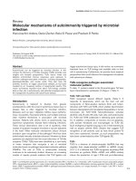
Isolation of microparticles
MPs were isolated from synovial fluids obtained from patients
with RA (n = 7), OA (n = 5), MC (n = 3), and AR (n = 5) by
differential centrifugation. Characteristics of the patients are
presented in Table 1. In this system, exosomes were elimi-
nated after the first centrifugation at 17,000 g. Quantitative
determinations of MPs were carried out using a prothrombi-
nase assay after capture on immobilized annexin V [28]. As
shown in Figure 1a, synovial fluids from all patients tested con-
tained MPs but their number was significantly higher in syno-
vial fluids isolated from RA and MC patients (32 ± 4 and 34 ±
5 nM PhtdSer Eq) compared with levels measured in synovial
fluids obtained from OA and AR patients (16 ± 3 and 18 ± 3
nM PhtdSer Eq, respectively). We also performed activation
experiments with MPs isolated from CEM lymphocyte and
THP-1 cells. CEM lymphocyte cells were treated with ActD
(0.5 g/mL) or with a combination of PHA (5 g/mL), ActD
(0.5 g/mL), and PMA (20 ng/mL) as described in Materials
and methods. THP-1 cells were stimulated with LPS from Sal-
monella abortus equi (15 g/mL) and MPs were then isolated
and quantified as described in Materials and methods. MP
capture assay on annexin V showed that LPS treatment
increased the number of MPs released from activated THP-1
cells (362 ± 76 nM PhtdSer Eq) as compared with untreated
THP-1 cells (254 ± 69 nM PhtdSer Eq) (P < 0.05). MP cap-
ture on immobilized CD14 antibody indicated that stimulated
THP-1 cells significantly released a higher proportion of MPs
bearing CD14 (390 ± 75 nM PhtdSer Eq) than unstimulated
cells (63 ± 12 nM PhtdSer Eq) (Figure 1b).
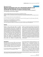
Microparticles promoted the synthesis of B cell-
activating factor by activated rheumatoid arthritis
fibroblast-like synoviocytes
To determine whether MPs are biologically active, we evalu-
ated their ability to induce BAFF release by activated RA FLSs.
In parallel, we also determined IL-6 and IL-8 production by MP-
treated RA FLSs. FLSs were incubated for 20 and 72 hours
with MPs isolated from RA synovial fluids, and IL-6, IL-8, and
BAFF release was determined by ELISA. As shown in Figure
2a,b, incubation of FLSs with MPs at a concentration of 40 nM
PhtdSer Eq increased IL-6 and IL-8 release by RA FLSs. IL-6
and IL-8 release reached 770 ± 110 and 1,150 ± 130 pg/mL,
respectively, after 20-hour incubation as compared with con-
trol medium (145 ± 40 and 150 ± 46 pg/mL) and control
supernatants (300 ± 30 and 50 ± 23 pg/mL) (Figure 2a,b).
Arthritis Research & Therapy Vol 11 No 2 Messer et al.
Page 4 of 10
(page number not for citation purposes)
Equal concentrations of OA MPs had a comparable effect on
IL-6 and IL-8 release.
We next determined whether MPs isolated from OA and RA
synovial fluids might also play a role in BAFF release by FLSs.
RA FLSs were incubated for 72 hours with MPs used at con-
centrations mentioned above, and BAFF release was evalu-
ated in culture supernatants by ELISA. As shown in Figure 2c,
BAFF secretion was increased in response to either OA or RA
MPs after 72 hours and reached 320 ± 76 pg/mL in response
to RA MPs and 250 ± 34 pg/mL in response to OA MPs as
compared with control medium (30 ± 9 pg/mL) and control
supernatants (70 ± 12 and 50 ± 21 pg/mL, respectively). IFN-
(180 ± 23 pg/mL) was used as a positive control. Taken
together, these results indicate that MPs isolated from either
OA and RA synovial fluids were able to activate IL-6, IL-8, and
BAFF release by activated FLSs.
It was previously demonstrated that most MPs present in the
synovial fluid of inflamed joints were leukocyte-derived MPs
[23]. In the present study, we investigated the ability of MPs
isolated from activated CEM lymphocyte and THP-1 cells to
induce BAFF synthesis by activated RA FLSs.
IL-6 and IL-8 release was enhanced by MPs from ActD/PMA/
PHA-treated CEM lymphocytes (1,050 ± 100 and 1,200 ±
210 pg/mL for IL-6 and IL-8, respectively) when MPs were
used at the concentration of 40 nM PhtdSer Eq (Figure 3a,b).
Table 1
Characteristics of patients
Diagnosis Patient Gender Age, years Disease duration, years Disease activity Medications
RA 1 Male 75 10 DAS28 = 3.9
CRP = 19 mg/L
Methotrexate 15 mg
Prednisone 5
2 Female 86 5 DAS28 = 4.7
CRP = 12 mg/L
Methotrexate 10 mg
Prednisone 7 mg
3Female79 20 DAS28 = 6
CRP = 78 mg/L
Methotrexate 15 mg
Prednisone 10 mg
4 Female 46 15 DAS28 = 4.1
CRP = 17 mg/L
Methotrexate 10 mg
Prednisone 2 mg
5 Female 55 6 DAS28 = 3.7
CRP = 30 mg/L
Methotrexate 17.5 mg
Prednisone 5 mg
6 Male 75 2 DAS28 = 4.2
CRP = 67 mg/L
Methotrexate 10 mg
Prednisone 7 mg
7 Male 46 15 DAS28 = 3.6
CRP = 150 mg/L
Methotrexate 10 mg
Prednisone 5 mg
Infliximab (3 mg/kg)
OA 1 Male 54 8 CRP = 4 mg/L Paracetamol, NSAID
2 Female 56 3 CRP = 6 mg/L Paracetamol
3 Female 46 5 CRP = 4 mg/L Paracetamol, NSAID
4 Female 65 7 CRP = 6 mg/L Paracetamol
5 Male 55 5 CRP = 4 mg/L Paracetamol, Tramadol
MC 1 Female 55 3 CRP = 80 mg/L Paracetamol, NSAID
2 Male 50 1 CRP = 34 mg/L Colchicine
3Male74 1 CRP = 90 mg/LNSAID
AR 1 Male 75 1 CRP = 54 mg/L NSAID
2Male30 2 CRP = 82 mg/LNSAID
3Male45 1 CRP = 20 mg/LNSAID
4 Female 20 1 CRP = 34 mg/L Paracetamol
5 Male 52 1 CRP = 25 mg/L Paracetamol
Patients were diagnosed with rheumatoid arthritis based on American College of Rheumatology 1987 diagnostic criteria at least 3 months before
study entry and have active disease. The diagnosis of microcristalline arthritis was based on clinical and laboratory features. All patients fulfilled
the American College of Rheumatology criteria for acute gouty or pseudogout. All patients gave informed consent. AR, reactive arthritis; CRP, C-
reactive protein; DAS28, disease activity score using 28 joint counts; MC, microcristalline arthritis; NSAID, nonsteroidal anti-inflammatory drugs;
OA, osteoarthritis; RA, rheumatoid arthritis.
Available online />Page 5 of 10
(page number not for citation purposes)
Such MPs did not increase BAFF production at the same con-
centration. No induction of BAFF release was observed with a
10-fold increase in MP concentration (P < 0.01). IFN- (170 ±
25 pg/mL) was used as a positive control of BAFF secretion
(Figure 3c). Similar concentrations of MPs derived from ActD-
treated CEM lymphocytes exhibited no significant effect on IL-
6, IL-8, or BAFF release by activated RA FLSs (Figure 3c).
As monocytes/macrophages are considered major instigators
of joint inflammation, we also analyzed whether MPs isolated
from LPS-activated THP-1 cells first stimulate IL-6 and IL-8
release by RA FLSs. As shown in Figure 4a,b, such MPs
induced IL-6 and IL-8 release to a similar extent (3,000 ± 350
pg/mL) in RA FLS supernatants. An induction of BAFF release
was observed after incubation with MPs (40 nM PhtdSer Eq)
derived from LPS-activated THP-1 cells (Figure 4c). BAFF
release could not be observed in negative controls.
Microparticles promoted the synthesis of thymic stroma
lymphopoietin and secretory leukocyte protease
inhibitor by activated rheumatoid arthritis fibroblast-like
synoviocytes
Having found that FLSs released BAFF in response to MP, we
sought to determine whether MP-activated FLSs could amplify
B-cell activation by stimulating dendritic cells to produce
BAFF via TSLP. RA FLSs were stimulated with MPs derived
from synovial fluids, CEM lymphocytes, and THP-1 cells at the
concentration of 40 nM PhtdSer Eq for 48 hours. As shown in
Figure 5, RA FLSs released soluble TSLP protein after expo-
sure to MPs. There were no significant differences between
FLSs activated by MPs derived from synovial fluids, CEM lym-
phocytes, and THP-1 cells. SLPI is an antiprotease that is
known to prevent BAFF-dependent class switching by inacti-
vating nuclear factor-kappa-B (NF-B). We detected SLPI in
the supernantant of MP-activated RA FLSs 72 hours after acti-
vation. Thus, FLSs produce this homeostatic regulator of class
switching after sensing MPs. Taken together, these results
demonstrate that MPs derived either from synovial fluids from
RA and OA patients or from leukocytes are able to induce
BAFF, TSLP, and SLPI release by activated RA FLSs and
could participate in cell-cell interactions leading to the proin-
flammatory response of FLSs as well as in their implication in
the B-cell autoimmune response.
Discussion
BAFF is a cytokine that plays a pivotal role in B-cell survival, dif-
ferentiation, and activation. We and others recently demon-
strated that resident cells of the synovial membrane synthesize
and release BAFF in response to stimulation of innate immune
receptors such as integrin 5
1
and IFN- receptors [20,21].
Beside this established key BAFF, other molecules might influ-
ence directly or indirectly B cells such as TSLP and SLPI.
Figure 1
Microparticle (MP) assessment in synovial fluids from arthritic patients and cell supernatantsMicroparticle (MP) assessment in synovial fluids from arthritic patients and cell supernatants. (a) Concentrations of MPs in synovial fluids from
patients with rheumatoid arthritis (RA) (n = 7), osteoarthritis (OA) (n = 5), microcristalline arthritis (MC) (n = 3), and reactive arthritis (AR) (n = 5)
were determined by a solid-phase capture assay on immobilized annexin V by use of a prothrombinase assay (nM PhtdSer Eq). (b) Concentrations
of MPs isolated from THP-1 cells stimulated with lipopolysaccharide (LPS) (15 g/mL) for 18 hours were determined by a solid-phase capture assay
on immobilized annexin V or on immobilized CD14 antibody by use of a prothrombinase assay (nM PhtdSer Eq). The control (C) corresponded to
untreated cells. Data are expressed as the mean of triplicate samples ± standard deviation and are representative of three independent experiments.
*P < 0.05. PhtdSer Eq, phosphatidylserine equivalents.
Arthritis Research & Therapy Vol 11 No 2 Messer et al.
Page 6 of 10
(page number not for citation purposes)
Direct evidence of the capacity of these factors to promote B-
cell activation was demonstrated following TLR activation of
oral epithelial cells [22].
As an increasing body of evidence suggested that MPs have
potent proinflammatory activities and are potentially important
mediators of inflammatory and autoimmune diseases [29-33],
we have investigated in this study the role of MPs on BAFF,
TSLP, and SLPI secretion by FLSs isolated from RA patients.
MPs are produced during cell death but they may also arise
during cell activation. They can be produced by virtually all cell
types, but in contrast to MPs isolated from blood, MPs isolated
from synovial fluids from RA patients are derived mainly from
inflammatory and immune cells [23].
We first evaluated MP levels in synovial fluids obtained from
patients with RA (7), OA (5), MC (3), and AR (5). All synovial
fluids contained MPs although their levels seemed to be
higher in RA and MC synovial fluids. These results are in
accordance with observations indicating that the number of
MPs is increased during inflammatory states in vivo [23].
As Distler and colleagues [16] demonstrated that MPs serve
as important triggering elements to promote cytokine, chem-
okine, and MMP release from RA synovial fibroblasts, we then
explored the role of RA and OA synovial fluid-derived MPs in
inducing BAFF synthesis by activated FLSs. In the present
study, we demonstrated a new mechanism by which FLSs
could contribute to the adaptative autoimmune response. We
showed that MPs, which are produced in synovial fluids during
RA and OA, are potent stimuli of BAFF synthesis in a similar
degree to IFN-, which is used as positive control. To our
knowledge, this is the first time that BAFF induction by MPs
has been demonstrated. This effect was observed with both
RA and OA MPs, suggesting that MPs isolated from joints of
patients with degenerative joint diseases such as OA have the
same effect as MPs present in the joints of patients with RA
and therefore that this action is not disease-dependent. The
main difference regarding the assessed effect concerns the
levels of MPs, which are much lower in synovial fluids of OA
than of RA. However, it can be speculated that other types of
cytokines and other functional effects of MPs which were not
assessed in the present study might be different. It must be
noted that, in contrast to other studies, this work was per-
Figure 2
Induction of interleukin (IL)-6, IL-8, and B cell-activating factor (BAFF) synthesis by microparticles (MPs) isolated from synovial fluidsInduction of interleukin (IL)-6, IL-8, and B cell-activating factor (BAFF) synthesis by microparticles (MPs) isolated from synovial fluids. Rheumatoid
arthritis (RA) fibroblast-like synoviocytes were stimulated with 40 nM phosphatidylserine equivalents of MPs isolated from synovial fluids of osteoar-
thritis (OA) and RA patients for 24 and 72 hours. IL-6 (a), IL-8 (b), and BAFF (c) release was determined by enzyme-linked immunosorbent assay.
Data are expressed as the mean of triplicate samples ± standard deviation and are representative of three independent experiments. C, control
medium; IFN-, interferon-gamma; SN, control supernatants.
Available online />Page 7 of 10
(page number not for citation purposes)
formed with purified MPs free of exosomes, which are pre-
formed vesicles of endosomal origin which are stored
intracellularly in multivesicular bodies and released by exocy-
tosis. Exosomes, which are investigated mainly in the regula-
tion of immune responses, do not expose phosphatidyserine,
they share a common set of membrane molecules like tet-
raspanins, and they harbor unique subsets of proteins linked
to cell type-associated functions [34,35]. Results suggest that
MPs could contribute to the interplay between FLSs and B
cells through BAFF synthesis. This induction of BAFF might
also contribute to the increased proliferation of FLSs in RA,
which might also be related to the autocrine effect of BAFF on
FLSs. Indeed, FLSs not only secrete BAFF, they also bear
BAFF receptors [36]. Berckmans and colleagues [15,37]
found that, in patients with RA, most of the MPs present in the
synovial fluid are produced by monocytes/macrophages, T
cells, and granulocytes. MPs deriving from B cells, platelets,
and erythrocytes are present only in low numbers.
To gain further information about the parental cells possibly
involved in MP-mediated BAFF synthesis, we explored the
effect of MPs isolated from CEM lymphocyte and THP-1 cells
activated under various conditions. As previously shown by
studies performed with LPS-treated U937 cells [38], THP-1
cell-derived MPs exhibit strong proinflammatory activities.
Moreover, they were able to induce BAFF release by activated
FLSs. In view of the abundance of these cells in the synovial
cavity in RA, our results suggest that macrophages could
serve as important triggering elements in promoting the inflam-
matory response and cooperation with B cells through the
release of MPs. In contrast, we observed that MPs derived
from activated CEM lymphocytes, which are inducers of IL-6
and IL-8 release, did not promote BAFF synthesis. BAFF
release was not observed even with a 10-fold increase in MP
concentration. T cells also occur abundantly in synovium and
synovial fluids; nevertheless, our data demonstrate that MPs
eventually produced by activated lymphocytes T in vivo cannot
Figure 3
Induction of interleukin (IL)-6, IL-8, and B cell-activating factor (BAFF) synthesis by microparticles (MPs) isolated from CEM lymphocytesInduction of interleukin (IL)-6, IL-8, and B cell-activating factor (BAFF) synthesis by microparticles (MPs) isolated from CEM lymphocytes. Rheuma-
toid arthritis fibroblast-like synoviocytes were stimulated with 40 and 400 nM phosphatidylserine equivalents of MPs isolated from CEM lymphocytes
treated either with actinomycin D (ActD) alone (0.5 g/mL) for 18 hours or with a combination of PHA, PMA and Adt-D (PPA) (phytohemagglutinin)
(5 g/mL) for 72 hours followed by ActD (0.5 g/mL) and phorbolmyristate acetate (20 ng/mL) for an additional 18-hour incubation period. Culture
supernatants were harvested 24 hours after stimulation for IL-6 (a) and IL-8 (b) determination and 72 hours after stimulation for BAFF evaluation (c)
by enzyme-linked immunosorbent assay. Lipopolysaccharide (IL-6 and IL-8) and interferon-gamma (IFN-) (BAFF) stimulation was used as a positive
control. Data are expressed as the mean of triplicate samples ± standard deviation and are representative of three independent experiments. **P <
0.01. Act, actinomycin; C, control medium; Pser, phosphatidylserine; TMP, control Hanks' balanced saline solution.
Arthritis Research & Therapy Vol 11 No 2 Messer et al.
Page 8 of 10
(page number not for citation purposes)
be considered key contributors in the induction of BAFF. We
cannot rule out that MPs released by other cells present in the
synovial cavity such as FLSs could interfere in this process. In
fact, we have performed some preliminary experiments with
MPs isolated from LPS-activated FLSs and have observed that
these MPs act in an autocrine pathway and induce IL-6 release
by activated FLSs (data not shown).
We reported also that MPs isolated from synovial fluids, CEM
lymphocytes, or THP-1 cells were strong inducers of TSLP.
TSLP is an IL-7-like cytokine that stimulates dendritic cells to
produce more BAFF and constitutes a Th2-independent path-
way for antibody production. It was demonstrated that epithe-
lial cells lining tonsillar crypts released AID (activation-induced
cytidine deaminase)-inducing factors, including BAFF, IL-10,
and TSLP, after sensing viral RNA through TLR3 [22]. The
resultant class switching caused the production of broadly
IgG and IgA antibodies, including antibodies to self antigens.
RA FLSs also release TSLP in response to LPS and poly I:C,
and this effect is downregulated by IFN- and dexamethasone
[39]. Our findings indicate that, like LPS and poly I:C, MPs
could indirectly participate in B-cell activation by activating
dendritic cells through TSLP release by RA FLSs and may be
involved in the physiopathology of inflammatory arthritis.
However, we also showed that RA FLSs activated with MPs
isolated from synovial fluids, CEM lymphocytes, or THP-1 cells
release SLPI. SLPI was originally identified as a protein synthe-
sized by macrophages which antagonized LPS activation of
NF-B. In B lymphocytes, SLPI inhibits class switching by
interfering with NF-B-dependent pathways and with the
upregulation of AID induced by BAFF and viral RNA. As SLPI
is released at a later time point, it may restrain the intrasynovial
production of potentially pathogenic IgG antibodies. This
needs to be demonstrated.
Conclusions
Our studies indicated that MPs are potent inducers of proin-
flammatory factors as well as B-cell survival and promote the
release of activation factors such as BAFF or TSLP by RA
Figure 4
Induction of interleukin (IL)-6, IL-8, and B cell-activating factor (BAFF) synthesis by microparticles (MPs) isolated from lipopolysaccharide (LPS)-acti-vated THP-1 cellsInduction of interleukin (IL)-6, IL-8, and B cell-activating factor (BAFF) synthesis by microparticles (MPs) isolated from lipopolysaccharide (LPS)-acti-
vated THP-1 cells. THP-1 cells were treated with LPS (15 g/mL) for 18 hours, and MPs were isolated as described in Materials and methods.
Rheumatoid arthritis (RA) FLSs were stimulated with 40 nM phosphatidylserine equivalents of MPs isolated either from THP-1 cells (MP THP-1) or
from LPS-activated THP-1 cells (MP THP-1 LPS). Culture supernatants were harvested 24 hours after stimulation for IL-6 (a) and IL-8 (b) determi-
nation and 72 hours after stimulation for BAFF secretion (c) by enzyme-linked immunosorbent assay. LPS (IL-6 and IL-8) and interferon-gamma (IFN-
) (BAFF) were used as positive controls. Data are expressed as the mean of triplicate samples ± standard deviation and are representative of three
independent experiments. C, control medium; TMP, control Hanks' balanced saline solution.
Available online />Page 9 of 10
(page number not for citation purposes)
FLSs. This cellular response is regulated by SLPI release.
Thus, MPs might play a fundamental role and behave as sen-
sors in the control of humoral B-cell responses in RA
synovium.
Competing interests
The authors declare that they have no competing interests.
Authors' contributions
GA participated in designing and performing all experiments
and in drafting the manuscript. LM participated in designing
and performing all experiments and in drafting the manuscript
and collected patient samples. FT-O supervised the character-
ization of MPs and the production of cell-derived MPs of vari-
ous origins and edited the manuscript. FZ performed cell
culture and MP quantitative determinations in patients and cell
supernatants. IL performed cell culture and MP quantitative
determinations in patients and cell supernatants. JS conceived
the study. J-MF assisted in designing the study. J-EG edited
the manuscript. DW conceived the study and drafted and
edited the manuscript. All authors read and approved the final
manuscript.
Acknowledgements
The work of DW was supported by grants from Bristol-Myers Squibb
Company (Princeton, NJ, USA), Roche (Basel, Switzerland), Pfizer Inc
(New York, NY, USA), the Courtin Foundation, and CAMPLP.
References
1. Firestein GS: Evolving concept of rheumatoid arthritis. Nature
2003, 423:356-361.
2. Muller-Ladner U, Ospelt C, Gay S, Distler O, Pap T: Cells of the
synovium in rheumatoid arthritis synovial fibroblasts. Arthritis
Res Ther 2007, 9:223-230.
3. Al-Okla S, Chatenay-Rivauday C, Klein JP, Wachsmann D:
Involvement of alpha5beta1 integrins in interleukin 8 produc-
tion induced by oral viridans streptococcal protein I/IIf in cul-
tured endothelial cells. Cell Microbiol 1999, 1:157-168.
4. Zeisel BM, Druet V, Wachsmann D, Sibilia J: MMP-3 expression
and release by rhumatoid arthritis fibroblast-like synoviocytes
induced with a bacterial ligand of integrin 5R. Arthritis Res
Ther 2005, 7:R118.
Figure 5
Induction of thymic stroma lymphopoietin (TSLP) and secretory leukocyte protease inhibitor (SLPI) synthesis by activated rheumatoid arthritis (RA) fibroblast-like synoviocytes (FLSs)Induction of thymic stroma lymphopoietin (TSLP) and secretory leukocyte protease inhibitor (SLPI) synthesis by activated rheumatoid arthritis (RA)
fibroblast-like synoviocytes (FLSs). TSLP (a) and SLPI (b) release was determined by enzyme-linked immunosorbent assay in supernatants of RA
FLSs stimulated 48 hours (TSLP) and 72 hours (SLPI) with microparticles (MPs) (40 nM phosphatidylserine equivalents) isolated from RA synovial
fluids, lipopolysaccharide (LPS)-treated THP-1 cells, and PHA, PMA and Adt-D (PPA)-treated CCRF-CEM cells. Data are expressed as the mean of
triplicate samples ± standard deviation and are representative of three independent experiments. *P < 0.05. C, control medium; IFN-, interferon-
gamma; SN, control supernatants; TMP, control Hanks' balanced saline solution.
Arthritis Research & Therapy Vol 11 No 2 Messer et al.
Page 10 of 10
(page number not for citation purposes)
5. Morel O, Toti F, Bakouboula B, Grunebaum L, Freyssinet JM: Pro-
coagulant microparticles: 'criminal partners' in atherothrom-
bosis and deleterious cellular exchanges. Pathophysiol
Haemost Thromb 2006, 35:15-22.
6. Martinez MC, Kunzelmann C, Freyssinet JM: Plasma membrane
remodelling and cell stimulation. Med Sci (Paris) 2004,
20:189-195.
7. Denzer K, van Eijk M, Kleijmeer MJ, Jakobson E, de Groot C, Geuze
HJ: Follicular dendritic cells carry MHC class II-expressing
microvesicles at their surface. J Immunol 2000,
165:1259-1265.
8. Morel O, Toti F, Hugel B, Freyssinet JM: Cellular microparticles:
a disseminated storage pool of bioactive vascular effectors.
Curr Opin Hematol 2004, 11:156-164.
9. Morel O, Toti F, Hugel B, Bakouboula B, Camoin-Jau L, Dignat-
George F, Freyssinet JM: Procoagulant microparticles: disrupt-
ing the vascular homeostasis equation? Arterioscler Thromb
Vasc Biol 2006, 26:2594-2604.
10. Ardoin SP, Shanahan JC, Pisetsky DS: The role of microparticles
in inflammation and thrombosis. Scand J Immunol 2007,
66:159-165.
11. Andoh A, Tsujikawa T, Hata K, Araki Y, Kitoh K, Sasaki M, Yoshida
T, Fujiyama Y: Elevated circulating platelet-derived microparti-
cles in patients with active inflammatory bowel disease. Am J
Gastroenterol 2005, 100:2042-2048.
12. Nieuwland R, Berckmans RJ, McGregor S, Böing AN, Romijn FP,
Westendorp RG, Hack CE, Sturk A: Cellular origin and procoag-
ulant properties of microparticles in meningococcal sepsis.
Blood 2000, 95:930-935.
13. Morel N, Morel O, Delabranche X, Jesel L, Sztark F, Dabadie P, Fre-
yssinet JM, Toti F: Microparticles during sepsis and trauma. A
link between inflammation and thrombotic processes. Ann Fr
Anesth Reanim 2006, 25:955-966.
14. Minagar A, Jy W, Jimenez JJ, Sheremata WA, Mauro LM, Mao WW,
Horstman LL, Ahn YS: Elevated plasma endothelial microparti-
cles in multiple sclerosis. Neurology 2001, 56:1319-1324.
15. Berckmans RJ, Nieuwland R, Kraan MC, Schaap MC, Pots D,
Smeets TJ, Sturk A, Tak PP:
Synovial microparticles from
arthritic patients modulate chemokine and cytokine release by
synoviocytes. Arthritis Res Ther 2005, 7:R536-R544.
16. Distler JHW, Jüngel A, Huber LC, Seemayer CA, Reich CF III, Gay
RE, Michel BA, Fontana A, Gay S, Pisetsky DS, Distler O: The
induction of matrix mettaloproteinase and cytokine expres-
sion in synovial fibroblast stimulated with immune cell
microparticles. Proc Natl Acad Sci USA 2005, 102:2892-2897.
17. Burger JA, Zvaifler NJ, Tsukada N, Firestein GS, Kipps TJ: Fibrob-
last-like synoviocytes support B-cell pseudoperiempolesis via
a stromal cell-derived factor-1 and CD106 (VCAM-1)-depend-
ent mechanism. J Clin Invest 2001, 107:305-315.
18. Shi K, Hayashida K, Kaneko M, Hashimoto J, Tomita T, Lipsky PE,
Yoshikawa H, Ochi T: Lymphoid chemokine B-cell attracting
chemokine-1 (CXCL13) is expressed in germinal center of
ectopic lymphoid follicles within the synovium of chronic
arthritis patients. J Immunol 2001, 166:650-655.
19. Weyand CM, Goronzy JJ: Ectopic germinal center formation in
rheumatoid synovitis. Ann NY Acad Sci 2003, 987:140-149.
20. Ohata J, Zvaifler NJ, Nishio M, Boyle DL, Kalled SL, Carson DA,
Kipps TJ: Fibroblast-like synoviocytes of mesenchymal origin
express functional B cell-activating factor of the TNF family in
response to proinflammatory cytokines. J Immunol 2005,
174:864-870.
21. Alsaleh G, Messer L, Semaan N, Boulanger N, Gottenberg JE,
Sibilia J, Wachsmann D: BAFF synthesis by rheumatoid synovi-
ocytes is positively controlled by alpha5beta1 integrin stimu-
lation and is negatively regulated by tumor necrosis factor
alpha and Toll-like receptor ligands. Arthritis Rheum 2007,
56:3202-3214.
22. Xu W, He B, Chiu A, Chadburn A, Shan M, Buldys M, Ding A,
Knowles DM, Santini PA, Cerutti A: Epithelial cells trigger front-
line immunoglobulin class switching through a pathway regu-
lated by the inhibitor SLPI. Nat Immunol 2007, 8:294-303.
23. Jorg H, Distler W, Huber LC, Gay S, Distler O, Pisetsky DS: Micro-
particles as mediators of cellular cross-talk in inflammatory
disease. Autoimmunity 2006, 39:683-690.
24. Dechanet J, Taupin JL, Chomarat P, Rissoan MC, Moreau JF,
Banchereau J, Miossec P: Interleukin-4 but not interleukin-10
inhibits the production of leukemia inhibitory factor by rheu-
matoid synovium and synoviocytes. Eur J Immunol 1994,
24:3222-3228.
25. Frank C, Arnett FC, Edworthy SM, Bloch DA, McShane DJ, Fries
JF, Cooper NS, Healey LA, Kaplan SR, Liang MH, Luthra HS,
Medsger TA Jr, Mitchell DM, Neustadt DH, Pinals RS, Schaller JG,
Sharp JT, Wilder RL, Hunder GG: The American Rheumatism
Association 1987 revised criteria for the classification of rheu-
matoid arthritis. Arthritis Rheum 1988, 31:315-324.
26. Neff L, Zeisel M, Sibilia J, Scholler-Guinard M, Klein JP, Wachs-
mann D: NF-kappaB and the MAP kinases/AP-1 pathways are
both involved in interleukin-6 and interleukin-8 expression in
fibroblast-like synoviocytes stimulated by protein I/II, a mod-
ulin from oral streptococci. Cell Microbiol 2001, 3:703-712.
27. Mosmann T: Rapid colorimetric assay for cellular growth and
survival: application to proliferation and cytotoxicity assays. J
Immunol Methods 1983, 65:55-63.
28. Jy W, Horstman LL, Jimenez JJ, Ahn YS, Biró E, Nieuwland R, Sturk
A, Dignat-George F, Sabatier F, Camoin-Jau L, Sampol J, Hugel B,
Zobairi F, Freyssinet JM, Nomura S, Shet AS, Key NS, Hebbel RP:
Measuring circulating cell-derived microparticles. J Thromb
Haemost 2004, 2:1842-1851.
29. Jüngel A, Distler O, Schulze-Horsel U, Huber LC, Ha HR, Simmen
B, Kalden JR, Pisetsky DS, Gay S, Distler JH: Microparticles stim-
ulate the synthesis of prostaglandin E(2) via induction of
cyclooxygenase 2 and microsomal prostaglandin E synthase
1. Arthritis Rheum 2007, 56:3564-3574.
30. Morel O, Morel N, Hugel B, Jesel L, Vinzio S, Goichot B, Bakou-
boula B, Grunebaum L, Freyssinet JM, Toti F: The significance of
circulating microparticles in physiology, inflammatory and
thrombotic diseases. Rev Med Interne 2005, 26:791-801.
31. Mause SF, von Hundelshausen P, Zernecke A, Koenen RR, Weber
C: Platelet microparticles: a transcellular delivery system for
RANTES promoting monocyte recruitment on endothelium.
Arterioscler Thromb Vasc Biol 2005, 25:1512-1518.
32. Gasser O, Schifferli JA: Activated polymorphonuclear neu-
trophils disseminate anti-inflammatory microparticles by
ectocytosis. Blood 2004, 104:2543-2548.
33. Van Wijk MJ, van Bavel E, Sturk A, Nieuwland R: Microparticles in
cardiovascular diseases. Cardiovasc Res 2003, 59:277-287.
34. Keller S, Sanderson MP, Stoeck A, Altevogt P: Exosomes: from
biogenesis and secretion to biological function. Immunol Lett
2006, 107:102-108.
35. Johnstone RM: Exosomes biological significance: a concise
review. Blood Cells Mol Dis 2006, 36:315-321.
36. Nagatani K, Itoh K, Nakajima K, Kuroki H, Katsuragawa Y, Mochi-
zuki M, Aotsuka S, Mimori A: Rheumatoid arthritis fibroblast-like
synoviocytes express BCMA and are stimulated by APRIL.
Arthritis Rheum 2007, 56:3554-3563.
37. Berckmans RJ, Nieuwland R, Tak PP, Boing AN, Romijin FP, Kraan
MC, Breedveld FC, Hack CE, Sturk A: Cell-derived microparti-
cles in synovial fluid from inflamed arthritic joints support
coagulation exclusively via a factor VII-dependent mechanism.
Arthritis Rheum 2002, 46:2857-2866.
38. Xie GL, Nomura S, Fukuhara S: Annexin V expression and mem-
brane vesiculation during activation of leukemic cell lines.
Haemostasis 1997, 27:259-268.
39. Ozawa T, Koyama K, Ando T, Ohnuma Y, Hatsushika K, Ohba T,
Sugiyama H, Hamada Y, Ogawa H, Okumura K, Nakao A: Thymic
stromal lymphopoietin secretion of synovial fibroblasts is pos-
itively and negatively regulated by Toll-like receptors/nuclear
factor-kappaB pathway and interferon-gamma/dexametha-
sone. Mod Rheumatol 2007, 17:459-463.