Báo cáo y học: "Increased risk of peripheral arterial disease in polymyalgia rheumatica: a population-based cohort study" ppsx
Bạn đang xem bản rút gọn của tài liệu. Xem và tải ngay bản đầy đủ của tài liệu tại đây (179.12 KB, 6 trang )
Open Access
Available online />Page 1 of 6
(page number not for citation purposes)
Vol 11 No 2
Research article
Increased risk of peripheral arterial disease in polymyalgia
rheumatica: a population-based cohort study
Kenneth J Warrington
1
, Elena P Jarpa
1
, Cynthia S Crowson
2
, Leslie T Cooper
3
, Gene G Hunder
1
,
EricLMatteson
1
and Sherine E Gabriel
4
1
Division of Rheumatology, Department of Medicine, Mayo Clinic, 200 First Street SW, Rochester, MN 55905, USA
2
Division of Biostatistics, Department of Health Sciences Research, Mayo Clinic, 200 First Street SW, Rochester, MN 55905, USA
3
Division of Cardiovascular Diseases, Department of Medicine, Mayo Clinic, 200 First Street SW, Rochester, MN 55905, USA
4
Division of Epidemiology, Department of Health Sciences Research, Mayo Clinic, 200 First Street SW, Rochester, MN 55905, USA
Corresponding author: Kenneth J Warrington,
Received: 13 Dec 2008 Revisions requested: 30 Jan 2009 Revisions received: 5 Mar 2009 Accepted: 31 Mar 2009 Published: 31 Mar 2009
Arthritis Research & Therapy 2009, 11:R50 (doi:10.1186/ar2664)
This article is online at: />© 2009 Warrington et al.; licensee BioMed Central Ltd.
This is an open access article distributed under the terms of the Creative Commons Attribution License ( />),
which permits unrestricted use, distribution, and reproduction in any medium, provided the original work is properly cited.
Abstract
Introduction The present study was conducted to determine
whether patients with polymyalgia rheumatica (PMR) are at an
increased risk of peripheral arterial disease (PAD).
Methods An inception cohort of all Olmsted County, Minnesota
residents diagnosed with PMR between 1 January 1970 and 31
December 1999 was compared with non-PMR subjects (two for
each PMR subject) from among residents. Both cohorts were
followed longitudinally by complete medical record review from
the incidence date of PMR (or index date for the non-PMR
cohort) until death, incident PAD, migration, or 31 December
2006. PMR-related disease characteristics, traditional
cardiovascular risk factors and diagnosis of PAD were
abstracted from the medical record. Cumulative incidence of
PAD was estimated using Kaplan–Meier methods. Cox
proportional hazards models were used to assess the risk of
PAD in PMR compared with non-PMR.
Results A total of 353 PMR patients (mean age 73.3 years,
67% women) and 705 non-PMR subjects (mean age 73.2
years, 68% female) were followed for a median of 11.0 years.
PAD developed in 38 patients (10-year cumulative incidence,
8.5%) with PMR and in 28 non-PMR subjects (10-year
cumulative incidence, 4.1%) (hazard ratio (95% confidence
interval), 2.40 (1.47, 3.92)). After adjusting for traditional
cardiovascular risk factors, patients with PMR still had a
significantly higher risk for PAD (hazard ratio, 2.50 (1.53, 4.08))
compared with controls. Giant cell arteritis occurred in 63
(18%) PMR patients but was not predictive of PAD (P = 0.15).
There was no difference between mortality in PMR and the non-
PMR cohorts nor in PMR patients with and those without PAD
(P = 0.16).
Conclusions Patients with PMR appear to have an increased
risk of PAD.
Introduction
Polymyalgia rheumatica (PMR) is an inflammatory condition
affecting middle-aged and older persons that is characterized
by aching and stiffness, typically in the cervical region, shoul-
ders, hips and proximal extremities. Most patients with PMR
have laboratory evidence of an acute phase response, includ-
ing elevation of the erythrocyte sedimentation rate and C-reac-
tive protein (CRP). Synovitis in proximal joints and periarticular
structures appears to be responsible for the musculoskeletal
symptoms in this condition [1-3]. Approximately 16 to 21% of
patients with PMR develop giant cell arteritis (GCA), an inflam-
matory vasculopathy that affects large and medium-size arter-
ies. The vascular bed of the extracranial arteries is typically
involved in GCA, but the aorta and its primary and secondary
branches can also be affected [3,4]. PMR is characteristically
very responsive to treatment with corticosteroids; however,
some patients have a chronic, relapsing course that lasts for
several years [5].
Some chronic inflammatory disorders such as rheumatoid
arthritis and systemic lupus erythematosus are associated
with an increased incidence of cardiovascular disease,
thought to be due to the effect of inflammation on progression
of atherosclerosis [6-8]. Similarly, there are data to suggest
CRP: C-reactive protein; GCA: giant cell arteritis; PAD: peripheral arterial disease; PMR: polymyalgia rheumatica.
Arthritis Research & Therapy Vol 11 No 2 Warrington et al.
Page 2 of 6
(page number not for citation purposes)
that PMR is also associated with an increased risk of cardio-
vascular disease [9].
Clinically evident peripheral arterial disease (PAD) is common
among older people and the prevalence of this condition
increases markedly with age, ranging from 2.5% for those
between the ages of 50 and 59 up to 14.5% in those aged 70
and older [10]. Among the traditional cardiovascular risk fac-
tors, diabetes mellitus and tobacco use are the strongest risk
factors for the development and progression of PAD. Hyper-
tension and dyslipidemia are weaker risk factors [11]. Inflam-
mation is also a recognized risk factor for PAD. In particular,
elevated fibrinogen and CRP levels have also been associated
with PAD [10].
We hypothesized that chronic inflammation due to PMR may
be associated with an increased risk of PAD. The present
study was undertaken to examine the incidence of clinically
significant PAD in patients with PMR compared with a control
cohort from the same geographic area. Risk factors for PAD
and the effect of PAD on survival were also evaluated.
Materials and methods
The population-based retrospective cohort study among resi-
dents of Olmsted County, Minnesota was approved by the
Mayo Clinic and Olmsted Medical Center Institutional Review
Boards. Since this is a retrospective study that poses minimal
risk to subjects, the Institutional Review Boards granted a
waiver of written informed consent for this study. All study sub-
jects, however, had authorized access to their medical record
for research purposes.
Polymyalgia rheumatica cohort
An inception cohort of all Olmsted County residents first diag-
nosed with PMR between 1 January 1970 and 31 December
1999 was assembled. This cohort has been described previ-
ously [5,9,12]. Individuals were included as subjects with
PMR if they fulfilled the following three criteria: age ≥ 50 years;
bilateral aching and morning stiffness (lasting 30 minutes or
more) persisting for at least 1 month involving two areas from
the neck or torso, the shoulders or proximal regions of the
arms, and the hips or proximal aspects of the thighs; and an
erythrocyte sedimentation rate >40 mm/hour (erythrocyte sed-
imentation rate, Westergren method). Patients with sugges-
tive clinical findings who fulfilled the first two of the three
criteria, and who had a prompt response (definite improve-
ment in symptoms within 24 hours) to low-dose corticosteroid
therapy (20 mg prednisone/day or less), were also considered
to have PMR. The presence of another disease that could
explain the symptoms, such as active rheumatoid arthritis, was
considered an exclusion criterion. A concomitant diagnosis of
GCA was documented if subjects fulfilled the 1990 American
College of Rheumatology criteria [13].
Non-polymyalgia rheumatica cohort
A comparison cohort of non-PMR subjects (two subjects for
each PMR subject) was also assembled from among Olmsted
County residents. Each non-PMR subject had a similar birth
year (± 3 years), sex, and length of medical history as the PMR
subject. Each subject in the non-PMR cohort was assigned an
index date corresponding to the PMR incidence date (base-
line) of the corresponding PMR subject.
Both the PMR and non-PMR cohorts were followed longitudi-
nally by complete (inpatient and outpatient) medical record
review, starting at the incidence date of PMR (or the index date
for the non-PMR cohort) and continuing until death, incident
PAD, migration, or 31 December 2006. PMR-related disease
characteristics were abstracted from the medical record,
including the following variables: aching neck, aching shoul-
ders/arms, aching hip/thigh, pain distal to arms, pain to the
chest/abdomen, pain to upper/lower back, tenderness in
upper arms/shoulders, tenderness in hips/thighs, tenderness
in elbow/wrist/hand/finger, tenderness in knee/leg/foot/ankle/
toe, anorexia, malaise/fatigue, weight loss. The diagnosis of
PAD was abstracted from the medical record. PAD was
defined as present if one of the following criteria were met:
ankle-brachial index ≤ 0.90 in either leg; symptoms of intermit-
tent claudication or ischemic pain at rest with examination find-
ings including documented absence of pedal and posterior
tibial artery pulses or bruits over the femoral arteries; presence
of prior peripheral artery surgery or lower extremity amputation
for PAD; aortoiliac stenosis on imaging studies (magnetic res-
onance angiography, computed tomography angiography,
conventional angiography).
An ankle-brachial index ≤ 0.90 is 90% sensitive and 95% spe-
cific for PAD [14]. We used a clinical definition of PAD to
avoid missing cases that may not have undergone noninvasive
arterial studies. The above criteria for PAD have been used
individually or in combination in previous epidemiological stud-
ies of PAD [15].
Diagnoses of traditional cardiovascular risk factors (diabetes
mellitus, hypertension and dyslipidemia) were obtained from
diagnostic indices using the ICD9 codes (diabetes, 250;
hypertension, 401 to 405; dyslipidemia, 272).
Statistical analysis
Descriptive statistics (means, standard deviations, propor-
tions) were used to summarize the data. The cumulative inci-
dence of PAD and survival following PAD were estimated
using Kaplan–Meier methods [16]. Cox proportional hazards
models were used to assess the risk of PAD in PMR subjects
compared with non-PMR subjects, as well as to examine the
influence of PMR disease characteristics on the development
of PAD within the PMR cohort. A time-dependent covariate
was used to examine the effect of GCA in order to account for
development of GCA during follow-up. Similarly, time-depend-
Available online />Page 3 of 6
(page number not for citation purposes)
ent covariates were used to adjust for diabetes mellitus, hyper-
tension and dyslipidemia.
Results
The PMR cohort included 364 patients while the non-PMR
cohort consisted of 728 individuals (two per case). We
excluded any individuals (from either cohort) with a diagnosis
of PAD prior to study entry. Our study population therefore
included a total of 353 PMR patients (mean age 73.3 years,
67% women) and 705 non-PMR subjects (mean age 73.2
years, 68% women). There was no significant difference at
baseline in the prevalence of diabetes mellitus, hypertension
and dyslipidemia between the PMR and the non-PMR cohorts
(Table 1).
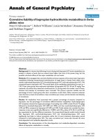
These individuals were followed for a median of 11.0 years.
During the follow-up period, 38 patients (10-year cumulative
incidence, 8.5%) with PMR developed PAD while 28 of the
non-PMR subjects (10-year cumulative incidence, 4.1%)
developed PAD (hazard ratio (95% confidence interval), 2.40
(1.47, 3.92)) (Figure 1).
After adjusting for hypertension, dyslipidemia, and diabetes
mellitus, patients with PMR still had a significantly higher risk
for PAD (hazard ratio (95% confidence interval), 2.50 (1.53,
4.08)) compared with those who did not have PMR (Table 2).
All patients with PAD (both in the PMR group and the non-
PMR group) had symptoms of lower extremity claudication,
and the majority had abnormal pedal pulses. The use of non-
invasive Doppler vascular studies and the prevalence of abnor-
mal ankle-brachial indexes were similar in both groups of
patients. PMR patients with PAD were less likely to undergo
vascular imaging studies (P = 0.01). It is therefore unlikely that
our results are due to detection bias related to increased sur-
veillance for PAD in the PMR cohort. Owing to the small sam-
ple size of patients undergoing detailed imaging studies, we
were unable to determine whether there is a difference in
severity of PAD in PMR patients versus non-PMR patients.
PMR patients with PAD, however, underwent less revascular-
ization procedures compared with the non-PMR cohort (P =
0.038) (Table 3).
We did not identify any PMR-related disease characteristics
(for example, location of musculoskeletal symptoms, constitu-
tional symptoms) that were significantly associated with the
development of PAD (data not shown).
Since inflammation may be playing a role in progression of
atherosclerosis, we examined whether the erythrocyte sedi-
mentation rate (as a continuous variable) at the time of PMR
diagnosis was associated with development of PAD. The
erythrocyte sedimentation rate value closest to the PMR diag-
nosis date (data for approximately 98% of patients was availa-
ble) was not predictive of PAD development.
While receiver operating characteristic analyses could shed
further light on this relationship, the data were inadequate to
support such an analysis. Concomitant GCA was not signifi-
cantly associated with the development of PAD in this cohort
(P = 0.15). A total of 63 (18%) PMR patients had GCA.
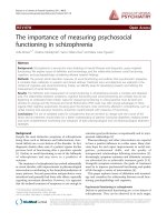
There was no difference between overall mortality in PMR
patients compared with the non-PMR cohort. In addition, and
as previously reported, there was no difference in survival for
PMR patients comparing those with PAD with those without
PAD (P = 0.16). Similarly, among those individuals who devel-
oped PAD, a prior diagnosis of PMR had no effect on survival
(P = 0.20) (Figure 2).
Discussion
Results from this population-based cohort study suggest that
individuals with PMR have an increased risk of PAD. This
increased risk may be due to premature atherosclerosis
Table 1
Demographics and risk factors in polymyalgia rheumatica
(PMR) patients and non-PMR patients
Variable Non-PMR (n = 705) PMR (n = 353) P value
Age (years) 73.2 ± 8.6 73.3 ± 8.6
Follow-up (years) 10.2 ± 7.4 11.7 ± 6.5
Female 476 (68%) 237 (67%)
Diabetes mellitus 170 (24%) 91 (26%) 0.55
Hypertension 470 (67%) 248 (70%) 0.24
Dyslipidemia 228 (32%) 127 (36%) 0.24
Data presented as mean ± standard deviation or n (%).
Figure 1
Cumulative incidence of peripheral arterial disease in the study cohortsCumulative incidence of peripheral arterial disease in the study cohorts.
Cumulative incidence of peripheral arterial disease (PAD) in 353 poly-
myalgia rheumatica (PMR) patients and 705 non-PMR patients.
Arthritis Research & Therapy Vol 11 No 2 Warrington et al.
Page 4 of 6
(page number not for citation purposes)
related to chronic inflammation. Alternatively, PAD in patients
with PMR may represent unrecognized or subclinical vasculi-
tis.
Inflammation clearly plays a major role in the pathogenesis of
atherosclerosis. Indeed, chronic inflammatory conditions such
as rheumatoid arthritis and systemic lupus erythematosus are
associated with accelerated atherosclerosis and premature
coronary artery disease [6-8]. Chronic inflammation is consid-
ered a major risk factor for both coronary and noncardiac vas-
cular disease in rheumatoid arthritis [17].
Among apparently healthy men, baseline levels of CRP predict
future risk of developing symptomatic PAD [18]. In a recent,
prospective study, however, CRP at baseline in patients with
established PAD was not predictive of disease progression
over a 3-year follow-up period [19]. Another study concluded
that high-sensitivity CRP was predictive of large-vessel PAD
[20]. Higher CRP levels are associated with greater functional
impairment in patients with PAD [21]. In the general popula-
tion, patients who have PAD and increased inflammation are at
high risk for adverse cardiovascular outcomes [22].
Overt vasculitis occurs in PMR and GCA. In these cases,
direct vascular immune-mediated injury may account for the
increased incidence of PAD in this patient population. While
we did not detect an association between concomitant GCA
and the risk of PAD, we cannot definitely rule out such an
association due to limited statistical power. It is possible that
the incidence of concomitant GCA, which rarely clinically
involves the lower extremity arteries, was underestimated in
Table 2
Risk of developing peripheral arterial disease in polymyalgia rheumatica and non-polymyalgia rheumatica
Model adjustors Hazard ratio (95% confidence interval)
Age and sex 2.40 (1.47, 3.92)
Age, sex, diabetes mellitus, hypertension, dyslipidemia 2.50 (1.53, 4.08)
Table 3
Characteristics and imaging studies of peripheral arterial disease in polymyalgia rheumatica (PMR) and non-PMR cohorts
Variable Non-PMR cohort (n = 28) PMR cohort (n = 38) P value
Claudication symptoms 28 (100%) 38 (100%)
Dorsalis pedis pulse 0.09
Abnormal 24 (92%) 37 (100%)
Normal 2 (8%) 0 (0%)
Noninvasive arterial study done 17 (61%) 19 (50%) 0.39
Ankle-brachial index < 0.90 in either leg 13 (76%) 17 (89%) 0.30
Computed tomography angiogram done 12 (43%) 5 (13%) 0.006
Magnetic resonance angiogram done 8 (29%) 3 (8%) 0.026
Conventional angiogram done 11 (39%) 6 (16%) 0.031
Any vascular imaging done 14 (50%) 8 (21%) 0.014
Stenosis on imaging 0.08
Yes 5 (36%) 6 (75%)
No 9 (64%) 2 (25%)
Revascularization for peripheral arterial disease 9 (32%) 4 (11%) 0.038
Percutaneous intervention/stent 2 (7%) 0 (0%) 0.09
Vascular bypass surgery 6 (22%) 4 (11%) 0.21
Amputation for peripheral arterial disease 2 (7%) 1 (3%) 0.40
Data presented as n (%).
Available online />Page 5 of 6
(page number not for citation purposes)
this cohort. Biopsy-proven GCA can occasionally occur in
patients without cranial symptoms [23].
PMR patients without clinically overt GCA may also frequently
have subclinical vasculitis, and therefore the complications of
GCA may also be encountered in patients with PMR. Indeed,
Weyand and colleagues demonstrated the presence of mac-
rophage-derived and T-cell-derived cytokines in temporal
artery biopsy specimens from patients with PMR but without
arteritis [24]. Activation and cytokine production from circulat-
ing monocytes is common to both patients with GCA and
PMR [25].
Using fluorodeoxyglucose positron emission tomography
scanning, Blockmans and colleagues found that three out of
five patients with PMR had increased fluorodeoxyglucose
uptake in the lower extremity vasculature, suggesting that sub-
clinical or asymptomatic vasculitis is frequently present in PMR
[26]. This was confirmed in a similar, more recent, study [27].
The strength of the present study is that it takes advantage of
the population-based medical data available through the
Rochester Epidemiology Project resources [28,29]. This
linked medical-records system allows access to accurate and
detailed clinical and laboratory data over many years, which
are not typically available in other databases or research set-
tings [30]. The primary limitation of the present study is due to
its retrospective nature. This study relies on complete and
accurate recording of pertinent information in the medical
record. Several major cardiovascular risk factors have been
incorporated into our analysis. Other elements, such as com-
plete medication (steroid) usage and smoking history, were
not analyzed. In a recent study, however, use of glucocorti-
coids for PMR was not associated with increased cardiovas-
cular events and is unlikely to account for our findings [31].
Specifically, Maradit-Kremers and colleagues reported that
glucocorticoid exposure in patients with PMR was not associ-
ated with a higher risk of PAD. Moreover, timing of glucocorti-
coid exposure and cumulative glucocorticoid dose was also
not associated with a greater risk of PAD in these patients
[31].
It should be noted that our findings are applicable primarily to
the US white population since the Olmsted County population
during the time period of the study was >95% white.
A significant number of patients with PAD are asymptomatic
and therefore we may have underestimated the incidence of
PAD in our study. This potential detection bias should be equal
for the PMR cohort and the non-PMR cohort, however, partic-
ularly since it has not been previously suspected that patients
with PMR are at higher risk for PAD and physicians would
therefore not have made more effort to detect PAD in this
cohort. Moreover, our focus was on clinically significant, symp-
tomatic PAD that could impact the patients' quality of life – as
opposed to asymptomatic disease.
The present study adds to the growing body of literature per-
taining to the risk of cardiovascular disease in chronic inflam-
matory conditions. Patients with PMR and PAD should be
monitored closely and modifiable cardiovascular risk factors
should be managed aggressively.
Conclusions
Patients with PMR appear to have an increased risk of PAD.
Future studies will be required to understand the mechanisms
underlying the excess risk of PAD in PMR in order to identify
and optimally treat at-risk PMR patients.
Competing interests
The authors declare that they have no competing interests.
Authors' contributions
KJW participated in the design of the study, reviewed medical
records, and drafted the manuscript. EPJ reviewed medical
records for acquisition of data and was involved in drafting the
manuscript. CSC participated in the design of the study and
performed the statistical analyses. LTC participated in the
design of the study and revised the manuscript for important
intellectual content. GGH participated in the design of the
study and revised the manuscript for important intellectual
content. ELM participated in the design of the study and data
interpretation, and revised the manuscript for important intel-
lectual content. SEG conceived of the study, and participated
in its design and coordination and helped to draft the manu-
script. All authors read and approved the final manuscript.
Acknowledgements
The present publication was supported by the Mayo Foundation, a grant
from the Arthritis Foundation (AF-19) and in part by the US Public Health
Figure 2
Survival following development of peripheral arterial disease in the study cohortsSurvival following development of peripheral arterial disease in the
study cohorts. Survival following development of peripheral arterial dis-
ease (PAD) in 38 polymyalgia rheumatica (PMR) patients and 28 non-
PMR patients.
Arthritis Research & Therapy Vol 11 No 2 Warrington et al.
Page 6 of 6
(page number not for citation purposes)
Service (AR-30582). This publication was made possible by Grant
Number 1 UL1 RR024150 from the National Center for Research
Resources (NCRR), a component of the National Institutes of Health
(NIH), and the NIH Roadmap for Medical Research [32,33]. The article's
contents are solely the responsibility of the authors and do not neces-
sarily represent the official view of the NCRR or the NIH.
References
1. Salvarani C, Cantini F, Boiardi L, Hunder GG: Polymyalgia rheu-
matica and giant-cell arteritis. N Engl J Med 2002,
347:261-271.
2. Salvarani C, Cantini F, Boiardi L, Hunder GG: Polymyalgia rheu-
matica. Best Pract Res Clin Rheumatol 2004, 18:705-722.
3. Weyand CM, Goronzy JJ: Giant-cell arteritis and polymyalgia
rheumatica. Ann Intern Med 2003, 139:505-515.
4. Weyand CM, Goronzy JJ: Medium- and large-vessel vasculitis.
N Engl J Med 2003, 349:160-169.
5. Kremers HM, Reinalda MS, Crowson CS, Zinsmeister AR, Hunder
GG, Gabriel SE: Relapse in a population based cohort of
patients with polymyalgia rheumatica. J Rheumatol 2005,
32:65-73.
6. Roman MJ, Shanker BA, Davis A, Lockshin MD, Sammaritano L,
Simantov R, Crow MK, Schwartz JE, Paget SA, Devereux RB,
Salmon JE: Prevalence and correlates of accelerated athero-
sclerosis in systemic lupus erythematosus. N Engl J Med
2003, 349:2399-2406.
7. Van Doornum S, McColl G, Wicks IP: Accelerated atherosclero-
sis: an extraarticular feature of rheumatoid arthritis? Arthritis
Rheum 2002, 46:862-873.
8. Maradit-Kremers H, Nicola PJ, Crowson CS, Ballman KV, Gabriel
SE: Cardiovascular death in rheumatoid arthritis: a population-
based study. Arthritis Rheum 2005, 52:722-732.
9. Kremers HM, Reinalda MS, Crowson CS, Zinsmeister AR, Hunder
GG, Gabriel SE: Direct medical costs of polymyalgia rheumat-
ica. Arthritis Rheum 2005, 53:578-584.
10. Selvin E, Erlinger TP: Prevalence of and risk factors for periph-
eral arterial disease in the United States: results from the
National Health and Nutrition Examination Survey, 1999–2000.
Circulation 2004, 110:738-743.
11. Murabito JM, D'Agostino RB, Silbershatz H, Wilson PWF: Inter-
mittent claudication: a risk profile from the Framingham Heart
Study. Circulation 1997, 96:
44-49.
12. Doran MF, Crowson CS, O'Fallon WM, Hunder GG, Gabriel SE:
Trends in the incidence of polymyalgia rheumatica over a 30
year period in Olmsted County, Minnesota, USA. J Rheumatol
2002, 29:1694-1697.
13. Hunder GG, Bloch DA, Michel BA, Stevens MB, Arend WP, Cala-
brese LH, Edworthy SM, Fauci AS, Leavitt RY, Lie JT, et al.: The
American College of Rheumatology 1990 criteria for the clas-
sification of giant cell arteritis. Arthritis Rheum 1990,
33:1122-1128.
14. Greenland P, Abrams J, Aurigemma GP, Bond MG, Clark LT,
Criqui MH, Crouse JR 3rd, Friedman L, Fuster V, Herrington DM,
Kuller LH, Ridker PM, Roberts WC, Stanford W, Stone N, Swan
HJ, Taubert KA, Wexler L: Prevention Conference V: beyond
secondary prevention: identifying the high-risk patient for pri-
mary prevention: noninvasive tests of atherosclerotic burden:
Writing Group III. Circulation 2000, 101:E16-E22.
15. Golomb BA, Dang TT, Criqui MH: Peripheral arterial disease:
morbidity and mortality implications. Circulation 2006,
114:688-699.
16. Kaplan E, Meier P: Non-parametric estimation from incomplete
observations. J Am Stat Assoc 1958, 53:457-481.
17. Liang KP, Liang KV, Matteson EL, McClelland RL, Christianson TJ,
Turesson C: Incidence of noncardiac vascular disease in rheu-
matoid arthritis and relationship to extraarticular disease man-
ifestations. Arthritis Rheum 2006, 54:642-648.
18. Ridker PM, Cushman M, Stampfer MJ, Tracy RP, Hennekens CH:
Plasma concentration of C-reactive protein and risk of devel-
oping peripheral vascular disease. Circulation 1998,
97:425-428.
19. Musicant SE, Taylor LM Jr, Peters D, Schuff RA, Urankar R, Landry
GJ, Moneta GL: Prospective evaluation of the relationship
between C-reactive protein, D-dimer and progression of
peripheral arterial disease. J Vasc Surg 2006, 43:772-780. dis-
cussion 780.
20. Aboyans V, Criqui MH, Denenberg JO, Knoke JD, Ridker PM,
Fronek A: Risk factors for progression of peripheral arterial
disease in large and small vessels. Circulation 2006,
113:2623-2629.
21. McDermott MM, Greenland P, Green D, Guralnik JM, Criqui MH,
Liu K, Chan C, Pearce WH, Taylor L, Ridker PM, Schneider JR,
Martin G, Rifai N, Quann M, Fornage M:
D-dimer, inflammatory
markers, and lower extremity functioning in patients with and
without peripheral arterial disease. Circulation 2003,
107:3191-3198.
22. Beckman JA, Preis O, Ridker PM, Gerhard-Herman M: Compari-
son of usefulness of inflammatory markers in patients with
versus without peripheral arterial disease in predicting
adverse cardiovascular outcomes (myocardial infarction,
stroke, and death). Am J Cardiol 2005, 96:1374-1378.
23. Gonzalez-Gay MA, Garcia-Porrua C, Amor-Dorado JC, Llorca J:
Giant cell arteritis without clinically evident vascular involve-
ment in a defined population. Arthritis Rheum 2004,
51:274-277.
24. Weyand CM, Hicok KC, Hunder GG, Goronzy JJ: Tissue cytokine
patterns in patients with polymyalgia rheumatica and giant cell
arteritis. Ann Intern Med 1994, 121:484-491.
25. Wagner AD, Goronzy JJ, Weyand CM: Functional profile of tis-
sue-infiltrating and circulating CD68
+
cells in giant cell arteri-
tis. Evidence for two components of the disease. J Clin Invest
1994, 94:1134-1140.
26. Blockmans D, Maes A, Stroobants S, Nuyts J, Bormans G, Knock-
aert D, Bobbaers H, Mortelmans L: New arguments for a vascu-
litic nature of polymyalgia rheumatica using positron emission
tomography. Rheumatology (Oxford, England) 1999,
38:444-447.
27. Blockmans D, De Ceuninck L, Vanderschueren S, Knockaert D,
Mortelmans L, Bobbaers H: Repetitive 18-fluorodeoxyglucose
positron emission tomography in isolated polymyalgia rheu-
matica: a prospective study in 35 patients. Rheumatology
(Oxford) 2007, 46:672-677.
28. Kurland LT, Molgaard CA: The patient record in epidemiology.
Sci Am 1981, 245:54-63.
29. Melton LJ 3rd: History of the Rochester Epidemiology Project.
Mayo Clin Proc 1996, 71:266-274.
30. Maradit Kremers H, Crowson CS, Gabriel SE: Rochester Epide-
miology Project: a unique resource for research in the rheu-
matic diseases. Rheum Dis Clin North Am 2004, 30:819-834.
vii.
31. Maradit Kremers H, Reinalda MS, Crowson CS, Davis JM 3rd,
Hunder GG, Gabriel SE: Glucocorticoids and cardiovascular
and cerebrovascular events in polymyalgia rheumatica. Arthri-
tis Rheum 2007, 57:279-286.
32. National Centre for Research Resources [http://
www.ncrr.nih.gov]
33. NIH Roadmap for Medical Research [road
map.nih.gov]