Báo cáo Y học: Coordinated action of protein tyrosine phosphatases in insulin signal transduction potx
Bạn đang xem bản rút gọn của tài liệu. Xem và tải ngay bản đầy đủ của tài liệu tại đây (247.29 KB, 10 trang )
MINIREVIEW
Coordinated action of protein tyrosine phosphatases
in insulin signal transduction
Alan Cheng, Nadia Dube
´
, Feng Gu and Michel L. Tremblay
Department of Biochemistry and McGill Cancer Center, McGill University, Montreal, Quebec, Canada
Insulin is the principal regulatory hormone involved in the
tight regulation of fuel metabolism. In response to blood
glucose l evels, it is secrete d by the b cells o f t he pancreas and
exerts its effects by binding to cell surface receptors that are
present on virtually all cell types and tissues. In humans,
perturbations in insulin function and/or secretion lead to
diabetes mellitus, a severe disorder primarily characterized
by an inability to maintain blood glucose homeostasis.
Furthermore, it is estimated that 90–95% of diabetic patients
exhibit resistance to insulin action. Thus an understanding of
insulin signal transduction and insulin resistance at the
molecular level is crucial to the understanding of the patho-
genesis of this disease. The in sulin receptor (IR) is a trans-
membrane tyrosine kinase that becomes activated upon
ligand binding. Consequently, the receptor and its down-
stream substrates become tyrosine p hosphorylated. This
activates a series of intracellular signaling cascades which
coordinately initiate the appropriate biological response.
One important mechanism by which insulin signalin g is
regulated involves the protein t yrosine phosphatases (PTPs),
which may either act on the IR itself and/or its substrates.
Two w ell characterized examples include leuckocyte antigen
related (LAR) and protein tyrosine phosphatase-1B (PTP-
1B). The present review will discuss the current knowledge of
these two and othe r potential PTPs involved in the insulin
signaling pathway.
Keywords: diabetes; insulin receptor; protein tyrosine phos-
phatase; knockout mice; signaling.
INTRODUCTION
Insulin is the most potent anabolic hormone identified to
date. It is produced and secreted in a regulated fashion by
the b cells in the pancreatic islet. Practic ally all cell types are
responsive to insulin, although the term Ôinsulin sensitive
tissuesÕ often r efer to the liver, muscle a nd adipose. The
primary biological effect of insulin is to maintain glucose
homeostasis. It acutely promotes glucose uptake in muscle
and adipose tissue, while suppressing hepatic glucose
production. However, in sulin also stimulates lipogenesis,
protein synthesis, and has been shown to be a mitogen for
certain cell types.
The importance of insulin function is highlighted by its
disregulation in diabetes mellitus, a human disease charac-
terized by an impairment in insulin secretion (type I; insulin
dependent) and/or action (type II; noninsulin dependent).
Currently, d iabetes is recognized as the world’s most
common metabolic disorder, affecting people globally and
of all age groups. For the year 2000, it was estimated that
over 175 million people worldwide, were affl icted with this
disease (International Diabetes Institute, World Health
Organisation). Clinically, diabetes is primarily characterized
by fasting hyperglycemia, is often associated with cardio-
vascular risk factors, and may lead to severe complications.
At present, type I diabetes comprises about 5–10% of all
diagnosed cases. The molecular complexity of this disorder
is well documented, and current therapies revolve around
exogenous insulin supplementation [1,2]. On the other hand,
type II diabetes accounts for the remaining 90–95% of the
cases. At the molecular level, a postreceptor defe ct of insulin
signaling is mainly thought to und erlie the basis of insulin
resistance in type II diabetes [3]. Consequently, understand-
ing the mechanisms by which this may occur will provide
invaluable insight for the development of novel therapies. In
the present review, we will summarize the current under-
standing of insulin signaling with particular focus on how
protein tyrosine phosphatases regulate this process.
BRIEF OVERVIEW OF INSULIN
SIGNALING
Insulin is a pleiotropic hormone with multiple integrated
signaling pathways. For brevity, we will only describe those
relevant to this review. The insulin receptor (IR) belongs to a
subclass of the large family of protein tyrosine kinases [4]. It
is a t ransmembrane protein c omprising two extracellular
a subu nits and two tran smembrane b subunits (Fig. 1).
Upon binding to insulin, the intrinsic kinase activity of the
receptor is increased, and the IR undergoes autophospho-
rylation on several tyrosine residues located on the cyto-
plasmic portion of the b subunits [5]. Subsequently, these
phosphotyrosine residues, in their surrounding seq uence
Correspondence to M. L. Tremblay, Department of Biochemistry
and McGill Cancer Center, McGill University, 3655 Promenade
Sir William Osler, Room 715, Montreal, Quebec, Canada, H 3G 1Y6.
Fax: + 1 514 398 6769, Tel.: + 1 514 398 7290,
E-mail:
Abbreviations: GLUT4, g lucose transporter 4; IR, insulin receptor;
IRS, insulin receptor substrate; PTP, protein tyrosine phosphatase;
SH2, Src homology 2; PI3-kinase, phosphatidyl inositol 3-kinase.
(Received 6 August 2001, accepted 20 September 2001)
Eur. J. Biochem. 269, 1050–1059 (2002) Ó FEBS 2002
context, recruit signaling m olecules containin g SH2 ( Src
homology 2) or PTB (phosphotyrosine binding) domains
[6]. Although not limited to, most of the recruited proteins
belong to a class of adapte r p roteins of the insulin receptor
substrate (IRS) family [7,8]. The best characterized examples
include Shc, which is primarily involved in the activation of
the mitogen activated protein kinase (MAPK) pathway for
mitogenic effect; and IRS-1, which can transmit insulin
signaling to both metabolic and mitogenic processes [9,10].
The primary function of insulin is to maintain glucose
homeostasis. For most ce lls, this is ach ieved via the insulin
dependent translocation of the glucose transporter,
GLUT4, from intracellular vesicles to the cell surface [11–
13]. Upon recruitment of IRS-1 to the activated IR, IRS-1
becomes heavily tyrosine phosphorylated and serves as a
large scaffolding protein by binding to several SH2
containing proteins. The most prominent e xample is the
p85 regulatory s ubunit of phosphatidylinositol 3-kinase
(PI3-kinase). Binding to p85 recruits the p110 catalytic
subunit of PI3-kinase, resulting in the activation of the PI3-
kinase pathway, a necessary component involved in
GLUT4 translocation. However, activation of PI3-kinase
alone is insufficient, as platelet-derived growth factor which
also activates PI3-kinase, does not promote glucose trans-
port [14,15]. In f act, recent studies in 3T3-L1 adipocytes
have demonstrated a pathway parallel to PI3-kinase, which
is required for insulin-mediated G LUT4 translocation.
Activation of the IR results in the formation of a protein
complex involving the scaffolding proteins CAP and Cbl.
Subsequent tyrosine phosphorylation of C bl by the IR leads
to translocation of the Cbl–CAP complex to lipid rafts, a
process that is necessary for GLUT4 translocation [14,15].
In contrast to the activation and propagation of insulin
signal transduction, the negative regulatory components
that attenuate insulin signaling are less well defined. Because
tyrosine phosphorylation o f t he IR corr elates with its
activity and function, the protein tyrosine phosphatases [16]
are prominent candidates t o negatively regulate i nsulin
action. Indeed, vanadium compounds, known inhibitors of
PTPs, have long been known t o possess insulin mimetic or
enhancing effects [17–19]. However, it should also be noted
that there is evidence f or serine phosphorylation [5] and
O-glycosylation [20] in attenuating insulin signaling, but
these will not be discussed here.
THE PROTEIN TYROSINE
PHOSPHATASE FAMILY
PTPs represent a large family of enz ymes t hat rival the
PTKs in both functional and structural diversity. Members
of this group can b e classified into receptor vs. nonreceptor
PTPs. Common to all members is a highly conserved core of
about 250 amino acids that make up the catalytic domain.
The PTP signature motif, V/IHCSAGXGRXG sequence
contains an invariant cysteine residue that is critical for PTP
activity [21]. In addition, s ome receptor PTPs (rPTPs)
possess two such d omains, although only one is usually
active [22–24]. Apart from the catalytic domain, the rest of
the protein is quite divergent amongst PTPs. For the sake of
simplicity, and to illustrate the point, we will only depict
several PTPs relevant to insulin signaling (Fig. 2).
A s ubset of rPTPs contain structural motifs such as
immunoglobulin-like and fibronectin type III elem ents.
These structures have been found in cell-adhesion molecules
and suggest a role for these PTPs in cell–cell contact or cell–
extracellular matrix interactions. On the other h and, all
intracellular PTPs possess a single conserved phosphatase
domain, that is flanked at either the N- or C-terminus by
noncatalytic segments. T hese segments play regulatory
roles, either by binding each PTP to its substrate(s) or to
adapter molecules through domains that regulate protein–
protein interactions, by targeting the PTP to a particular
subcellular compartment, or by keeping the enzyme in an
inactive conformation.
To elucidate the function of PTPs and t heir mechanisms
of action, identification of their substrates is critical. Over
the past decade, many studies on PTPs have utilized a
strategy called Ôsubstrate trappingÕ [25]. In this method,
mutagenesis of the conserved cysteine to serine (CfiS) or an
aspartate to alanine (DfiA) within the catalytic domain of
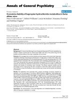
Fig. 1. Scheme of the major insulin signaling
pathways. The activated insulin receptor
phosphorylates tyrosine residu es on IRS p ro-
teins, Shc, CAP and other intracellular sub-
strates. These substrates then bind to various
downstream signaling effectors, transmitting
the metabolic and mitogenic signal of insulin.
CAP, c-Cbl-associating protein; FRAP/
mTOR, mammalian target of rapamycin;
MAP, mitogen activa ted protein ; MAPK,
MAP kinase, MEK, MAP/ERK kinase; PI3-
kinase, phosphatidylinositol 3-kinase; PKB/
Akt, protein kinase B; SHP-2, SH2 containing
phosphatase-2; Sos, Son of sevenless. Refer to
text for more details.
Ó FEBS 2002 Protein tyrosine phosphatases in insulin signaling (Eur. J. Biochem. 269) 1051
PTPs eliminates their e nzymatic ac tivity. However the
resulting mutated enzymes are still able to recognize their
specific targets with complete loss of or reduced ability to
catalyze the removal of the phosphate moiety from tyrosine.
Thus, these Ôtrapping mutantsÕ provide a convenient means
to isolate potential substrates of PTPs in a rapid and
efficient way. In a modified approach, this trapping strategy
was used in combination with gene targeting technology to
identify physiological substrates of PTPs [26].
CANDIDATE IR PTPs
As a first step to identify PTP s that play a regulatory role in
insulin signaling pathway, several groups studied the
expression profile of PTPs expressed in the major insulin
sensitive t issues. For example, rPTPa,LAR,SHP-2and
PTP-1B have been identified as the four major PTPs in rat
adipocytes [27]. Moreover, immunodepletion studies in rat
skeletal muscle demonstrated that LAR, SHP-2 and PTP-
1B were the t hree major enzymes responsible for PTP
activity [28]. Further compelling eviden ce for these PTPs in
insulin signaling stems from the f act t hat the expression
levels and/or activity these specific PTPs are increased in
insulin resistant obese patients [29].
LAR
LAR belongs t o a subfami ly of r PTPs that also include
PTPr and PTPd. Members o f this subfamily are expressed
as preproteins and undergo proteolytic processing to
generate a molecule containing two cytoplasmic c atalytic
domains linked through a single hydrophobic transmem-
brane stretch to a large extracellular segment (Fig. 2). An
additional proteolytic cleavage site near the transmembrane
stretch a llows shedding of the extracellular domain, and
has been s uggested to be a mechanism c ontrolling LAR
function [30,31]. The extracellular segment consists of three
immunoglobulin-like r epeats and four to eight type-III
fibronectin repeats. On the cell surface the two subunits of
LAR form a complex of two noncovalently associated
subunits [32,33].
The localization of this rPTP makes it a logical candidate
for d ephosphorylation of the IR. Indeed, an a ssociation
between LAR and the I R h as been demonstrated by
coimmunoprecipitaition studies in cells [34]. Furthermore,
these studies also showed that insulin treatment increased
the amount of IR/LAR complex detected. Consistent with
these results, overexpression or antisense suppression stud-
ies of LAR sh owed that this rPTP could negatively regulate
IR, IRS-1 and Shc phosphorylation, as wel l as the P I3-
kinase and M APK pathways [35– 37]. In C HO-hIR cells
expression of LAR reduced insulin stimulated tyrosine
phosphorylation of IR and IRS-1, as well as DNA synthesis
[38]. Importantly, proper membrane localization of LAR
seems to be required, as expression of the cytoplasmic
domain of LAR alone does not recapitulate these effects.
Studies with knockout mice indicate that, although L AR
is not required for embryonic development, it seems to be
necessary for mammary gland development [39]. The effect
of LAR deficiency on insulin signaling has yet t o be
reported in these mice. Using a different strategy, Skarnes
et al. g enerated transgenic mice expressing reduced (near
undetectable) levels of LAR transcript [40,41]. Studies in
this model were performed to provid e some in vivo evid ence
that LAR is involved in glucose homeostasis and insulin
signaling [42]. However, insulin stimulated receptor phos-
phorylation and basal PI3-kinase activity were only
modestly increased under reduced LAR expression. Further-
more, I RS-1 tyrosine phosphorylation was unaffected.
In contrast, insulin stimulated PI3-kinase activity was
diminished in these mice compared to controls. It should
be noted that the importance of LAR for p roper neuronal
development [41,43] makes the situation a complex one.
To overcome the phys iological complexity of LAR,
transgenic mice overexpressing this rPTP in skeletal muscle
(MCK-hLAR mice) were developed [44]. Importantly, this
model was intended to approximate the increased expres-
sion of LAR in insulin-resistant humans. MCK-hLAR mice
maintain glucose levels at higher plasma insulin levels, and
glucose uptake is reduced in skeletal muscles, compared to
controls. In muscle tissue of these mice, insulin induced IR
and IRS-1 phosphorylation is normal, but IRS-2 phos-
phorylation is decreased. Although IRS-1 can be dephos-
phorylated by LAR in vitro [45], studies on IRS-2 have yet
to be performed. Furthermore, IRS-1 o r IRS-2 associated
PI3-kinase activity was also diminished. Taken together,
these results suggest that LAR negatively regulates insulin
signaling primarily through d ephosphorylation of IRS-2 (or
other IRS proteins), although IR and IRS-1 may be affected
in other tissues or physiological states. Finally, MCK-
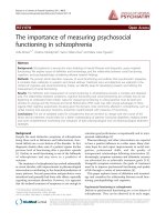
Fig. 2. The prominent protein tyrosine phos-
phatases implicated in insulin signal transduc-
tion. Structure of several phosph atases
implicated in insulin signal transduction.
rPTP-a, LAR, P TP-1B an d SHP-2 are the
major phosphatases acting on the insulin
signaling pathw ay. rPTP-e, r PTP-r and
TC-PTP are candidates shown by in vitro
binding and dephosp horylation a ssays or
suggested by their structure similarity to
phosphatases involved in the insulin
signaling path way.
1052 A. Cheng et al. (Eur. J. Biochem. 269) Ó FEBS 2002
hLAR mice also provide an important model t o understand
the role of LAR in the pathogenesis of insulin resistance.
RPTPa
rPTPa mRNA is expressed in m ost tissues, w ith highest
expression in brain and kidney, suggesting that this PTP
could play a fundamental role in the physiology of all cells
[46]. The best-characterized substrate of rPTPa is the Src
kinase, with particular emphasis in the context of transfor-
mation and neuronal differentiation [47,48]. In addition,
p130Cas [49] and the IR [50] have also been shown to be
potential substrates of rPTPa.ExpressionofrPTPa in cells
can inhibit some of insulin mediated effects. For example,
expression of rPTPa in BHK-IR cells inhibits insulin-
mediated cell rounding and growth inhibition of those cells,
concomitant with increased IR phosphorylation [51]. Fur-
thermore, in rat adipocytes, rPTPa decreases in sulin stimu-
lated GLUT4 cell surface translocation [ 52]. In contrast,
antisense studies in 3T3-L1 adipocytes showed that rPTPa
was dispensable for insulin induced MAPK activation and
DNA synthesis [53]. Although mice deficient in rPTPa have
demonstrated the importance of this P TP in the activation
of Src kinases [54,55], its physiological importance in insulin
signaling remains unclear.
SHP-2
SHP-2 [56] is a widely expressed nonreceptor PTP that
contains two N-terminal SH2 domains, a C-terminal
catalytic domain and a C-terminal s egment containing
two tyrosyl phosphorylation sites (Fig. 2). The SH2
domains of SHP-2 bind many a ctivated growth factor
receptors as well as IRS-1 [57–59]. It has been suggested that
these associations displace intramolecular i nteractions of
SHP-2, leading to a conformationally more open state and
increased catalytic activity [6 0–62]. In contrast to many
other growth factor receptor associated PTPs, S HP-2 does
not seem to dephosphorylate the receptor. In fact, genetic
studies indicate that SHP-2 is a positive effector of growth
factor receptor signaling [63]. However, Kuhne et al. [64]
proposed that the binding of IRS-1 to SHP-2 enhances its
phosphatase activity toward IRS-1, resulting in its dep-
hosphorylation in vivo. Thus SHP-2, in contrast with most
other PTPs, may a ct as ei ther a positive o r n egative
regulator of growth f actor signaling.
Many studies suggest that SHP-2 binds to both the IR and
IRS-1. Forexample, t he IR and SHP-2 interact in ayeast t wo-
hybrid assay [65]. Transfection experiments demonstrated
that this association is mediated be tween the proximal SH2
domain of SHP-2 and phosphotyrosine 1146 of the activated
insulin receptor [66]. Insulin also induces the formation of a
complex of IRS-1 and SHP-2, requiring the tyrosines 1172
and 1222 of IRS-1 [59,67]. However, others have suggested
that SHP-2 is not the major protein complexed with IRS-1 in
insulin stimulated 3T3-L1 adipocytes [68].
Microinjection of in terfering molecules [69], overexpres-
sion of dominant negative mutants [70–72], a nd genetic
studies [63] indicate that SHP-2 is required for activation of
the MAPK p athway by a variety of growth factors,
including insulin. However, the requirement for SHP-2
binding to IRS-1 for this pathway is unclear [69,73]. SHP-2
canalsobindtoIRS-2,IRS-3[74]andIRS-4,suggesting
possible functional redundancy for the SHP-2/IRS-1
association. In addition, SHP-2 binding to SHPS-1 [SH2-
domain bearing protein tyrosine phosphatase (SHP) sub-
strate-1] [75] may provide an additional pathway for insulin
induced MAPK activation. In ins ulin-induced metabolic
signaling, a SHP-2 C-S mutant slightly impaired GLUT4
translocation in primary adipocytes, whereas wild-type
SHP-2 did not [76].
Genetic studies in mice indicate that SHP-2 is required for
embryonic d evelopment [ 77,78]. S HP-2 heterozygous
knockout mice are v iable, and in these mice, plasma insulin
and glucose uptake were normal [78]. Moreover tyrosine
phosphorylation of IR and IRS-1 from muscle tissue was
similar to that of wild-type controls. These results suggest
that SHP-2 may play a minor role in the metabolic effects of
insulin that may not be detectable unless SHP-2 function is
completely removed. Perhaps tissue specific knockouts of
SHP-2 can further address the issue.
In another approach, t he transgenic expression of a
mutant SHP-2 was studied [79]. This mutant (DeltaPTP)
contains the two SH2 domains but lacks the PTP domain and
the C-terminal tyrosines. In DeltaPTP mice, insulin induced
association of endogenous SHP-2 with IRS-1 was reduced,
suggesting a dominant negative effect of the mutant SHP-2
protein. Furthermore, DeltaPTP mice are insulin resistant,
and i nsulin-mediated t yrosine phosphorylation of IRS-1,
stimulation of PI3-kinase and Akt activities were attenuated
in muscle and liver. T hus, t he inhibition of endogenous
SHP-2 by these dominant negative studies suggests a positive
role for SHP-2 in insulin-induced metabolic signaling.
Because DeltaPTP mice are viable, it suggests that the SH2
domains of SHP-2 a lone, are able to mediate a spects of
signaling required for embryonic development.
PTP-1B
PTP-1B was the first mammalian PTP identified and
purified to homogeneity [80]. This phosphatase is widely
expressed and localizes predominantly to the ER through a
cleavable proline-rich C-terminal segment (Fig. 2) [81,82].
Moreover, the C-terminal 35 amino acids of PTP-1B were
found both necessary and sufficient for its targeting to the
ER [81]. Cleavage of this segment appears to release the
enzyme from the ER and increase its specific activity [83]. By
in situ hybridization, Brown-Shimer et al. [84] identified
PTP-1B as a single-copy gene that mapped to the long arm
of human chromosome 20 in the r egion q13.1–q13.2.
Interestingly this region was identified as a quantitative trait
locus linked to obesity and insulin [85].
Studies using the CfiSmutantofPTP-1B(PTP-1B
C215S) have demonstrated an association of PTP-1B with
the IR [86,87]. Upon insulin treatment, PTP-1B becomes
tyrosine phosphorylated at three sites (Tyr 66, 152, 153),
and mutation of any of these residues impairs its association
with the activated IR [87]. Within the IR, the binding occurs
in a region containing residues 1142–1153 [88], and muta-
tion of tyrosines 114 6, 1150, and 1151 diminish the
association [ 87,89]. Indeed, crystal structure and kinetic
studies provide evidence t hat P TP-1B preferentially dep-
hosphorylates tyrosines 1150 a nd 1151 of the IR [90].
In addition to the IR, IRS-1 might also be a substrate of
PTP-1B [45]. Furthermore, in the presence of Grb2, IRS-1
dephosphorylation by PTP-1B is accelerated. Thus, these
Ó FEBS 2002 Protein tyrosine phosphatases in insulin signaling (Eur. J. Biochem. 269) 1053
results suggest that PTP-1B could negatively regu late insulin
signaling by acting on t wo different componen ts of the
pathway.
A plethora of s tudies demonstrate that PTP-1B can
attenuate insulin signaling. Microinjection of homogeneous
preparations of PTP-1B protein into Xenopus oo cytes
decreases t yrosine phosphorylation of proteins correspond-
ing to the molecular m ass of the IR. Correspondingly,
insulin-induced S6 kinase activity and meiotic cell division
were retarded as well [91,92]. In mammalian cells, osmotic
loading of PTP-1B antibodies decreases insulin induced
IRS-1 phosphorylation, PI3-kinase activity, as well as DNA
synthesis [93]. Finally, overexpression of PTP-1B reduces
glucose uptake and GLUT4 translocation to the cell
membra ne [ 76, 94].
Regulation of insulin s ignaling b y PTP-1B appears to be
tissue specific. Overexpression of PTP-1B in 3T3-L1 adipo-
cytes attenuates insulin induced IR, IRS-1 phosphorylation,
as well as PI3-kinase and MAPK activation [95]. However,
neither Akt activation nor glucose transport seemed to be
affected. Thus, it i s possible that PTP-1B may regulate
insulin-mediated mitogenic, as opposed to metabolic events
in this cell type. In contrast, overexpression of PTP-1B in L6
myocytes and Fao hepatoma cells attenuated insulin-
induced Akt activation and glycogen synthesis [96].
An increasing amount of evidence suggests that insulin
signaling can inhibit PTP-1B activity, perhaps as part of a
negative feedback loop. For example, insulin stimulation of
3T3-L1 adipocytes induces a burst of intracellular hydrogen
peroxide that is thought to reversibly oxidize an d t hus
inactivate the invariant cysteine in the catalytic domain of
PTP-1B [97]. In another study, it was suggested that insulin
could also down-regulate PTP-1B activity by suppressing
serine phosphorylation and activation on the phosphatase
via an unidentified mechanism [98].
Knockout studies in mice provided in vivo confirmation
that PTP-1B is a bona fide phosphatase of the IR [99,100].
Despite its involvement in a variety of signaling p rocesses,
PTP-1B is surprisingly not required for embryonic devel-
opment, and PTP-1B-deficient mice grow and d evelop
normally with similar lifespans to wild-type littermates.
However, PTP-1B-deficient mice display increased insulin
induced IR phosphorylation i n liver and muscle but n ot
adipose tissue. IRS-1 phosphorylation was also increased in
muscle, but it is unclear whether this is because IRS-1 is a
substrate of PTP-1B, or an increased IR activity in
knockout mice. Furthermore, PTP-1B-deficient mice are
hypersensitive as assayed b y oral g lucose tolerance t ests,
intraperitoneal insulin tolerance tests, and blood levels of
glucose and insulin.
Importantly, PTP-1B-deficient mice remained insulin
sensitive when f ed a high fat diet. Strikingly, though, they
were also resistant to obesity, due in part to a decrease in fat
cell mass and increased energy expenditure. These results
suggest that PTP-1B is a major modulator of insulin
sensitivity and fuel metabolism, and point to PTP-1B as a
potential therapeutic target f or the treatment of type II
diabetes and obesity. Importantly the insulin receptor
phosphorylation appears to be modified in liver and muscle
tissues but not in adipose tissue, suggesting that although
PTP-1B is a major modulator of the IR, other PTPs may
have tissue specific preferences for the insulin receptor, in
particular in adipocytes.
OTHER CANDIDATE PTPs
RPTPr
Evidence for a functional r ole of rPTPr in insulin signaling
has not been reported to date. However, its similarity with
LAR, and the fact that it is expressed i n relatively high levels
in insulin sensitive tissues (higher than LAR) [101] make
rPTPr a possible c andid ate to regulate insulin signaling.
Genetic studies wit h rPTPr deficient mice reveal the
presence of primarily neuroendocrine defects [102,103].
However, preliminary s tudies also indicate that rPTP r
deficiency leads to insulin hypersensitivity f rom measure-
ments of fasting glucose and insulin levels (X. Elchebly &
M. L. Tremblay, unpublished observations). As in the case
with the LAR knockout it cannot be ruled out that the
effects on insulin signaling are secondary to the neuroendo-
crine status. Thus generation of transgenic lines over-
expressing this PTP or the creation of tissue specific
knockouts should answer this question.
rPTPe
rPTPe is similar in structure to rPTPa. In addition to
rPTPa, expression of rPTP e in BHK-IR cells also inhibits
insulin mediated cell rounding and g rowth in hibition o f
BHK-IR cells, and requires membrane localization of the
PTP [51,104].
TC-PTP, rPTPd and Sap-1
In an attem pt to f urther extend the list of c andidate IR
PTPs, a mass screen approach utilizing in vitro binding and
dephosphorylation a ssays were performed on a large list o f
PTPs [105]. In this study, in addition to PTP-1B, three other
PTPs were suggested to be important for IR dephosphory-
lation: TC-PTP, rPTPd and Sap-1. Although not all PTPs
tested performed well in these assays, one must consider that
for each PTP, their physiological situation is unique and
several other factors are implicated. For example, these may
include: t issue distribution, subcellular localization, as well
as the significance of additional binding partners to form
functional multiprotein complexes.
COMPARTMENTALIZATION
OF IR AND PTPs
An increasing amount of evidence suggests that regulation
of insulin signaling by PTPs may also occur at the level of
compartmentalization. In the absence of ligand, the I R
normally resides a t the plas ma membrane. U pon insulin
binding, the ligand–receptor complex is rapidly sequestered
from the p lasma membrane a nd internalized into endo-
somes within several minutes [106,107]. Here, the acidic pH
of endosomes induces the dissociation of insulin from IR
and allows the degradation of insulin by endosomal acidic
insulinase [108]. The IR is then recycled back to the cell
surface. However, under conditions of prolonged stimula-
tion with saturating levels of insulin, a subset of the IRs are
transported t o the late endosome a nd lysosome for degra-
dation [109,110].
Although the IR kinase activity is required for ligand-
stimulated IR internalization, the role of IR internalization
1054 A. Cheng et al. (Eur. J. Biochem. 269) Ó FEBS 2002
on insulin receptor signaling remains unclear. The endoso-
mally associated IRs h ave been reported to exhibit a
transient elevation of the tyrosine phosphorylation and to
achieve the full activation of the receptor itself as well as th e
activation of IRS-1 and PI3-kinase (reviewed in [111]). In
contrast, studies using a dominant negative dynamin
molecule that blocks IR internalization showed that the
inhibition o f IR endocytosis had no major effect on IR
autophosphorylation and IRS-1 tyrosine phosphorylation
[112]. Even though a 50% decrease in the insulin activated
PI3-kinase activity was observed when IR internalization is
blocked, it did not affect the subsequent Akt phosphory-
lation and activation. The only major defect caused by
inhibition of IR internalization was impaired Shc tyrosine
phosphorylation and MAPK activation. In summary, most
of the acute actions of insulin could be initiated by activation
of the plasma me mbrane-localized insulin receptor.
While the activation of the insulin signaling cascade
appears to be independent of IR internalization, tyrosine
phosphatase activity towards IR is l argely observed in
endosomes (reviewed in [111,113]). Studies in rat hepatoma
cells demonstrated that internalized IRs w ere dephosphory-
lated and inactivated prior t o recycling back to the plasma
membrane [114]. Using isolated rat liver endosomes, Faure
et al. sh owed that a substantial amount of IR PTP activities
is tightly associated with the endosomes. This activity resists
the 0.6
M
KCl treatment of the endosomal membrane, but
Triton X-100 totally abolishes dephosphorylation [115,116].
These studies strongly suggest that t he endosome is a major
site of IR dephosphorylation. However, due to the large
number of phosphotyrosine residues o n the IR, and the
complexity of insulin signaling, the plasma membrane
localized IR should not be discarded as an important site of
PTP action.
For rPTPs such as LAR and rPTPa, the plasma
membrane is the obvious location for IR dephosphorylation.
As previously discussed, membrane targeting of these rPTPs
seems to be necessary for IR dephosphorylation. Yet, LAR
has also been detected i n r at hepati c e ndosomes [34].
Although the kinetics of LAR internalization upon insulin
administration was much slower ( 30 min), compared to
that of IR ( 2–5 min), incubation of endosomal fractions
with antibodies against LAR reduced IR dephosphorylation
by about 28%. In addition, subcellular fractionation of rat
adipocytes showed that both LAR and rPTPa are present in
heavy microsomes [117]. Although the identity of these h eavy
microsomes was not determined, the presence of increased
IR in the same fraction after insulin stimulation suggests that
these membranes likely contain endosomal compartments.
Amongst the intracellular P TPs, PTP-1B is a c lear
physiological regulator of the IR, and perhaps IRS proteins
as well. Although a truncated form of PTP-1B was initially
identified in the cytosolic fraction of human placenta, the
subcellular characterization of full length PTP-1B demon-
strated its predominan t localization in the ER through an
association with its C-terminal 35 amino acids [81,82]. Other
Fig. 3. Model of coordinated PTP action on the insulin signaling pathway. Different PTPs may act on the IR in various compartments within the cell.
For example, the transm embrane phosphatases LAR and rPTP-a may p redomin antly act at the plasma membrane on either the receptor or
downstream substrates. On the other hand, PTP-1B could act on IR and IRS-1 at the plasma membrane and/or endosomes. Finally, the cytosolic
PTP, SHP-2, could potentially be re cruited to man y sites of insu lin action. Howeve r, the role of SHP-2 is m ainly to transmit positive s ignals from the
IR. How these PTPs coordinate their ac tion on the insulin signaling pathway remains to be determined.
Ó FEBS 2002 Protein tyrosine phosphatases in insulin signaling (Eur. J. Biochem. 269) 1055
reports indicated that s ubstantial a mounts o f full length
PTP-1B are also found in the cytosol of rat fibroblasts and
skeletal muscle [28]. In rat adipocytes, PTP-1B was found in
the light microsome f raction a nd to a l esser extent, the
cytosol and heavy m icrosomes [117]. H owever, immuno-
blotting failed to reveal th e presence of PTP-1B in rat liver
endosomes [116]. Thus PTP-1B could dephosphorylate the
IR in the light and heavy microsomes, or cytosolic PTP-1B
may be recruited to th e appropriate site. The precise
localization where PTP-1B dephosphorylates the IR and the
mechanism of P TP-1B translocation to t he site o f IR
dephosphorylation remain to be elucidated.
Finally, in r esponse to i nsulin, s ignificant amounts o f
IRS-1 and IRS-2 a re also associated with internal mem-
branes in rat a dipocytes [1 17–119]. Furthermore, i nsulin
stimulation in rat liver increases the association of active
IRS-1, IRS-2, and PI3-kinase to endosomal fractions [120].
These d ata further show that there is a complex spatial
control in insulin receptor signaling of t he various molecules
that are i nvolved a nd support an important role of the
subcellular localization of both the tyrosine kinases, their
substrates and the PTPs involved in insulin signaling.
CONCLUSIONS AND PROSPECTS
In contrast to what we have depicted in Fig. 1, the
metabolic and mitogenic pathways emanating from the IR
are diverse and complex [121]. Although not limited to this,
the action of PTPs represents an important aspect in both
the transmission and attenuation of insulin signaling.
Indeed, many s tudies have been aimed at developing
inhibitors towards these PTPs that might c ircumvent insulin
resistance and treat type II diabetes. Currently, both PTP-
1B and L AR are s trong candidates for inhibitor design
studies, although PTP-1B has been the major focus due to
its s maller s ize, the remarkable d ata from the knockout
mice, and the availability of structural and kinetic data.
An emerging theme that requires further study is how the
coordinate actions of several PTPs may regulate insulin
signaling (Fig. 3). As a cytosolic protein, SHP-2 could
potentially be recruited t o many sites of insulin action and
positively participate in signal transduction, either through
direct binding with the IR, or through adapter molecules
such as IRS proteins. On the other hand, the rPTPs probably
dephosphorylate the IR (or I RS proteins) a t the plas ma
membrane, a lthough evidence s uggests other subcellular
compartments are a possibility as well. For PTP-1B, several
sites of insulin action seem possible. It will be interesting to
determine how different PTPs m ight temporally, as well as
spatially, regulate insulin signaling under normal physiolo-
gical conditions and in pathophysiological states such as
diabetes. Finally, it still remains t o be determined whether
different PTPs may act on specific phosphotyro sine residues
on the IR, thus providing another level of specificity.
ACKNOWLEDGEMENTS
We wish to thank John Wagner for critical reading of the manuscript
and helpful discussions. A. C. is a recipient of a Medical Research
Council studentship. N. D. is a recipient of a Canadian Institutes of
Health Research doctoral award. F. G. is a recipient of a Human
Frontiers postdoctoral fellowship. M. L. T. is a Canadian Institutes of
Health Research Scientist.
REFERENCES
1. Bennett, S.T. & Todd, J.A. (1996) Human type 1 diabetes and the
insulin gene: principles of mapping polygenes. Annu. Rev. Genet.
30, 343–370.
2. Gottlieb, P.A. & Eisenbarth, G.S. (1998) Diagnosis and treatment
of pre-insulin dependent diabet es. Annu. Rev. Med. 49, 391–405.
3. Saltiel, A.R. (2001) New perspectives into the molecular patho-
genesis and treatmen t of type 2 d iabetes. Cell. 104, 517–529.
4. Schlessinger, J. (2000) Cell signaling by receptor tyrosine kinases.
Cell. 103, 211–225.
5. White, M.F. & Kahn, C.R. (1994) The insulin signaling system.
J. Biol. Chem. 269,1–4.
6. Mayer, B.J. & Gupta, R. (1998) Functions of SH2 and SH3
domains. Curr. Top Microbiol Immunol. 228, 1–22.
7. White, M.F. (1 998) The I RS-signalling system: a network of
docking proteins that m ediate insulin action . Mol. Cell. Biochem.
182, 3–11.
8. Ogawa, W., M atozaki, T. & Kasuga, M. (1998) Role of binding
proteins to IRS-1 in insulin signalling. Mol. Cell. Biochem. 182,
13–22.
9. Pronk,G.J.,McGlade,J.,Pelicci,G.,Pawson,T.&Bos,J.L.
(1993) Insulin-induced phosphorylation of the 46- and 52-kDa Shc
proteins. J. Biol. Chem. 268, 5748–5753.
10. Skolnik, E.Y., Lee, C.H., Batzer, A., Vicentini, L.M., Zhou, M.,
Daly, R., Myers, M.J. Jr,, Backer, J.M., Ullrich, A. & White, M.F.
et al. (1993) The SH2/SH3 domain-containing protein GRB2
interacts with tyrosine- phosphorylated IRS1 and Shc: implica-
tions for insulin control of ras signalling. EMBO J. 12, 1929–1936.
11. Pessin, J.E., Thurmond, D.C., Elmendorf, J.S., Coker, K.J. &
Okada, S. (1999) Molecular basis of insulin-stimulated GLUT4
vesicle trafficking. Location! Location! Location! J. Biol. Chem.
274, 2593–2596.
12. Charron, M.J., Katz, E.B. & Olson, A.L. (1999) GLUT4 gene
regulation and manipulation. J. Biol. Chem. 274, 3253–3256.
13. She pherd, P.R. & Kahn, B.B. (1999) Glucose transporters and
insulin action – implications for insulin resistance and diabetes
mellitus. N.Engl.J.Med.341, 248–257.
14. Chiang, S.H., Baumann, C.A., Kanzaki, M., Thurmond, D.C.,
Watson,R.T.,Neudauer,C.L.,Macara,I.G.,Pessin,J.E.&Saltiel,
A.R. (2001) Insulin-stimulated GLUT4 translocation requires the
CAP-dependent activation o f TC10. Nature 410, 944–948.
15. Baum ann, C.A., Ribon, V., K anzak i, M., T hurm ond, D.C.,
Mora, S., Shigematsu, S., Bickel, P.E., Pessin, J.E. & Saltiel, A.R.
(2000) CAP defines a second signalling pathway required for
insulin-stimulated glucose transport. Nature 407, 202–207.
16. Tonks, N.K. & Neel, B.G. (2001) Combinatorial control of the
specificity of protein tyrosine phosphatases. Curr. Opin. Cell Biol.
13, 182–195.
17. Fantus, I.G. & Tsiani, E. (1998) Multifunctional actions of
vanadium compounds on insulin signaling pathways: evidence for
preferential enhancement of metabolic versus mitogenic effects.
Mol. Cell. Biochem. 182, 109–119.
18. Bevan, A.P., Drake, P.G., Yale, J.F., Shaver, A. & Posner, B.I.
(1995) Peroxovanadium c ompounds: biological actions and
mechanism of insulin-mimesis. Mol. Cell . B io che m. 153, 49–58.
19. Shaver, A., Ng, J .B., H all, D.A. & P osner, B.I. (1995) The
chemistry of peroxovanadium compounds relevant to insulin
mimesis. Mol. Cell. Biochem. 153, 5–15.
20. Wells, L., Vosseller, K. & Hart, G.W. (2001) Glycosylation of
nucleocytoplasmic proteins: signal transduction and O-GlcNAc.
Science 291, 2376–2378.
21. Walton, K.M. & Dixon, J.E. (1993) Protein tyrosine phospha-
tases. Annu.Rev.Biochem.62, 101–120.
22. Streuli,M.,Krueger,N.X.,Thai,T.,Tang,M.&Saito,H.(1990)
Distinct functional roles of the two intracellular phosphatase like
domains of the receptor-linked protein tyrosine phosphatases
LCA and LAR. EMBO J. 9, 2399–2407.
1056 A. Cheng et al. (Eur. J. Biochem. 269) Ó FEBS 2002
23. Itoh, M., Streuli, M., Krueger, N.X. & Saito, H. (1992) Purifi-
cation an d characterization o f the catalytic domains of the
human receptor-linked protein tyrosine phosphatases HPTP be ta,
leukocyte common antigen (LCA), and leukocyte common anti-
gen-related molecule (LAR). J. Biol. Chem. 267, 12356–12363.
24. Wagner, J., Boerboom, D. & Tremblay, M.L. (1994) Molecular
cloning and tissue-specific RNA processing of a murine receptor-
type protein tyrosine ph osphatase . Eur. J. Biochem. 22 6, 773–782.
25. Flint, A.J., Tiganis, T., Barford, D. & Tonks, N.K. (1997)
Development of Ôsubstrate-trappingÕ mutants to identify physio-
logical substrates of protein tyrosine phosphatases. Proc. Natl
Acad.Sci.USA94, 1680–1685.
26. C
^
oote
´
, J.F., Charest, A., Wagner, J. & Tremblay, M.L. (1998)
Combination of gene targeting and substrate trapping to identify
substrates of protein tyrosine phosphatases using PTP-PEST as a
model. Bioch em ist ry 37, 13128–13137.
27. Ding, W., Zhang, W.R., Sullivan, K., Hashimoto, N. & Goldstein,
B.J. (1994) Identification of protein-tyrosine phosphatases pre-
valent in adipocytes by molecular cloning. Biochem. Biophys. Res.
Commun. 202, 902–907.
28. A hmad, F. & Goldstein, B.J. (1995) Purification, identification
and subcellular distribution of three predominant protein-tyrosine
phosphatase enzymes in skeletal muscle tissue. Biochim. Biophys.
Acta. 1248, 57–69.
29. Ahmad, F., Azevedo, J.L., Cortright, R., Dohm, G.L. & Gold-
stein, B.J. (1997) Alterations in skeletal muscle protein-tyrosine
phosphatase activity and expression in insulin-resistant human
obesity and diabetes. J. Clin. Invest. 100 , 449–458.
30. S erra-Pages, C., Saito, H. & Streuli, M. (1994) Mutational analysis
of proprotein processing, subunit association, and shedding of t he
LAR transmembrane protein tyrosine phosphatase. J. Biol. Chem.
269, 23632–23641.
31. Aicher, B., Lerch, M.M., Muller, T., Schilling, J. & Ullrich, A.
(1997) Cellular redistribution of protein tyrosine phosphatases
LAR and PTPsigma by inducible proteolytic processing. J. Cell.
Biol. 138, 681–696.
32. S treuli, M., Krueger, N.X., Ariniello, P.D., Tang, M., Munro,
J.M., Blattler, W.A., Adler, D.A., Disteche, C.M. & Saito, H.
(1992) Expression of the receptor-linked prot ein tyrosine phos-
phatase LAR: proteolytic cleavage and shedding of the CAM-like
extracellular region. EMBO J. 11, 897–907.
33. YuQ., Lenardo, T. & Weinberg, R.A. (1992) The N-terminal and
C-terminal domains of a receptor tyrosine phosphatase are asso-
ciated by non-covalent link age. Oncogene. 7, 1051–1057.
34. A hmad, F. & Goldstein, B.J. (1997) Functional association
between the insulin receptor and the transmembran e protein-tyro-
sine phosphatase LAR in intact cells. J. Biol. Chem. 272, 448–457.
35. Li, P.M., Zhang, W.R. & Goldstein, B.J. (1996) Suppression of
insulin receptor activation by overexpression of the protein-
tyrosine phosphatase L AR in he patoma cells. Cell Signal. 8,
467–473.
36. Kulas, D.T., Zhang, W.R., Goldstein, B.J., Furlanetto, R.W. &
Mooney, R.A. (1995) Insulin receptor signaling is augmented by
antisense inhibition of the protein tyrosine phosphatase LAR.
J. Biol. Chem. 270, 2435–2438.
37. Kulas, D.T., Goldstein, B.J. & Mooney, R.A. (1996) The trans-
membrane protein-tyrosine phosphatase LAR m odulates signaling
by multiple receptor tyrosine kinases. J. Biol. Chem. 271, 748–754.
38. Zhang, W.R., Li, P.M., Oswald, M.A. & Goldstein, B.J. (1996)
Modulation of insulin sign al transduc tion by eutop ic over-
expression of the receptor-type protein-tyrosine phosphatase
LAR. Mol. Endocrinol. 10, 575–584.
39. Schaapveld, R.Q., Schepens, J.T., Robinson, G.W., Attema, J.,
Oerlemans, F.T., Fransen, J.A., Streuli, M., Wieringa, B., Hen-
nighausen, L. & Hendriks, W.J. (1997) Impaired mammary g land
development and function in mice lacking LAR rec eptor -like
tyrosine phosphatase activity. Dev Biol. 188, 134–146.
40.Skarnes,W.C.,Moss,J.E.,Hurtley,S.M.&Beddington,R.S.
(1995) Capturing genes encoding membrane and sec reted proteins
important for mouse development. Proc. Natl Acad. Sci. USA 92,
6592–6596.
41. Yeo,T.T.,Yang,T.,Massa,S.M.,Zhang,J.S.,Honkaniemi,J.,
Butcher, L.L. & Longo, F.M. (1997) Deficient LAR expression
decreases basal forebrain cholinergic neuronal size and hippo-
campal cholinergic innervation. J. Neurosci. Res. 47, 348–360.
42. Ren, J.M., Li, P.M., Zhang, W.R., Sweet, L.J., Cline, G., Shul-
man, G.I., Livingston, J.N. & Goldstein, B.J. (1998) Transgenic
mice deficient in the LAR protein-tyrosin e phosph atase exhibit
profound defects in glucose homeostasis. Diabetes 47, 493–497.
43. Van Lieshout, E.M., Van der Heijden, I., Hendriks, W.J. & Van
der Zee, C.E. (2001) A decrease in size and number of basal
forebrain cholinergic neurons is paralleled by diminished hippo-
campal cholinergic innervation in mice lacking leukocyte
common antigen-related protein tyrosine phosphatase activity.
Neuroscience 102, 833–841.
44. Z abolotny, J.M., Kim, Y.B., Peroni, O.D., Kim, J.K., Pani, M.A.,
Boss, O., Klaman, L.D., Kamatkar, S., Shulman, G.I., Kahn, B.B.
& Neel, B.G. (2001) Overexpression of the LAR (leukocyte anti-
gen-related) protein-tyrosine phosphatase in muscle causes insulin
resistance. Proc.NatlAcad.Sci.USA98, 5187–5192.
45. G oldstein, B.J., Bittner-Kowalczyk, A., White, M.F. & Harbeck,
M. (2000) Tyrosine dephosphorylation and deactivation of insulin
receptor substrate-1 by protein-tyrosine phosphatase 1B. Possible
facilitation by the formation of a ternary complex with th e Grb2
adaptor protein. J. Biol. Che m. 275, 4283–4289.
46. Sap, J., D ’Eustachio, P., Givol, D. & Schlessinger, J . (1990)
Cloning and expression of a widely expressed receptor tyrosine
phosphatase. Proc. Natl Acad. Sci. USA 87, 6112–6116.
47. Zheng, X.M., W ang, Y. & Pallen, C.J. (1992) Cell transformation
and activation of pp60c-src by overexpression of a protein tyrosine
phosphatase. Nature 359, 336–339.
48. d en Hertog, J., Pals, C.E., Peppelenbosch, M.P., Tertoolen, L.G.,
de Laat, S.W. & Kruijer, W. (1993) Receptor protein tyrosine
phosphatase alpha activates pp60c-src and is involved in neuronal
differentiation. EMBO J. 12, 3789–3798.
49. Buist, A., Blanchetot, C., Tertoolen, L.G. & den Hertog, J. (2000)
Identification of p130cas as an in v ivo substrate of receptor pro-
tein- tyrosine phosphatase alpha. J. Biol. Chem. 275, 20754–20761.
50. Lammers, R., Moller, N.P. & Ullrich, A. (1997) The transmem-
brane protein tyrosine phosphatase alpha de phosphorylates the
insulin receptor in intact cells. FEBS Lett. 404, 37–40.
51. Moller, N.P., Moller, K.B., Lammers, R., Kharitonenkov, A.,
Hoppe,E.,Wiberg,F.C.,Sures,I.&Ullrich,A.(1995)Selective
down-regulation of the insulin receptor signal by protein- tyrosine
phosphatases alpha and ep silon. J. Biol. Chem. 270, 23126–23131.
52. Cong,L.N.,Chen,H.,Li,Y.,Lin,C.H.,Sap,J.&Quon,M.J.
(1999) Overexpression o f prote in tyrosine phosphatase-alpha
(PTP-alpha) but not PTP-kappa inhibits translocation of GLUT4
in rat adipose cells. Biochem. Biophys. Res. Commun. 255, 200–207.
53. Arnott, C.H., Sale, E.M., Miller, J. & Sale, G.J. (1999) Use of an
antisense strategy to dissect the signaling role of protein-tyrosine
phosphatase alpha. J. Biol. Chem. 274, 26105–26112.
54. Ponniah, S., W ang, D.Z., Lim, K.L. & Pallen, C.J. (1999) Tar-
geted disruption of the tyrosine phosphatase PTPalpha leads to
constitutive downregulation of the kinases Src and Fyn. Curr.
Biol. 9, 535–538.
55. Su, J., Muranjan, M. & Sap, J. (1999) Receptor protein tyrosine
phosphatase alpha activates Src-family kinases and contro ls
integrin-mediated r esponses in fibroblasts. Curr. Biol. 9, 505–511.
56. F eng, G.S. (1999) Shp-2 tyrosine phosphatase: signaling one cell
or many. Exp. Cell Res. 253, 47–54.
57.Feng,G.S.,Hui,C.C.&Pawson,T.(1993)SH2-containing
phosphotyrosine p hosphatase as a target of p rotein- t yrosine
kinases. Science 259, 1607–1611.
Ó FEBS 2002 Protein tyrosine phosphatases in insulin signaling (Eur. J. Biochem. 269) 1057
58. Vogel, W., Lammers, R., Huang, J. & Ullrich, A. (1993) Activa-
tion of a phosphotyrosine phosphatase by tyrosine phosphoryla-
tion. Science 259, 1611–1614.
59. Kuhne, M.R., Pawson, T., Lienhard, G.E. & Feng, G.S. (1993)
The insulin receptor substrate 1 associates with the SH2-contain-
ing phosphotyrosine phosphatase Syp. J . Biol. Chem. 268, 11479–
11481.
60. Lechleider, R.J., Sugimoto, S., Bennett, A.M., Kashishian, A.S.,
Cooper, J.A., Shoelson, S.E., Walsh, C.T. & Neel, B.G. (1993)
Activation of the SH2-con taining phosphotyrosine phosphatase
SH-PTP2 by its binding site, phosphotyrosine 1009, on the human
platelet-derived growth factor receptor. J. Biol. Chem. 268, 21478–
21481.
61. Pluskey, S., Wandless, T.J., Walsh, C.T. & Shoelson, S.E. (1995)
Potent stimulation of SH-PTP2 phosphatase activity by simulta-
neous occupancy of both SH2 domains. J. Biol. Chem. 270, 2897–
2900.
62. Zhao, Z., Larocque, R., Ho, W.T., Fischer, E.H. & Shen, S.H.
(1994) Purification a nd characterization of PTP2C, a widely dis-
tributed protein tyrosine phosphatase containing two SH2
domains. J. Biol. Chem. 269, 8780–8785.
63. Shi, Z.Q., Lu, W. & Feng, G.S. (1998) The Shp-2 tyrosine phos-
phatase has opposite effects in mediating the activation of extra-
cellular signal-regulated and c-Jun NH
2
-terminal mitogen-activated
protein kinases. J. Biol. Chem. 273, 4904–4908.
64.Kuhne,M.R.,Zhao,Z.,Rowles,J.,Lavan,B.E.,Shen,S.H.,
Fischer, E.H. & Lienhard, G.E. (1994) Dephosphorylation of
insulin re ceptor substrate 1 by the tyrosine phosphatase PTP2C.
J. Biol. Chem. 269, 15833–15837.
65. Rocchi, S., Tartare-Deckert, S., Sawka-Verhelle, D., Gamha, A. &
van Obberghen, E. (1996) Interaction of SH2-containing protein
tyrosine phosphatase 2 with the insulin recep tor and the insulin-
like growth factor-I receptor: studies of the domains involved
using the yeast two-hybrid system. Endocrinology 137, 4944–4952.
66. Khariton enkov, A., Schnekenburger, J., Chen, Z., Knyazev, P.,
Ali, S., Zwick, E., White, M. & Ullrich, A. (1995) Adapter func-
tion of protein-tyrosine phosphatase 1D in insulin receptor/insulin
receptor substrate-1 i nteraction . J. Biol. Chem. 270, 29189–29193.
67. Ro cchi, S., Tartare-Deckert, S., M othe, I. & Van Obberghen, E.
(1995) Identification by mutation of the tyrosine residues in the
insulin receptor substrate-1 affecting association with the tyrosine
phosphatase 2C and phosphatidylinositol 3-kinase. Endocrinology
136, 5291–5297.
68. Yamauchi, K., Ribon, V., Saltiel, A.R. & Pessin, J.E. (1995)
Identification of the major SHPTP2-binding protein that is tyro-
sine- phosphorylated in response to insulin. J. Biol. Chem. 270,
17716–17722.
69. Xiao, S., Rose, D.W., Sasaoka, T., Maegawa, H., Burke, T.R. Jr,,
Roller, P.P., Shoelson, S.E. & Olefsky, J.M. (1994) Syp (SH-
PTP2) is a positive mediator of growth factor-stimulated mito-
genic signal transduction. J. Biol. Chem. 269, 21244–21248.
70. Milarski, K.L. & S altiel, A.R. (1994) Expression of catalytically
inactive Syp phosphatase in 3T3 cells blocks stimulation of
mitogen-activated protein kinase by insulin. J. Biol. Chem. 269 ,
21239–21243.
71. Noguchi, T., Matozaki, T., Horita, K., Fujioka, Y. & Kasuga, M.
(1994) Role of SH-PTP2, a protein-tyrosine phosphatase with Src
homology 2 dom ains, in insulin-stimulated Ras activation. Mol.
Cell. Biol. 14, 6674–6682.
72. Yamauchi, K., Milarski, K.L., Saltiel, A.R. & Pessin, J.E. (1995)
Protein-tyrosine-pho sphatase SHPTP2 is a r equired positive
effector for insulin downstream signaling. Proc. Natl Acad. Sci.
USA 92, 664–668.
73. Mye rs, M.G. Jr,, Me ndez, R., Shi, P., Pierce, J.H., Rhoads, R. &
White, M.F. (1998) The COOH-terminal tyrosine phosphoryla-
tion sites o n IRS-1 bind SHP-2 and ne gatively re gulate insulin
signaling. J. Biol. Chem. 273, 26908–26914.
74. Ross, S.A., Lienhard, G.E. & Lavan, B.E. (1998) Association of
insulin receptor substrate 3 with SH2 do main-containing proteins
in rat adipocytes. Biochem. Biophys. Res. Commun. 247, 487–492.
75. Takada, T., Matozaki, T., Takeda, H., Fuku naga, K., Noguchi,
T., Fujioka, Y., Okazaki, I., Tsuda, M., Yamao, T., Ochi, F. &
Kasuga, M. (1998) Roles of the complex formation of SHPS-1
with SHP-2 in insulin-stimulated mitogen-activated protein kinase
activation. J. Biol. Chem. 273, 9234–9242.
76. Chen, H., Wertheimer, S.J., Lin, C.H., Katz, S.L., Amrein, K.E.,
Burn, P . & Quon, M.J. (1997) Protein-tyrosine phosphatases
PTP1B and syp are modulators of insulin- stimulated transloca-
tion of GLUT4 in transfected rat adipose cells. J. Biol. Chem. 272,
8026–8031.
77. Saxt on, T.M., Henkemeyer, M., Gasca, S., Shen, R., Rossi, D.J.,
Shalaby, F., Feng, G.S. & Pawson, T. (1997) Abnormal mesoderm
patterning in mouse em bryos mutant for the SH2 tyrosine phos-
phatase Shp-2. EMBO J. 16, 2352–2364.
78. Arrandale, J.M., Gore- Willse, A., Rocks, S., Ren, J.M., Zhu, J.,
Davis, A., Livingston, J.N. & Rabin, D.U. (1996) Insulin signaling
in mice expressing reduced levels of Syp. J. Biol. Chem. 271,
21353–21358.
79. Maegawa, H. , Hasegawa, M., Sugai, S., O b ata, T., Ugi, S.,
Morino,K.,Egawa,K.,Fujita,T.,Sakamoto,T.,Nishio,Y.,
Kojima,H.,Haneda,M.,Yasuda,H.,Kikkawa,R.&Kashiwagi,
A. (1999) Expression of a dominant negative SHP -2 in transgenic
mice induces insu lin resistan ce. J. Biol. Chem. 274, 30236–30243.
80. Tonks, N.K., Diltz, C.D. & Fischer, E.H. (1988) Characterization
of the major protein-tyrosine-phosphatases of human placenta.
J. Biol. Chem. 263, 6731–6737.
81. Frangioni, J.V., Beahm, P.H., Shifrin, V., Jost, C.A. & Neel, B.G.
(1992) The nontransmembrane tyrosine phosphatase PTP-1B
localizes to the endoplasmic reticulum via its 35 amino acid
C-terminal sequence. Cell 68, 545–560.
82. Charbon neau, H., Tonks, N.K., Kumar, S., Diltz, C.D., Harry-
lock, M., Cool, D.E., Krebs, E.G., Fischer, E.H. & Walsh, K.A.
(1989) Human placenta protein-tyrosine-phosphatase: amino acid
sequence and relationship to a family of receptor-like proteins.
Proc. Natl Acad. Sci. USA. 86, 5252–5256.
83. Frangioni,J.V.,Oda,A.,Smith,M.,Salzman,E.W.&Neel,B.G.
(1993) Calpain-catalyzed cleavage and subcellular relocation of
protein phosphotyrosine ph osphatase 1 B (PTP-1B) in human
platelets. EMBO J. 12 , 4843–4856.
84. Brown -Shimer, S., Johnson, K.A., Lawrence, J.B., Johnson, C.,
Bruskin, A., Green, N .R. & Hill, D.E. (1990) Molecular cloning
and chromosome map ping of th e human ge ne enco ding pr otein
phosphotyrosyl phosphatase 1B. Proc. Natl Acad. Sci. USA 87,
5148–5152.
85. Lembertas,A.V.,Perusse,L.,Chagnon,Y.C.,Fisler,J.S.,War-
den, C.H., Purc ell-Huynh, D.A., Dionne, F .T., Gagnon, J.,
Nadeau, A., Lusis, A.J. & Bouchard, C. (1997) Identification of an
obesity quantitative trait locus on mouse chr omosome 2 and evi-
dence of linkage to bod y fat and insulin on the hu man homo-
logous region 20q. J. Clin. Invest. 100, 1240–1247.
86. Seely, B.L., Staubs, P.A., Reichart, D.R., B erhanu, P ., Milarski,
K.L.,Saltiel,A.R.,Kusari,J.&Olefsky,J.M.(1996)Protein
tyrosine phosphatase 1B interacts with the activated insulin
receptor. Diabetes 45, 1379–1385.
87. Band yopadhyay, D., Kusari, A., Kenner, K.A., Liu, F., Chernoff,
J., Gustafson, T.A. & Kusari, J. (1997) Protein–tyrosine phos-
phatase 1B complexes with the insulin receptor in vivo and is tyro-
sine-phosphorylated in the presence of insulin. J. Biol. Chem. 272,
1639–1645.
88. Liotta, A.S., Kole, H.K., Fales, H.M., Roth, J. & Bernier, M.
(1994) A synthetic tris-sulfotyrosyl dodecapeptide analogue of the
insulin receptor 1146-k inase domain in hibits tyrosine de ph os-
phorylation of the insulin receptor in situ. J. Biol. Chem. 269,
22996–23001.
1058 A. Cheng et al. (Eur. J. Biochem. 269) Ó FEBS 2002
89. Dadke, S., Kusari, J. & Chernoff, J. (2000) Down-regulation of
insulin signaling by protein-tyrosine phosphatase 1B is media-
ted by an N-terminal binding region. J. Biol. Chem. 275, 23642–
23647.
90. S almeen, A., Andersen, J.N., Myers, M.P., Tonks, N.K. & Bar-
ford, D. (2000) Molecular basis for the dephosphorylation of the
activation segment of the insulin receptor by protein tyrosine
phosphatase 1B. Mol. Cell. 6, 1401–1412.
91. T onks, N.K., Cicirelli, M.F., Diltz, C.D., Krebs, E.G. & Fischer,
E.H. (1990) Effect of microinjection of a low-Mr human placenta
protein tyrosine phosphatase on induction of meiotic cell division
in Xenopus oocytes. Mol. Cell. Biol. 10, 458–463.
92. Cicirelli, M.F., Tonks, N.K., Diltz, C.D., Weiel, J.E., Fischer,
E.H. & Krebs, E.G. (1990) Microinjection of a protein-tyrosine-
phosphatase inhibits insulin action in Xenopus oocytes. Proc. Natl
Acad.Sci.USA87, 5514–5518.
93. Ahmad, F., Li, P.M., Meyerovitch, J. & G oldstein, B.J. (1995)
Osmotic loading of neutralizing antibodies demonstrates a role for
protein-tyrosine phosphatase 1B in negative regulation of the
insulin action pathway. J. Biol. Chem. 270, 20503–20508.
94. Kenner, K.A., Anyanwu, E., Olefsky, J.M. & Kusari, J. (1996)
Protein-tyrosine phosphatase 1B is a negative regulator of insulin-
and insulin-like growth factor-I-stimulated signaling. J. Biol.
Chem. 271, 19810–19816.
95. Venable, C.L., Frevert, E.U., Kim, Y.B., Fischer, B., Kamatkar,
S., Neel, B.G. & Kahn, B.B. (2000) Overexpression of protein
tyrosine phosphatase 1B in adipocytes inh ibits insulin-stimulated
phosphoinositide 3 kinase a ctivity witho ut alterin g glucose
transport or akt/pkb activation. J. Biol. Chem. 275, 18318–
18326.
96. Egawa,K.,Maegawa,H.,Shimizu,S.,Morino,K.,Nishio,Y.,
Bryer-Ash, M., Cheung, A.T., Kolls, J.K., Kikkawa, R. &
Kashiwagi, A. (2001) Protein-tyrosine phosphatase-1B negatively
regulates insulin signaling in l6 myocytes and Fao hepatoma cells.
J. Biol. Chem. 276, 10207–10211.
97. Mahadev, K., Zilbering, A., Zhu, L. & Goldstein, B.J. (2001)
Insulin-stimulated hydrogen peroxide reversibly inhibits protein-
tyrosine phosphatase 1b in vivo and enhances the early insulin
action cascade. J. Biol. Chem. 276, 21938–21942.
98. T ao, J., Malbon, C.C. & Wang, H.Y. (2001) Insulin stimulates
tyrosine ph osph orylation and inactivation of protein tyrosine
phosphatase 1B in vivo. J. Biol. Chem. 276, 29520–29525.
99. E lchebly, M., Payette, P., Michaliszyn, E., Cromlish, W., Collins,
S., Loy, A.L., Normandin, D., Cheng, A., Himms-Hagen, J.,
Chan, C.C., Ramachandran, C., Gresser, M.J., Tremblay, M.L. &
Kennedy, B.P. (1999) Increased insulin sensitivity and obesity
resistance in mice lacking the protein tyrosine phosphatase-1B
gene. Science 283, 1544–1548.
100. Klaman, L.D., Boss, O., Peroni, O.D., Kim, J.K., Martino, J.L.,
Zabolotny, J.M., Moghal, N., Lubkin, M., Kim, Y.B., Sharpe,
A.H., S tricker-Krongrad, A., Sh ulman, G.I., Neel, B.G. & Kahn,
B.B. (2000) Increased energy expenditure, decreased adiposity,
and tissue-specific insulin sensitivity in protein-tyrosine phospha-
tase 1B-deficient mice . Mol. Cell. Biol. 20, 5479–5489.
101. Norris, K., Norris, F., Kono, D.H., Vestergaard, H., Pedersen, O.,
Theofilopoulos, A.N. & Moller, N.P. (1997) Exp ression of pro-
tein-tyrosine p ho sphatases in the major insulin target tissues.
FEBS Lett. 415 , 243–248.
102. Elchebly, M., Wagner, J., Kennedy, T.E., Lanctot, C., Mich-
aliszyn, E., Itie, A., Drouin, J. & Tremblay, M.L. (1999) Neu-
roendocrine d ysplasia in mice lacking protein tyrosine
phosphatase sigma. Nat. Genet. 21, 330–333.
103. Wallace, M.J., Batt, J., Fladd, C.A., Henderson, J.T., Skarnes, W.
& R o tin, D. (1999) Neuronal de fec ts and po sterior pituitary
hypoplasia in mi ce lacking t he receptor tyrosine ph osphatase
PTPsigma. Nat. Genet. 21, 334–338.
104. Andersen, J.N., Elson, A., Lammers, R., Romer, J., Clausen, J.T.,
Moller, K.B. & Moller, N.P. (2001) Comparative study of protein
tyrosine phosphatase-epsilon isoforms: membrane localization
confers specificity in cellular signalling. Biochem. J. 354 , 581–590.
105. Walchli, S., Curchod, M.L., Gobert, R.P., Arkinstall, S. & Hooft
van Huijsduijnen, R. (2000) Identification of tyrosine phospha-
tases that dephosphorylate the insulin receptor. A brute force
approach based on Ôsubstrate-trappingÕ mutants. J. Biol. Chem.
275, 9792–9796.
106. Burgess, J.W., Wada, I., Ling, N., Khan, M.N., Bergeron, J.J. &
Posner, B.I. (1992) Decrease in beta-subunit pho spho tyrosine
correlates with internalization and activation of the endosomal
insulin receptor kinase. J. Biol. Chem . 267, 10077–10086.
107. K han, M.N., Baquiran, G., Brule, C., Burgess, J., Foster, B.,
Bergeron, J.J. & Posner, B.I. (1989) Internalization and activation
of the rat liver insulin receptor k inase in vivo. J. Biol. Chem. 264,
12931–12940.
108. A uthier, F., Rachubinski, R.A., Posner, B.I. & Bergeron, J.J.
(1994) Endosomal proteolysis of insulin by an acid ic thiol
metalloprotease unrelated to insulin degrading enzyme. J. Biol.
Chem. 269, 3010–3016.
109. Doherty, J.J., nd, Kay, D.G., Lai, W.H., Posner, B.I. & Bergeron,
J.J. (1990) Selective degradation of insulin within rat liver endo-
somes. J. Cell. Biol. 110, 35–42.
110. Backer, J.M., Kahn, C.R. & White, M.F. (1990) The dissociation
and degradation of internalized insulin occur in the endosomes of
rat hepatoma cells. J. Biol. Chem. 265, 14828–14835.
111. Drake, P.G. & Posner, B.I. (1998) Insulin receptor-associated
protein tyrosine phosphatase(s): role in insulin action. Mol. Cell.
Biochem. 182, 79–89.
112. Ceresa, B.P., Kao, A.W., Santeler, S.R. & Pessin, J.E. (1998)
Inhibition of clathrin-mediated en docytosis selectively attenuates
specific insulin receptor signal transduction pathways. Mol. Cell.
Biol. 18, 3862–3870.
113. D i Guglielmo, G.M., Drake, P.G., Baass, P.C., Authier, F., Pos-
ner, B.I. & Bergeron, J.J. (1998) Insulin receptor internalization
and signalling. Mol. Cell. Biochem. 182, 59–63.
114. Backer, J.M., Kahn, C.R. & White, M.F. (1989) Tyrosine phos-
phorylation o f the insulin receptor during insulin-stimulated
internalization in rat hepatoma cells. J. Biol. Chem. 264 , 1694–1701.
115. Bevan, A.P., Burgess, J.W., Drake, P.G., Shaver, A., Bergeron,
J.J. & Posner, B.I. (1995) Selective activation of the rat hepatic
endosomal insulin receptor kinase. Role for the endosome in
insulin signaling. J. Biol. Chem. 270, 10784–10791.
116. Faure,R.,Baquiran,G.,Bergeron,J.J.&Posner,B.I.(1992)The
dephosphorylation of insulin and epidermal growth factor
receptors. Role of endosome-associated phosphotyrosine phos-
phatase(s). J. Biol. Chem. 267, 11215–11221.
117. Calera, M.R., Vallega, G. & Pilch, P.F. (2000) Dynamics of pro-
tein-tyrosine phosphatases in rat adipocytes. J. Biol. Chem. 275,
6308–6312.
118. Kublaoui, B., Lee, J. & Pilch, P.F. (1995) Dynamics of signaling
during insulin-stimulated endocytosis of its receptor in adipocytes.
J. Biol. Chem. 270, 59–65.
119. T suji, Y., Kaburagi, Y., Terauchi, Y., Satoh, S., Kubota, N.,
Tamemoto, H., Kraemer, F.B., Sekihara, H., Aizawa, S., Aka-
numa,Y.,Tobe,K.,Kimura,S.&Kadowaki,T.(2001)Sub-
cellular localization of insulin receptor substrate family proteins
associated with phosphatidylinositol 3-kinase activity and alter-
ations in lipolysis in primary mouse adipocytes from IRS-1 null
mice. Diabetes 50, 1455–1463.
120. Balbis, A., Baquiran, G., Bergeron, J.J. & Posner, B.I. (2000)
Compartmentalization a nd insulin-induced t ranslocations of
insulin rec eptor s ubstrates, ph osph atidylinositol 3-kinase, and
protein kinase B in rat liver. Endocrinology 141, 4041–4049.
121. Bevan, P. (2001) Insulin signalling. J. Cell Sci. 114, 1429–1430.
Ó FEBS 2002 Protein tyrosine phosphatases in insulin signaling (Eur. J. Biochem. 269) 1059