Báo cáo y học: "RANKL increases the level of Mcl-1 in osteoclasts and reduces bisphosphonate-induced osteoclast apoptosis in vitro" pps
Bạn đang xem bản rút gọn của tài liệu. Xem và tải ngay bản đầy đủ của tài liệu tại đây (2.02 MB, 9 trang )
Open Access
Available online />Page 1 of 9
(page number not for citation purposes)
Vol 11 No 2
Research article
RANKL increases the level of Mcl-1 in osteoclasts and reduces
bisphosphonate-induced osteoclast apoptosis in vitro
Karen A Sutherland*, Helena L Rogers*, Denise Tosh and Michael J Rogers
Bone & Musculoskeletal Research Programme, School of Medicine & Dentistry, Institute of Medical Sciences, University of Aberdeen, Foresterhill,
Aberdeen, AB25 2ZD, UK
* Contributed equally
Corresponding author: Michael J Rogers,
Received: 25 Jun 2008 Revisions requested: 31 Jul 2008 Revisions received: 8 Apr 2009 Accepted: 30 Apr 2009 Published: 30 Apr 2009
Arthritis Research & Therapy 2009, 11:R58 (doi:10.1186/ar2681)
This article is online at: />© 2009 Sutherland et al.; licensee BioMed Central Ltd.
This is an open access article distributed under the terms of the Creative Commons Attribution License ( />),
which permits unrestricted use, distribution, and reproduction in any medium, provided the original work is properly cited.
Abstract
Introduction Bisphosphonates are the most widely used class
of drug for inhibiting osteoclast-mediated bone loss, but their
effectiveness at preventing joint destruction in rheumatoid
arthritis has generally been disappointing. We examined
whether the ability of bisphosphonates to induce osteoclast
apoptosis and inhibit bone resorption in vitro is influenced by the
cytokine receptor activator of nuclear factor-kappa B ligand
(RANKL), an important mediator of inflammation-induced bone
loss.
Methods Rabbit osteoclasts were treated with the
bisphosphonates clodronate or alendronate for up to 48 hours
in the absence or presence of RANKL. Changes in cell
morphology and induction of apoptosis were examined by
scanning electron microscopy, whilst resorptive activity was
determined by measuring the area of resorption cavities.
Changes in the level of anti-apoptotic proteins, including Mcl-1,
Bcl-2, and Bcl-x
>L
, were determined in rabbit osteoclasts and in
cytokine-starved mouse osteoclasts by Western blotting.
Results RANKL significantly attenuated the ability of both
clodronate and alendronate to induce osteoclast apoptosis and
inhibit bone resorption. Treatment of rabbit osteoclasts with
RANKL was associated with an increase in the anti-apoptotic
protein Mcl-1 but not Bcl-2. A role for Mcl-1 in osteoclast
survival was suggested using osteoclasts generated from
mouse bone marrow macrophages in the presence of RANKL +
macrophage colony-stimulating factor (M-CSF) since cytokine
deprivation of mouse osteoclasts caused a rapid loss of Mcl-1
(but not Bcl-2 or Bcl-x
L
), which preceded the biochemical and
morphological changes associated with apoptosis. Loss of Mcl-
1 from mouse osteoclasts could be prevented by factors known
to promote osteoclast survival (RANKL, M-CSF, tumour
necrosis factor-alpha [TNF-], or lipopolysaccharide [LPS]).
Conclusions RANKL protects osteoclasts from the apoptosis-
inducing and anti-resorptive effects of bisphosphonates in vitro.
The ability of RANKL (and other pro-inflammatory factors such
as TNF- and LPS) to increase the level of Mcl-1 in osteoclasts
may explain the lack of effectiveness of some bisphosphonates
in preventing inflammation-induced bone loss.
Introduction
The molecular mechanisms by which bisphosphonate (BP)
drugs inhibit osteoclast-mediated bone resorption have been
clarified in recent years [1]. After targeting bone mineral and
internalisation by osteoclasts, simple BPs such as clodronate
are metabolised intracellularly by osteoclasts to form non-
hydrolysable analogues of ATP which induce osteoclast apop-
tosis [2]. By contrast, the nitrogen-containing BPs such as
alendronate and zoledronate do not appear to be metabolised
but are potent inhibitors of farnesyl diphosphate (FPP) syn-
thase, thereby preventing the post-translational prenylation of
small GTPases that are necessary for osteoclast polarisation,
bone resorption, and cell survival [3,4]. Both simple BPs and
nitrogen-containing BPs are therefore capable of causing
osteoclast apoptosis, in vitro and in vivo [5], but by different
molecular mechanisms. The regulation of osteoclast apoptosis
-MEM: alpha-minimum essential medium; ALN: 4-amino-1-hydroxy-butylidene-1,1-bisphosphonate (alendronate); BP: bisphosphonate; CLO: dichlo-
romethylene-1,1-bisphosphonate (clodronate); DAPI: 4,6-diamidino-2-phenylindole; EM: electron microscopy; FCS: fetal calf serum; FPP: farnesyl
diphosphate; LPS: lipopolysaccharide; MAPK: mitogen-activated protein kinase; M-CSF: macrophage colony-stimulating factor; mTOR: mammalian
target of rapamycin; PBS: phosphate-buffered saline; RA: rheumatoid arthritis; RANKL: receptor activator of nuclear factor-kappa-B ligand; TNF-:
tumour necrosis factor-alpha; TRAP: tartrate-resistant acid phosphatase.
Arthritis Research & Therapy Vol 11 No 2 Sutherland et al.
Page 2 of 9
(page number not for citation purposes)
appears to be an important mechanism of physiological bone
homeostasis since a variety of growth factors and cytokines
that stimulate bone resorption (such as receptor activator of
nuclear factor-kappa B ligand [RANKL], interleukin-1, and
tumour necrosis factor-alpha [TNF-]) also prevent osteoclast
apoptosis (reviewed elsewhere [6,7]). In this study, we exam-
ined the extent to which RANKL might antagonise the anti-
resorptive activity of clodronate and alendronate in vitro. This
is of particular relevance in the context of rheumatoid arthritis
(RA), in which high levels of RANKL expressed by synovial
fibroblasts and T lymphocytes contribute to osteoclast-medi-
ated joint destruction [8-10]. Some BPs have been shown to
prevent local and systemic bone loss in some animal models
of inflammation-induced arthritis [11-14] and to preserve joint
architecture in a recent clinical trial [15]. However, the effec-
tiveness of BPs at preventing joint destruction in other clinical
studies in patients with RA has been disappointing [16-19].
The reasons for this are not completely clear but could involve
factors in the local environment of the inflamed joint, such as
RANKL, that might antagonise the anti-resorptive action of
BPs.
Materials and methods
Reagents
Clodronate (dichloromethylene-1,1-bisphosphonate) (CLO)
and alendronate (4-amino-1-hydroxy-butylidene-1,1-bisphos-
phonate) (ALN) were kindly provided by Procter & Gamble
Pharmaceuticals (Cincinnati, OH, USA). Stock solutions were
prepared in phosphate-buffered saline (PBS) (the pH adjusted
to pH 7.4 with 5 M sodium hydroxide) and filter-sterilised prior
to use. Cell culture reagents were from Sigma-Aldrich (Poole,
UK).
Quantification of osteoclast apoptosis
Mature osteoclasts were isolated from rabbit long bones and
seeded into 24-well plates as previously described [3]. The
following day, the plates were washed several times with PBS
to remove the majority of stromal cells, leaving cultures of
approximately 95% pure osteoclasts (tartrate-resistant acid
phosphatase [TRAP]-positive multinucleated cells). Cultures
were incubated with alpha-minimum essential medium (-
MEM) containing 10% (vol/vol) fetal calf serum (FCS), 100 U/
mL penicillin, 100 g/mL streptomycin, and 100 M CLO or
ALN in the absence or presence of 100 ng/mL recombinant
human RANKL (PeproTech, Rocky Hill, NJ, USA) (three wells
per treatment). After 48 hours, the culture media were
removed and adherent cells were fixed with 4% formaldehyde
and either stained with 1 g/mL 4,6-diamidino-2-phenylindole
(DAPI) in PBS or stained for TRAP [20]. The number of TRAP-
positive multinucleated osteoclasts per well or the proportion
of osteoclasts with DAPI-stained nuclei showing characteristic
apoptotic nuclear morphology (chromatin condensation and
nuclear fragmentation) [21] was determined using a Zeiss
Axiovert 135 microscope and × 20 objective (Carl Zeiss, Jena,
Germany).
Analysis of osteoclast morphology by scanning electron
microscopy
Bone marrow cells from rabbit long bones were seeded onto
discs of elephant ivory in 96-well plates [20] and cultured with
-MEM containing 10% (vol/vol) FCS with 50 M CLO or
ALN in the absence or presence of 100 ng/mL recombinant
human RANKL. After 24 hours, cells were fixed in 2.5% (vol/
vol) glutaraldehyde and 2.5 mM MgCl
2
in 0.089 M phosphate
buffer (pH 7.2) for 3 hours at room temperature. Discs were
washed overnight in 0.1 M phosphate buffer (pH 7.2), post-
fixed in osmium tetroxide for 1 hour, washed in distilled water,
and dehydrated through a graded series of ethanol solutions.
The samples were critical-point-dried from CO
2
, glued onto
aluminium stubs with colloidal silver adhesive, sputter-coated
with 20 nm platinum, and examined in a Jeol JSM-35CF scan-
ning electron microscope (EM) (Jeol Ltd., Tokyo, Japan) oper-
ating at 10 kV.
Quantification of osteoclast-mediated bone resorption
Rabbit bone marrow cells were seeded onto ivory discs as
described above and cultured with -MEM containing 10%
(vol/vol) FCS with 100 M CLO or ALN in the absence or
presence of 100 ng/mL recombinant human RANKL (four
wells per treatment). After 48 hours, the media were removed,
cells were wiped from the ivory discs, and the total area of min-
eral resorbed per disc was determined using a reflected light
microscope [3].
Measurement of caspase-9 activity in osteoclasts
Rabbit osteoclasts, purified as described above, were cul-
tured with -MEM containing 100 M ALN ± 100 ng/mL
RANKL for 48 hours. Unfixed, adherent cells were stained
using an Apofluor Green Caspase Activity Assay kit (Enzyme
Systems Products, Livermore, CA, USA). This involves the
covalent binding of a fluorescently labelled, cell-permeable
caspase inhibitor to active caspase-9, thus allowing the detec-
tion of cells with caspase-9 activity. Cells were counterstained
with Hoechst 33342, washed to remove excess stain, and vis-
ualised using a Zeiss Axiovert 135 microscope and × 20
objective.
Western blot analysis
Mature osteoclasts were isolated from rabbit long bones,
seeded into 10-cm-diameter Petri dishes, and purified as pre-
viously described [3]. Purified osteoclasts were cultured for
48 hours with 100 ng/mL RANKL or with 100 M ALN ± 100
ng/mL RANKL (four dishes per treatment). Dishes were rinsed
with PBS, and osteoclasts were lysed in 300 L of RIPA buffer
(1% [vol/vol] NP-40, 0.5% [wt/vol] sodium deoxycholate, and
0.1% [wt/vol] SDS) containing 20 L of Sigma-Aldrich pro-
tease inhibitor cocktail (P-8340). Protein (40 g) from each
sample was electrophoresed under reducing conditions on a
12.5% polyacrylamide/SDS gel and then transferred onto pol-
yvinyldifluoride membrane. Blots were hybridised with goat
polyclonal anti-Rap1A (sc1482; Santa Cruz Biotechnology,
Available online />Page 3 of 9
(page number not for citation purposes)
Inc., Santa Cruz, CA, USA), rabbit polyclonal anti-Mcl-1 or
mouse monoclonal anti-Bcl-2 (Santa Cruz Biotechnology,
Inc.), anti--actin (Sigma-Aldrich), or rabbit polyclonal antibod-
ies to cIAP-1, XIAP, or cIAP-2 (R&D Systems, Inc., Minneapo-
lis, MN, USA), followed by horseradish peroxidise-conjugated
secondary antibodies. Blots were visualised after chemilumi-
nescence detection using a Bio-Rad FluorS Max imager (Bio-
Rad Laboratories, Inc., Hercules, CA, USA).
Generation and cytokine starvation of mouse
osteoclasts
Mouse osteoclasts were generated in vitro from macrophage
colony-stimulating factor (M-CSF)-dependent bone marrow
macrophages. Bone marrow cells were flushed into 10-cm
Petri dishes (Falcon, now part of BD Biosciences, San Jose,
CA, USA) from the tibiae and femorae of adult male C57BL/6
mice and cultured in -MEM containing 100 U/mL penicillin,
100 g/mL streptomycin, 1 mM glutamine, 10% FCS, and
100 ng/mL murine M-CSF (R&D Systems, Inc.). After 2 days,
non-adherent cells were removed and the adherent cells were
re-seeded into 24-well plates (Corning Life Sciences, Acton,
MA, USA) at a density of 2.5 × 10
4
cells per well in the medium
described above containing 25 ng/mL M-CSF and 20 ng/mL
murine RANKL (R&D Systems, Inc.). Multinucleated, TRAP-
positive osteoclasts formed after 5 days.
These cultures are amenable to studies on osteoclast survival
since (unlike rabbit osteoclasts) the cells are highly dependent
on the presence of exogenous pro-survival factors such as M-
CSF, RANKL, TNF-, or lipopolysaccharide (LPS). To induce
osteoclast apoptosis, the medium was removed and replaced
with fresh medium lacking M-CSF/RANKL or with medium
containing 100 ng/mL M-CSF, 100 ng/mL RANKL, 10 ng/mL
TNF-, 0.1 M LPS, or M-CSF + RANKL (control). After 2 to
12 hours, cell lysates were analysed by Western blotting as
described above using antibodies to Mcl-1, Bcl-2, Bcl-x
L
(Santa Cruz Biotechnology, Inc.), and cleaved caspase-3
(Promega Corporation, Madison, WI, USA). After 8 hours of
cytokine starvation, osteoclasts grown on glass coverslips
were also fixed and processed for analysis by scanning EM as
described above.
Statistical analysis
The effects of BPs and RANKL on osteoclast number, osteo-
clast apoptosis, bone resorption, and caspase-9 activity were
analysed by analysis of variance with a Bonferroni post hoc
test (SPSS version 9.0; SPSS Inc., Chicago, IL, USA).
Results
RANKL attenuates osteoclast apoptosis induced by
clodronate or alendronate
Treatment with 100 M ALN or CLO reduced the number of
adherent osteoclasts (TRAP-positive cells with at least three
nuclei) in plastic culture dishes to approximately 52% and
57% of control cultures, respectively (Figure 1a). In the pres-
Figure 1
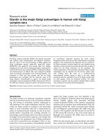
RANKL attenuates the effect of bisphosphonates on osteoclast number, apoptosis, and bone resorption in vitroRANKL attenuates the effect of bisphosphonates on osteoclast
number, apoptosis, and bone resorption in vitro. Cultures of mature
osteoclasts from rabbit bones were treated with 100 M ALN or CLO,
± 50 ng/mL RANKL for 48 hours. Cells were then fixed, stained for tar-
trate-resistant acid phosphatase (TRAP), and counterstained with
DAPI. (a) Results are the mean number of TRAP-positive multinucle-
ated osteoclasts (more than three nuclei per cell) ± standard error of
the mean (SEM) (n = 3) or (b) the percentage of non-apoptotic and
apoptotic osteoclasts. Data are expressed as the mean ± SEM (n = 3
replicates). **P = 0.01, ***P = 0.001 compared with ALN or CLO alone
(analysis of variance).
#
Treatment with ALN or CLO alone caused a sig-
nificant decrease in osteoclast number compared with control (CTL)
cultures (P = 0.01) and a significant increase in osteoclast apoptosis
compared with control cultures (P = 0.001). (c) Values of resorption
area are the mean resorbed area (mm
2
) per slice ± SEM (n = 6 slices).
***P = 0.001 compared with CLO alone and **P = 0.01 compared with
ALN alone (analysis of variance).
#
Treatment with ALN or CLO alone
caused a significant decrease in osteoclastic bone resorption com-
pared with control cultures (P = 0.001). The data shown are represent-
ative of three independent experiments. ALN, 4-amino-1-hydroxy-
butylidene-1,1-bisphosphonate (alendronate); CLO, dichloromethyl-
ene-1,1-bisphosphonate (clodronate); DAPI, 4,6-diamidino-2-phenylin-
dole; RANKL, receptor activator of nuclear factor-kappa-B ligand.
Arthritis Research & Therapy Vol 11 No 2 Sutherland et al.
Page 4 of 9
(page number not for citation purposes)
ence of 50 ng/mL RANKL, the reduction in osteoclast number
was significantly attenuated (to approximately 77% and 85%
of control cultures, respectively) (P < 0.01).
Osteoclasts in culture dishes that were undergoing apoptosis
but remained adherent were identified on the basis of charac-
teristic morphological features (chromatin condensation and
nuclear fragmentation) after staining with DAPI [21]. Approxi-
mately 17% of adherent osteoclasts in culture dishes were
apoptotic after treatment with 100 M ALN or CLO for 48
hours. This was significantly reduced (to approximately 9%) in
the presence of 50 ng/mL RANKL (P < 0.001), similar to the
proportion of osteoclasts undergoing apoptosis in cultures
without BPs (Figure 1b). RANKL alone did not significantly
alter the number of adherent osteoclasts or the proportion of
apoptotic osteoclasts in cultures in the absence of BPs.
RANKL protects osteoclasts from the anti-resorptive
effects of bisphosphonates
The effect of RANKL on the morphology of BP-treated rabbit
osteoclasts was also studied by scanning EM. After 24 hours
of culture on ivory discs, 71% of osteoclasts in control cultures
were spread on the mineral surface, often located in or adja-
cent to extensive and deep resorption cavities (Figure 2a).
However, after treatment with 50 M CLO for 24 hours, few,
shallow resorption pits were present and 50% of the osteo-
clasts were rounded and lacked areas of spreading. Many of
these rounded osteoclasts lacked membrane ruffles or micro-
villi but contained numerous blebs, indicative of cells undergo-
ing apoptosis (Figure 2b). The morphology of other cell types,
such as stromal cells, in the culture did not appear to be
affected by CLO. The appearance of apoptotic osteoclasts
was prevented by the presence of RANKL since 30% of oste-
oclasts were rounded when cultured with CLO + RANKL, few
of these had membrane blebbing, and 70% of the osteoclasts
appeared similar to those in control cultures, associated with
numerous resorption pits (Figure 2c).
Treatment with 50 M ALN for 24 hours caused the appear-
ance of osteoclasts that (although still spread and adherent to
the mineral) appeared retracted, with long cell processes (Fig-
ure 2d), and were associated (if at all) with only minor resorp-
tion pits. These osteoclasts often lacked microvilli but
exhibited ridges on the basolateral membrane, and few (<5%)
Figure 2
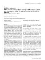
Scanning EM analysis of the effect of CLO, ALN, and RANKL on the morphology of mature osteoclasts cultured in vitroScanning EM analysis of the effect of CLO, ALN, and RANKL on the morphology of mature osteoclasts cultured in vitro. Rabbit osteoclasts were
cultured on ivory discs for 24 hours with 50 M CLO or 50 M ALN, ± 100 ng/mL RANKL. (a) Control. (b) CLO. (c) CLO + RANKL. (d) ALN. (e)
ALN + RANKL. Osteoclasts were fixed and processed for scanning EM analysis. Bars = 10 m. ALN, 4-amino-1-hydroxy-butylidene-1,1-bisphos-
phonate (alendronate); CLO, dichloromethylene-1,1-bisphosphonate (clodronate); RANKL, receptor activator of nuclear factor-kappa-B ligand.
Available online />Page 5 of 9
(page number not for citation purposes)
apoptotic osteoclasts were observed with the obvious mem-
brane blebbing observed in CLO-treated cultures. After cul-
ture with ALN + RANKL, osteoclasts were mostly well spread
with membrane ruffles and surface microvilli, and resorption
cavities were more evident (Figure 2e). Some osteoclasts also
retained the presence of membrane ridges on the basolateral
surface, but few (<5%) had the retracted morphology of ALN-
treated cells or the blebbed morphology of CLO-treated cells.
As expected, when the area of bone resorption was quantified,
100 M ALN or CLO significantly inhibited the resorptive
activity of rabbit osteoclasts cultured on ivory discs in vitro.
However, the inhibitory effect of ALN or CLO on bone resorp-
tion was significantly overcome by the presence of 100 ng/mL
RANKL (Figure 1c).
RANKL reduces caspase-9 activity in osteoclasts
A fluorescent, cell-permeable caspase-9 inhibitor was used to
identify single cells with caspase-9 activity (Figure 3a). In con-
trol cultures of purified rabbit osteoclasts, approximately 10%
of the cells (Figure 3b) had detectable caspase-9 activity (sim-
ilar to the proportion of cells in control cultures that were iden-
tified as apoptotic on the basis of nuclear morphology) (Figure
1b). After treatment with 100 M ALN for 48 hours, the pro-
portion of caspase-9-positive osteoclasts increased signifi-
cantly (to about 30%). This was significantly reduced, almost
to the proportion in control cultures, in the presence of 100
ng/mL RANKL.
RANKL does not prevent accumulation of unprenylated
Rap1A in osteoclasts
In accord with our previous studies, treatment of purified rabbit
osteoclasts with 100 M ALN for 48 hours caused the accu-
mulation of the unprenylated form of the small GTPase Rap1A,
thereby demonstrating that ALN inhibits protein prenylation in
osteoclasts [2,22]. Incubation of osteoclasts with 100 M
ALN in the presence of 100 ng/mL RANKL for 48 hours did
not prevent the accumulation of unprenylated Rap1A (Figure
3c).
RANKL increases the level of Mcl-1 in rabbit osteoclasts
Western blot analysis of purified rabbit osteoclasts showed
that treatment with 100 ng/mL RANKL, 100 M ALN, or ALN
+ RANKL had no effect on the level of Bcl-2 protein (Figure
3d). However, RANKL alone consistently caused a threefold
increase in the level of Mcl-1 protein in osteoclasts. Treatment
with ALN caused a decrease of approximately 90% in Mcl-1,
although co-treatment with RANKL almost completely pre-
vented this effect and maintained the level of Mcl-1 similar to
that in control cells (Figure 3d).
Loss of Mcl-1 precedes apoptosis during cytokine
deprivation of mouse osteoclasts but is prevented by
pro-survival factors
To further examine the importance of Mcl-1 in osteoclast sur-
vival, multinucleated osteoclasts were generated from M-CSF-
Figure 3
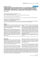
RANKL prevents activation of caspase-9 and increases Mcl-1 in osteo-clasts but does not prevent inhibition of protein prenylationRANKL prevents activation of caspase-9 and increases Mcl-1 in osteo-
clasts but does not prevent inhibition of protein prenylation. Cultures of
mature osteoclasts from rabbit bones were treated with 100 M ALN ±
100 ng/mL RANKL for 48 hours and stained using an Apofluor Green
Caspase-9 Activity Assay kit and Hoechst 33342. (a) A representative
non-apoptotic and apoptotic osteoclast. (b) Quantification of caspase-
9-positive osteoclasts after treatment with alendronate ± RANKL for 48
hours. *P 0.05 compared with ALN alone or
#
P 0.05 compared
with control (CTL) (analysis of variance). Values are the mean ± stand-
ard error of the mean (n = 3 replicates) (100 to 150 cells counted per
well). The data shown are representative of three independent experi-
ments. (c) Purified rabbit osteoclasts were treated for 48 hours with
100 M ALN ± 100 ng/mL RANKL or with RANKL alone. Cell lysates
were then analysed by Western blotting for the unprenylated form of
Rap1A and for -actin. (d) Purified rabbit osteoclasts were treated for
48 hours with 100 M ALN ± 100 ng/mL RANKL or with 100 ng/mL
RANKL alone. Cell lysates were then analysed by Western blotting for
Mcl-1 and Bcl-2. The level of Mcl-2 or Bcl-2 was quantified by densito-
metric analysis and expressed as a ratio of the level in control cells.
Data shown are representative of three independent experiments. ALN,
4-amino-1-hydroxy-butylidene-1,1-bisphosphonate (alendronate);
RANKL, receptor activator of nuclear factor-kappa-B ligand.
Arthritis Research & Therapy Vol 11 No 2 Sutherland et al.
Page 6 of 9
(page number not for citation purposes)
dependent mouse bone marrow macrophages by culturing the
latter cells for 5 days with M-CSF + RANKL. When the osteo-
clasts were starved of these cytokines, morphological
changes indicative of apoptosis (Figure 4a) were apparent
after 6 to 8 hours, consistent with the appearance in Western
blots of the cleaved form of caspase-3 after 6 hours of
cytokine starvation (Figure 4b). The appearance of apoptotic
osteoclasts and cleaved caspase-3 was preceded by a
decrease in the level of Mcl-1 (noticeable after 4 hours). Mcl-
1 was almost completely absent after 12 hours of cytokine
starvation, although the levels of Bcl-1 and Bcl-x
L
did not
change during this time (Figure 4b). The loss of Mcl-1 that
occurred in mouse osteoclasts following cytokine starvation
could be prevented by the addition of M-CSF, RANKL, TNF-,
or LPS (Figure 4c).
Discussion
BPs have become the mainstay of treatment for post-meno-
pausal osteoporosis, Paget disease, and tumour-associated
osteolysis and have been shown to prevent generalised bone
loss in patients with RA treated with corticosteroids (reviewed
recently by Breuil and Euller-Ziegler [16]). However, apart
Figure 4
Mcl-1 levels decrease rapidly in mouse osteoclasts following cytokine starvation but are restored by pro-survival factorsMcl-1 levels decrease rapidly in mouse osteoclasts following cytokine starvation but are restored by pro-survival factors. Mouse osteoclasts were
generated by culturing bone marrow macrophages for 5 days with macrophage colony-stimulating factor (M-CSF) + receptor activator of nuclear
factor-kappa-B ligand (RANKL). (a) Osteoclasts were starved of M-CSF and RANKL for 8 hours and then fixed and processed for scanning EM anal-
ysis. Representative osteoclasts from non-starved (control) or starved cultures are shown. Bars = 10 m. (b) Mouse osteoclasts were starved of M-
CSF and RANKL for 2 to 12 hours. Western blot analysis was then used to determine the level of Mcl-1, cleaved caspase-3, Bcl-2, and Bcl-x
L
at
each time point. (c) After osteoclasts were generated, the medium was replaced with normal medium (control [Ctrl]: M-CSF + RANKL), with medium
lacking cytokines (starved), or with medium containing recombinant M-CSF, RANKL, tumour necrosis factor-alpha (TNF-), or lipopolysaccharide
(LPS). After 12 hours, Western blotting was used to determine the level of Mcl-1 in 40 g of cell lysate. Data shown are representative of three inde-
pendent experiments.
Available online />Page 7 of 9
(page number not for citation purposes)
from a recent clinical study using the highly potent BP
zoledronic acid in patients with RA [15] and two studies of
zoledronic acid in animal models of RA [12,13], the effective-
ness of BPs at preventing focal bone loss has been less con-
vincing [16-19]. It has recently been suggested that the
reason for this relative lack of effect on local, inflammatory
bone loss is due to factors in the inflamed joint, such as TNF-
, that antagonise the ability of BPs to inhibit osteoclasts.
Zhang and colleagues [23], using TNF- transgenic mice,
showed that Bcl-x
L
levels were markedly higher in osteoclasts,
an effect that appeared to be caused by TNF--induced
expression of Ets-2. Furthermore, overexpression of Ets-2 or
Bcl-x
L
protected osteoclasts from ALN-induced apoptosis in
vitro. RANKL is also abundant in the rheumatoid microenviron-
ment and drives the enhanced osteoclastogenesis and hence
excessive osteoclast-mediated destruction of bone [8-10]. In
our study, we demonstrate that RANKL also protects osteo-
clasts from the apoptosis-inducing and anti-resorptive effects
of ALN or CLO in vitro. The number of apoptotic rabbit osteo-
clasts was significantly lower in cultures treated for 48 hours
with ALN or CLO in the presence of RANKL than in cultures
treated with the BPs alone. Consistent with this, RANKL pre-
served the total number of osteoclasts in cultures treated with
the BPs and also significantly overcame the anti-resorptive
effect of the BPs when osteoclasts were cultured on dentine
discs. This ability of RANKL to rescue osteoclasts from the
effects of BPs was also observed morphologically by using
scanning EM. Treatment of osteoclasts with CLO for 24 hours
caused morphological changes associated with apoptotic cell
death, consistent with the ability of CLO to induce osteoclast
apoptosis via the formation and rapid accumulation of a cyto-
toxic metabolite [2,24,25]. The morphological changes asso-
ciated with ALN treatment for 24 hours (cell retraction and
alterations in plasma membrane morphology) are consistent
with the inhibition of FPP synthase. This leads to the inhibition
of protein prenylation and the slow accumulation of unpre-
nylated small GTPases such as Rap1A [1,26]. The morpho-
logical appearance of osteoclasts after 24 hours of treatment
with ALN therefore probably indicates cells with altered
cytoskeletal arrangement and vesicular trafficking, as well as
cells in the very early stages of apoptosis, as a result of abnor-
mal small GTPase signalling [4,27]. In the presence of
RANKL, these effects of ALN and CLO on osteoclast morphol-
ogy were largely overcome, at least over a 24-hour culture
period. In the case of ALN, this was not due to any ability of
RANKL to prevent inhibition of FPP synthase, since RANKL
treatment did not prevent the accumulation of unprenylated
Rap1A in osteoclasts.
We have previously shown that BP-induced osteoclast apop-
tosis involves loss of mitochondrial membrane potential, lead-
ing (presumably as a result of subsequent release of
cytochrome C from mitochondria and activation of procas-
pase-9) to the cleavage and activation of procaspase-3 [21].
To identify a potential mechanism by which RANKL prevents
BP-induced apoptosis, we examined in more detail the events
involved in ALN-induced apoptosis. Co-treatment with RANKL
prevented the increase in the proportion of osteoclasts with
active caspase-9 seen after ALN treatment, suggesting that
RANKL either prevents the mitochondrial changes that lead to
activation of procaspase-9 [28] or prevents procaspase-9
activation subsequent to the release of cytochrome C (for
example, by increasing the expression of XIAP [29]). Although
expression of XIAP and cIAP1/2 can be stimulated via nuclear
factor-kappa-B [30], which is a major signalling pathway acti-
vated by RANKL [7], we did not observe any effect of RANKL
on the level of XIAP, cIAP1, or cIAP2 in rabbit osteoclasts by
Western blotting (data not shown), consistent with earlier
studies on murine osteoclasts [31]. This suggests that RANKL
probably prevents apoptosis by preventing mitochondrial
changes prior to caspase activation, perhaps via members of
the Bcl-2 family of proteins that regulate the mitochondrial
pathway of apoptosis [32,33].
RANKL did not alter the level of Bcl-2 in rabbit osteoclasts but
caused a marked increase in the level of Mcl-1, an anti-apop-
totic member of the Bcl-2 family that is expressed in cells of
the myeloid lineage such as macrophages and neutrophils
[34,35], and is also known to be increased in synovial macro-
phages and fibroblasts in RA [36,37] as well as in synovial
fluid lymphocytes in juvenile idiopathic arthritis [38]. In myeloid
cells, Mcl-1 prevents apoptosis and its expression is highly
regulated by survival-promoting factors via the PI3K/Akt/
mammlian target of rapamycin (mTOR)/S6 kinase as well as
mitogen-activated protein kinase (MAPK) signalling pathways
[39,40]. The latter are also known to be activated by M-CSF,
TNF-, and RANKL [7,41], which promote osteoclast survival
[6]. Whilst our work was in progress, Bradley and colleagues
[42] showed that Mcl-1 mRNA and protein are upregulated in
osteoclasts by M-CSF via activation of MEK/ERK (MAPK
kinase/extracellular regulated kinase) and increased expres-
sion of Egr2. Hence, although the exact mechanism by which
RANKL upregulates Mcl-1 in osteoclasts remains to be
proven, it is highly likely that this also involves MAPK and/or
mTOR signalling pathways. ALN treatment alone caused a
decrease in Mcl-1 levels, perhaps since the activation of
mTOR/S6 kinase in osteoclasts appears to involve geranylger-
anylated proteins such as Rac [41], and ALN is known to
effectively prevent protein geranylgeranylation [1,4]. With
myeloma cells, others have also shown that the inhibition of
protein geranylgeranylation causes the loss of Mcl-1 [43]. In
our study, RANKL restored the level of Mcl-1 to that in control
osteoclasts, but this was still less than the level of Mcl-1 in the
presence of RANKL alone. Hence, ALN and RANKL may have
opposing effects on the same signalling pathways (perhaps
involving mTOR) that are required for Mcl-1 expression in
osteoclasts.
In further support of an important role for Mcl-1 in osteoclast
survival, we found that the level of Mcl-1 rapidly decreased fol-
Arthritis Research & Therapy Vol 11 No 2 Sutherland et al.
Page 8 of 9
(page number not for citation purposes)
lowing the removal of M-CSF and RANKL from cultures of
mouse osteoclasts, which are highly dependent on the pres-
ence of such survival factors. In this model, the loss of Mcl-1
preceded the appearance of morphological and biochemical
features of apoptosis and occurred in the absence of changes
in the level of Bcl-2 or Bcl-x
L
. In addition, the loss of Mcl-1 in
mouse osteoclasts could be prevented by the addition of fac-
tors (LPS, TNF-, M-CSF, and RANKL) that are known to pro-
mote osteoclast survival [6,7,44]. The loss of Mcl-1 in
osteoclasts probably occurred by proteasomal degradation
since we also found that the proteasome inhibitor MG132
caused an increase in the level of Mcl-1 in mouse osteoclasts
(data not shown). This is consistent with a requirement in oste-
oclasts for continual protein synthesis to prevent apoptosis
[41,45]. Together, these observations indicate that Mcl-1
plays an important role in maintaining osteoclast survival.
Mcl-1 inhibits activation of the mitochondrial pathway of apop-
tosis by interacting with pro-apoptotic Bcl-2 family proteins
such as Bak and Bim, thereby preventing the increased mito-
chondrial permeability that leads to caspase activation
[33,46,47]. Lack of Bim leads to enhanced osteoclast survival
and the pro-survival factor M-CSF decreases Bim levels in
osteoclasts and osteoclast precursors [48,49], probably by
increasing ubiquitin-dependent degradation of Bim via upreg-
ulation of cCbl [42]. The role of Bim in BP-induced osteoclast
apoptosis is not known, although a recent study showed that
apoptosis of MCF-7 breast cancer cells induced by the BP
risedronate involved increased levels of Bim [50]. Upregula-
tion of Mcl-1 (and therefore antagonism of Bim) by RANKL is
therefore a likely mechanism by which RANKL prevents BP-
induced apoptosis of osteoclasts.
Conclusions
We have shown that RANKL protects osteoclasts from the
pro-apoptotic and hence anti-resorptive effects of the BPs
CLO and ALN in vitro. This protective effect of RANKL is likely
to be mediated at least in part by an increase in the level of the
anti-apoptotic protein Mcl-1. The ability of RANKL, together
with other factors such as TNF- [23], to rescue osteoclasts
from the pro-apoptotic effects of BPs may account for the
apparent lack of effectiveness of BPs (particularly the less
potent BPs such as CLO and ALN) in preventing local, inflam-
mation-induced bone loss in RA.
Competing interests
MJR has received research funding from Procter & Gamble
(Cincinnati, OH, USA), Novartis (Basel, Switzerland), and
Roche (Basel, Switzerland) and acts as a consultant for
Novartis and Procter & Gamble. The other authors declare that
they have no competing interests.
Authors' contributions
MJR conceived and designed the study and wrote the manu-
script. KAS, HLR, and DT helped to design, and performed,
the experiments. All authors read and approved the final
manuscript.
Acknowledgements
We are grateful to Debbie Marshall for technical assistance with scan-
ning EM and to Ruth Craig (Dartmouth Medical School, Hanover, NH,
USA) and Brendan Boyce (University of Rochester Medical Center,
Rochester, NY, USA) for helpful discussions. This work was funded by
a studentship to KAS from the Arthritis Research Campaign.
References
1. Rogers MJ: New insights into the molecular mechanisms of
action of bisphosphonates. Curr Pharm Des 2003,
9:2643-2658.
2. Frith JC, Monkkonen J, Auriola S, Monkkonen H, Rogers MJ: The
molecular mechanism of action of the anti-resorptive and anti-
inflammatory drug clodronate: evidence for the formation in
vivo of a metabolite that inhibits bone resorption and causes
osteoclast and macrophage apoptosis. Arthritis Rheum 2001,
44:2201-2210.
3. Coxon FP, Helfrich MH, Van't Hof R, Sebti S, Ralston SH, Hamilton
A, Rogers MJ: Protein geranylgeranylation is required for oste-
oclast formation, function, and survival: inhibition by bisphos-
phonates and GGTI-298. J Bone Miner Res 2000,
15:1467-1476.
4. Coxon FP, Thompson K, Rogers MJ: Recent advances in under-
standing the mechanism of action of bisphosphonates. Curr
Opin Pharmacol 2006, 6:307-312.
5. Hughes DE, Wright KR, Uy HL, Sasaki A, Yoneda T, Roodman GD,
Mundy GR, Boyce BF: Bisphosphonates promote apoptosis in
murine osteoclasts in vitro and in vivo. J Bone Miner Res 1995,
10:1478-1487.
6. Xing L, Boyce BF: Regulation of apoptosis in osteoclasts and
osteoblastic cells. Biochem Biophys Res Commun 2005,
328:709-720.
7. Asagiri M, Takayanagi H: The molecular understanding of oste-
oclast differentiation. Bone 2007, 40:251-264.
8. Schett G, Hayer S, Zwerina J, Redlich K, Smolen JS: Mechanisms
of disease: the link between RANKL and arthritic bone
disease. Nat Clin Pract Rheumatol 2005, 1:47-54.
9. Takayanagi H: Osteoimmunological insight into bone damage
in rheumatoid arthritis. Mod Rheumatol 2005, 15:225-231.
10. Teitelbaum SL: Osteoclasts; culprits in inflammatory
osteolysis. Arthritis Res Ther 2006, 8:201.
11. Zhao H, Liu S, Huang D, Xu Q, Shuto T, Iwamoto Y:
The protec-
tive effects of incadronate on inflammation and joint destruc-
tion in established rat adjuvant arthritis. Rheumatol Int 2006,
26:732-740.
12. Herrak P, Gortz B, Hayer S, Redlich K, Reiter E, Gasser J, Berg-
meister H, Kollias G, Smolen JS, Schett G: Zoledronic acid pro-
tects against local and systemic bone loss in tumor necrosis
factor-mediated arthritis. Arthritis Rheum 2004, 50:2327-2337.
13. Sims NA, Green JR, Glatt M, Schlict S, Martin TJ, Gillespie MT,
Romas E: Targeting osteoclasts with zoledronic acid prevents
bone destruction in collagen-induced arthritis. Arthritis Rheum
2004, 50:2338-2346.
14. Matsuo A, Shuto T, Hirata G, Satoh H, Matsumoto Y, Zhao H,
Iwamoto Y: Antiinflammatory and chondroprotective effects of
the aminobisphosphonate incadronate (YM175) in adjuvant
induced arthritis. J Rheumatol 2003, 30:1280-1290.
15. Jarrett SJ, Conaghan PG, Sloan VS, Papanastasiou P, Ortmann
CE, O'Connor PJ, Grainger AJ, Emery P: Preliminary evidence for
a structural benefit of the new bisphosphonate zoledronic acid
in early rheumatoid arthritis. Arthritis Rheum 2006,
54:1410-1414.
16. Breuil V, Euller-Ziegler L: Bisphosphonate therapy in rheuma-
toid arthritis. Joint Bone Spine 2006, 73:349-354.
17. Ritchlin CT, Schwarz EM, O'Keefe RJ, Looney RJ: RANK, RANKL
and OPG in inflammatory arthritis and periprosthetic
osteolysis. J Musculoskelet Neuronal Interact 2004, 4:276-284.
18. Maksymowych WP: Bisphosphonates for arthritis – a confusing
rationale. J Rheumatol 2003, 30:430-434.
Available online />Page 9 of 9
(page number not for citation purposes)
19. Eggelmeijer F, Papapoulos SE, van Paassen HC, Dijkmans BA,
Valkema R, Westedt ML, Landman JO, Pauwels EK, Breedveld FC:
Increased bone mass with pamidronate treatment in rheuma-
toid arthritis. Results of a three-year randomized, double-blind
trial. Arthritis Rheum 1996, 39:396-402.
20. Coxon FP, Frith JC, Benford HL, Rogers MJ: Isolation and purifi-
cation of rabbit osteoclasts. In Bone Research Protocols Edited
by: Helfrich MH, Ralston SH. Totowa, NJ: Humana Press;
2003:89-99.
21. Benford HL, McGowan NW, Helfrich MH, Nuttall ME, Rogers MJ:
Visualization of bisphosphonate-induced caspase-3 activity in
apoptotic osteoclasts in vitro. Bone 2001, 28:465-473.
22. Coxon FP, Helfrich MH, Larijani B, Muzylak M, Dunford JE, Marshall
D, McKinnon AD, Nesbitt SA, Horton MA, Seabra MC, Ebetino FH,
Rogers MJ: Identification of a novel phosphonocarboxylate
inhibitor of Rab geranylgeranyl transferase that specifically
prevents Rab prenylation in osteoclasts and macrophages. J
Biol Chem 2001, 276:48213-48222.
23. Zhang Q, Badell IR, Schwarz EM, Boulukos KE, Yao Z, Boyce BF,
Xing L: Tumor necrosis factor prevents alendronate-induced
osteoclast apoptosis in vivo by stimulating Bcl-x
L
expression
through Ets-2. Arthritis Rheum 2005, 52:2708-2718.
24. Monkkonen H, Rogers MJ, Makkonen N, Niva S, Auriola S, Monk-
konen J: The cellular uptake and metabolism of clodronate in
RAW 264 macrophages. Pharm Res 2001, 18:1550-1555.
25. Lehenkari PP, Kellinsalmi M, Näpänkangas JP, Ylitalo KV,
Mönkkönen J, Rogers MJ, Azhayev A, Väänänen HK, Hassinen IE:
Further insight into mechanism of action of clodronate: inhibi-
tion of mitochondrial ADP/ATP translocase by a nonhydrolyz-
able, adenine-containing metabolite. Mol Pharmacol 2002,
61:1255-1262.
26. Roelofs AJ, Thompson K, Gordon S, Rogers MJ: Molecular mech-
anisms of action of bisphosphonates: current status. Clin
Cancer Res 2006, 12:6222s-6230s.
27. Dunford JE, Rogers MJ, Ebetino FH, Phipps RJ, Coxon FP: Inhibi-
tion of protein prenylation by bisphosphonates causes sus-
tained activation of Rac, Cdc42 and Rho GTPases. J Bone
Miner Res 2006, 21:684-694.
28. Kroemer G, Galluzzi L, Brenner C: Mitochondrial membrane per-
meabilization in cell death. Physiol Rev 2007, 87:99-163.
29. Eckelman BP, Salvesen GS, Scott FL: Human inhibitor of apop-
tosis proteins: why XIAP is the black sheep of the family.
EMBO Rep 2006, 7:988-994.
30. Karin M, Lin A: NF-kappaB at the crossroads of life and death.
Nat Immunol 2002, 3:221-227.
31. Kanaoka K, Kobayashi Y, Hashimoto F, Nakashima T, Shibata M,
Kobayashi K, Kato Y, Sakai H: A common downstream signaling
activity of osteoclast survival factors that prevent nitric oxide-
promoted osteoclast apoptosis. Endocrinology 2000,
141:2995-3005.
32. van Delft MF, Huang DC: How the Bcl-2 family of proteins inter-
act to regulate apoptosis. Cell Res 2006, 16:203-213.
33. Reed JC: Proapoptotic multidomain Bcl-2/Bax-family proteins:
mechanisms, physiological roles, and therapeutic
opportunities. Cell Death Differ 2006, 13:1378-1386.
34. Kozopas KM, Yang T, Buchan HL, Zhou P, Craig RW:
MCL1, a
gene expressed in programmed myeloid cell differentiation,
has sequence similarity to BCL2. Proc Natl Acad Sci USA
1993, 90:3516-3520.
35. Craig RW: MCL1 provides a window on the role of the BCL2
family in cell proliferation, differentiation and tumorigenesis.
Leukemia 2002, 16:444-454.
36. Liu H, Huang Q, Shi B, Eksarko P, Temkin V, Pope RM: Regulation
of Mcl-1 expression in rheumatoid arthritis synovial
macrophages. Arthritis Rheum 2006, 54:3174-3181.
37. Liu H, Eksarko P, Temkin V, Haines GK 3rd, Perlman H, Koch AE,
Thimmapaya B, Pope RM: Mcl-1 is essential for the survival of
synovial fibroblasts in rheumatoid arthritis. J Immunol 2005,
175:8337-8345.
38. Smolewska E, Stanczyk J, Robak T, Smolewski P: Inhibited apop-
tosis of synovial fluid lymphocytes in children with juvenile idi-
opathic arthritis is associated with increased expression of
myeloid cell leukemia 1 and XIAP proteins. J Rheumatol 2006,
33:1684-1690.
39. Domina AM, Vrana JA, Gregory MA, Hann SR, Craig RW: MCL1 is
phosphorylated in the PEST region and stabilized upon ERK
activation in viable cells, and at additional sites with cytotoxic
okadaic acid or taxol. Oncogene 2004, 23:5301-5315.
40. Zhou P, Qian L, Kozopas KM, Craig RW: Mcl-1, a Bcl-2 family
member, delays the death of hematopoietic cells under a vari-
ety of apoptosis-inducing conditions. Blood 1997,
89:630-643.
41. Glantschnig H, Fisher JE, Wesolowski G, Rodan GA, Reszka AA:
M-CSF, TNFalpha and RANK ligand promote osteoclast sur-
vival by signaling through mTOR/S6 kinase. Cell Death Differ
2003, 10:1165-1177.
42. Bradley EW, Ruan MM, Oursler MJ: Novel pro-survival functions
of the Kruppel-like transcription factor Egr2 in promotion of
macrophage colony-stimulating factor-mediated osteoclast
survival downstream of the MEK/ERK pathway. J Biol Chem
2008, 283:8055-8064.
43. Donk NW van de, Kamphuis MM, van Kessel B, Lokhorst HM,
Bloem AC: Inhibition of protein geranylgeranylation induces
apoptosis in myeloma plasma cells by reducing Mcl-1 protein
levels. Blood 2003, 102:3354-3362.
44. Suda K, Woo JT, Takami M, Sexton PM, Nagai K: Lipopolysaccha-
ride supports survival and fusion of preosteoclasts independ-
ent of TNF-alpha, IL-1, and RANKL. J Cell Physiol
2002,
190:101-108.
45. Oursler MJ, Bradley EW, Elfering SL, Giulivi C: Native, not
nitrated, cytochrome c and mitochondria-derived hydrogen
peroxide drive osteoclast apoptosis. Am J Physiol Cell Physiol
2005, 288:C156-C168.
46. Yang-Yen HF: Mcl-1: a highly regulated cell death and survival
controller. J Biomed Sci 2006, 13:201-204.
47. Gelinas C, White E: BH3-only proteins in control: specificity
regulates MCL-1 and BAK-mediated apoptosis. Genes Dev
2005, 19:1263-1268.
48. Akiyama T, Bouillet P, Miyazaki T, Kadono Y, Chikuda H, Chung UI,
Fukuda A, Hikita A, Seto H, Okada T, Inaba T, Sanjay A, Baron R,
Kawaguchi H, Oda H, Nakamura K, Strasser A, Tanaka S: Regu-
lation of osteoclast apoptosis by ubiquitylation of proapoptotic
BH3-only Bcl-2 family member Bim. EMBO J 2003,
22:6653-6664.
49. Sugatani T, Hruska KA: Akt1/Akt2 and mammalian target of
rapamycin/Bim play critical roles in osteoclast differentiation
and survival, respectively, whereas Akt is dispensable for cell
survival in isolated osteoclast precursors. J Biol Chem 2005,
280:3583-3589.
50. Suyama K, Noguchi Y, Tanaka T, Yoshida T, Shibata T, Saito Y,
Tatsuno I: Isoprenoid-independent pathway is involved in
apoptosis induced by risedronate, a bisphosphonate, in which
Bim plays a critical role in breast cancer cell line MCF-7. Oncol
Rep 2007, 18:1291-1298.