Báo cáo y học: "Apigenin, a non-mutagenic dietary flavonoid, suppresses lupus by inhibiting autoantigen presentation for expansion of autoreactive Th1 and Th17 cells" potx
Bạn đang xem bản rút gọn của tài liệu. Xem và tải ngay bản đầy đủ của tài liệu tại đây (1.66 MB, 13 trang )
Open Access
Available online />Page 1 of 13
(page number not for citation purposes)
Vol 11 No 2
Research article
Apigenin, a non-mutagenic dietary flavonoid, suppresses lupus by
inhibiting autoantigen presentation for expansion of autoreactive
Th1 and Th17 cells
Hee-Kap Kang, Diane Ecklund, Michael Liu and Syamal K Datta
Division of Rheumatology, Departments of Medicine and Microbiology-Immunology, Northwestern University Feinberg School of Medicine, 240 East
Huron Street, Chicago, IL 60611, USA
Corresponding author: Syamal K Datta,
Received: 15 Jan 2009 Revisions requested: 4 Mar 2009 Revisions received: 26 Mar 2009 Accepted: 30 Apr 2009 Published: 30 Apr 2009
Arthritis Research & Therapy 2009, 11:R59 (doi:10.1186/ar2682)
This article is online at: />© 2009 Kang et al.; licensee BioMed Central Ltd.
This is an open access article distributed under the terms of the Creative Commons Attribution License ( />),
which permits unrestricted use, distribution, and reproduction in any medium, provided the original work is properly cited.
Abstract
Introduction Lupus patients need alternatives to steroids and
cytotoxic drugs. We recently found that apigenin, a non-
mutagenic dietary flavonoid, can sensitize recurrently activated,
normal human T cells to apoptosis by inhibiting nuclear factor-
kappa-B (NF-B)-regulated Bcl-x
L
, cyclooxygenase 2 (COX-2),
and cellular FLICE-like inhibitory protein (c-FLIP) expression.
Because sustained immune activation and hyperexpression of
COX-2 and c-FLIP contribute to lupus, we treated SNF1 mice
that spontaneously develop human lupus-like disease with
apigenin.
Methods SNF1 mice with established lupus-like disease were
injected with 20 mg/kg of apigenin daily and then monitored for
development of severe nephritis. Histopathologic changes in
kidneys, IgG autoantibodies to nuclear autoantigens in serum
and in cultures of splenocytes, along with nucleosome-specific
T helper 1 (Th1) and Th17 responses, COX-2 expression, and
apoptosis of lupus immune cells were analyzed after apigenin
treatment.
Results Apigenin in culture suppressed responses of Th1 and
Th17 cells to major lupus autoantigen (nucleosomes) up to 98%
and 92%, respectively, and inhibited the ability of lupus B cells
to produce IgG class-switched anti-nuclear autoantibodies
helped by these Th cells in presence of nucleosomes by up to
82%. Apigenin therapy of SNF1 mice with established lupus
suppressed serum levels of pathogenic autoantibodies to
nuclear antigens up to 97% and markedly delayed development
of severe glomerulonephritis. Apigenin downregulated COX-2
expression in lupus T cells, B cells, and antigen-presenting cells
(APCs) and caused their apoptosis. Autoantigen presentation
and Th17-inducing cytokine production by dendritic cells were
more sensitive to the inhibitory effect of apigenin in culture, as
evident at 0.3 to 3 M, compared with concentrations (10 to
100 M) required for inducing apoptosis.
Conclusions Apigenin inhibits autoantigen-presenting and
stimulatory functions of APCs necessary for the activation and
expansion of autoreactive Th1 and Th17 cells and B cells in
lupus. Apigenin also causes apoptosis of hyperactive lupus
APCs and T and B cells, probably by inhibiting expression of NF-
B-regulated anti-apoptotic molecules, especially COX-2 and c-
FLIP, which are persistently hyperexpressed by lupus immune
cells. Increasing the bioavailability of dietary plant-derived COX-
2 and NF-B inhibitors, such as apigenin, could be valuable for
suppressing inflammation in lupus and other Th17-mediated
diseases like rheumatoid arthritis, Crohn disease, and psoriasis
and in prevention of inflammation-based tumors overexpressing
COX-2 (colon, breast).
ACUC: Animal Care and Use Committee; AICD: activation-induced cell death; AP: alkaline phosphatase; APC: antigen-presenting cell; c-FLIP: cel-
lular FLICE-like inhibitory protein; COX-2: cyclooxygenase 2; DC: dendritic cell; DMSO: dimethyl sulfoxide; dsDNA: double-stranded DNA; ELISA:
enzyme-linked immunosorbent assay; ELISPOT: enzyme-linked immunosorbent spot; IFN-: interferon-gamma; IL: interleukin; NF-B: nuclear factor-
kappa-B; PBS: phosphate-buffered saline; RNP: ribonucleoprotein; SNF1: (SWR × NZB)F1; ssDNA: single-stranded DNA; Th: T helper; TLR: Toll-
like receptor; T
reg
: regulatory T.
Arthritis Research & Therapy Vol 11 No 2 Kang et al.
Page 2 of 13
(page number not for citation purposes)
Introduction
In lupus, intrinsic 'hyperactivity' of the immune system is asso-
ciated with persistent interactions between certain autoim-
mune T helper (Th) cells and B cells, leading to the production
of IgG autoantibodies against apoptotic nuclear antigens and
the formation of pathogenic immune complexes [1,2]. Nor-
mally, autoreactive T and B cells are eliminated by functional
inactivation (anergy) and activation-induced cell death (AICD)
(apoptosis) [3]. However, autoimmune Th cells of human lupus
resist AICD by upregulating the expression of cyclooxygenase
2 (COX-2) and the anti-apoptotic molecule c-FLIP (cellular
FLICE-like inhibitory protein) in a sustained manner [4]. COX-
2 is also overexpressed and is important for survival and func-
tion of other cells involved in the autoimmune inflammatory
responses for pathogenesis of lupus [5,6]. Therefore, COX-2
and associated molecules are critical targets for developing
non-mutagenic steroid-sparing drugs for lupus therapy.
Indeed, intermittent therapy with low doses of the COX-2
inhibitor celecoxib (Celebrex) has beneficial effects in murine
models of lupus [6,7], and preliminary results are encouraging
in lupus patients [8].
Apigenin (4',5,7-trihydroxyflavone) is a non-toxic non-muta-
genic flavonoid that is widely distributed in dietary plants,
especially in parsley, thyme, peppermint, olives, and herbs like
chamomile, and it can block COX-2 expression in cancer cells
[9]. We found that, in chronically activated but not in freshly
activated human T cells, relatively non-toxic apigenin can sup-
press PI3K-Akt-mediated nuclear factor-kappa-B (NF-B) acti-
vation and, consequently, NF-B-regulated anti-apoptotic
pathways, especially inhibiting c-FLIP and COX-2 expression
that are important for functioning and maintenance of immune
cells in inflammation, autoimmunity, and lymphoproliferation
[5]. Although apigenin decreases COX-2 expression, it does
not counteract COX-2 enzymatic activity itself. Moreover,
unlike the conventional COX-2 inhibitors, celecoxib (Cele-
brex), rofecoxib (Vioxx), or other non-steroidal anti-inflamma-
tory drugs, apigenin has vasorelaxing, anti-platelet, and anti-
oxidant properties, which could reduce the risk of coronary
disease and improve endothelial function [10-13]. Herein, we
treated spontaneously developing systemic lupus erythemato-
sus in the (SWR × NZB)F1 (SNF1) mouse model [14,15] with
apigenin and studied its mechanistic effects on the lupus
immune system.
Materials and methods
Mice
NZB and SWR mice were purchased from The Jackson Lab-
oratory (Bar Harbor, ME, USA). Lupus-prone SNF1 hybrids
were bred and females were used with the approval of the Ani-
mal Care and Use Committee (ACUC).
Administration of apigenin
Apigenin was purchased from Sigma-Aldrich (St. Louis, MO,
USA) and dissolved in dimethyl sulfoxide (DMSO) and then
diluted in phosphate-buffered saline (PBS) for experiments.
Twelve-week-old SNF1 mice were injected intraperitoneally
with apigenin (3, 6, or 20 mg/kg) daily. The control group was
injected with the same amount of vehicle solution (DMSO-
PBS). All mice were monitored weekly for the development of
proteinurea by testing with Albustix (VWR International, West
Chester, PA, USA) and for survival. The treatment lasted until
the mice were 52 weeks old. To study early immunologic
changes after treatment with apigenin, additional batches of
12-week-old SNF1 mice (five mice per group) were treated
with the same regimens as described above and then sacri-
ficed after 8 weeks.
Quantitation of total IgG and IgG autoantibodies
IgG class autoantibodies to single-stranded DNA (ssDNA),
double-stranded DNA (dsDNA), histone, and nucleosome
(histone-DNA complex) were measured by enzyme-linked
immunosorbent assay (ELISA) [16,17]. Two months after api-
genin treatment (at 5 months of age), the SNF1 mice were
bled for autoantibody measurement in serum. Total IgG and
IgG subclasses in sera of apigenin- or vehicle-treated SNF1
mice were also quantitated by ELISA [16-18]. Briefly, 96-well
plates were coated with goat anti-mouse IgG antibody (South-
ernBiotech, Birmingham, AL, USA). Serially diluted serum
samples were added and incubated overnight and then total
IgG or IgG subclasses were measured by using goat anti-
mouse IgG-alkaline phosphatase (AP) conjugate or anti-
mouse IgG isotype-specific antibody-AP conjugates.
Measurement of intracellular cyclooxygenase 2 and
analysis surface marker staining by flow cytometry
Three-month-old SNF1 female mice were treated with api-
genin (20 mg/kg) or vehicle solution for 8 weeks. Total spleen
cells from apigenin- or vehicle-treated SNF1 mice were
stained with fluorescein isothiocyanate-conjugated antibodies
to mouse CD4 (for T cells), mouse CD19 and CD86 for acti-
vated B cells, mouse CD11c for dendritic cells (DCs), and
mouse F4/80 for macrophages (BD Pharmingen, San Diego,
CA, USA, or eBioscience, San Diego, CA, USA, respectively)
at 4°C for 30 minutes. Antibody to CD11b was also used, but
it is a marker shared by DC subsets, macrophages, and other
cell types. After washing and fixation, cells were permeabilized
and stained with goat anti-mouse COX-2 antibody or its iso-
type control conjugated with phycoerythrin (Santa Cruz Bio-
technology, Inc., Santa Cruz, CA, USA) at room temperature
for 30 minutes. To lower the background for intracellular stain-
ing, we used cell fixation and permeabilization reagents from
eBioscience (00-5523) and a different antibody for COX-2
staining (sc-1745; Santa Cruz Biotechnology, Inc.) than in a
previous study [6]. For analysis, isotype-matched control stain-
ings were used for marking positive and negative cell popula-
tions. Usually, 200,000 events were collected after live cell
gating, using FACSCalibur, and analyzed by CellQuest (BD
Pharmingen) or FlowJo software (TreeStar Inc., FlowJo LLC,
Ashland, OR, USA).
Available online />Page 3 of 13
(page number not for citation purposes)
Induction of apoptosis
Spleen cells from 6-month-old SNF1 mice were cocultured for
24 hours with various concentrations of apigenin to measure
experimental apoptosis or vehicle to measure spontaneous,
control apoptosis. Apoptotic cells were detected by staining
of whole splenocytes with annexin V and propidium iodide (BD
Pharmingen), accompanied by simultaneous staining with
appropriate fluorochrome-conjugated antibodies to CD4,
B220, CD19, CD11c, or CD11b. Apoptosis in the specific
cell subset, as gated by flow cytometry, was calculated as
(percentage of experimental apoptosis – percentage of spon-
taneous apoptosis)/(100 – percentage of spontaneous apop-
tosis) [4].
Enzyme-linked immunosorbent spot assay
Enzyme-linked immunosorbent spot (ELISPOT) assay plates
(Cellular Technology Ltd., Shaker Heights, OH, USA) were
coated with capture antibodies against interferon-gamma
(IFN-) (BD Pharmingen) in PBS at 4°C overnight. Splenic T
cells (1 × 10
6
) from treated mice were cultured with irradiated
(3,000 rad) splenic antigen-presenting cells (APCs) (non-T
cells) from 1-month-old SNF1 mice in the presence of nucleo-
somes or their peptide autoepitopes or of PBS control. Cells
were removed after 24 hours of incubation for IFN- or after 48
hours for interleukin (IL)-17 production, and the responses
were visualized by the addition of the individual anti-cytokine
antibody-biotin and subsequent horseradish peroxidase-con-
jugated streptavidin. Cytokine-expressing cells were detected
by Immunospot scanning and analysis (Cellular Technology
Ltd.). To test the effect of apigenin on nuclear autoantigen
presentation, apigenin- or vehicle-pulsed splenic T cells (5 ×
10
5
per well) were cocultured with apigenin- or vehicle-pulsed
APCs (5 × 10
5
per well) for 1 hour before being added to IFN-
or IL-17 ELISPOT plates. The cultures were performed in the
presence of 0.1 to 30 g/mL nucleosomes.
Cytokine enzyme-linked immunosorbent assay
DCs (5 × 10
5
) from apigenin- or vehicle-treated SNF1 mice
were isolated as described [18] and stimulated with nucleo-
somes (1 to 60 g/mL) or Toll-like receptor (TLR)-9 ligand
CpG or TLR-7 ligand R837 (1 to 100 g/mL) obtained from
InvivoGen (San Diego, CA, USA). After 60 hours, amounts of
IL-6 in culture supernatants were measured by BD OptEIA™
ELISA set (BD Pharmingen).
Helper assays for IgG autoantibody production
To test the effect of apigenin on IgG autoantibody production
in vitro, whole splenocytes (1 × 10
6
cells per well) were stim-
ulated with 10 g/mL nucleosomes in the presence of various
amounts of apigenin or vehicle solution. After 7 days of culture,
supernatants were collected and assayed by ELISA for IgG
antibodies against dsDNA, ssDNA, histones, and nucleo-
somes as described [17].
Histopathologic analysis of kidneys
Halves of each kidney from apigenin- or control vehicle-treated
mice were fixed in 10% formalin and paraffin-embedded. To
determine the extent of renal disease, sections were stained
with hematoxylin and eosin and periododic acid-Schiff and
graded in a blinded fashion from 0 to 4+ for pathologic
changes (as described in [17,19-21]).
Statistical analysis
The log-rank test and the Student two-tailed t test were used.
Results are expressed as mean ± standard error of the mean
unless noted otherwise.
Results
Apigenin suppresses interferon-gamma response to
nuclear autoantigen and IgG autoantibody production in
vitro
T cells in unmanipulated SNF1 mice are spontaneously primed
to nuclear autoantigens in early life and respond to them ex
vivo by proliferation and production of IFN- without further
immunization [17,22]. Splenocytes from 5- to 6-month-old
SNF1 mice with overt lupus renal disease were stimulated in
vitro with nucleosomes (3 g/mL) in the presence of various
amounts of apigenin (1 to 100 M) and then analyzed for IFN-
production by ELISPOT. IFN- responses to nucleosomes
were markedly reduced by apigenin as compared with vehicle
(Figure 1a, P < 0.01 to 0.001). Exposure to 1 M apigenin
reduced the response to autoantigen by 57%, 3 M apigenin
inhibited response by 85%, and apigenin at concentrations of
10 M or above reduced the autoimmune response by 98%
(Figure 1a).
We also found that the levels of IgG class anti-dsDNA, anti-
ssDNA, anti-nucleosome, and anti-histone autoantibodies in
culture supernantants of nucleosome-stimulated SNF1 mouse
splenocytes were significantly reduced (up to 77%, 76%,
82%, and 66%, respectively) in the presence of apigenin (0.3
to 100 M) in comparison with vehicle (Figure 1b, P < 0.05 to
0.001). In this helper assay, the splenocytes were cultured for
7 days, and apigenin or vehicle was added once at the begin-
ning of culture. Thus, significant suppression of IFN-
responses to nucleosomes and reduction of IgG class-
switched autoantibody production occurred with 0.3 to 100
M apigenin (Figure 1).
Optimal dose of apigenin in vivo for suppression of
interferon-gamma response to nucleosomes
We used the suppressive effect of apigenin on lupus spleen
cells' IFN- response to nucleosomes (Figure 1) to determine
the optimal dose for in vivo treatment. We injected unmanipu-
lated 3-month-old SNF1 mice intraperitoneally with apigenin
daily at 3 mg/kg (13.89 M), 6 mg/kg (27.8 M), and 20 mg/
kg (0.93 mM). At this age, the SNF1 mice have elevated levels
of anti-nuclear autoantibodies in serum, but they do not have
overt proteinuria. After 2 weeks of treatment, we tested splen-
Arthritis Research & Therapy Vol 11 No 2 Kang et al.
Page 4 of 13
(page number not for citation purposes)
ocytes from treated mice for IFN- response to various
amounts of nucleosomes ex vivo. Although injection treatment
with the lowest dose had an inhibitory effect on IFN- response
to the autoantigen ex vivo, the 20 mg/kg dose showed the
most marked suppression in responses even at higher doses
of the autoantigen (Figure 2, P < 0.05 to 0.001). Therefore, we
decided to use a concentration of 20 mg/kg (0.93 mM) for in
vivo treatment. Moreover, apigenin administration was found
to be non-toxic at 20 mg/kg in other situations [23].
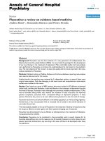
Figure 1
Apigenin suppressed nucleosome-specific interferon-gamma (IFN-) response and IgG-autoantibody productionApigenin suppressed nucleosome-specific interferon-gamma (IFN-) response and IgG-autoantibody production. Splenocytes from 5- to 6-month-
old unmanipulated SNF1 mice were stimulated with nucleosomes in the presence of various amounts of apigenin or vehicle (dimethyl sulfoxide-phos-
phate-buffered saline). (a) Apigenin markedly suppressed IFN- responses by nucleosome-specific T cells in enzyme-linked immunosorbent spot
assay. (b) Apigenin significantly reduced the level of IgG class autoantibodies in nucleosome-stimulated lupus Th cell-B cell coculture assays. *P <
0.001, **P < 0.01, and
x
P < 0.02. dsDNA, double-stranded DNA; SNF1, (SWR × NZB)F1; ssDNA, single-stranded DNA.
Available online />Page 5 of 13
(page number not for citation purposes)
In vivo treatment with apigenin suppresses interferon-
gamma and interleukin-17 responses and IgG
autoantibody production to nucleosomes
Three-month-old SNF1 mice were treated by intraperitoneal
injection with 20 mg/kg apigenin or vehicle daily. After 2
months of treatment, we analyzed IFN- and IL-17 responses
of nucleosome-specific T cells and IgG autoantibody
responses by culturing splenocytes from apigenin- or vehicle-
treated SNF1 in the presence of various concentrations of
nucleosomes. We found that IFN- and IL-17 responses to
nucleosome by lupus T cells were markedly reduced as com-
pared with vehicle treatment (up to 79% and 88%, respec-
tively) (Figure 3a, P < 0.05 to 0.001). However, polyclonal Th1
and Th17 responses with low-dose or optimal anti-CD3 (0.2
g/mL) stimulation were not suppressed by apigenin treat-
ment (Figure 3a). Moreover, we did not observe any significant
differences in viability of spleen cells isolated from apigenin-
treated and vehicle-treated mice. We also observed significant
reductions (up to 83%, 84%, 97%, and 94%, respectively) in
the levels of IgG class anti-dsDNA, anti-ssDNA, anti-nucleo-
somes, and anti-histone autoantibodies in culture supernant-
ants of nucleosome-stimulated splenocytes from apigenin-
treated SNF1 mice as compared with vehicle-treated mice
(Figure 3b, P < 0.02 to 0.001).
Apigenin therapy suppresses IgG autoantibody levels in
serum and delays incidence of severe renal disease
We injected apigenin (20 mg/kg) into 3-month-old unmanipu-
lated SNF1 mice intraperitoneally. After 1 month and 2 months
of treatment with daily intraperitoneal injections of apigenin,
we measured IgG autoantibody levels in serum by ELISA.
Treatment for 1 month reduced IgG class autoantibodies to
dsDNA, ssDNA, and nucleosomes by 65%, 57%, and 81%,
respectively (Figure 4a, P < 0.02, P < 0.001, and P < 0.001,
respectively), and after 2 months of treatment, the levels of the
respective IgG autoantibodies were reduced by 37%, 66%,
83%, and 97% (Figure 4b, P < 0.01, P < 0.001, P < 0.001,
and P < 0.001, respectively). However, apigenin treatment did
not result in reduction of total IgG levels in serum (Figure 4e),
and the distribution of total IgG isotypes was not changed by
apigenin treatment as compared with vehicle-treated control
mice (data not shown).
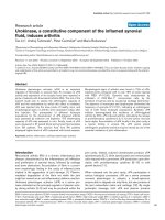
Figure 2
Dose response for in vivo treatment with apigenin for suppressing interferon-gamma (IFN-) response to nucleosomesDose response for in vivo treatment with apigenin for suppressing interferon-gamma (IFN-) response to nucleosomes. Three-month-old unmanipu-
lated SNF1 mice were treated daily with apigenin at 3 mg/kg (13.89 M), 6 mg/kg (27.8 M), and 20 mg/kg (0.93 mM). Treatment with 20 mg/kg
apigenin for 2 weeks markedly suppressed IFN- response to nuclesosomes ex vivo. Values are mean ± standard error of the mean. *P < 0.001, **P
< 0.01,
x
P < 0.02, and
+
P < 0.05. SNF1, (SWR × NZB)F1.
Arthritis Research & Therapy Vol 11 No 2 Kang et al.
Page 6 of 13
(page number not for citation purposes)
Another batch of 3-month-old pre-nephritic SNF1 mice (10
mice per group) were injected intraperitoneally daily with api-
genin (20 mg/kg) or DMSO-PBS vehicle as control. The con-
trol group started developing severe nephritis from 20 weeks
of age, as documented by persistent proteinurea of greater
than 100 mg/dL (Figure 4b, log-rank test, P = 0.00313) and a
renal pathology grade of 3 to 4+ (Figure 4c, P < 0.01). From
18 to 24 weeks of age, 40% of control group mice developed
severe nephritis, whereas apigenin-injected mice did not
develop overt renal disease. At 36 weeks of age, 100% of
control group mice had developed severe nephritis, whereas
only 40% of apigenin-injected group developed severe
nephritis.
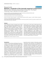
Figure 3
In vivo treatment with apigenin reduced nucleosome-specific Th1, Th17, and IgG autoantibody productionIn vivo treatment with apigenin reduced nucleosome-specific Th1, Th17, and IgG autoantibody production. In vivo treatment with apigenin (20 mg/
kg) for 2 months markedly reduced nucleosome-specific Th1 and Th17 responses and IgG autoantibody production ex vivo as compared with vehi-
cle-treated SNF1 mice. (a) Splenocytes from apigenin- or vehicle-treated SNF1 mice were stimulated with nucleosomes and analyzed for Th1(left
panel) and Th17 (right panel) responses by enzyme-linked immunosorbent spot assay. 'CD3' indicates results upon stimulation with optimal amount
of anti-CD3 antibody (0.2 g/mL). (b) IgG autoantibody levels of anti-dsDNA, anti-ssDNA, anti-nucleosome, and anti-histone in culture supernatants
of lupus Th cell-B cell-nucleosome cocultures were analyzed by enzyme-linked immunosorbent assay. *P < 0.001, **P < 0.01,
x
P < 0.02, and
+
P <
0.05. dsDNA, double-stranded DNA; IL-17, interleukin-17; SNF1, (SWR × NZB)F1; ssDNA, single-stranded DNA; Th, T helper.
Available online />Page 7 of 13
(page number not for citation purposes)
At 42 to 52 weeks of age, 20% of DMSO-PBS-treated mice
were dead, whereas 100% of apigenin-treated mice were
alive. However, survival curves of mice followed until death
cannot be shown as moribund mice with severe nephritis had
to be euthanized according to ACUC rules. There were no
gross signs of toxicity or apparent loss of weight in the api-
genin-treated mice as compared with age-matched normal
strains, such as SWR or C57B/L6 mice, consistent with other
studies [23]. Weight gain, apparently due to fluid retention and
Figure 4
Apigenin treatment in vivo suppresses IgG anti-nuclear autoantibodies and lupus nephritisApigenin treatment in vivo suppresses IgG anti-nuclear autoantibodies and lupus nephritis. (a) Treatment for 1 month and 2 months resulted in sig-
nificant reduction of IgG autoantibody levels in serum of SNF1 mice as compared with vehicle treatment. (b) Another group of mice was treated with
apigenin or vehicle and monitored for the incidence of severe nephritis. Apigenin treatment markedly delayed incidence of nephritis (log-rank test,
++
P = 0.00313). (c) With treatment regimens identical to those in (b), renal histopathologic features of lupus nephritis were evaluated. Apigenin
treatment significantly lowered the histopathology score of nephritis. (d) Representative histopathology figures of kidneys with treatment regimens
identical to those in (b); hematoxylin and eosin stain (× 200). (e) Total IgG levels in serum of apigenin- or vehicle-treated mice were measured by
enzyme-linked immunosorbent assay. *P < 0.001, **P < 0.01, and
x
P < 0.02. Ag, antigen; AutoAb, autoantibody; DMSO-PBS, dimethyl sulfoxide-
phosphate-buffered saline; dsDNA, double-stranded DNA; Nuc, nucleosome; SNF1, (SWR × NZB)F1; ssDNA, single-stranded DNA.
Arthritis Research & Therapy Vol 11 No 2 Kang et al.
Page 8 of 13
(page number not for citation purposes)
lethargy, was observed in mice of either group after they had
developed severe nephritis and proteinuria.
For assessment of renal pathologic features at the earliest
stages (before persistent proteinuria sets in), another group of
3-month-old mice was treated for 6 weeks. Kidney sections
from control and apigenin-treated mice were examined and
graded for typical lesions of lupus glomerulonephritis such as
glomerular enlargement, hypercellularity, crescent formation,
mesangial thickening, glomerulosclerosis, and interstitial infil-
tration with mononuclear cells [17,19-21]. Six weeks after api-
genin treatment, kidney sections from control mice had an
overall score of 3 ± 0.7 for nephritis, whereas the apigenin-
treated group showed 1.1 ± 0.4 as the overall score (Figures
4c and 4d, P < 0.001).
Antigen-presenting cells are more sensitive to apigenin than T
cells in suppression of nucleosome-specific interferon-gamma
and interleukin-17 responsesWe tested which cells are sensi-
tive to apigenin in suppression of autoantigen response. We
pulsed APCs and T cells isolated from splenocytes from 4- to
5-month-old SNF1 mice with apigenin or vehicle for 1 hour
and then cultured apigenin-treated APCs with vehicle-treated
T cells, and apigenin-treated T cells with vehicle-treated APCs
in the presence of various amounts of nucleosomes, and then
analyzed for IFN- and IL-17 ELISPOT responses. APCs were
more sensitive to apigenin than T cells. Apigenin-pulsed APCs
showed marked reduction of nucleosome-specific IFN-
response at 10 to 100 M, whereas apigenin-pulsed T cells
showed marked reduction in IFN- response at 30 to 100 M.
In the case of nucleosome-specific IL-17 response, both api-
genin-pulsed APCs and T cells showed marked reduction at
10 to 100 M, but apigenin-pulsed APCs showed more
reduction than T cells (Figures 5a and 5b, P < 0.02 to 0.001).
At a concentration of 10 M, apigenin pre-treated APCs
showed 87% reduction of autoimmune IFN- response as
compared with that of vehicle-treated APCs, whereas api-
genin pre-treated T cells showed only 6% reduction, and at
the same concentration, apigenin pre-treated APCs showed
92% reduction of autoimmune IL-17 responses, whereas api-
genin pre-treated T cells showed 75% reduction.
Apigenin treatment reduces the level of cyclooxygenase
2 in lupus CD4
+
T cells, B cells, dendritic cells, and
macrophages
Since SNF1 mouse T cells, activated B cells, DCs, and mac-
rophages express higher basal levels of COX-2 as compared
with those in non-autoimmune SWR or BALB/c strains and
hyperexpression of COX-2 contributes to lupus autoimmunity
[4,6], we tested whether apigenin could reduce hyperexpres-
sion of COX-2 in cells of autoimmune SNF1 mice. After 3
months of treatment with apigenin (20 mg/kg daily), COX-2
expression was markedly reduced in CD4
+
T cells, B cells,
DCs, and macrophages (but there were no differences in total
CD11b
+
cells or CD8
+
cells) (Figures 6a and 6c, P < 0.05 to
0.01). A high proportion of activated lupus cells (particularly
CD4 T cells and DCs) expressed COX-2 (Figure 6c), and it
appeared that apigenin caused depletion of these COX-2-
positive cells. However, apigenin treatment resulted in the
apparent removal of only the cells expressing high levels of
COX-2 (Figure 6b). CD4
+
T cells in apigenin-treated mice
were still expressing low levels of COX-2. Apigenin sup-
presses the expression of COX-2 at the transcriptional and
post-transcriptional levels [5,9]; thus, apigenin might have ren-
dered the activated lupus cells dull-positive for COX-2 stain-
ing as well.
Apigenin induces apoptosis of lupus immune cells
Apigenin is known to induce apoptosis of cancer cells [24,25],
and it potentiates AICD in normal human T cells that are recur-
rently activated [5], which would guard against autoreactivity.
We therefore examined the ability of apigenin to induce apop-
Figure 5
Effect of apigenin on nucleosome-induced Th1 and Th17 responses and antigen presentation function of antigen-presenting cells (APCs)Effect of apigenin on nucleosome-induced Th1 and Th17 responses and antigen presentation function of antigen-presenting cells (APCs). T cells
and APCs from 3-month-old unmanipulated SNF1 mice were pulsed with various amounts of apigenin or vehicle for 1 hour, and crisscross cocul-
tures were done in the presence of nucleososome (10 g/mL). Apigenin pre-exposure suppressed autoantigen-presenting ability of APCs and
resulted in inhibition of Th1 (a) and Th17 (b) responses more markedly than pre-exposure of the responding T cells to apigenin. *P < 0.001, **P <
0.01, and
x
P < 0.02. Api, apigenin; IFN-, interferon-gamma; IL-17, interleukin-17; SNF1, (SWR × NZB)F1; Th, T helper.
Available online />Page 9 of 13
(page number not for citation purposes)
tosis of lupus immune cells, which are spontaneously activated
in vivo from ongoing autoimmune response. Treatment with
apigenin in vitro at 30 M induced significant apoptosis of T
cells, B cells, DCs, and macrophages of SNF1 mice after 24
hours of incubation as compared with cultures with vehicle
(Figure 6d, *P < 0.001, **P < 0.01). At a concentration of 30
M, apigenin induced twofold more apoptosis in DCs and
macrophages than in T and B cells. At a concentration of 10
M, apigenin did not induce significant apoptosis of T cells,
but B cells, DCs, and macrophages were affected.
Apigenin suppressed interleukin-6 production induced
through Toll-like receptor-7 and -9 pathways
IL-6 produced by APCs is important for generating Th17 cells
[26], and apigenin suppressed Th17 responses in SNF1 mice
(Figure 3a right panel and Figure 5b). Moreover, DNA and
RNA in the major lupus autoantigens, nucleosomes and ribo-
nucleoprotein (RNP), can act as TLR-9 and TLR-7 ligands,
respectively [27]. We therefore tested whether apigenin could
suppress IL-6 production stimulated by nucleosomes, CpG
(TLR-9 ligand), and R837 (TLR-7 ligand) in SNF1 mice. Api-
genin at a concentration of 30 M suppressed IL-6 production
induced by nucleosome, CpG, and R837 completely (Figure
7, P < 0.01 to 0.001), but significant inhibition was seen even
at 1 M (for response to CpG) and 3 M (for nucleosome).
Apigenin at concentrations of 30 to 100 M also suppressed
IFN- production by DCs stimulated with 2.5 g/mL CpG (P
< 0.001), but not at concentrations of 1 to 10 M (data not
shown).
Apigenin did not increase suppressive function of
CD4
+
CD25
+
regulatory T cells
Since IL-6 inhibits regulatory T (T
reg
) cells while promoting
Th17 cell expansion and we observed that apigenin sup-
pressed IL-6 production by APCs, we analyzed whether api-
genin could increase CD4
+
CD25
+
T
reg
cell activity. After 2
months of treatment, CD4
+
CD25
+
T cells from apigenin- or
vehicle-treated SNF1 mice were cocultured with splenocytes
from 4.5-month-old unmanipulated SNF1 mice in an autoanti-
gen-specific suppression assay described previously [18,28].
As compared with CD4
+
CD25
+
T
reg
cells from vehicle-treated
SNF1 mice, apigenin treatment did not increase the suppres-
sive function of CD4
+
CD25
+
T cells on nucleosome-specific
Th1 and Th17 responses (P < 0.05, data not shown).
Discussion
Using SNF1 mice that spontaneously develop human lupus-
like disease, we show that apigenin treatment in vitro and in
vivo markedly inhibited autoimmune responses of Th1 and
Th17 cells that are spontaneously primed to nucleosomes, the
major nuclear autoantigen in lupus. Both IFN--producing Th1
cells and IL-17-producing Th17 cells are critical for help in the
production of pathogenic autoantibodies [17,22,29,30] and
development of lupus nephritis [18,31-34]. Moreover, the
spontaneously pre-primed, autoimmune Th17 cells in SNF1
mice with lupus-like disease can expand when challenged with
nucleosomes ex vivo without requiring any polarizing cytokine
conditions or PMA (phorbol myristate acetate)-ionomycin
additions that are used widely to detect such pathogenic Th
cells in other systems [18]. Apigenin suppressed production
of the Th17-inducing cytokine, IL-6, by APCs stimulated by
nucleosomes, CpG (TLR-9 ligand), and R837 (TLR-7 ligand).
This is relevant because DNA and RNA in the major lupus
autoantigens, nucleosomes and RNP, can stimulate APCs via
TLR-9 and TLR-7 pathways, respectively [27]. Consequent to
the inhibition of lupus Th cells, apigenin treatment suppressed
the production of IgG class-switched pathogenic autoantibod-
ies to nuclear antigens and significantly delayed the develop-
ment of severe glomerulonephritis (Figures 1, 2, 3 and 4).
However, autoantigen-presenting function of APCs appeared
to be more sensitive to the inhibitory effect of apigenin,
although apigenin has been shown to inhibit NF-B activation
pathways in both T cells [5] and macrophages [35,36]. Mac-
rophages and myeloid DCs are important for ongoing presen-
tation of nucleosome-derived epitopes to autoreactive T cells
in mice with established lupus [37,38], and hyperactive APCs
are a characteristic feature of lupus, playing a critical role in ini-
tiation and pathogenesis [39-43]. By inhibiting NF-kB activa-
tion, not only does apigenin inhibit the autoantigen-presenting
and stimulatory functions of the APCs necessary for activation
and expansion of autoreactive Th and B cells, but it causes
apoptosis of the hyperactive lupus APCs (this study), probably
by inhibiting NF-kB-regulated anti-apoptotic molecules, espe-
cially COX-2 and c-FLIP [5,6]. However, the functional inhibi-
tory effect of apigenin in vitro could be seen in concentrations
of as low as 0.3 to 3 mM (Figures 1a and 7), which were well
below the concentrations (10 to 30 mM) required for inducing
significant apoptosis (Figure 6c).
Despite the fact that apigenin is widely distributed in fruits and
herbs, diet is insufficient for bioavailable therapeutic levels of
apigenin due to first-pass metabolism (glucuronidation) in gut
and liver, although some systemic effects of diets rich in api-
genin are detectable [44]. Bioavailability has been improved in
the case of other drugs by the pharmaceutical industry, and
similar attempts are being applied to related flavone com-
pounds [45]. Thus, apigenin, a non-mutagenic plant flavone, is
a strong inhibitor of NF-B activation and COX-2 expression
in activated autoimmune cells, but it also has properties that
might reduce the risk of coronary disease, as mentioned
above. Obviously, relatively benign COX-2 and NF-B inhibi-
tors such as apigenin and other herbal products [46] might be
of value in lupus therapy.
Conclusions
Apigenin inhibits autoantigen-presenting and stimulatory func-
tions of the APCs necessary for activation and expansion of
autoreactive Th1 and Th17 cells and B cells in lupus. Apigenin
also causes apoptosis of the hyperactive lupus APCs, T cells,
Arthritis Research & Therapy Vol 11 No 2 Kang et al.
Page 10 of 13
(page number not for citation purposes)
Figure 6
Effect of apigenin on cyclooxygenase 2 (COX-2) expression and apoptosisEffect of apigenin on cyclooxygenase 2 (COX-2) expression and apoptosis. Intracellular COX-2 expression followed treatment with apigenin or vehi-
cle for 3 months. (a) COX-2 expression in representative histograms of spleen cell subsets. (b) Representative dot plot of gated CD4 T cells (per-
centage shown in right upper quadrant). (c) Compiled results from three experiments. Treatment with apigenin markedly suppressed COX-2
expression in gated CD4
+
T cells, B cells, dendritic cells (DCs), and macrophages, but there was no difference in total CD11b
+
cells or CD8
+
T
cells. (d) In vitro treatment with apigenin induced apoptosis of lupus T cells, B cells, DCs, and macrophages from SNF1 mice after 24-hour incuba-
tion. Culture with 30 M apigenin resulted in a twofold increase in percentage of specific apoptosis in DCs and macrophages than in T and B cells.
Apoptotic cells were analyzed in gated cell subsets to calculate percentage of specific apoptosis, as described in Materials and methods (n = 5 per
stain). *P < 0.001, **P < 0.01,
x
P < 0.02, and
+
P < 0.05 for (c) and (d). SNF1, (SWR × NZB)F1.
Available online />Page 11 of 13
(page number not for citation purposes)
and B cells, probably by inhibiting expression of NF-B-regu-
lated anti-apoptotic molecules, especially COX-2 and c-FLIP,
which are persistently hyperexpressed by the lupus immune
cells. Although apigenin, a non-mutagenic plant flavone, inhib-
its COX-2 expression in activated autoimmune cells, it also
has properties that might reduce the risk of coronary disease
in contrast to conventional COX-2 inhibitors. Increasing the
bioavailability of simple dietary plant-derived COX-2 and NF-
B inhibitors, such as apigenin, might be of value in lupus ther-
apy as well as for suppressing inflammation in other Th17-
mediated inflammatory diseases like rheumatoid arthritis,
Crohn disease, and psoriasis and in prevention of inflamma-
tion-based tumors overexpressing COX-2 (colon, breast).
Competing interests
The authors declare that they have no competing interests.
Authors' contributions
H-KK participated in study design, apigenin therapy, cellular
immunologic assays, acquisition of data, statistical analysis,
and drafting of the manuscript. DE measured levels of autoan-
tibodies and assisted in apigenin therapy injections, cellular
immunologic assays, and data acquisition. ML assisted in cel-
lular immunologic assays. SKD conceived of the study and
participated in its design and coordination, data analysis, and
manuscript preparation. All authors read and approved the
final manuscript.
Acknowledgements
This work was supported by grants from the National Institutes of Health
(R37-AR39157 and RO1-AI41985) and Solovy Arthritis Research
Society (SKD).
References
1. Rahman A, Isenberg DA: Systemic lupus erythematosus. N
Engl J Med 2008, 358:929-939.
2. Kotzin BL: Systemic lupus erythematosus. Cell 1996,
85:303-306.
3. Refaeli Y, Van Parijs L, London CA, Tschopp J, Abbas AK: Bio-
chemical mechanisms of IL-2-regulated Fas-mediated T cell
apoptosis. Immunity 1998, 8:615-623.
Figure 7
Apigenin markedly decreased interleukin-6 (IL-6) production by lupus dendritic cells (DCs)Apigenin markedly decreased interleukin-6 (IL-6) production by lupus dendritic cells (DCs). DCs from unmanipulated 3-month-old SNF1 mice were
stimulated with nucleosomes (30 g/mL), CpG (2.5 g/mL), and R837 (1 g/mL) in the presence of various amounts of apigenin. IL-6 in culture
supernatant was measured by enzyme-linked immunosorbent assay. *P < 0.001, **P < 0.01, and
+
P < 0.05. Nuc, nucleosome; SNF1, (SWR ×
NZB)F1.
Arthritis Research & Therapy Vol 11 No 2 Kang et al.
Page 12 of 13
(page number not for citation purposes)
4. Xu L, Zhang L, Yi Y, Kang HK, Datta SK: Human lupus T cells
resist inactivation and escape death by upregulating COX-2.
Nat Med 2004, 10:411-415.
5. Xu L, Zhang L, Bertucci AM, Pope RM, Datta SK: Apigenin, a die-
tary flavonoid, sensitizes human T cells for activation-induced
cell death by inhibiting PKB/Akt and NF-kappaB activation
pathway. Immunol Lett 2008, 121:74-83.
6. Zhang L, Bertucci AM, Smith KA, Xu L, Datta SK: Hyperexpres-
sion of cyclooxygenase 2 in the lupus immune system and
effect of cyclooxygenase 2 inhibitor diet therapy in a murine
model of systemic lupus erythematosus. Arthritis Rheum
2007, 56:4132-4141.
7. Yang P, Zhang Y, Ping L, Gao XM: Apoptosis of murine lupus T
cells induced by the selective cyclooxygenase-2 inhibitor
celecoxib: molecular mechanisms and therapeutic potential.
Int Immunopharmacol 2007, 7:1414-1421.
8. Wallace DJ: Celecoxib for lupus. Arthritis Rheum 2008,
58:2923.
9. Tong X, Van Dross RT, Abu-Yousif A, Morrison AR, Pelling JC: Api-
genin prevents UVB-induced cyclooxygenase 2 expression:
coupled mRNA stabilization and translational inhibition. Mol
Cell Biol 2007, 27:283-296.
10. Woodman OL, Chan E: Vascular and anti-oxidant actions of fla-
vonols and flavones. Clin Exp Pharmacol Physiol 2004,
31:786-790.
11. Olszanecki R, Gebska A, Kozlovski VI, Gryglewski RJ: Flavonoids
and nitric oxide synthase. J Physiol Pharmacol 2002,
53:571-584.
12. Guerrero JA, Lozano ML, Castillo J, Benavente-Garcia O, Vicente
V, Rivera J: Flavonoids inhibit platelet function through binding
to the thromboxane A2 receptor. J Thromb Haemost 2005,
3:369-376.
13. Zhang YH, Park YS, Kim TJ, Fang LH, Ahn HY, Hong JT, Kim Y, Lee
CK, Yun YP: Endothelium-dependent vasorelaxant and anti-
proliferative effects of apigenin. Gen Pharmacol 2000,
35:341-347.
14. Datta SK, Schwartz RS: Genetics of expression of xenotropic
virus and autoimmunity in NZB mice. Nature 1976,
263:412-415.
15. Datta SK, McConahey PJ, Manny N, Theofilopoulos AN, Dixon FJ,
Schwartz RS: Genetic studies of autoimmunity and retrovirus
expression in crosses of New Zealand black mice. II. The viral
envelope glycoprotein gp70.
J Exp Med 1978, 147:872-881.
16. Mohan C, Adams S, Stanik V, Datta SK: Nucleosome: a major
immunogen for pathogenic autoantibody-inducing T cells of
lupus. J Exp Med 1993, 177:1367-1381.
17. Kaliyaperumal A, Michaels MA, Datta SK: Naturally processed
chromatin peptides reveal a major autoepitope that primes
pathogenic T and B cells of lupus. J Immunol 2002,
168:2530-2537.
18. Kang HK, Liu M, Datta SK: Low-dose peptide tolerance therapy
of lupus generates plasmacytoid dendritic cells that cause
expansion of autoantigen-specific regulatory T cells and con-
traction of inflammatory Th17 cells. J Immunol 2007,
178:7849-7858.
19. Kaliyaperumal A, Michaels MA, Datta SK: Antigen-specific ther-
apy of murine lupus nephritis using nucleosomal peptides:
tolerance spreading impairs pathogenic function of autoim-
mune T and B cells. J Immunol 1999, 162:5775-5783.
20. Singh RR, Saxena V, Zang S, Li L, Finkelman FD, Witte DP, Jacob
CO: Differential contribution of IL-4 and STAT6 vs STAT4 to
the development of lupus nephritis. J Immunol 2003,
170:4818-4825.
21. Schiffer L, Sinha J, Wang X, Huang W, von Gersdorff G, Schiffer
M, Madaio MP, Davidson A: Short term administration of cos-
timulatory blockade and cyclophosphamide induces remis-
sion of systemic lupus erythematosus nephritis in NZB/W F1
mice by a mechanism downstream of renal immune complex
deposition. J Immunol 2003, 171:489-497.
22. Kaliyaperumal A, Mohan C, Wu W, Datta SK: Nucleosomal pep-
tide epitopes for nephritis-inducing T helper cells of murine
lupus. J Exp Med 1996, 183:2459-2469.
23. Singh JP, Selvendiran K, Banu SM, Padmavathi R, Sakthisekaran
D: Protective role of Apigenin on the status of lipid peroxida-
tion and antioxidant defense against hepatocarcinogenesis in
Wistar albino rats. Phytomedicine 2004, 11:309-314.
24. Wang IK, Lin-Shiau SY, Lin JK: Induction of apoptosis by api-
genin and related flavonoids through cytochrome c release
and activation of caspase-9 and caspase-3 in leukaemia HL-
60 cells. Eur J Cancer 1999, 35:1517-1525.
25. Vargo MA, Voss OH, Poustka F, Cardounel AJ, Grotewold E, Dos-
eff AI: Apigenin-induced-apoptosis is mediated by the activa-
tion of PKCdelta and caspases in leukemia cells.
Biochem
Pharmacol 2006, 72:681-692.
26. Bettelli E, Carrier Y, Gao W, Korn T, Strom TB, Oukka M, Weiner
HL, Kuchroo VK: Reciprocal developmental pathways for the
generation of pathogenic effector TH17 and regulatory T cells.
Nature 2006, 441:235-238.
27. Marshak-Rothstein A, Rifkin IR: Immunologically active autoanti-
gens: the role of toll-like receptors in the development of
chronic inflammatory disease. Annu Rev Immunol 2007,
25:419-441.
28. Kang HK, Michaels MA, Berner BR, Datta SK: Very low-dose tol-
erance with nucleosomal peptides controls lupus and induces
potent regulatory T cell subsets. J Immunol 2005,
174:3247-3255.
29. Lu L, Kaliyaperumal A, Boumpas DT, Datta SK: Major peptide
autoepitopes for nucleosome-specific T cells of human lupus.
J Clin Invest 1999, 104:345-355.
30. Hsu HC, Yang P, Wang J, Wu Q, Myers R, Chen J, Yi J, Guentert
T, Tousson A, Stanus AL, Le TV, Lorenz RG, Xu H, Kolls JK, Carter
RH, Chaplin DD, Williams RW, Mountz JD: Interleukin 17-pro-
ducing T helper cells and interleukin 17 orchestrate autoreac-
tive germinal center development in autoimmune BXD2 mice.
Nat Immunol 2008, 9:166-175.
31. Haas C, Ryffel B, Le Hir M: IFN-gamma receptor deletion pre-
vents autoantibody production and glomerulonephritis in
lupus-prone (NZB × NZW)F1 mice. J Immunol 1998,
160:3713-3718.
32. Balomenos D, Rumold R, Theofilopoulos AN: Interferon-gamma
is required for lupus-like disease and lymphoaccumulation in
MRL-lpr mice. J Clin Invest 1998, 101:364-371.
33. Wong CK, Lit LC, Tam LS, Li EK, Wong PT, Lam CW: Hyperpro-
duction of IL-23 and IL-17 in patients with systemic lupus ery-
thematosus: implications for Th17-mediated inflammation in
auto-immunity. Clin Immunol 2008, 127:385-393.
34. Crispin JC, Oukka M, Bayliss G, Cohen RA, Van Beek CA, Stillman
IE, Kyttaris VC, Juang YT, Tsokos GC: Expanded double nega-
tive T cells in patients with systemic lupus erythematosus pro-
duce IL-17 and infiltrate the kidneys. J Immunol 2008,
181:8761-8766.
35. Nicholas C, Batra S, Vargo MA, Voss OH, Gavrilin MA, Wewers
MD, Guttridge DC, Grotewold E, Doseff AI: Apigenin blocks
lipopolysaccharide-induced lethality in vivo and proinflamma-
tory cytokines expression by inactivating NF-kappaB through
the suppression of p65 phosphorylation. J Immunol 2007,
179:7121-7127.
36. Liang YC, Huang YT, Tsai SH, Lin-Shiau SY, Chen CF, Lin JK:
Suppression of inducible cyclooxygenase and inducible nitric
oxide synthase by apigenin and related flavonoids in mouse
macrophages. Carcinogenesis 1999, 20:1945-1952.
37. Okamoto A, Fujio K, van Rooijen N, Tsuno NH, Takahashi K, Tsurui
H, Hirose S, Elkon KB, Yamamoto K: Splenic phagocytes pro-
mote responses to nucleosomes in (NZB × NZW) F1 mice. J
Immunol 2008, 181:5264-5271.
38. Ronnefarth VM, Erbacher AI, Lamkemeyer T, Madlung J, Nordheim
A, Rammensee HG, Decker P: TLR2/TLR4-independent neu-
trophil activation and recruitment upon endocytosis of
nucleosomes reveals a new pathway of innate immunity in
systemic lupus erythematosus. J Immunol 2006,
177:7740-7749.
39. Blanco P, Palucka AK, Gill M, Pascual V, Banchereau J: Induction
of dendritic cell differentiation by IFN-alpha in systemic lupus
erythematosus. Science 2001, 294:1540-1543.
40. Kalled SL, Cutler AH, Burkly LC: Apoptosis and altered dendritic
cell homeostasis in lupus nephritis are limited by anti-CD154
treatment. J Immunol 2001, 167:1740-1747.
41. Zhu J, Liu X, Xie C, Yan M, Yu Y, Sobel ES, Wakeland EK, Mohan
C: T cell hyperactivity in lupus as a consequence of hyperstim-
ulatory antigen-presenting cells. J Clin Invest 2005,
115:1869-1878.
Available online />Page 13 of 13
(page number not for citation purposes)
42. Wan S, Xia C, Morel L: IL-6 produced by dendritic cells from
lupus-prone mice inhibits CD4
+
CD25
+
T cell regulatory
functions. J Immunol 2007, 178:271-279.
43. Holdsworth SR, Tipping PG: Leukocytes in glomerular injury.
Semin Immunopathol 2007, 29:355-374.
44. Nielsen SE, Young JF, Daneshvar B, Lauridsen ST, Knuthsen P,
Sandstrom B, Dragsted LO: Effect of parsley (Petroselinum
crispum) intake on urinary apigenin excretion, blood antioxi-
dant enzymes and biomarkers for oxidative stress in human
subjects. Br J Nutr 1999, 81:447-455.
45. Lahey TP, Rajadhyasksha VJ: Inhibition by 3-deoxyflavonoids of
T-lymphocyte activation and therapies related thereto. US pat-
ent 6,774,142 2004.
46. Tao X, Fan F, Hoffmann V, Gao CY, Longo NS, Zerfas P, Lipsky
PE: Effective therapy for nephritis in (NZB × NZW)F1 mice with
triptolide and tripdiolide, the principal active components of
the Chinese herbal remedy Tripterygium wilfordii Hook F.
Arthritis Rheum 2008, 58:1774-1783.