Báo cáo y học: "Different properties of ACPA and IgM-RF derived from a large dataset: further evidence of two distinct autoantibody systems" potx
Bạn đang xem bản rút gọn của tài liệu. Xem và tải ngay bản đầy đủ của tài liệu tại đây (233.73 KB, 6 trang )
Open Access
Available online />Page 1 of 6
(page number not for citation purposes)
Vol 11 No 3
Research article
Different properties of ACPA and IgM-RF derived from a large
dataset: further evidence of two distinct autoantibody systems
Jennie Ursum
1
, Wouter H Bos
1
, Rob J van de Stadt
1
, Ben AC Dijkmans
2
and Dirkjan van
Schaardenburg
1,2
1
Jan van Breemen Institute, Dr. Jan van Breemenstraat 2, 1056 AB Amsterdam, The Netherlands
2
VU University Medical Centre, Postbus 7057, 1007 MB Amsterdam, The Netherlands
Corresponding author: Dirkjan van Schaardenburg,
Received: 5 Jan 2009 Revisions requested: 11 Feb 2009 Revisions received: 24 Feb 2009 Accepted: 21 May 2009 Published: 21 May 2009
Arthritis Research & Therapy 2009, 11:R75 (doi:10.1186/ar2704)
This article is online at: />© 2009 Ursum et al.; licensee BioMed Central Ltd.
This is an open access article distributed under the terms of the Creative Commons Attribution License ( />),
which permits unrestricted use, distribution, and reproduction in any medium, provided the original work is properly cited.
Abstract
Introduction The aim of this study was to examine
seroconversion and the relationship with age and inflammation
of autoantibodies in a large group of patients attending an
outpatient rheumatology clinic.
Methods Levels of antibodies to citrullinated proteins/peptides
(ACPAs) and IgM rheumatoid factor (IgM-RF) were determined
in 22,427 samples collected from 18,658 patients. The
diagnosis was derived from a diagnosis registration system. The
degree of seroconversion in repeated samples and the
correlation of levels with age and inflammatory markers were
determined for ACPA and IgM-RF in rheumatoid arthritis (RA)
and non-RA patients.
Results Seventy-one percent of RA patients (n = 1,524) were
ACPA-positive and 53% were IgM-RF-positive; in non-RA
patients (n = 2,245), the corresponding values were 2% and
4%, respectively. In patients with at least two samples (n =
3,769), ACPA status was more stable than IgM-RF status in RA
patients. ACPA- or IgM-RF-negative non-RA patients seldom
became positive. ACPA positivity was unrelated to age in both
RA and non-RA patients. IgM-RF positivity was unrelated to age
in RA patients; however, it increased with age in non-RA
patients. The correlation between autoantibody levels and
inflammatory markers was low in general and was somewhat
higher for IgM-RF than for ACPA.
Conclusions ACPA status is more stable in time and with
increasing age than IgM-RF status, further establishing its role
as a disease-specific marker. ACPA and IgM-RF levels are only
moderately correlated with markers of inflammation.
Introduction
One of the frequent characteristics of rheumatoid arthritis (RA)
is the presence of antibodies to citrullinated proteins/peptides
(ACPAs) and/or IgM rheumatoid factor (IgM-RF) [1]. IgM-RF
targets the Fc fragment of IgG and is observed in about 60%
to 65% of RA patients, but it is also frequently observed in
other inflammatory diseases [2,3]. ACPAs comprise a group
of antibodies that are highly specific for RA: among those are
antibodies against cyclic citrullinated peptide (CCP) [4].
ACPAs target citrullinated proteins and are observed in
around 70% of RA patients. In contrast to IgM-RF, ACPA is
highly specific for RA (specificity 80% versus 96%, respec-
tively) [3].
Besides their well-established superior specificity for RA, sev-
eral other properties of ACPA are distinct from IgM-RF. About
50% to 70% of early-RA patients are ACPA-positive, and this
phenotype remains fairly stable thereafter [2,5,6], even during
treatment with tumour necrosis factor (TNF)-blocking agents
[7]. On the other hand, IgM-RF levels decrease during
antirheumatic treatment [8] and 17% of IgM-RF-positive RA
patients turned negative after 6 months of anti-TNF treatment
[9].
Furthermore, IgM-RF [10], but not ACPA [11], is sometimes
present in healthy older persons, suggesting that RF can be a
consequence of nonspecific immune activation. Moreover, it
ACPA: antibody to citrullinated proteins/peptides; AU: arbitrary units; CCP: cyclic citrullinated peptide; CRP: C-reactive protein; ELISA: enzyme-
linked immunosorbent assay; ESR: erythrocyte sedimentation rate; IgM-RF: IgM rheumatoid factor; IQR: interquartile range; IU: international units;
RA: rheumatoid arthritis; RF: rheumatoid factor; TNF: tumour necrosis factor.
Arthritis Research & Therapy Vol 11 No 3 Ursum et al.
Page 2 of 6
(page number not for citation purposes)
has been suggested that IgM-RF production also is a conse-
quence of the rheumatoid inflammation whereas ACPA may
have pathophysiological properties. Evidence supporting this
concept is emerging [12]. For instance, ACPA precedes IgM-
RF in the preclinical phase [13] and the change in IgM-RF lev-
els during anti-TNF treatment is associated with the change in
acute-phase response; this is not observed for ACPA [9].
These data suggest that ACPA and IgM-RF represent two dif-
ferent autoantibody systems. ACPAs are disease-specific,
their presence is fairly stable in time and does not increase
with age, and ACPA levels are not correlated with the acute-
phase response. On the other hand, IgM-RF is less disease-
specific, its presence increases with age in healthy/non-RA
individuals, and its levels are correlated with the acute-phase
response.
Most of these data have emerged from studies of selected
populations with small sample sizes. In the present study, we
sought to confirm the stability of ACPA in time, the increased
IgM-RF frequency with age, and the correlation of IgM-RF with
the acute-phase response using a repository of over 22,000
serum samples collected from over 18,000 patients attending
a rheumatology clinic network in The Netherlands.
Materials and methods
ACPA and IgM-RF levels were determined in 22,427 samples,
which were collected from 18,658 patients between August
2003 and August 2007. These patients attended one of the
outpatient rheumatology clinics of the Jan van Breemen Insti-
tute in the Amsterdam region of The Netherlands. Each
patient's final diagnosis was obtained from the International
Classification of Diseases version 10 diagnosis registration
system, which reflects the opinion of the treating rheumatolo-
gist. The diagnosis was categorized into five groups according
to the following codes: RA, polyarthritis or oligoarthritis,
spondylarthropathy (including ankylosing spondylitis, reactive
arthritis, psoriatic arthritis, arthritis associated with inflamma-
tory bowel disease, and undifferentiated spondyloarthropa-
thy), osteoarthritis, and other (including arthralgia,
fibromyalgia, and no final diagnosis). The latter four groups
were also combined and classified as 'non-RA'. The disease
duration at the time of autoantibody testing was variable and
unknown. For the association between age and autoantibody
positivity, patients were grouped according to their age at the
first available sample: younger than 30, 30 to 39, 40 to 49, 50
to 59, 60 to 69, 70 to 79, and 80 years old or older. The local
ethics committee approved the study protocol and waived the
need for informed patient consent.
Laboratory investigations
All measurements were routinely performed at the certified
clinical laboratory of the Jan van Breemen Institute. After the
first sample, sequential samples were obtained as a part of
routine or protocollar care. In the case of routine care, samples
were obtained at the request of the rheumatologist at a non-
specific time point. In the case of protocollar care, samples
were obtained annually. ACPA levels were determined by sec-
ond-generation anti-CCP enzyme-linked immunosorbent
assay (ELISA) (Axis-Shield, Dundee, UK). Sera reaching
1,000 arbitrary units (AU) were not further diluted. The cutoff
level for ACPA positivity was set at 5 AU/mL in accordance
with the instructions of the manufacturer. IgM-RF levels were
determined by an in-house ELISA. The cutoff level for IgM-RF
positivity was set at 30 international units (IU)/mL, which was
determined on the basis of receiver operating characteristic
curves described previously [14]. Erythrocyte sedimentation
rate (ESR) was measured according to the Westergren
method using a Starrsed analyser (Mechatronics, Zwaag, The
Netherlands), and the reference value was less than 15 mm
per first hour. C-reactive protein (CRP) was measured on a
Cobas 6000 analyser (Roche, Woerden, The Netherlands) in
accordance with the instructions of the manufacturer, and the
reference value was less than 10 mg/L.
Analysis
The effect of age on ACPA and IgM-RF positivity was deter-
mined using the chi-square test. Correlations between levels
of antibodies and levels of markers of inflammation were deter-
mined with Spearman correlation. All analyses were performed
using SPSS 16.0 software (SPSS Inc., Chicago, IL, USA).
Results
Serum samples were available in 18,658 patients: 3,116
patients with RA, 1,063 with polyarthritis or oligoarthritis, 818
with spondylarthropathy, 2,736 with osteoarthritis, and
10,925 classified as other. A second sample was available in
1,524 patients with RA, 419 with polyarthritis or oligoarthritis,
195 with spondylarthropathy, 333 with osteoarthritis, and
1,298 classified as other. Of all second samples, 35% were
obtained annually as part of a protocol, and the others were
obtained according to physician request. These 35% can be
divided into patients receiving routine care (24%), described
elsewhere [15], and those receiving anti-TNF treatment
(11%). In the first sample of the 18,658 patients, the percent-
ages of patients with positive ACPA were 71% in the RA
group and 2% in the non-RA group. Rates of IgM-RF positivity
were 53% in the RA group and 4% in the non-RA group. In the
second sample (n = 3,769), the percentages of patients with
positive ACPA were 70% in the RA group and 7% in the non-
RA group. Rates of IgM-RF positivity were 49% in the RA
group and 10% in the non-RA group.
Switch in antibody to citrullinated proteins/peptides or
IgM rheumatoid factor status between first and second
samples
In patients with at least two samples, the stability of the
autoantibody status was assessed (n = 3,769). The median
times between the first and second samples were similar for
RA patients (n = 1,524) and non-RA patients (n = 2,245): 11
months (interquartile range [IQR] 4 to 13 months) and 9
Available online />Page 3 of 6
(page number not for citation purposes)
months (IQR 3 to 16 months), respectively. In RA patients, the
percentages of patients switching from ACPA positivity to
negativity and from ACPA negativity to positivity were lower
compared with percentage changes in IgM-RF. In initially
ACPA-positive RA patients, 1% of the second sample was
negative, whereas 13% of the second sample in IgM-RF-pos-
itive RA was negative (P < 0.001). In initially ACPA-negative
RA patients, 4% of the second sample was ACPA-positive,
whereas 8% of the initially IgM-RF-negative RA patients
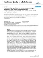
became positive (P < 0.001). Furthermore, autoantibody-pos-
itive non-RA patients frequently became negative in the sec-
ond sample (9% for ACPA and 17% for IgM-RF, respectively),
whereas autoantibody-negative non-RA patients seldom
became positive (Figure 1). When levels in RA patients were
the focus, initially ACPA-positive patients showed a nonsignif-
icant median increase from 74 AU/mL (IQR 25 to 252) to 80
AU/mL (IQR 24 to 229), whereas in initially IgM-RF-positive
RA patients, median IgM-RF levels decreased (P < 0.001)
from 94 IU/mL (IQR 51 to 188) to 81 IU/mL (IQR 41 to 178).
Autoantibody positivity in relation to age
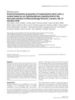
To explore an effect of age on ACPA and IgM-RF positivity, we
grouped patients according to their age at the first sample. In
RA, ACPA positivity was more frequent than IgM-RF positivity
in all age groups. In non-RA, however, the frequency of ACPA
positivity was lower than IgM-RF positivity in all age groups
except in patients younger than 30 years old. ACPA positivity
was similar among the different age groups in RA (Figure 2) as
well as non-RA (Figure 3), whereas IgM-RF positivity
increased with age in the non-RA group but not in the RA
group. The chi-square test for linear trend revealed a signifi-
cant (P < 0.001) linear trend; IgM-RF positivity increased with
age in non-RA patients.
Autoantibody levels in relation to markers of
inflammation
In RA patients, a low correlation between autoantibodies and
the levels of markers of inflammation (as measured by ESR
and CRP) was found. The correlation between IgM-RF levels
and the levels of markers of inflammation (ESR: r = 0.23; CRP:
r = 0.21, both P < 0.01) was somewhat stronger compared
with the correlation of ACPA levels and the levels of markers
of inflammation (ESR: r = 0.14; CRP: r = 0.14, both P < 0.01)
(Table 1). In a second analysis, the correlation of the changes
of levels in time of both antibodies and markers of inflammation
– that is, the correlation between (a) the difference between
the first and second ACPA/RF levels and (b) the difference
between the first and second ESR/CRP levels – was meas-
ured. The correlation between change in ACPA levels and
change of levels in markers of inflammation (ESR: r = 0.16;
CRP: r = 0.13, both P < 0.01) was similar to the correlation at
a single time point. For IgM-RF, the correlation of change of
levels in time with change of levels in markers of inflammation
(ESR: r = 0.31; CRP: r = 0.28, both P < 0.01) was slightly
higher compared with the correlation at a single time point. In
non-RA patients, no correlation between autoantibody levels
and the levels of markers of inflammation was found at a single
time point or between changes in ACPA or IgM-RF and mark-
ers of inflammation.
Discussion
Characteristics of ACPA and IgM-RF were studied in a large
group of RA and non-RA patients. ACPA status was more sta-
ble than IgM-RF status in RA and non-RA patients. ACPA pos-
itivity did not increase with age in any group, whereas IgM-RF
positivity was stable with age in RA but more frequent in older
versus younger non-RA patients. The correlation between
autoantibody levels and markers of inflammation was low in RA
and absent in non-RA patients.
The results of this study show a low percentage of ACPA sero-
conversion in both directions compared with IgM-RF. In very
early RA, the seroconversion to positivity might occur more fre-
quently [5]. Previous studies of prolonged follow-up of early-
arthritis patients seem to show that qualitative changes in
ACPA are rare [2,6], although the numbers of patients (n = 96
and 279) were relatively small. In RA, ACPA seroconversion
data are available from treatment cohorts, mostly with a follow-
up period of less than a year. Most, but not all, studies [16-18]
reported a modest decrease in ACPA levels; however, down-
ward seroconversion does not seem to occur. Data on IgM-RF
seroconversion in early RA are scarce. In very early RA, a
decrease in the percentage of patients positive for IgM-RF
was reported [5], and in a study of early RA, 11% of the
patients had variable IgM-RF status during 6-year follow-up
[19]. In RA, IgM-RF downward seroconversion during anti-
Figure 1
Percentage of rheumatoid arthritis (RA) and non-RA patients with a change in positivity in antibodies to citrullinated proteins/peptides (ACPA) and IgM rheumatoid factor (IgM-RF) between the first and sec-ond samplesPercentage of rheumatoid arthritis (RA) and non-RA patients with a
change in positivity in antibodies to citrullinated proteins/peptides
(ACPA) and IgM rheumatoid factor (IgM-RF) between the first and sec-
ond samples.
Arthritis Research & Therapy Vol 11 No 3 Ursum et al.
Page 4 of 6
(page number not for citation purposes)
TNF treatment has been reported in up to 50% of patients
[7,9,20].
With regard to age and autoantibody status, ACPA positivity
was stable in both RA and non-RA, whereas IgM-RF positivity
increased with age in non-RA, but not in RA. A formal compar-
ison cannot be made, but these results seem to be in line with
earlier observations made in healthy older persons. IgM-RF
positivity increases with age; in one study, up to 25% of per-
sons more than 85 years old were IgM-RF-positive [10]. In a
similar study, ACPA positivity was observed in only 1 out of
300 healthy individuals over 75 years of age [11]. The fact that
IgM-RF positivity increases with age in non-RA but not in RA
supports the notion that low-affinity RFs associated with infec-
tion and older age appear to play an important role in the host
response to many infectious organisms and are likely to con-
Figure 2
Percentage of rheumatoid arthritis patients with positive antibodies to citrullinated proteins/peptides (ACPA) or IgM rheumatoid factor (IgM-RF) sta-tus at the first samplePercentage of rheumatoid arthritis patients with positive antibodies to citrullinated proteins/peptides (ACPA) or IgM rheumatoid factor (IgM-RF) sta-
tus at the first sample. Patients are grouped according to age.
Figure 3
Percentage of non-rheumatoid arthritis patients with positive antibodies to citrullinated proteins/peptides (ACPA) or IgM rheumatoid factor (IgM-RF) status at the first samplePercentage of non-rheumatoid arthritis patients with positive antibodies to citrullinated proteins/peptides (ACPA) or IgM rheumatoid factor (IgM-RF)
status at the first sample. Patients are grouped according to age.
Available online />Page 5 of 6
(page number not for citation purposes)
tribute to host defence. By contrast, high-affinity RFs in RA
represent an 'autoimmune humoral signature' that may be
independent of age [21]. In a previous study reporting on IgM-
RF and ACPA levels in RA, no association was found with age
[22].
The present data show modest, but significant, correlations
between (changes in) ACPA and IgM-RF levels and the inflam-
matory indices ESR and CRP. The correlation of changes in
autoantibody levels with changes in acute-phase markers was
stronger for IgM-RF than for ACPA, which is in line with results
seen during anti-TNF treatment [9]. It has been suggested that
ACPA is a disease-specific marker because of the stable phe-
notype, which is confirmed in our results, and IgM-RF acts as
a marker of inflammation because it fluctuates with disease
activity [9]. Observations of decreasing ACPA or IgM-RF lev-
els were made mainly during treatment with TNF blockers [7].
In this large population, regardless of treatment, ACPA had a
low correlation with ESR and CRP and IgM-RF also had a low
correlation with ESR and CRP, although the latter correlation
was slightly higher both at a single time point and in the course
of time. Matsui and colleagues [23] reported similar correla-
tions in a group of RA patients. The slightly stronger correla-
tion of IgM-RF with ESR and CRP supports the notion that
IgM-RF acts as a marker of inflammation.
This study has some limitations. Diagnoses were derived from
a diagnosis registration system based on the rheumatologist's
opinion, used for insurance purposes, and not on standardized
criteria. These diagnoses could change over time; the latest
diagnosis was used in the study. Furthermore, differences in
disease duration and antirheumatic treatment might have influ-
enced these results but this information was unavailable. Anti-
TNF treatment may influence ACPA levels; however, it is not
likely that this was the cause of the observed change in ACPA
positivity since a relatively small number of patients were
treated with these agents. Moreover, non-RA patients positive
for IgM-RF or ACPA might eventually develop RA since these
autoantibodies are present in the preclinical phase [13]. The
strength of the study is the large number of observations from
routine clinical practice.
Conclusions
This study shows that, in a large group of rheumatology clinic
patients, seroconversion of ACPA in either direction is less fre-
quent than that of IgM-RF. Furthermore, IgM-RF positivity
increases with age in non-RA patients, but not in RA patients,
whereas ACPA status is stable at different ages. Finally, IgM-
RF levels, and to a lesser extent ACPA levels, modestly corre-
late with markers of the acute-phase response.
Competing interests
The authors declare that they have no competing interests.
Authors' contributions
JU performed analysis and interpretation of data and drafted
the manuscript. WHB contributed to the interpretation of data
and the drafting of the manuscript. RJvdS collected the data
and was involved in the design of the study. BACD helped
design the study and draft the manuscript. DvS performed
study design, interpretation of data, and drafting of the manu-
script. All authors read and approved the final manuscript.
References
1. Nishimura K, Sugiyama D, Kogata Y, Tsuji G, Nakazawa T, Kawano
S, Saigo K, Morinobu A, Koshiba M, Kuntz KM, Kamae I, Kumagai
S: Meta-analysis: diagnostic accuracy of anti-cyclic citrulli-
nated peptide antibody and rheumatoid factor for rheumatoid
arthritis. Ann Intern Med 2007, 146:797-808.
2. Kastbom A, Strandberg G, Lindroos A, Skogh T: Anti-CCP anti-
body test predicts the disease course during 3 years in early
rheumatoid arthritis (the Swedish TIRA project). Ann Rheum
Dis 2004, 63:1085-1089.
3. Avouac J, Gossec L, Dougados M: Diagnostic and predictive
value of anti-cyclic citrullinated protein antibodies in rheuma-
toid arthritis: a systematic literature review. Ann Rheum Dis
2006, 65:845-851.
4. Schellekens GA, de Jong BA, Hoogen FH van den, Putte LB van
de, van Venrooij WJ: Citrulline is an essential constituent of
antigenic determinants recognized by rheumatoid arthritis-
specific autoantibodies. J Clin Invest 1998, 101:273-281.
5. Nell-Duxneuner V, Machold K, Stamm T, Eberl G, Heinzl H, Hoefler
E, Smolen JS, Steiner G: Autoantibody profiling in patients with
very early rheumatoid arthritis – a follow-up study. Ann Rheum
Dis 2009 in press.
6. Rönnelid J, Wick MC, Lampa J, Lindblad S, Nordmark B, Klareskog
L, van Vollenhoven RF: Longitudinal analysis of citrullinated pro-
tein/peptide antibodies (anti-CP) during 5 year follow up in
early rheumatoid arthritis: anti-CP status predicts worse dis-
ease activity and greater radiological progression. Ann Rheum
Dis 2005, 64:1744-1749.
7. Bobbio-Pallavicini F, Caporali R, Alpini C, Moratti R, Montecucco
C: Predictive value of antibodies to citrullinated peptides and
rheumatoid factors in anti-TNF-alpha treated patients. Ann N
Y Acad Sci 2007, 1109:287-295.
Table 1
Correlations between autoantibody levels and markers of inflammation
Erythrocyte sedimentation rate C-reactive protein
Diagnosis group ACPA IgM-RF ACPA IgM-RF
RA (n = 1,130) 0.16
a
0.31
a
0.13
a
0.28
a
Non-RA (n = 1,190) 0.07
b
0.06 0.02 0.06
a
P < 0.01;
b
P < 0.05. ACPA, antibody to citrullinated proteins/peptides; IgM-RF, IgM rheumatoid factor; RA, rheumatoid arthritis.
Arthritis Research & Therapy Vol 11 No 3 Ursum et al.
Page 6 of 6
(page number not for citation purposes)
8. Knijff-Dutmer E, Drossaers-Bakker W, Verhoeven A, Sluijs Veer G
van der, Boers M, Linden S van der, Laar M van de: Rheumatoid
factor measured by fluoroimmunoassay: a responsive meas-
ure of rheumatoid arthritis disease activity that is associated
with joint damage. Ann Rheum Dis 2002, 61:603-607.
9. Bos WH, Bartelds GM, Wolbink GJ, de Koning MH, Stadt RJ van
de, van Schaardenburg D, Dijkmans BA, Nurmohamed MT: Differ-
ential response of the rheumatoid factor and anticitrullinated
protein antibodies during adalimumab treatment in patients
with rheumatoid arthritis. J Rheumatol 2008, 35:1972-1977.
10. van Schaardenburg D, Lagaay AM, Otten HG, Breedveld FC: The
relation between class-specific serum rheumatoid factors and
age in the general population. Br J Rheumatol 1993,
32:546-549.
11. Palosuo T, Tilvis R, Strandberg T, Aho K: Filaggrin related anti-
bodies among the aged. Ann Rheum Dis 2003, 62:261-263.
12. Klareskog L, Ronnelid J, Lundberg K, Padyukov L, Alfredsson L:
Immunity to citrullinated proteins in rheumatoid arthritis. Annu
Rev Immunol 2008, 26:651-675.
13. Nielen MM, van Schaardenburg D, Reesink HW, Stadt RJ van de,
Horst-Bruinsma IE van der, de Koning MH, Habibuw MR, Vanden-
broucke JP, Dijkmans BA: Specific autoantibodies precede the
symptoms of rheumatoid arthritis: a study of serial measure-
ments in blood donors. Arthritis Rheum 2004, 50:380-386.
14. Nielen MM, van Schaardenburg D, Reesink HW, Twisk JW, Stadt
RJ van de, Horst-Bruinsma IE van der, de Gast T, Habibuw MR,
Vandenbroucke JP, Dijkmans BA: Increased levels of C-reactive
protein in serum from blood donors before the onset of rheu-
matoid arthritis. Arthritis Rheum 2004, 50:2423-2427.
15. Goekoop-Ruiterman YP, Vries-Bouwstra JK, Kerstens PJ, Nielen
MM, Vos K, van Schaardenburg D, Speyer I, Seys PE, Breedveld
FC, Allaart CF, Dijkmans BA: DAS-driven therapy versus routine
care in patients with recent-onset active Rheumatoid Arthritis.
Ann Rheum Dis 2009 in press.
16. Atzeni F, Sarzi-Puttini P, Dell' Acqua D, de Portu S, Cecchini G,
Cruini C, Carrabba M, Meroni PL: Adalimumab clinical efficacy
is associated with rheumatoid factor and anti-cyclic citrulli-
nated peptide antibody titer reduction: a one-year prospective
study. Arthritis Res Ther 2006, 8:R3.
17. Alessandri C, Bombardieri M, Papa N, Cinquini M, Magrini L, Tin-
cani A, Valesini G: Decrease of anti-cyclic citrullinated peptide
antibodies and rheumatoid factor following anti-TNFalpha
therapy (infliximab) in rheumatoid arthritis is associated with
clinical improvement.
Ann Rheum Dis 2004, 63:1218-1221.
18. Bobbio-Pallavicini F, Alpini C, Caporali R, Avalle S, Bugatti S, Mon-
tecucco C: Autoantibody profile in rheumatoid arthritis during
long-term infliximab treatment. Arthritis Res Ther 2004,
6:R264-R272.
19. van Zeben D, Hazes JM, Zwinderman AH, Cats A, Voort EA van
der, Breedveld FC: Clinical significance of rheumatoid factors
in early rheumatoid arthritis: results of a follow up study. Ann
Rheum Dis 1992, 51:1029-1035.
20. Vis M, Bos WH, Wolbink G, Voskuyl AE, Twisk JW, Stadt RJ van
de, Hamann D, Dijkmans BA, Lems WF: IgM-rheumatoid factor,
anti-cyclic citrullinated peptide, and anti-citrullinated human
fibrinogen antibodies decrease during treatment with the
tumor necrosis factor blocker infliximab in patients with rheu-
matoid arthritis. J Rheumatol 2008, 35:425-428.
21. Dörner T, Egerer K, Feist E, Burmester GR: Rheumatoid factor
revisited. Curr Opin Rheumatol 2004, 16:246-253.
22. Lee DM, Phillips R, Hagan EM, Chibnik LB, Costenbader KHMM,
Schur PH: Quantifying anti-cyclic citrullinated peptide titres:
clinical utility and association with tobacco exposure in
patients with rheumatoid arthritis. Ann Rheum Dis 2009,
68:201-208.
23. Matsui T, Shimada K, Ozawa N, Hayakawa H, Hagiwara F,
Nakayama H, Sugii S, Ozawa Y, Tohma S: Diagnostic utility of
anti-cyclic citrullinated peptide antibodies for very early rheu-
matoid arthritis. J Rheumatol 2006, 33:2390-2397.