Báo cáo y học: "Effect of methotrexate and anti-TNF on Epstein-Barr virus T-cell response and viral load in patients with rheumatoid arthritis or spondylarthropathies" ppt
Bạn đang xem bản rút gọn của tài liệu. Xem và tải ngay bản đầy đủ của tài liệu tại đây (578.84 KB, 9 trang )
Open Access
Available online />Page 1 of 9
(page number not for citation purposes)
Vol 11 No 3
Research article
Effect of methotrexate and anti-TNF on Epstein-Barr virus T-cell
response and viral load in patients with rheumatoid arthritis or
spondylarthropathies
Corinne Miceli-Richard
1,2
*, Nicolas Gestermann
2
*, Corinne Amiel
3
, Jérémie Sellam
1,2
, Marc Ittah
2
,
Stephan Pavy
1
, Alejandra Urrutia
2
, Isabelle Girauld
2
, Guislaine Carcelain
4
, Alain Venet
2
and
Xavier Mariette
1,2
1
Rhumatologie, Hôpital Bicêtre, Assistance Publique-Hôpitaux de Paris (AP-HP), 78 rue du Général Leclerc, 94275 Le Kremlin Bicêtre, France
2
Institut Pour la Santé et la Recherche Médicale (INSERM) U 802, Université Paris-Sud 11, 64 rue Gabriel Péri, 94275 Le Kremlin Bicêtre, France
3
Virologie, Hôpital Tenon, AP-HP, 4 rue de la Chine, 75020 Paris, France
4
INSERM U543, Hôpital La Pitié Salpétrière, AP-HP, 47 Boulevard de l'Hôpital, 75013 Paris, France
* Contributed equally
Corresponding author: Xavier Mariette,
Received: 24 Dec 2008 Revisions requested: 17 Feb 2009 Revisions received: 31 Mar 2009 Accepted: 26 May 2009 Published: 26 May 2009
Arthritis Research & Therapy 2009, 11:R77 (doi:10.1186/ar2708)
This article is online at: />© 2009 Miceli-Richard et al.; licensee BioMed Central Ltd.
This is an open access article distributed under the terms of the Creative Commons Attribution License ( />),
which permits unrestricted use, distribution, and reproduction in any medium, provided the original work is properly cited.
Abstract
Introduction There is a suspicion of increased risk of Epstein-
Barr virus (EBV)-associated lymphoproliferations in patients
with inflammatory arthritides receiving immunosuppressive
drugs. We investigated the EBV load and EBV-specific T-cell
response in patients treated with methotrexate (MTX) or anti-
TNF therapy.
Methods Data for patients with rheumatoid arthritis (RA) (n =
58) or spondylarthropathy (SpA) (n = 28) were analyzed at
baseline in comparison with controls (n = 22) and after 3
months of MTX or anti-TNF therapy for EBV load and EBV-
specific IFNγ-producing T cells in response to EBV latent-cycle
and lytic-cycle peptides.
Results The EBV load and the number of IFNγ-producing T-cells
after peptide stimulation were not significantly different between
groups at baseline (P = 0.61 and P = 0.89, respectively). The
EBV load was not significantly modified by treatment, for RA
with MTX (P = 0.74) or anti-TNF therapy (P = 0.94) or for SpA
with anti-TNF therapy (P = 1.00). The number of EBV-specific T
cells was not significantly modified by treatment, for RA with
MTX (P = 0.58) or anti-TNF drugs (P = 0.19) or for SpA with
anti-TNF therapy (P = 0.39). For all patients, the EBV load and
EBV-specific T cells were significantly correlated (P = 0.017; R
= 0.21). For most patients, short-term exposure (3 months) to
MTX or anti-TNF did not alter the EBV load or EBV-specific T-
cell response but two patients had discordant evolution.
Conclusions These data are reassuring and suggest there is no
short-term defect in EBV-immune surveillance in patients
receiving MTX or anti-TNF drugs. However, in these patients,
long term follow-up of EBV-specific T-cell response is necessary
and the role of non-EBV-related mechanisms of
lymphomagenesis is not excluded.
Introduction
Rheumatoid arthritis (RA) is associated with a twofold
increase of non-Hodgkin's lymphoma [1] and a threefold
increase of Hodgkin's lymphoma [2]. The effect of immuno-
suppressive drugs on the risk of lymphoma is debated. Most
recent studies did not find an overall increased risk of non-
Hodgkin's lymphoma in RA patients treated with methotrexate
(MTX). Several reports, however, showed that MTX can rarely
induce Epstein-Barr virus (EBV)-associated lymphoprolifera-
tion regressive after withdrawal of the drug [3,4].
bp: base pairs; DMARD: disease-modifying anti-rheumatic drug; EBV: Epstein-Barr virus; FCS: fetal calf serum; HLA: human leukocyte antigen; IFN:
interferon; MTX: methotrexate; PBMC: peripheral blood mononuclear cell; PCR: polymerase chain reaction; RA: rheumatoid arthritis; SpA: spondy-
larthropathy; SFC: spot-forming cell; TNF: tumor necrosis factor.
Arthritis Research & Therapy Vol 11 No 3 Miceli-Richard et al.
Page 2 of 9
(page number not for citation purposes)
Recent concerns about possible treatment effects and lym-
phoma have focused on anti-TNF drugs because of their pro-
found immunoregulatory effect. A recent meta-analysis of
randomized controlled trials of infliximab and adalimumab
identified 10 cases of lymphoma (four cases in the randomized
phase of the trials and six cases in the extension phase) in the
treated groups (3,493 patient-years) and none in the placebo
groups (1,512 patient-years) [5]. Inflammatory activity of the
underlying disease is the main risk factor of lymphoma in RA
[6], however, and anti-TNF therapy is used for patients with the
most active disease. Results for three large cohorts of RA
patients did not reveal any increased risk of lymphoma in RA
patients receiving anti-TNF drugs versus RA patients receiving
classical disease-modifying anti-rheumatic drugs (DMARDs).
In most of these cohorts, however, increased risk of lymphoma
persisted as compared with that in the general population [7-
9].
Cases of EBV-associated lymphoproliferation that regressed
after withdrawal of MTX have been described [3,4]. Case
reports of lymphoma associated or not with EBV, treated with
anti-TNF drugs and regressing after withdrawal of therapy
have also been reported [10,11]. These cases may mimic
post-transplant lymphoproliferative disorder, a severe compli-
cation of EBV reactivation linked to impaired EBV control by
CD8 T cells and arising in allograft recipients receiving immu-
nosuppressive drugs [12].
Taken together, such data provide reliable arguments to inves-
tigate a potential EBV reactivation during MTX and/or TNFα
antagonist therapy as a possible first step of lymphoma induc-
tion. During primary EBV infection, specific cytotoxic CD8
+
T
cells expand and recognize epitopes from lytic-cycle antigens
and, to a lesser extent, from latent-cycle antigens. A small pop-
ulation of EBV-specific memory CD8
+
T cells further persists
[13] and plays a crucial role in the control of persistent EBV
infection [14]. An impaired EBV-specific T-cell response could
constitute one of the first steps of lymphoma induction with
immunosuppressive drug therapy.
The present study aimed to determine the EBV viral load and
the specific effector CD8
+
T-cell response against EBV anti-
gens in patients with RA and spondylarthropathy (SpA) receiv-
ing MTX or anti-TNF drugs, to shed some light on a possible
impaired EBV-specific T-cell response as the triggering mech-
anism of lymphomagenesis in this population.
Materials and methods
Study population
All studied subjects were seropositive for EBV. The present
study consisted of two parts. In the cross-sectional first part of
the study we investigated EBV-specific IFNγ-producing T cells
at baseline (week 0) in 87 patients: 32 MTX naïve RA patients
(mean age 60 ± 16 years, mean duration of disease 4.5 ± 6.6
years), 27 patients with RA receiving MTX who were not
responders to the drug (mean age 53 ± 11 years, mean dura-
tion of disease 9.5 ± 10.5 years) and 28 patients with SpA (14
not receiving DMARDs and 14 receiving MTX; mean age 36 ±
11 years, mean duration of disease 9.6 ± 9.7 years). Patients
with RA fulfilled the 1987 American College of Rheumatology
criteria [15] and those with SpA fulfilled the European Spond-
ylarthropathy Study Group criteria [16]. All RA patients were
rheumatoid factor positive and/or anti-cyclic citrullinated pep-
tide positive. The Disease Activity Score for 28 joints was 4.8
± 1.2 in naïve RA patients and was 5.6 ± 1.2 in RA patients
who were nonresponders to MTX. The Bath Ankylosing
Spondylitis Disease Activity Index score [17] was 55 ± 22 in
SpA patients. The control group comprised 22 patients with
mechanic radiculopathic conditions (mean age 47 ± 15
years).
From the 87 patients included in the cross-sectional part of the
study, 62 underwent the second longitudinal part of the study
for EBV-specific IFNγ-producing T cells after 3 months (week
12) of MTX or anti-TNF treatment. Forty patients (21 SpA and
19 RA) received anti-TNF drugs. All RA patients and 10/21
SpA patients had anti-TNF + MTX. Twenty-two MTX naive RA
patients received MTX. EBV viral load data were also available
for 67 patients and 15 control individuals at week 0, and for
52 patients at week 12.
The present study was performed with approval of the local
ethics committee (CPP Ile de France 7), and informed consent
was obtained from all study participants.
Isolation of peripheral blood mononuclear cells
Peripheral blood mononuclear cells (PBMCs) were isolated by
density gradient centrifugation using Ficoll-Hypaque 1.107
(Biochrom, Berlin, Germany). The PBMCs were then frozen in
FCS containing 10% dimethyl sulfoxide (Sigma, Saint-Quentin
Fallavier, France) and stored in liquid nitrogen until use.
Epstein-Barr virus peptides
A set of 39 9-mer latent-cycle peptides was used, correspond-
ing to known human leukocyte antigen (HLA) class I-restricted
cytotoxic T lymphocyte epitopes. Considering that the HLA
status of our study patients and control individuals was
unknown, these peptides were chosen as being recognized by
a broad range of class I molecules [18]. The latent-cycle pep-
tides used were immunodominant sequences from EBNA1,
EBNA3A, EBNA3B, EBNA3C and LMP2 already tested in
four different laboratories [18]. Lytic-cycle EBV antigens were
represented by a BMLF 9-mer peptide and a collection of 47
overlapping 15-mer lytic-cycle peptides spanning the entire
sequence of BZLF1 protein. The BMLF 9-mer is a peptide
from the replicative phase of EBV previously reported to be an
immunodominant HLA-A2-restricted epitope [19,20] (Table
1). Lyophilized peptides were dissolved in sterile water sup-
plemented with 10% dimethyl sulfoxide at 40 μg/ml and were
stored at -20°C. For peptide pulsing, target cells were incu-
Available online />Page 3 of 9
(page number not for citation purposes)
Table 1
Human leukocyte antigen class I-restricted cytotoxic T-lymphocyte Epstein-Barr virus epitopes
Human leukocyte antigen Protein Epitope position Epitope sequence
A2 EBNA3A 596 to 604 SVRDRLARL
A2.01 EBNA3C 284 to 293 LLDFVRFMGV
A2.01 LMP2 329 to 337 LLWTLVVLL
A2.01 LMP2 426 to 434 CLGGLLTMV
A2.01 BMLF1 280 to 288 GLCTLVAML
A2.06 LMP2 453 to 461 LTAGFLIFL
A3 EBNA3A 603 to 611 RLRAEAQVK
A11 EBNA3B 399 to 408 AVFDRKSDAK
A11 EBNA3B 416 to 424 IVTDVSVIK
A11 LMP2 340 to 350 SSCSSCPLSKI
A23 LMP2 131 to 139 PYLFWLAAI
A24 EBNA3A 246 to 253 LYSIFFDY
A24 LMP2 419 to 427 TYGPVFMCL
A24.02 EBNA3B 217 to 225 TYSAGIVKI
A25 LMP2 442 to 451 VMSNTLLSAW
A29 EBNA3A 491 to 499 VFSDGRVAC
A30.02 EBNA3A 176 to 184 AYSSWMYSY
B7 EBNA3A 502 to 510 GPAPAGPIV
B7 EBNA3A 379 to 387 RPPIFIRRL
B7 EBNA3C 881 to 889 QPRAPIRPI
B8 EBNA3A 158 to 166 QAKWRLQTL
B8 EBNA3A 325 to 333 FLRGRAYGL
B8 BZLF1 190 to 197 RAKFKQLL
B27.02 EBNA3B 244 to 254 RRARSLSAERY
B27.02/.04/.05 EBNA3C 258 to 266 RRIYDLIEL
B27.04 LMP2 236 to 244 RRRWRRLTV
B27.05 EBNA3B 149 to 157 HRCQAIRKK
B27.05 EBNA3C 249 to 258 LRGKWQRRYR
B27.05 EBNA3C 343 to 351 FRKAQIQGL
B35 EBNA3A 458 to 466 YPLHEQHGM
B35 EBNA3B 488 to 496 AVLLHEESM
B35 BZLF1 EPLPQGQLTAY
B35.01 EBNA1 407 to 417 HPVGEADYFEY
B39 EBNA3C 271 to 278 HHIWQNLL
B44 EBNA3B 567 to 666 VEITPYKPTW
B44.02 EBNA3C 281 to 290 EENLLDFVRF
B44.02 EBNA3C 335 to 343 KEHVIQNAF
B44.03 EBNA3C 163 to 171 EGGVGWRHW
B60 LMP2 200 to 208 IEDPPFNSL
B62 EBNA3A 406 to 414 LEKARGSTY
B62 EBNA3B 831 to 839 GQGGSPTAM
B62 EBNA3C 213 to 222 QNGALAINTF
Arthritis Research & Therapy Vol 11 No 3 Miceli-Richard et al.
Page 4 of 9
(page number not for citation purposes)
bated with peptides (final concentration 2 μg/ml). Individual
responses to latent-cycle peptides and lytic-cycle peptides
were summed and analyzed as a whole, and were also ana-
lyzed separately.
ELISPOT assay
The ELISPOT-IFNγ assay was used to determine the fre-
quency of T cells that produced IFNγ in response to a brief
exposure to EBV antigens, as previously published [21].
Briefly, nitrocellulose ELISPOT plates (Millipore, Guyancourt,
France) were coated with anti-IFNγ antibody (1 μg/ml, 100 μl/
well in PBS; 1-D1K; Mabtech, Sophia Antipolis, France).
PBMCs were added in duplicate wells at 10
5
cells per well
with 2 μg/ml peptide. The second biotinylated anti-IFNγ mon-
oclonal antibody was then added (7-B6-biotin; Mabtech) and
IFNγ secreting cells were revealed with an enzymatic reaction
with streptavidin-conjugated alkaline phosphatase (Sigma-
Aldrich, Saint-Quentin Fallavier, France).
The number of specific T-cell responders per 10
6
PBMCs was
calculated after subtraction of the background, which corre-
sponded to the mean value of IFNγ spots associated with non-
stimulated PBMCs (PBMCs in the presence of medium
alone). Results were expressed as spot-forming cells (SFCs)
per 10
6
PBMCs and were calculated for each pool of peptides
as follows:
Results were presented as the individual response to the set
of latent-cycle peptides (9-mer peptides), to the set of lytic-
cycle peptides (BMLF 9-mer peptide added with the 15-mer
lytic-cycle peptides) or to both sets.
Wells were counted as positive if they contained at least 50
SFCs/10
6
PBMCs and exhibited at least twofold the mean
value of the background (per million PBMC). The median
number of IFNγ-producing PBMCs in the presence of medium
alone (background) was zero spots/well (range 0 to 4).
Epstein-Barr virus load in peripheral blood mononuclear
cells
The level of EBV DNA copies in PBMCs was measured by
Taqman real-time quantitative PCR as previously described
[22]. For each quantification, 500,000 to 10
6
PBMCs were
thawed and DNA extractions were further performed. The
PCR primers were selected to amplify a 121 bp product in the
thymidine kinase gene. A pcDNA 3.1 vector (Invitrogen, Gron-
ingen, the Netherlands) containing one copy of the EBV target
region was used as standard for EBV quantification. The level
of albumin DNA copies in PBMC samples estimated by real-
time PCR was used as the endogenous reference to normalize
the variations in PBMC number or DNA extraction. All stand-
ard dilutions, control samples and PBMC samples were run in
parallel and in duplicate for EBV and albumin DNA quantifica-
tions. The normalized value of the cell-associated EBV DNA
load corresponding to the ratio EBV average copy number/
albumin average copy number × 2.10
6
was finally expressed
as the number of EBV DNA copies per 10
6
PBMC.
Statistical analysis
Results are given as the percentage of patients with positive
EBV T-cell response, as well as the mean response ± standard
deviation. Statistical analyses involved use of StatView 5.0
(Abacus Concepts, Berkeley, CA, USA). Nonparametric tests
were used. Comparisons between groups involved the
Kruskal-Wallis test. Cross-sectional comparison of EBV T
spots or the EBV copy number distribution involved the Mann-
Whitney rank-sum test. Longitudinal comparison of EBV T
spots or the EBV copy number between week 0 and week 12
involved the Wilcoxon test. Correlation studies involved
Spearman's correlation. P < 0.05 was considered statistically
significant.
Results
Cross-sectional study
Epstein-Barr virus load in peripheral blood mononuclear
cells
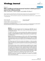
The proportion of patients with positive EBV viral load did not
differ among groups (control individuals, 80%; SpA patients,
65%; RA patients with MTX, 79%; and RA patients without
DMARD treatment, 85% (P = 0.42, chi-square test)), nor did
they differ when considering the distribution of all viral loads in
the four groups of patients (P = 0.61, Kruskal-Wallis test) (Fig-
SFCs/10 PBMCs 10
(mean SFCs/10 cells from two antigen-st
6
5
=×
iimulated wells mean SFC/10 cells from four unstimulated
5
− wwells).
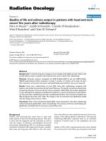
Figure 1
Epstein-Barr virus load in peripheral blood mononuclear cells in the cross-sectional studyEpstein-Barr virus load in peripheral blood mononuclear cells in the
cross-sectional study. Epstein-Barr virus (EBV) load distribution in con-
trol individuals (n = 15), spondylarthropathy (SpA) patients (n = 23),
rheumatoid arthritis (RA) patients receiving methotrexate (MTX) (n =
18) and RA patients not receiving disease-modifying anti-rheumatic
drug therapy (n = 26). Mean values of EBV viral load are represented
by a black line. *Kruskall-Wallis test. PBMC, peripheral blood mononu-
clear cell.
Available online />Page 5 of 9
(page number not for citation purposes)
ure 1). Likewise, control individuals did not differ from any
other group in viral load (Mann-Whitney test). The mean (±
standard deviation) viral loads in each group were as follows:
control individuals, 197 ± 433 copies/10
6
cells; SpA patients,
353 ± 905 copies/10
6
cells; RA patients with MTX, 1,596 ±
4,533 copies/10
6
cells; and MTX naïve RA patients, 387 ±
893 copies/10
6
cells. The median viral loads were 113 for
control individuals, 55 for SpA patients, 58 RA patients with
MTX, and 114 for MTX naïve RA patients.
We found no significant correlation between the EBV viral load
and disease activity (Disease Activity Score for 28 joints for
RA patients, P = 0.54; Bath Ankylosing Spondylitis Disease
Activity Index for SpA patients, P = 0.84) or disease duration
(P = 0.29).
Epstein-Barr virus-specific IFN
γ
-producing T cells
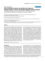
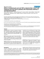
The proportion of patients with positive EBV-specific IFNγ-pro-
ducing T cells did not differ among groups (control individuals,
73%; SpA patients, 71%; RA patients with MTX, 59%; and
RA patients without DMARD treatment, 72% (P = 0.68, chi-
square test)) (Figure 2a). No significant differences were
observed between groups when considering T-cell responses
to the whole set of peptides (P = 0.86) or restricted to latent
peptides (P = 0.92) or lytic peptides (P = 0.34) (Kruskal-Wal-
lis test) (Figure 2b, c). The control group did not differ from
each other treatment group either when considering pulses
with the whole set of peptides, or when considering pulses
with latent or lytic peptides (Mann-Whitney test) (Figure 2a to
2c).
We found no significant correlation between the number of
EBV-specific IFNγ-producing T cells and disease activity (Dis-
Figure 2
Epstein-Barr virus-specific IFNγ-producing T cellsEpstein-Barr virus-specific IFNγ-producing T cells. Number of IFNγ-producing T cells per 10
6
peripheral blood mononuclear cells (PBMCs). (a) After
pulsing with the full set of Epstein-Barr virus peptides. (b) After pulsing with latent-cycle peptides. (c) After pulsing with lytic-cycle peptides. Mean
IFNγ-producing T cells per 10
6
PBMCs are represented by a black line. *Kruskall-Wallis test. MTX, methotrexate; RA, rheumatoid arthritis; SpA,
spondylarthropathy.
Arthritis Research & Therapy Vol 11 No 3 Miceli-Richard et al.
Page 6 of 9
(page number not for citation purposes)
ease Activity Score for 28 joints for RA patients (n = 64), P =
0.32; Bath Ankylosing Spondylitis Disease Activity Index for
SpA patients (n = 21), P = 0.47) or disease duration (n = 88,
P = 0.40).
Longitudinal study
Epstein-Barr virus load in peripheral blood mononuclear
cells
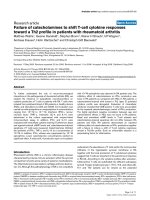
When pooling all treatment groups, longitudinal observation of
the EBV viral load showed no significant change between
baseline (week 0) and week 12 (P = 0.33) (Wilcoxon test)
(Figure 3a). Similar results were obtained when analyzing each
treatment group longitudinally: SpA patients receiving anti-
TNF drugs (P = 1.00), RA patients receiving anti-TNF drugs (P
= 0.94) and RA patients receiving MTX (P = 0.74). Patients
receiving anti-TNF drugs showed no difference in EBV viral
load according to the class of TNF used: monoclonal antibody
(infliximab and adalimumab) (n = 9, P = 0.31) or soluble recep-
tor (etanercept) (n = 18, P = 0.63).
Epstein-Barr virus-specific IFN
γ
-producing T cells
In response to the full set of peptides, the number of IFNγ-pro-
ducing cells was not significantly modified by immunosuppres-
sive treatment (SpA patients receiving anti-TNF drugs (n =
21), P = 0.39; RA patients receiving anti-TNF drugs (n = 19),
P = 0.19; RA patients receiving MTX (n = 22), P = 0.58) (Fig-
ure 3b), nor was the number of EBV-specific IFNγ-producing
T cells modified when considering each set of peptides
(latent-cycle peptides or lytic-cycle peptides) (data not
shown). Among patients treated with TNF blockers, there was
no difference according to the class of molecule used: mono-
clonal antibody (infliximab and adalimumab) (n = 16, P = 0.74)
or soluble receptor (etanercept) (n = 24, P = 0.92).
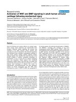
Correlation between Epstein-Barr virus load and T spots
For correlation studies between the EBV viral load and EBV-
specific T-cell response, 113 patients were studied (66 at
week 0 and 47 at week 12). We found a positive correlation
between the EBV viral load and the number of EBV-specific
IFN-γ-producing T cells in response to the full set of peptides
(n = 113, P = 0.017, R = 0.21) (Figure 4), to latent-cycle pep-
tides (P = 0.035, R = 0.16) and to lytic-cycle peptides (P =
0.011, R = 0.16) (data not shown).
Unadapted Epstein-Barr virus-specific T-cell IFN
γ
production under treatment
Five patients demonstrated inappropriate EBV-specific T-cell
IFNγ production (<100 IFNγ secreted T cells and >1,000 EBV
copies per 10
6
PBMCs). Three of these patients had no or
very low IFNγ secreted T cells at week 0 and week 12. Two
other patients had an accurate in vitro effector function at
week 0 but a large decrease of EBV-specific IFNγ secreted T
cell number at week 12 despite a concomitant increased level
of EBV copy numbers above 1,000 copies per 10
6
PBMCs
(Figure 5a, b). These two patients were treated with anti-TNF
monoclonal antibody associated with MTX: one SpA patient
with infliximab, and one RA patient with adalimumab. For both
patients, an EBV-specific T-cell response to latent peptides
was detectable at baseline but was not detectable at week 12.
Nevertheless, the response to lytic peptides was persistent in
both cases at week 12.
Figure 3
Epstein-Barr virus load in peripheral blood mononuclear cells in the lon-gitudinal studyEpstein-Barr virus load in peripheral blood mononuclear cells in the lon-
gitudinal study. (a) Epstein-Barr virus (EBV) load between week 0
(W0) and week 12 (W12) for all treated patients (n = 42). (b) EBV-
specific IFNγ-producing T cells per 10
6
peripheral blood mononuclear
cells between W0 and W12 for all patients receiving anti-TNF drugs (n
= 40). PBMCs, peripheral blood mononuclear cells.
Available online />Page 7 of 9
(page number not for citation purposes)
Discussion
The present large cross-sectional and longitudinal study
showed no abnormality in EBV viral load or EBV-specific T-cell
response in patients with RA or SpA at baseline or after treat-
ment with MTX or anti-TNF drugs.
In contrast to other studies [23,24], we did not find increased
EBV viral load in PBMCs of patients with RA or SpA. A casual
high EBV DNA prevalence in our control group (80%) could
account for the differing results, and/or the highly sensitive
PCR used in our study might explain such differences. Our
cross-sectional study revealed no significant differences
between patients and control individuals in the proportion of
subjects with positive EBV-specific IFNγ-producing T cells in
PBMCs, the mean number of SFCs or the SFC distribution.
Two studies have assessed the EBV-specific T-cell response
in PBMCs of RA patients [25,26]. The first study found no dif-
ference between 49 RA patients and 26 control individuals in
the frequency of T cells directed against two immunodominant
EBV peptides, but did observe a reduced ability to produce
INFγ in RA patients; the effect of immunosuppressive treat-
ment was not assessed [25]. In the second study, EBV-spe-
cific effector CD8 T cells were higher in number in RA patients
(n = 25) than in control individuals (n = 20), but this study con-
cerned a low number of patients and was only cross-sectional
[26]. Actually, this increase in IFNγ secreted T cells was
related to increased viral load, which we did not observe in our
study.
Our longitudinal results did not reveal any influence of immu-
nosuppressive treatment on the EBV viral load. These results
are in accordance with several studies on Crohn disease or
RA patients assessing the evolution of EBV viral load under
immunosuppressive treatment [27-29]. To the best of our
knowledge, no published study has specifically evaluated the
longitudinal effect of MTX and anti-TNF drug on EBV-specific
T-cell effector functions in patients with RA or SpA. At 3-
month follow-up, neither MTX treatment in RA patients nor
anti-TNF therapy in RA and/or SpA patients modified these
effector functions, regardless of the EBV peptide used for
pulsing – latent-cycle peptides or lytic-cycle peptides or the
full set of peptides. The lack of increase in the EBV viral load
during the same period in all groups of patients agrees with
the preserved specific T-cell effector function, which was con-
firmed by a global correlation between EBV viral load and
EBV-specific T-cell response.
Interestingly, in five patients treated with anti-TNF, an inade-
quate in vitro EBV-specific IFNγ production was observed
after specific pulse with EBV peptides despite an in vivo high
viral load. Among those patients, two different profiles were
observed. The first profile corresponded to patients without
any IFNγ production in spite of high EBV viral loads (>1,000/
10
6
PBMCs), both at week 0 and week 12. In such cases, the
lack of adequate HLA for presenting one of the EBV peptides
probably accounted for the absence of IFNγ production after
specific pulse. The second profile corresponded to two
patients having EBV-specific T-cell IFNγ production at base-
line but a discordant evolution between an IFNγ secreted T-
cell decrease and an EBV viral load increase after 12 weeks of
treatment. In these two patients, immunosuppressive therapy
might have impaired EBV-specific T-cell effector functions
leading to the lack of control of the EBV viral load. These two
patients having been treated with the association of anti-TNF
antibody and MTX makes it impossible to differentiate a possi-
ble effect of one drug individually. Nevertheless, we never saw
profound discrepancies, such as those observed in a pediatric
sample in whom post-transplant lymphoproliferative disorder
developed after liver transplantation [30]. Since we analyzed
data only 12 weeks after the introduction of immunosuppres-
sive treatment, however, we cannot exclude that MTX or anti-
TNF therapy could induce impaired EBV control after longer-
term treatment. This relative short time duration of immunosup-
pressive treatment exposure might be considered as a limita-
tion of our study. Nevertheless, post-transplant
lymphoproliferative diseases occurring in children, for exam-
ple, have been reported to occur after a short-term exposure
to immunosuppressive treatments (median delay of 12 weeks,
range 6 to 56 weeks) [30]. Moreover, with the same method-
ology (ELISPOT assay), we detected a significant decrease of
the specific anti-tuberculosis T-cell response in patients after
12 weeks of anti-TNF therapy [31]. Lastly, in the present study,
patients treated with MTX were treated for several years on
average, and their results were no different from the MTX naïve
patients at baseline.
Figure 4
Correlation between the Epstein-Barr virus viral load and Epstein-Barr virus-specific T-cell responseCorrelation between the Epstein-Barr virus viral load and Epstein-Barr
virus-specific T-cell response. Correlation between the number of IFNγ-
producing T cells and the Epstein-Barr virus (EBV) viral load. EBV-spe-
cific IFNγ-producing T cells were pulsed with the full set of peptides
(latent-cycle peptides and lytic-cycle peptides). PBMCs, peripheral
blood mononuclear cells.
Arthritis Research & Therapy Vol 11 No 3 Miceli-Richard et al.
Page 8 of 9
(page number not for citation purposes)
Conclusions
In patients with RA or SpA, short-term (3-month) exposure to
MTX or anti-TNF therapy does not alter the EBV viral load or
the EBV-specific T-cell response. These findings are rather
reassuring in light of a suggested increased risk of EBV-asso-
ciated lymphoma in patients receiving immunosuppressive
therapy. Long-term follow-up of the EBV-specific T-cell
response, however, is necessary. Moreover, control of EBV is
only one mechanism of control of lymphomagenesis and the
different epidemiologic studies currently available do not elim-
inate the possibility of increased risk of non-EBV-associated
lymphoma in patients receiving immunosuppressive therapy.
Competing interests
The authors declare that they have no competing interests.
Authors' contributions
XM was responsible for the study design, manuscript prepara-
tion and interpretation of the data. CM-R was responsible for
sample blood collection, manuscript preparation, interpreta-
tion of data and statistical analyses. NG performed the ELIS-
POT assays and statistical analyses. CA performed the EBV
quantitative PCR. JS, MI and SP contributed to the blood sam-
ple collection. AU and IG contributed to the ELISPOT assay
analyses. GC and AV contributed to interpretation of the data.
Acknowledgements
The set of EBV peptides was kindly provided by Prof. D Olive (INSERM
Action-Thématique-Concertée). The authors thank Dr Martine Sinet and
Prof. Martine Raphael for helpful discussions. The present work was
supported by Assistance Publique Hôpitaux de Paris, Département de
la Recherche Clinique, Paris, France (Contrat d'Initiation à la Recherche
Clinique) and by Société Française de Rhumatologie.
Figure 5
Unadapted Epstein-Barr virus-specific T-cell IFNγ production under treatmentUnadapted Epstein-Barr virus-specific T-cell IFNγ production under treatment. Patients with inappropriate Epstein-Barr virus (EBV)-specific T-cell
IFNγ production in response to high EBV viral load under treatment. (a) Spondylarthropathy patient with infliximab + methotrexate. (b) Rheumatoid
arthritis patient with adalimumab + methotrexate. Both responses to latent peptides and lytic peptides are represented. PBMCs, peripheral blood
mononuclear cells; W0, week 0; W12, week 12.
Available online />Page 9 of 9
(page number not for citation purposes)
References
1. Smedby KE, Hjalgrim H, Askling J, Chang ET, Gregersen H, Por-
wit-MacDonald A, Sundstrom C, Akerman M, Melbye M, Glimelius
B, Adami HO: Autoimmune and chronic inflammatory disor-
ders and risk of non-Hodgkin lymphoma by subtype. J Natl
Cancer Inst 2006, 98:51-60.
2. Landgren O, Engels EA, Pfeiffer RM, Gridley G, Mellemkjaer L,
Olsen JH, Kerstann KF, Wheeler W, Hemminki K, Linet MS, Goldin
LR: Autoimmunity and susceptibility to Hodgkin lymphoma: a
population-based case-control study in Scandinavia. J Natl
Cancer Inst 2006, 98:1321-1330.
3. Kamel OW, Rijn M van de, Weiss LM, Del Zoppo GJ, Hench PK,
Robbins BA, Montgomery PG, Warnke RA, Dorfman RF: Brief
report: reversible lymphomas associated with Epstein-Barr
virus occurring during methotrexate therapy for rheumatoid
arthritis and dermatomyositis. N Engl J Med 1993,
328:1317-1321.
4. Salloum E, Cooper DL, Howe G, Lacy J, Tallini G, Crouch J,
Schultz M, Murren J: Spontaneous regression of lymphoprolif-
erative disorders in patients treated with methotrexate for
rheumatoid arthritis and other rheumatic diseases. J Clin
Oncol 1996, 14:1943-1949.
5. Bongartz T, Sutton AJ, Sweeting MJ, Buchan I, Matteson EL, Mon-
tori V: Anti-TNF antibody therapy in rheumatoid arthritis and
the risk of serious infections and malignancies: systematic
review and meta-analysis of rare harmful effects in rand-
omized controlled trials. JAMA 2006, 295:2275-2285.
6. Baecklund E, Iliadou A, Askling J, Ekbom A, Backlin C, Granath F,
Catrina AI, Rosenquist R, Feltelius N, Sundstrom C, Klareskog L:
Association of chronic inflammation, not its treatment, with
increased lymphoma risk in rheumatoid arthritis. Arthritis
Rheum 2006, 54:692-701.
7. Wolfe F, Michaud K: Lymphoma in rheumatoid arthritis: the
effect of methotrexate and anti-tumor necrosis factor therapy
in 18,572 patients. Arthritis Rheum 2004, 50:1740-1751.
8. Geborek P, Bladstrom A, Turesson C, Gulfe A, Petersson IF,
Saxne T, Olsson H, Jacobsson LT: Tumour necrosis factor
blockers do not increase overall tumour risk in patients with
rheumatoid arthritis, but may be associated with an increased
risk of lymphomas. Ann Rheum Dis 2005, 64:699-703.
9. Schiff MH, Burmester GR, Kent JD, Pangan AL, Kupper H, Fitz-
patrick SB, Donovan C: Safety analyses of adalimumab
(HUMIRA) in global clinical trials and US postmarketing sur-
veillance of patients with rheumatoid arthritis. Ann Rheum Dis
2006, 65:889-894.
10. Park SH, Kim CG, Kim JY, Choe JY: Spontaneous regression of
EBV-associated diffuse lymphoproliferative disease in a
patient with rheumatoid arthritis after discontinuation of
etanercept treatment. Rheumatol Int 2008, 28:475-477.
11. Thonhofer R, Gaugg M, Kriessmayr M, Neumann HJ, Erlacher L:
Spontaneous remission of marginal zone B cell lymphoma in
a patient with seropositive rheumatoid arthritis after discontin-
uation of infliximab-methotrexate treatment. Ann Rheum Dis
2005, 64:1098-1099.
12. Boubenider S, Hiesse C, Goupy C, Kriaa F, Marchand S, Charpen-
tier B: Incidence and consequences of post-transplantation
lymphoproliferative disorders. J Nephrol 1997, 10:136-145.
13. Hislop AD, Annels NE, Gudgeon NH, Leese AM, Rickinson AB:
Epitope-specific evolution of human CD8(+) T cell responses
from primary to persistent phases of Epstein-Barr virus infec-
tion. J Exp Med 2002, 195:893-905.
14. Dunne PJ, Faint JM, Gudgeon NH, Fletcher JM, Plunkett FJ, Soares
MV, Hislop AD, Annels NE, Rickinson AB, Salmon M, Akbar AN:
Epstein-Barr virus-specific CD8(+) T cells that re-express
CD45RA are apoptosis-resistant memory cells that retain rep-
licative potential. Blood 2002, 100:933-940.
15. Arnett FC, Edworthy SM, Bloch DA, McShane DJ, Fries JF, Cooper
NS, Healey LA, Kaplan SR, Liang MH, Luthra HS, Medsger TA,
Mitchell DM, Neustadt DH, Pinals RS, Schaller JG, Sharp JT,
Wilder RL, Hunder GG: The American Rheumatism Association
1987 revised criteria for the classification of rheumatoid arthri-
tis. Arthritis Rheum 1988, 31:315-324.
16. Dougados M, Linden S van der, Juhlin R, Huitfeldt B, Amor B, Calin
A, Cats A, Dijkmans B, Olivieri I, Pasero G, Veys E, Zeidler H: The
European Spondylarthropathy Study Group preliminary crite-
ria for the classification of spondylarthropathy. Arthritis Rheum
1991, 34:1218-1227.
17. Garrett S, Jenkinson T, Kennedy LG, Whitelock H, Gaisford P,
Calin A: A new approach to defining disease status in ankylos-
ing spondylitis: the Bath Ankylosing Spondylitis Disease Activ-
ity Index. J Rheumatol 1994, 21:2286-2291.
18. Samri A, Durier C, Urrutia A, Sanchez I, Gahery-Segard H, Imbart
S, Sinet M, Tartour E, Aboulker JP, Autran B, Venet A: Evaluation
of the interlaboratory concordance in quantification of human
immunodeficiency virus-specific T cells with a gamma inter-
feron enzyme-linked immunospot assay. Clin Vaccine Immu-
nol 2006, 13:684-697.
19. Yang J, Lemas VM, Flinn IW, Krone C, Ambinder RF: Application
of the ELISPOT assay to the characterization of CD8(+)
responses to Epstein-Barr virus antigens. Blood 2000,
95:241-248.
20. Steven NM, Annels NE, Kumar A, Leese AM, Kurilla MG, Rickinson
AB: Immediate early and early lytic cycle proteins are frequent
targets of the Epstein-Barr virus-induced cytotoxic T cell
response. J Exp Med 1997, 185:1605-1617.
21. Doisne JM, Urrutia A, Lacabaratz-Porret C, Goujard C, Meyer L,
Chaix ML, Sinet M, Venet A: CD8
+
T cells specific for EBV,
cytomegalovirus, and influenza virus are activated during pri-
mary HIV infection. J Immunol 2004, 173:2410-2418.
22. Besson C, Amiel C, Le-Pendeven C, Brice P, Ferme C, Carde P,
Hermine O, Raphael M, Abel L, Nicolas JC: Positive correlation
between Epstein-Barr virus viral load and anti-viral capsid
immunoglobulin G titers determined for Hodgkin's lymphoma
patients and their relatives. J Clin Microbiol 2006, 44:47-50.
23. Balandraud N, Meynard JB, Auger I, Sovran H, Mugnier B, Reviron
D, Roudier J, Roudier C: Epstein-Barr virus load in the periph-
eral blood of patients with rheumatoid arthritis: accurate quan-
tification using real-time polymerase chain reaction. Arthritis
Rheum 2003, 48:1223-1228.
24. Alvarez-Lafuente R, Fernandez-Gutierrez B, de Miguel S, Jover JA,
Rollin R, Loza E, Clemente D, Lamas JR: Potential relationship
between herpes viruses and rheumatoid arthritis: analysis
with quantitative real time polymerase chain reaction. Ann
Rheum Dis 2005, 64:1357-1359.
25. Klatt T, Ouyang Q, Flad T, Koetter I, Buhring HJ, Kalbacher H,
Pawelec G, Muller CA: Expansion of peripheral CD8
+
CD28-T
cells in response to Epstein-Barr virus in patients with rheu-
matoid arthritis. J Rheumatol 2005, 32:239-251.
26. Lunemann JD, Frey O, Eidner T, Baier M, Roberts S, Sashihara J,
Volkmer R, Cohen JI, Hein G, Kamradt T, Munz C: Increased fre-
quency of EBV-specific effector memory CD8
+
T cells corre-
lates with higher viral load in rheumatoid arthritis. J Immunol
2008, 181:991-1000.
27. Reijasse D, Le Pendeven C, Cosnes J, Dehee A, Gendre JP, Nico-
las JC, Beaugerie L: Epstein-Barr virus viral load in Crohn's dis-
ease: effect of immunosuppressive therapy. Inflamm Bowel
Dis 2004, 10:85-90.
28. Torre-Cisneros J, Del Castillo M, Caston JJ, Castro MC, Perez V,
Collantes E: Infliximab does not activate replication of lympho-
tropic herpesviruses in patients with refractory rheumatoid
arthritis. Rheumatology (Oxford) 2005, 44:1132-1135.
29. Balandraud N, Guis S, Meynard JB, Auger I, Roudier J, Roudier C:
Long-term treatment with methotrexate or tumor necrosis fac-
tor alpha inhibitors does not increase Epstein-Barr virus load
in patients with rheumatoid arthritis. Arthritis Rheum 2007,
57:762-767.
30. Smets F, Latinne D, Bazin H, Reding R, Otte JB, Buts JP, Sokal
EM: Ratio between Epstein-Barr viral load and anti-Epstein-
Barr virus specific T-cell response as a predictive marker of
posttransplant lymphoproliferative disease. Transplantation
2002, 73:1603-1610.
31. Hamdi H, Mariette X, Godot V, Weldingh K, Hamid AM, Prejean
MV, Baron G, Lemann M, Puechal X, Breban M, Berenbaum F,
Delchier JC, Flipo RM, Dautzenberg B, Salmon D, Humbert M,
Emilie D: Inhibition of anti-tuberculosis T-lymphocyte function
with tumour necrosis factor antagonists. Arthritis Res Ther
2006, 8:R114.