Báo cáo y học: "Alterations in peripheral blood memory B cells in patients with active rheumatoid arthritis are dependent on the action of tumour necrosis factor" potx
Bạn đang xem bản rút gọn của tài liệu. Xem và tải ngay bản đầy đủ của tài liệu tại đây (576.46 KB, 12 trang )
Open Access
Available online />Page 1 of 12
(page number not for citation purposes)
Vol 11 No 3
Research article
Alterations in peripheral blood memory B cells in patients with
active rheumatoid arthritis are dependent on the action of tumour
necrosis factor
M Margarida Souto-Carneiro
1
*, Vijayabhanu Mahadevan
2
*, Kazuki Takada
3
, Ruth Fritsch-Stork
4
,
Toshihiro Nanki
3
, Margaret Brown
5
, Thomas A Fleisher
5
, Mildred Wilson
2
, Raphaela Goldbach-
Mansky
2
and Peter E Lipsky
2
1
Centro de Neurociências e Biologia Celular, Department of Zoology, University of Coimbra, 3004-517 Coimbra, Portugal
2
National Institute of Arthritis and Musculoskeletal and Skin Diseases, NIH, 9000 Rockville Pike, Bethesda, MD 20892, USA
3
Department of Medicine and Rheumatology, Graduate School, Tokyo Medical and Dental University, 1-5-45, Yushima, Bunkyo-ku, Tokyo 113-8519,
Japan
4
Department of Rheumatology, UMC Utrecht, Heidelberglaan 100, 3584 CX Utrecht, The Netherlands
5
Department of Laboratory Medicine, Warren Magnuson Center, NIH, 9000 Rockville Pike, Bethesda, MD 20892, USA
* Contributed equally
Corresponding author: Peter E Lipsky,
Received: 23 Jan 2009 Revisions requested: 6 Mar 2009 Revisions received: 20 Apr 2009 Accepted: 5 Jun 2009 Published: 5 Jun 2009
Arthritis Research & Therapy 2009, 11:R84 (doi:10.1186/ar2718)
This article is online at: />© 2009 Souto-Carneiro et al.; licensee BioMed Central Ltd.
This is an open access article distributed under the terms of the Creative Commons Attribution License ( />),
which permits unrestricted use, distribution, and reproduction in any medium, provided the original work is properly cited.
Abstract
Introduction Disturbances in peripheral blood memory B cell
subpopulations have been observed in various autoimmune
diseases, but have not been fully delineated in rheumatoid
arthritis (RA). Additionally, the possible role of tumour necrosis
factor (TNF) in regulating changes in specific peripheral blood
memory B cell subsets in RA is still unclear.
Methods The frequency and distribution of B cell subsets in the
peripheral blood and synovial membrane of active RA patients
with long-standing disease have been analysed. Additionally, the
possible role of TNF in causing disturbances in memory B cell
subsets in RA patients was assessed in a clinical trial with the
specific TNF-neutralising antibody, infliximab.
Results RA patients, independent of disease duration, have a
significantly lower frequency of peripheral blood pre-switch
IgD
+
CD27
+
memory B cells than healthy individuals, whereas
post-switch IgD
-
CD27
+
accumulate with increased disease
duration. Notably, both pre-switch IgD
+
CD27
+
and post-switch
IgD
-
CD27
+
memory B cells accumulate in the synovial
membrane of RA patients. Finally, anti-TNF therapy increased
the frequency of pre-switch IgD
+
CD27 memory B cells in the
peripheral blood.
Conclusions The data suggest that decreases in peripheral
blood IgD
+
CD27
+
pre-switch memory B cells in RA reflect their
accumulation in the synovial tissue. Moreover, the significant
increase in the peripheral blood pre-switch memory B cells in
patients who underwent specific TNF-blockade with infliximab
indicates that trafficking of memory B cells into inflamed tissue
in RA patients is regulated by TNF and can be corrected by
neutralising TNF.
Introduction
Rheumatoid arthritis (RA) is a chronic systemic autoimmune
disease, characterised by inflammatory polyarthritis and joint
damage resulting in progressive disability [1]. The inflamma-
tory infiltrate in RA includes T cells, B cells and dendritic cells
[2-4], and in approximately 20% of patients lymphoid neogen-
APC: allophycocyanin; BSA: bovine serum albumin; CRP: C-reactive protein; DMARD: disease-modifying anti-rheumatic drugs; DMEM: Dulbecco's
Modified Eagle's Medium; ELISA: enzyme-linked immunosorbent assay; ESR: erythrocyte sedimentation rate; FCS: fetal calf serum; FDC: follicular
dendritic cell; FITC: fluorescein isothiocyanate; ICAM: intercellular adhesion molecule; IgV
H
: immunoglobulin heavy chain variable region; IL: inter-
leukin; LT: lymphotoxin; MAb: monoclonal antibodies; MTX: methotrexate; PBMC: peripheral blood mononuclear cells; PBS: phosphate-buffered
saline; PCR: polymerase chain reaction; PE: phycoerythrin; PerCpCy5.5: peridinin-chlorophyll-protein Cy5.5; RA: rheumatoid arthritis; SLE: systemic
lupus erythematosus; SS: Sjögren's syndrome; TNF: tumour necrosis factor; VCAM: vascular cell adhesion molecule; VEGF: vascular endothelial
growth factor.
Arthritis Research & Therapy Vol 11 No 3 Souto-Carneiro et al.
Page 2 of 12
(page number not for citation purposes)
esis develops with the formation of ectopic germinal centres
[5-8].
The importance of B cells in RA has been emphasised by the
success of therapeutic approaches using anti-CD20 mono-
clonal antibodies (mAbs) [9]. It is currently unknown whether
this approach to treatment is successful because of the pro-
duction of early plasma cells due to the loss of rheumatoid fac-
tor or because of other functions of B cells.
Functionally distinct B cell subsets can be defined by the sur-
face expression of immunoglobulin (Ig) D and CD27. These
include naïve IgD
+
CD27
-
; pre-switch memory IgD
+
CD27
+
;
and post-switch memory IgD
-
CD27
+
[10-12]. Importantly,
CD27 expression by B cells has been considered a hallmark
for cells that have undergone somatic hypermutation [13],
although recently a CD27
-
population of memory B cells with
mutated Ig genes has been described [14-16], which is ele-
vated in patients with systemic lupus erythematosus (SLE)
[15]. Abnormalities in the frequencies of peripheral blood
memory B cells have been reported in SLE [17], and Sjögren's
syndrome (SS) [18]. However, in RA the data on possible dis-
turbances of peripheral blood B cell distributions have not
been delineated as well. Part of this could relate to differences
in disease duration and therapy of the cohorts studied [19-21].
Treatment with TNF blockers ameliorates the signs and symp-
toms of RA and disease progression [22-25]. Recently, a
study of peripheral blood and tonsilar biopsies from RA
patients undergoing treatment with the combined TNF and
lymphotoxin α (LTα) antagonist, etanercept, suggested that
part of the success of this therapy in RA could be linked to a
disruption of follicular dendritic cell (FDC) networks in second-
ary lymphoid organs, thus impairing germinal centre formation,
and decreasing the number of CD27
+
memory B cells in the
blood [19]. However, this effect was noted in the tonsil, mak-
ing it uncertain whether etanercept would have a similar
impact on germinal centres in the spleen and lymph nodes.
Etanercept neutralises both TNF and LTα, so it is difficult to
determine the possible contribution of each cytokine to the
effects noted. TNF and LTα have many non-overlapping func-
tions and, therefore, distinct effects of blocking each of these
two cytokines on memory B cell homeostasis are possible. For
example, TNF is involved in the regulation of the expression of
adhesion molecules, such as vascular cell adhesion molecule
(VCAM-1), intercellular adhesion molecule (ICAM-1), P-selec-
tin, E-selectin, and L-selectin (reviewed in [26]) and also vas-
cular endothelial growth factor (VEGF)-C [27], suggesting
that it may play a crucial role in the neovascularisation of rheu-
matoid synovium and also recruitment of lymphocytes into the
inflamed synovium.
In order to study the changes in peripheral memory B cell sub-
populations in RA patients, and to understand the possible
role of TNF in regulating changes in specific memory B cells,
we analysed the frequency and distribution of B cell subsets
in the peripheral blood and synovial membrane of active RA
patients with long-standing disease. Subsequently, we
assessed whether treatment with the specific TNF-blocker, inf-
liximab, normalised the distribution of these peripheral B cell
subsets. Our results show, for the first time, that RA patients,
independent of disease duration, have a much lower fre-
quency of peripheral blood pre-switch IgD
+
CD27
+
memory B
cells than healthy individuals, whereas post-switch IgD
-
CD27
+
memory B cells accumulate with increased disease duration.
Additionally, we present evidence that pre-switch IgD
+
CD27
+
memory B cells accumulate in the synovial membrane of RA
patients, and that this accumulation might be related to the
influence of TNF, because anti-TNF therapy increased the fre-
quency of pre-switch IgD
+
CD27 memory B cells in the periph-
eral blood. These results document disease-related and TNF-
dependent abnormalities in memory B cell subsets in RA and
suggest that part of the success of TNF neutralising therapy
could relate to normalisation of memory B cell abnormalities.
Materials and methods
Patients and controls
Peripheral blood samples from 40 healthy donors (26 females,
14 males; mean age 44 years) were obtained from the
National Institutes of Health blood bank, and from 33 patients
(28 females, five males; mean age 57 years) with long-stand-
ing RA (median disease length, 13 years) enrolled in a natural
history protocol (00-AR-0222) at the Warren G. Magnuson
Clinical Center (National Institutes of Health, Bethesda, Mary-
land, USA).
In addition, blood samples were obtained from 23 patients (20
females, 3 males; mean age 48.5 years) with active RA
(defined as having greater than four tender and swollen joints,
erythrocyte sedimentation rate (ESR) greater than 20 mm/
hour or C-reactive protein (CRP) greater than 0.8 mg/dl) who
failed treatment with methotrexate (MTX; 12.5 to 15 mg/week)
and were entering a clinical trial of infliximab therapy (00-AR-
0220). For this trial, patients on prednisone had to be on 7.5
mg or less per day to be eligible to participate. Patients were
randomised to receive either monthly infliximab infusions (3
mg/kg infliximab with MTX 15 mg/week), or monthly control
infusions and weekly MTX alone (<25 mg/week). All patients
fulfilled the revised American College of Rheumatology criteria
for RA [28]. MB, carrying out the flow cytometric analysis, was
blinded to the measurements of clinical response and disease
activity scores.
The group of patients enrolled in the natural history protocol,
with a median disease length of 13 years, were considered as
the long-standing disease group. The group of patients
enrolled in the clinical trial for infliximab, with a median disease
duration of 4.4 years, were considered as the group with
shorter disease duration.
Available online />Page 3 of 12
(page number not for citation purposes)
Synovial specimens and peripheral blood samples were col-
lected at the Department of Rheumatology, Tokyo Medical and
Dental University from 10 RA subjects with long-standing dis-
ease (median disease length of 13.5 years).
The characteristics of all patients studied are shown in Table
1.
The local institutional review board or the ethics committees
(National Institutes of Health and Tokyo Medical and Dental
University) approved the studies and all patients signed an
informed consent before participating in this study. Patient's
management was performed in accordance with the local
standard practice and the study was conducted in accord-
ance with the regulations governing clinical trials, such as the
Declaration of Helsinki as amended in Edinburgh (2000).
Lymphocyte phenotyping
Peripheral Blood
Peripheral blood samples from the controls and natural history
patients were obtained during a single scheduled outpatient
visit. Peripheral blood mononuclear cells (PBMCs) were iso-
lated by Ficoll gradient centrifugation and re-suspended in 1.5
ml PBS and 1% BSA (1 × 10
6
cells/100 μL). Isolated PBMCs
were stained by standard methods with fluorescein isothiocy-
anate (FITC), phycoerythrin (PE), peridinin-chlorophyll-protein
Cy5.5 (PerCpCy5.5) or allophycocyanin (APC) conjugated
mAb specific for the following human cell surface markers:
anti-CD19 PerCpCy5.5, anti-CD27 PE, anti-IgD FITC and
anti-IgM FITC (all mAb were obtained from BD Pharmingen,
Franklin Lakes, NJ, USA). Data were acquired on a FACSCal-
ibur (BD Biosciences, Franklin Lakes, NJ, USA).
Peripheral blood samples from the RA patients treated with
MTX and infliximab or MTX alone were obtained before and
after treatment. Anticoagulated samples were stained for
three-colour flow cytometry using a whole blood staining
method at the National Institutes of Health Clinical Center lab-
oratory. B cells were identified by staining with anti-CD20
APC and anti-CD27 PE (BD Biosciences, San Jose, CA,
USA) and anti-IgD FITC (Caltag, Burlingame, CA, USA). T
cells were identified by anti-CD3 APC or PE, anti-CD4 PE,
anti-CD8 FITC or APC, anti-CD45RA FITC and anti-CD45R0
APC (BD Biosciences, San Jose, CA, USA).
To calculate absolute numbers of each lymphocyte subset, the
percentage of cells staining positively was multiplied by the
absolute peripheral blood lymphocyte count, which was deter-
mined by cell counting with a Celldyne 3500 (Abbott, Santa
Clara, CA, USA) blood cell counting machine. With all experi-
ments, peripheral blood from healthy adult patients was
stained and analysed as controls.
To determine the chemokine receptor expression by B cells
and their subsets, the following APC-conjugated anti-human
mAbs were used: anti-CXCR1, anti CXCR2 and anti-CCR2
(R&D Systems, Minneapolis, MN, USA); and anti-CXCR4 (BD
Biosciences, San Jose, CA, USA).
Irrelevant, directly conjugated, murine IgG1 (BD Biosciences,
San Jose, CA, USA) was used to ascertain background stain-
Table 1
Clinical and demographic characteristics of the RA patients
Treatment trial
Healthy controls Long-standing RA RA patients for synovium
collection
MTX only Infliximab and MTX
Number of subjects 40 33 10 8 15
Age (years) 44 ± 9 57 ± 12 62 ± 10 52 ± 16 45 ± 11
Female/male ratio 26:14 28:5 9:1 7:1 13:2
Disease duration (years) 13 ± 12 13.5 ± 11.0 5.7 ± 1.5 3.0 ± 0.5
% RF positive patients 79% 90% 50% 73%
ESR (mm/hour) 36 ± 27 36.9 ± 14.3 60.2 ± 30.3
CRP (mg/dl) <0.4 ± 9.75 2.8 ± 2.3 0.8 ± 1.1 1.8 ± 1.9
% Patients on MTX (dose) 85% (15 mg/wk) 60% (4 mg/wk) 100% (14 mg/wk) 100% (14 mg/wk)
% Patients on GC (dose) 49% (5 mg/day) 80% (5 mg/day) 50% (6 mg/day) 64% (6 mg/day)
% Patients on other DMARD 82% 50%
Data are means ± standard deviation.
CRP = C-reactive protein; DMARD = disease-modifying anti-rheumatic drug; ESR = erythrocyte sedimentation rate; GC = glucocorticoids; MTX
= methotrexate; RA = rheumatoid arthritis; RF = rheumatoid factor.
Arthritis Research & Therapy Vol 11 No 3 Souto-Carneiro et al.
Page 4 of 12
(page number not for citation purposes)
ing. Samples were run on a FACScan or a FACSCalibur (BD
Biosciences, San Jose, CA, USA). Data were analysed using
the WinList software, version 5.0, and FloJo software (TreeS-
tar, Stanford University, CA, USA). B cells (CD20
+
or CD19
+
)
were gated and the percentages of CD27
+
(total memory),
IgD
+
CD27
-
(naïve), IgD
+
CD27
+
(pre-switch memory) and IgD
-
CD27
+
(post-switch memory) populations in the gated B cells
were calculated. Although anti-CD20 mAb do not identify all
plasmablasts, most of which are CD19
+
CD27
++
IgD
-
, the
results from both staining protocols were pooled together,
because no significant differences in total or post-switch
memory B cells were observed when analysing the results
separately.
T cells (CD3
+
) were gated, and the precentages of CD4
+
(total helper), CD8
+
(total cytotoxic), CD4
+
CD45RA
+
(total
naïve helper) and CD4
+
CD45R0
+
(total memory helper) pop-
ulations within the T cell population were calculated.
Synovial specimens
Synovial tissues were obtained during joint replacement sur-
gery from 10 RA patients. Specimens were minced and incu-
bated with 0.3 mg/ml of collagenase (Sigma, St. Louis, MO,
USA) for one hour at 37°C in Dulbecco's Modified Eagle's
Medium (DMEM; Sigma, St. Louis, MO, USA). Partially
digested pieces of the tissue were pressed through a metal
screen to obtain single cell suspensions. Cells were stained
with anti-CD19 PECy5 (Beckman Coulter, Fullerton, CA,
USA), anti-CD27 FITC, anti-IgM PE and anti-IgD PE (all from
Becton Dickinson, Fullerton, CA, USA), anti-CXCR1 PE, anti-
CXCR2 PE, anti-CXCR4 PE and anti-CCR2 PE (all from R&D
Systems Inc., Minneapolis, MN. USA). Synovial tissue cells
were adjusted to 1 × 10
5
cells, and incubated with the above
mAbs for 30 minutes, rinsed with PBS-3% FCS, and analysed
with a FACSCalibur (Becton Dickinson, Fullerton, CA, USA).
Amplification of the IgV heavy chain by single-cell PCR
CD19
+
IgD
+
CD27
-
naïve B cells and CD19
+
IgD
+
CD27
+
mem-
ory B cells from four patients with RA were sorted using a
Beckton Dickinson FACS DIVA (Fullerton, CA, USA) or a
Dako Cytomation MoFlo (Dako Cytomation, Ft Collins, CO,
USA) and 1 to 1.5 cells/5 μL PBS, and then plated into 96-
well PCR plates containing 10 μL lysis buffer (2 × PCR buffer
+ 0.4 mg/ml proteinase K (Sigma, St. Louis, MO, USA)), sub-
jected to primer extension pre-amplification and then VH3 and
VH4 genes were amplified by nested PCR, as previously
described [29]. PCR products were purified using the Per-
forma
®
96-Well Standard Plate kit (Edge BioSystems, Gaith-
ersburg, MD, USA) and sequenced on a model 3100 capillary
sequencer (Applied Biosystems, Foster City, CA, USA) using
the Big Dye
®
Terminator v1.1 Cycle Sequencing Kit (Applied
Biosystems, Foster City, CA, USA). Ig variable heavy chain
rearrangements were analysed for somatic mutations using
the web-based algorithm JOINSOLVER
®
(NIAMS/CIT, Mary-
land, USA) [30].
Soluble CD27 ELISA
The level of soluble CD27 was determined in serum samples
from RA patients in the natural history protocol and healthy
controls using the PeliKline Compact human soluble CD27
ELISA kit (CLB, Central Laboratory of the Netherlands Red
Cross, Amsterdam, The Netherlands) according to the manu-
facturer's instructions.
Statistical analyses
Data were checked for a normal distribution in order to decide
whether to use parametric or non-parametric tests. Median
group values (with standard error of the mean) for percentage
and absolute numbers of the different B cell populations were
compared in patients and healthy controls using the nonpara-
metric unpaired Mann-Whitney test.
Mean values (with standard deviation) of the CD27
+
memory
B cell population were compared between the synovium and
peripheral blood of 10 RA patients undergoing synovectomy
using a paired Student's t-test.
Median group values (with standard error of the mean) of the
different B cell populations compared pre- and post-treatment
in the 23 RA patients who were treated with infliximab plus
MTX or MTX monotherapy using the nonparametric paired
Wilcoxon Signed Rank test. A P < 0.05 was considered sta-
tistically significant.
Results
Characteristics of the RA patients
The demographic and clinical characteristics of the RA patient
groups evaluated in this study are shown in Table 1. Most of
the 33 patients with long-standing RA were women with
chronic (median disease duration of 13 years), rheumatoid
factor-positive erosive disease. All patients were receiving
MTX alone or in combination with other disease-modifying
anti-rheumatic drugs (DMARDs). Most of the subjects from
whom synovial specimens were obtained were also older
women with chronic rheumatoid factor-positive RA.
The 23 RA patients enrolled in a clinical trial comparing MTX
plus infliximab with MTX alone had disease of shorter duration
(median 4.4, infliximab + MTX: 3.0 and MTX: 5.7 years).
RA patients have a reduced peripheral blood pre-switch
IgD
+
CD27
+
memory B cell population
The frequencies of B cell subsets defined by the expression of
IgD and CD27 in the peripheral blood of patients with long-
standing RA were compared with healthy donors (Figures 1a,
b). One striking finding was that the subjects with long-stand-
ing RA had a significantly (P = 0.0031) lower frequency of
IgD
+
CD27
+
pre-switch memory B cells than the healthy
donors (median RA 10.4 ± 1.3% vs control 15.1 ± 1.1%). This
significant difference (P = 0.0036) was maintained when ana-
lysing the absolute number of pre-switch memory B cells
Available online />Page 5 of 12
(page number not for citation purposes)
(median RA: 13.8 ± 4.7 cells/μl vs control: 21.3 ± 3.9 cells/
μl). On the other hand, the frequency – but not the absolute
numbers – of the IgD
-
CD27
+
post-switch memory population
was significantly (P = 0.0101) increased in subjects with long-
standing RA when compared with the control individuals
(median RA 19.6 ± 2.9% vs control 13.2 ± 1.0%). Interest-
ingly, no significant difference could be seen between RA
patients and controls in the frequency or absolute number of
the total CD27
+
memory B cell pool (median RA 31.3 ± 3.8%
vs control 30.3 ± 1.6%, P = 0.6258; median RA 41.0 ± 11.3
cells/μl vs control: 44.6 ± 5.0 cells/μl, P = 0.7022). Finally, the
frequency of IgD
+
CD27
-
naïve B cell population in the periph-
eral blood of subjects with long-standing RA was comparable
with the healthy donors (median RA 57.3 ± 4.1% vs control
65.6 ± 1.7%). However, the absolute number of naïve B cells
was significantly (P = 0.0231) lower in the long-standing RA
patients when compared with the control individuals (median
RA: 61.4 ± 28.6 cells/μl vs control: 100.5 ± 10.7 cells/μl).
These differences could not be the result of B cell lymphope-
nia, because the absolute number of the total B cell pool in the
long-standing RA patients was comparable to the healthy
Figure 1
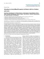
RA patients, irrespective of disease duration show marked shifts in the frequency of the peripheral blood B cell subsetsRA patients, irrespective of disease duration show marked shifts in the frequency of the peripheral blood B cell subsets. (a) Dot-plots of IgD versus
CD27 of peripheral blood CD19
+
B cells from representative healthy control and a long-standing rheumatoid arthritis (RA) patients illustrating the
differences in the frequency of each B cell subset. (b) Box-plots representing the 10th, 25th, 50th (median), 75th and 90th percentiles of the fre-
quencies of the total B cells (as a percentage of lymphocytes), total CD27
+
memory B cells, naïve IgD
+
CD27
-
B cells, pre-switch IgD
+
CD27
+
mem-
ory B cells and post-switch IgD-CD27+ memory B cells (each as a percentage of B cells) in the peripheral blood of healthy donors (n = 40, white
bars) and long-standing RA patients (n = 33, grey bars). *Significant (P < 0.01) difference from control donors.
Arthritis Research & Therapy Vol 11 No 3 Souto-Carneiro et al.
Page 6 of 12
(page number not for citation purposes)
donors (median RA 151.0 ± 35.8 cells/μl vs control: 148.5 ±
20.0 cells/μl).
In order to assess whether disease duration had an influence
on the B cell subset disparities between RA patients and
healthy individuals, the frequencies of the different B cell sub-
populations in the RA patients with long-standing disease
(natural history patients, median disease duration 13 years)
were compared with the baseline values of another group of
patients with shorter disease duration (median disease dura-
tion 4.4 years) who had enrolled in a clinical trial examining the
impact of MTX versus that of the combination of MTX and inf-
liximab (Table 1). Both groups had comparable frequencies
and absolute numbers of pre-switch memory B cells (median
shorter disease: 10.2 ± 1.5% vs long-standing disease: 10.4
± 1.3%, P = 0.8156; median shorter disease 14.4 ± 2.9 cells/
μl vs long-standing disease: 13.8 ± 4.7 cells/μl, P = 0.4003),
each of which was significantly (P < 0.03) lower than that
found in the healthy controls. Notably, however, the frequency
of post-switch memory B cells was significantly (P = 0.0025)
lower in the patient group with shorter disease duration when
compared with the long-standing group (median shorter dis-
ease: 9.7 ± 2.9% vs longer standing disease: 19.6 ± 2.9%).
Furthermore, the frequency of the total CD27
+
memory B cell
population was significantly (P = 0.0184) lower in the patient
group with shorter disease duration when compared with the
long-standing group (median shorter disease: 19.1 ± 3.2% vs
longer standing disease: 31.3 ± 3.8%).
Reduced peripheral blood pre-switch IgD
+
CD27
+
memory B cell population is not the result of CD27
shedding
Shedding of surface CD27 from peripheral blood pre-switch
memory B cells could account for the reduced frequency of
IgD
+
CD27
+
B cells in RA patients. Moreover, CD27
-
memory
B cells have been recently reported in healthy individuals
[14,16] and in SLE patients [15]. Therefore, to verify whether
the IgD
+
CD27
-
B cells in RA patients were actually naïve, sin-
gle-cell PCR analysis of immunoglobulin heavy chain variable
region (IgV
H
) genes from sorted peripheral blood, IgD
+
CD27
+
and IgD
+
CD27
-
RA B cells, was carried out to determine the
frequency of somatic mutations in those subsets. As expected,
most of the IgD
+
CD27
-
B cells expressed unmutated IgV
H
genes (76%) and those that were mutated contained few
mutations. Both the frequency of mutated Ig sequences and
the mutational frequency in the IgD
+
CD27
-
subset was signif-
icantly (P < 0.05) lower when compared with the IgD
+
CD27
+
subset. Moreover, the mean number of somatic mutations per
IgV
H
gene was significantly (P < 0.05) lower in the IgD
+
CD27
-
subset (Table 2). Thus, there was no evidence that the
IgD
+
CD27
-
population of RA patients contained a subgroup of
pre-switch memory B cells that failed to express CD27.
To confirm that CD27 shedding from the surface of memory B
cells was unlikely to be responsible for the reduction of the
pre-switch memory B cell population in the peripheral blood of
RA patients, the levels of soluble CD27 in the serum of RA
patients and control individuals were determined by ELISA. As
other studies have previously reported [31], no differences
could be detected between healthy donors and RA patients
(data not shown).
CD27
+
memory B cells accumulate in the synovial
membrane of RA patients
It is known that the rheumatoid synovial membrane is infiltrated
by B and plasma cells [3,6]. In order to determine the nature
of the B cell subsets that comprise the synovial B cell popula-
tion in long-standing RA, lymphocytes isolated from the syno-
vial membrane and from peripheral blood of RA patients with
long-standing disease were phenotyped by flow cytometry. As
depicted in Figure 2a for one representative patient, the major-
ity of the synovial B cells express CD27. In this patient cohort
the rheumatoid synovial membrane had significantly more
CD27-expressing B cells than the peripheral blood (61.4 ±
10.4% vs 25.1 ± 15.6% respectively; P < 0.0001; Figure 2b).
Both pre-switch IgD
+
IgM
+
memory cells and post-switch IgD
-
IgM
-
memory cells were found in the synovial tissue (data not
shown).
RA peripheral blood and synovial memory B cells
express abnormal chemokine receptor patterns
Chemokine receptor expression by RA B cells can be indica-
tive of their preferential capability for homing into the inflamed
tissues. To assess whether the expression of chemokine
receptors by RA synovial CD27
+
memory B cells could con-
tribute to the skewed distribution of the different B cell subsets
in the RA synovial membrane, the frequencies of CD27
+
mem-
ory B cells expressing specific chemokine receptors was also
Table 2
Mutational frequencies of B cell subsets in peripheral blood from patients with rheumatoid arthritis
Population Number of
Sequences
Frequency (number) of
mutated sequences
Mean (range) number of
mutated nucleotides
per mutated sequence
Overall mutational
frequency
Mutational frequency
for mutated
sequences
CD19
+
IgD
+
CD27
-
92 24%
a
(n = 22) 1.8 bp
a
(1 to 5 bp) 1.7 × 10
-3a
7.3 × 10
-3a
CD19
+
IgD
+
CD27
+
28 96% (n = 27) 12.3 bp (2 to 29 bp) 4.9 × 10
-2
5.1 × 10
-2
a
Significant difference (P < 0.05) between the CD19
+
IgD
+
CD27 and the CD19
+
IgD
+
CD27
+
subsets.
Available online />Page 7 of 12
(page number not for citation purposes)
determined. Notably, expression of CXCR1, CXCR2, CXCR4
and CCR2 was significantly elevated in synovial compared
with blood memory B cells (P < 0.00001) of either controls or
RA patients (Table 3). Moreover, RA blood B cells manifested
significantly enhanced expression of CXCR1, CXCR2 and
CCR2 and decreased expression of CXCR4 compared with
control blood (P < 0.02).
Anti-TNF therapy increases the pre-switch IgD
+
CD27
+
memory B cell population
As shown in Table 4, the group of patients receiving infliximab
plus MTX therapy exhibited significant (P < 0.05) improvement
of several laboratory and clinical parameters, including dis-
ease activity score, rheumatoid factor titres, ESR and the
number of swollen and tender joints. By contrast, after the sixth
treatment, the group receiving MTX monotherapy manifested
significant (P < 0.05) improvement only in disease activity
score and in the number of tender joints.
In order to determine whether anti-TNF therapy had an effect
on the distribution of the different peripheral blood B cell sub-
sets in RA, the frequencies of those subsets were calculated
after six administrations of study drug (Figure 3). At the time of
the first visit all the RA patients in both treatment groups had
comparable frequencies of IgD
+
CD27
-
naïve B cells,
IgD
+
CD27
+
pre-switch memory B cells and IgD
-
CD27
+
post-
switch B cells. Before any of the therapies had been initiated
all RA patients and healthy controls had a comparable fre-
quency of naïve B cells (median controls: 65.6 ± 1.7% vs inf-
liximab + MTX: 76.5 ± 4.4%; MTX: 81.4 ± 6.7%). Similarly,
the frequency of the post-switch memory B cell population in
all RA patients before treatment was comparable to healthy
donors (median control: 13.6 ± 1.0% vs infliximab + MTX: 9.7
± 2.6%; MTX: 8.7 ± 3.4%), whereas the pre-switch memory
B cell subset was significantly (P < 0.03) lower in RA patients
than in the control individuals (median control: 15.1 ± 1.1% vs
infliximab + MTX: 11.2 ± 1.5%; MTX: 5.4 ± 3.1%).
When compared with the baseline values from the first visit,
the RA patients receiving infliximab plus MTX significantly (P <
0.01) increased the frequency of total and pre-switch periph-
eral memory B cells after the sixth round of treatment (median
baseline 11.2 ± 1.5% vs after treatment 14.7 ± 1.8%) How-
ever, no changes could be observed in the patients receiving
MTX alone (median baseline 5.4 ± 3.1% vs after treatment 5.9
± 2.4%).
In order to determine whether the changes observed after
TNF-blockade exclusively involved B cells, the frequencies of
CD4 and CD8 T cells were analysed in both patient groups.
However, no changes were observed between visits in either
treatment group (data not shown).
Discussion
Abnormal distributions of peripheral B cell subsets, particu-
larly of CD27
+
memory B cells, have been reported in several
autoimmune diseases including RA [15-18,20,21]. However,
in RA there is no consensus about the nature of the abnormal-
ities, because some reports note an increase and others no
change in peripheral memory B cells [19-21]. In the present
study, we report that RA patients have a lower frequency of cir-
culating pre-switch IgD
+
CD27
+
memory B cells when com-
pared with healthy individuals. Importantly, memory B cells
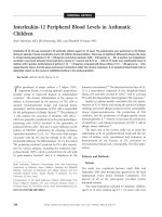
Figure 2
CD27
+
memory B cells tend to accumulate in the synovial membrane of RA patientsCD27
+
memory B cells tend to accumulate in the synovial membrane of
RA patients. (a) Histogram from a representative rheumatoid arthritis
(RA) patient showing the difference in CD27 expression in peripheral
blood and synovial CD19
+
B cells. Dashed line shows staining with the
isotype control and the solid line for CD27. (b) Box-plots representing
the 10th, 25th, 50th (median), 75th and 90th percentiles of the fre-
quency of CD27 expression by peripheral blood and synovial CD19
+
B
cells in long-standing RA (n = 10).
Arthritis Research & Therapy Vol 11 No 3 Souto-Carneiro et al.
Page 8 of 12
(page number not for citation purposes)
accumulate in the synovial membrane of subjects with RA,
suggesting that accumulation of pre-switch memory B cells
within inflamed tissue may contribute to a decrease in this B
cell subset in the blood. It should be pointed out that post-
switch IgD
-
CD27
+
memory B cells are also enriched in the
rheumatoid synovium, although these cells are not decreased
in the blood in early RA. This discrepancy may be explained by
the more complex homeostasis of post-switch memory B cells.
Although these cells accumulate in the synovium, they are also
generated in increased numbers in patients with RA [19]. As
a result, post-switch memory B cells accumulate not only in the
synovium but also in the blood of patients with long-standing
RA.
The human memory B cell population is heterogeneous, com-
prising mutated pre-switched IgD
+
CD27
+
and post-switch
IgD
-
CD27
+
B cells [13,32], that develop with age [33]. It is
widely accepted that post-switch IgD
-
CD27
+
memory B cells
are post-germinal centre highly mutated memory B cells
[11,13,32,34]. However, the function and the origin of the pre-
switch IgD
+
CD27
+
subpopulation is still a matter of contro-
versy. Despite some characterisation [35], it has not been
clearly established whether the IgD
+
CD27
+
memory popula-
tion only participates in T cell-independent immune responses,
because this population expresses heavily mutated Ig genes
[32]. The role of this population in autoimmune diseases has
been stressed by the finding that in the peripheral blood of
SLE and SS patients the pre-switch memory B cell subset is
markedly reduced [18,36]. However, in RA patients with long-
standing disease, previously published data suggested that
there might be an accumulation of CD27
+
memory cells in the
peripheral blood [20,21], and especially of the post-switch
IgD
-
CD27
+
memory subset, whereas the IgD
+
CD27
+
subset
was reported to be comparable to healthy donors [20]. We
were unable to confirm these findings. Instead, we observed
that patients with RA manifested a marked reduction in the
peripheral blood pre-switch IgD
+
CD27
+
subset. At baseline
the group of patients engaged in the clinical trial and those
with shorter disease duration had similarly lower frequencies
and absolute numbers of peripheral blood pre-switch
Table 3
Chemokine receptor expression by CD19
+
CD27
+
memory B cells from peripheral blood of healthy donors and RA patients and in RA
synovium
Peripheral blood Synovium
Chemokine receptor Control (n = 13) RA (n = 20) (n = 10)
CXCR1 5.0 ± 0.6% 16.9 ± 1.2%
a
39.0 ± 2.2%
b
CXCR2 3.4 ± 0.4% 7.7 ± 0.6%
a
31.2 ± 1.6%
b
CXCR4 60.0 ± 0.9% 47.5 ± 2.3%
a
92.1 ± 0.7%
b
CCR2 2.8 ± 0.4% 7.6 ± 0.6%
a
28.7 ± 1.8%
b
a
indicates significant difference (P < 0.02) between controls and patients with rheumatoid arthritis (RA);
b
indicates significant difference (P < 0.00001) between RA blood and synovium.
Data are means ± standard error of the mean.
Table 4
Laboratory and clinical parameters of the patients undergoing MTX or MTX plus infliximab therapy at baseline (first visit) and after
six treatments (seventh visit)
MTX (n = 8) MTX + infliximab (n = 15)
first visit seventh visit first visit seventh visit
DAS 44 5.2 ± 0.4 4.4 ± 0.4* 5.8 ± 0.5 3.9 ± 0.5*
RF (IU) 210.1 ± 117.9 146.4 ± 80.4 396.2 ± 178.4 257.3 ± 142.8*
CRP (mg/dl) 0.8 ± 0.3 0.6 ± 0.2 1.8 ± 0.5 1.1 ± 0.3
ESR (mm/hour) 36.9 ± 5.4 31.9 ± 5.0 60.2 ± 8.1 44.2 ± 5.5*
SJC 19.6 ± 4.6 14.0 ± 3.0 21.9 ± 3.1 11.6 ± 2.4*
TJC 25.8 ± 4.9 16.0 ± 4.3* 27.0 ± 4.5 12.1 ± 3.4*
Data are means ± standard error of the mean.
* P < 0.05 between first and seventh visit.
CRP = C-reactive protein; DAS = disease activity score; ESR = erythrocyte sedimentation rate; MTX = methotrexate; RF = rheumatoid factor;
SJC = swollen joints count; TJC = tender joints count.
Available online />Page 9 of 12
(page number not for citation purposes)
IgD
+
CD27
+
memory B cells compared with the group of
patients with long-standing disease, so this abnormality would
seem to be an integral feature of RA, independent of disease
duration. Preliminary assessment of a group of patients with
very early RA, who had disease duration of less than six weeks
and had received no DMARD therapy, also indicated a
decrease in IgD
+
CD27
+
pre-switch memory B cells and is
consistent with the conclusion that this abnormality in memory
B cell homeostasis is characteristic of RA independent of dis-
ease duration and DMARD therapy (R Moura and JE Fonseca,
unpublished data). Notably, in none of the analysed RA patient
groups did we observe an increase in the total CD27
+
memory
B cells when compared with control subjects. Nevertheless,
with long-standing disease in both the National Institutes of
Health and Japanese cohorts, the post-switch IgD
-
CD27
+
population was increased. This is likely to be related to the
increased production of post-switch memory B cells owing to
persistent immunological stimulation that is sufficient to over-
compensate for the enhanced sequestration of these cells in
the synovium.
The disparities between our data and the results previously
reported [19-21] may be explained by a number of factors,
including disease duration, cohort size and therapy. Impor-
tantly, most studies did not analyse patients in terms of dis-
ease duration, which as we report here can clearly affect
memory B cell subset distribution. It is notable that when total
CD27
+
memory B cells were analysed in patients receiving
only MTX therapy, a remarkably broad range of distributions
was noted, with some patients with very high and others with
low frequencies [19]. Finally, some studies did not separately
analyse the patients on TNF-blockers, which can alter periph-
Figure 3
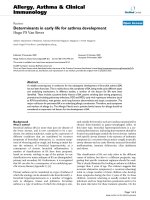
TNF blockade induces an increase in the frequency of peripheral blood total memory and pre-switch memory B cells, while reducing the circulating naïve B cellsTNF blockade induces an increase in the frequency of peripheral blood total memory and pre-switch memory B cells, while reducing the circulating
naïve B cells. (a) Box plots representing the 10th, 25th, 50th (median), 75th and 90th percentiles of the frequency of IgD
+
CD27
-
naïve B cells at the
time of the 1st and 7th visits for the patients in the infliximab plus methotrexate (MTX) group (n = 15, grey bars) and MTX monotherapy (n = 8, white
bars). (b) Box plots representing the 10th, 25th, 50th (median), 75th and 90th percentiles of the frequency of IgD
+
CD27
+
pre-switch memory B
cells at the time of the 1st and 7th visits for the patients in the infliximab plus MTX group (n = 15, grey bars) and MTX monotherapy (n = 8, white
bars). (c) Box plots representing the 10th, 25th, 50th (median), 75th and 90th percentiles of the frequency of IgD
-
CD27
+
post-switch memory B
cells at the time of the 1st and 7th visits for the patients in the infliximab plus MTX group (n = 15, grey bars) and MTX monotherapy (n = 8, white
bars). (d) Box plots representing the 10th, 25th, 50th (median), 75th and 90th percentiles of the frequency of total CD27
+
memory B cells at the
time of the 1st and 7th visits for the patients in the infliximab plus MTX group (n = 15, grey bars) and MTX monotherapy (n = 8, white bars). * Signif-
icant (P < 0.01) difference from 1st visit.
Arthritis Research & Therapy Vol 11 No 3 Souto-Carneiro et al.
Page 10 of 12
(page number not for citation purposes)
eral blood memory subset distribution as found here and
reported previously [19].
Elevated concentrations of matrix metalloproteinases, with the
capacity to cleave molecules of the TNF-family from the cell
surface [37], have been reported in RA synovial fluid [38], and
soluble CD27 is increased in the synovial fluid but not in the
serum of RA patients [31]. Therefore, it was possible that the
reduction of IgD
+
CD27
+
B cells in RA patients could be the
result of proteolytic cleavage of CD27 (a TNF-receptor family
member [39]) from the cell surface of pre-switch memory B
cells. However, the negligible number of somatic mutations in
the V
H
genes of the IgD
+
CD27
-
subset and the comparable
serological levels of soluble CD27 in RA patients and healthy
individuals discounted the possibility that CD27 had been
cleaved from the cell surface of pre-switch IgD
+
CD27
+
mem-
ory B cells, therefore giving them a false-naïve phenotype.
Chemokine receptor imbalances have been reported in sev-
eral autoimmune diseases [21,40-43]. Additionally, several
studies have provided strong evidence that in RA synovium
and synovial fluid, monocytes/macrophages, synovial fibrob-
lasts, FDC and mast cells have an increased expression of
either chemokines or their receptors responsible for B and T
cell recruitment [4,44-48]. Together with an accumulation of
both subsets of memory B cells in the synovial membrane of
RA patients with reduced peripheral blood IgD
+
CD27
+
B
cells, we have also observed significant shifts in the expres-
sion of several chemokine receptors in the RA peripheral
blood B cell subsets and in the synovial memory B cells: the
frequency of CD27
+
memory B cells expressing the pro-
inflammatory CXCR1, CXCR2 and CCR2 chemokine recep-
tors [49,50] was elevated in both peripheral blood and syn-
ovium; and contrary to RA peripheral blood, the large majority
of synovial membrane memory B cells expressed CXCR4. The
high frequency of RA peripheral blood and synovial membrane
memory B cells expressing pro-inflammatory chemokine
receptors, such as the IL8-receptors CXCR1 and CXCR2, or
CCR2 (a negative modulator of cytoskeleton rearrangement
and immature B cell migration [51]), stresses the potential role
of interactions of memory B cells with other effector cells of
the immune system that could contribute to the perpetuation
of chronic synovitis. An important, and novel, finding was the
abnormally increased frequency of CXCR4
+
memory B cells in
the RA synovium. CXCR4 is the receptor for the homeostatic
and pro-inflammatory chemokine CXCL12 and is expressed
by mature naïve B cells when they recirculate through germinal
centres of secondary lymphoid organs [52]. CXCR4 expres-
sion is essential for correct formation of the dark and light
zones of the germinal centre [53]. Moreover, CXCR4 is
involved in plasma cell function, because mice lacking CXCR4
expression present major abnormalities in plasma cell home-
ostasis [54], whereas human peripheral blood CD27
+
memory
B cells increase CXCR4 expression upon differentiation into
plasma cells [55]. The importance of CXCR4 expression in
synovial membrane inflammation has been emphasised by the
inhibition of collagen-induced arthritis by the CXCR4 antago-
nist T140, and by the finding of elevated CXCR4 gene expres-
sion in RA synovial biopsies with follicular-like lymphoid
structures [4]. Therefore, our data are consistent with the pos-
sibility that in RA elevated numbers of CXCR4
+
memory B
cells may be recruited into the synovial membrane where they
accumulate, and might be involved in seeding follicular-like
structures and/or differentiating into autoantibody-secreting
plasma cells, thus perpetuating the chronic synovitis.
Anti-TNF therapy in RA has been linked to a reduction of B
cells expressing the early activation marker CD23 in the
peripheral blood [56]. Neutralising TNF in RA diminishes the
production of pro-inflammatory cytokines in the joints, lowers
the levels of circulating IL1, IL6 and acute-phase proteins
[22,25], and decreases the serological levels of ICAM-1,
ICAM-3, VCAM-1, VEGF and E-selectin [57,58]. In the syn-
ovium, infliximab induces a major reduction of sublining T cells,
B cells and macrophages [59], decreases ectopic lymphoid
neogenesis [5], and lowers the expression of IL8 and MCP-1/
CCL2 [60]. A major finding in the present study was the nor-
malisation of the peripheral blood pre-switch memory B cell
population in RA patients who received anti-TNF therapy,
which was accompanied by a significant amelioration of sev-
eral clinical parameters. This recovery of circulating pre-switch
memory B cells after infliximab treatment contrasts with the
findings in a recent report using etanercept [19]. By blocking
TNF and LTα simultaneously with etanercept, the possible
effects of LTα on B cell homeostasis and GC formation make
it difficult to identify the individual contribution of TNF-block-
ade on the B cell compartment in RA. As a result of treating a
group of RA patients with infliximab, (that specifically blocks
TNF) we have found an increase in circulating pre-switch
memory B cells. It is difficult to be certain whether the increase
in pre-switch memory B cells specifically related to an
improvement in disease activity or specifically the blockade of
TNF. In this regard, the patients who received only MTX ther-
apy exhibited some improvement in clinical disease activity,
but it was not as profound as that noted in patients treated
with MTX and infliximab. Therefore, it is uncertain whether the
lack of significant change in circulating pre-switch memory B
cells in the patients receiving MTX resulted from a failure to
block TNF completely or from an incomplete clinical response.
The effect of TNF blockade, however, did appear to be spe-
cific for pre-switch memory B cells. Although, previous studies
reported a transient increase in T cell counts after four weeks
[61] and after repeated doses [62] of anti-TNF therapy, we
could not detect any significant changes in any of the T cell
populations in the patients treated with the combination of inf-
liximab and MTX. Therefore, these results suggest that neutral-
ising TNF alone might block the migration of memory B cells
into the synovium without a persistent effect on T cell traffick-
ing, which support recent findings showing that in RA patients
with disease remission after anti-TNF therapy (using either
Available online />Page 11 of 12
(page number not for citation purposes)
specific TNF blockers or simultaneous blockade of TNF and
LTα) the number and the density of B cells in the synovial
membrane and the frequency of ectopic germinal centres sig-
nificantly decreased [5].
Conclusions
In summary, the present findings indicate that the reduction of
circulating memory B cells in RA patients, particularly the pre-
switch memory subset, might be linked to their accumulation
in the inflamed rheumatoid synovium under the influence of
TNF.
Competing interests
The authors declare that they have no competing interests.
Authors' contributions
MMS-C collected data, designed experiments, carried out sta-
tistical analysis and wrote the manuscript. VM carried out the
clinical study and collected data. RG-M designed and carried
out the clinical study. RF, KT, TN, MW, MB and TAF collected
patient data. PEL designed and coordinated the study and
wrote the manuscript.
Acknowledgements
This work was supported by the National Institute of Arthritis and Musc-
uloskeletal and Skin Diseases Intramural Research Program. MMSC
was supported by the Marie Curie Intra-European Fellowship, LIF-
025885 and EULAR Young Investigator Award.
References
1. Firestein GS: Pathogenesis of rheumatoid arthritis: how early
is early? Arthritis Res Ther 2005, 7:157-159.
2. Kotzin BL: The role of B cells in the pathogenesis of rheuma-
toid arthritis. J Rheumatol Suppl 2005, 73:14-18.
3. Krenn V, Souto-Carneiro MM, Kim HJ, Berek C, Starostik P, Konig
A, Harms H, Müller-Hermelink HK: Histopathology and molecu-
lar pathology of synovial B-lymphocytes in rheumatoid arthri-
tis. Histol Histopathol 2000, 15:791-798.
4. Timmer TC, Baltus B, Vondenhoff M, Huizinga TW, Tak PP, Verweij
CL, Mebius RE, Pouw Kraan TC van der: Inflammation and
ectopic lymphoid structures in rheumatoid arthritis synovial
tissues dissected by genomics technology: identification of
the interleukin-7 signaling pathway in tissues with lymphoid
neogenesis. Arthritis Rheum 2007, 56:2492-2502.
5. Canete JD, Celis R, Moll C, Izquierdo E, Marsal S, Sanmarti R, Pal-
acin A, Lora D, de la Cruz J, Pablos JL: Clinical significance of
synovial lymphoid neogenesis and its reversal after anti-TNF-
{alpha} therapy in rheumatoid arthritis. Ann Rheum Dis 2009,
68:751-756.
6. Kim HJ, Krenn V, Steinhauser G, Berek C: Plasma cell develop-
ment in synovial germinal centers in patients with rheumatoid
and reactive arthritis. J Immunol 1999, 162:3053-3062.
7. Schroder AE, Greiner A, Seyfert C, Berek C: Differentiation of B
cells in the nonlymphoid tissue of the synovial membrane of
patients with rheumatoid arthritis. Proc Natl Acad Sci USA
1996, 93:221-225.
8. Weyand CM, Goronzy JJ: Ectopic germinal center formation in
rheumatoid synovitis. Ann N Y Acad Sci 2003, 987:140-149.
9. Edwards JC, Leandro MJ, Cambridge G: B lymphocyte depletion
in rheumatoid arthritis: targeting of CD20. Curr Dir Autoimmun
2005, 8:175-192.
10. Maurer D, Fischer GF, Fae I, Majdic O, Stuhlmeier K, Von Jeney N,
Holter W, Knapp W: IgM and IgG but not cytokine secretion is
restricted to the CD27+ B lymphocyte subset. J Immunol 1992,
148:3700-3705.
11. Shi Y, Agematsu K, Ochs HD, Sugane K: Functional analysis of
human memory B-cell subpopulations: IgD+CD27+ B cells
are crucial in secondary immune response by producing high
affinity IgM.
Clin Immunol 2003, 108:128-137.
12. Werner-Favre C, Bovia F, Schneider P, Holler N, Barnet M, Kindler
V, Tschopp J, Zubler RH: IgG subclass switch capacity is low in
switched and in IgM-only, but high in IgD+IgM+, post-germinal
center (CD27+) human B cells. Eur J Immunol 2001,
31:243-249.
13. Klein U, Rajewsky K, Kuppers R: Human immunoglobulin
(Ig)M+IgD+ peripheral blood B cells expressing the CD27 cell
surface antigen carry somatically mutated variable region
genes: CD27 as a general marker for somatically mutated
(memory) B cells. J Exp Med 1998, 188:1679-1689.
14. Fecteau JF, Cote G, Neron S: A new memory CD27-IgG+ B cell
population in peripheral blood expressing VH genes with low
frequency of somatic mutation. J Immunol 2006,
177:3728-3736.
15. Jacobi AM, Reiter K, Mackay M, Aranow C, Hiepe F, Radbruch A,
Hansen A, Burmester GR, Diamond B, Lipsky PE, Dörner T: Acti-
vated memory B cell subsets correlate with disease activity in
systemic lupus erythematosus: Delineation by expression of
CD27, IgD, and CD95. Arthritis Rheum 2008, 58:1762-1773.
16. Wei C, Anolik J, Cappione A, Zheng B, Pugh-Bernard A, Brooks J,
Lee EH, Milner EC, Sanz I: A new population of cells lacking
expression of CD27 represents a notable component of the B
cell memory compartment in systemic lupus erythematosus. J
Immunol 2007, 178:6624-6633.
17. Odendahl M, Keitzer R, Wahn U, Hiepe F, Radbruch A, Dörner T,
Bunikowski R: Perturbations of peripheral B lymphocyte
homoeostasis in children with systemic lupus erythematosus.
Ann Rheum Dis 2003, 62:851-858.
18. Hansen A, Odendahl M, Reiter K, Jacobi AM, Feist E, Scholze J,
Burmester GR, Lipsky PE, Dörner T: Diminished peripheral
blood memory B cells and accumulation of memory B cells in
the salivary glands of patients with Sjogren's syndrome.
Arthritis Rheum 2002, 46:2160-2171.
19. Anolik JH, Ravikumar R, Barnard J, Owen T, Almudevar A, Milner
EC, Miller CH, Dutcher PO, Hadley JA, Sanz I: Cutting edge: anti-
tumor necrosis factor therapy in rheumatoid arthritis inhibits
memory B lymphocytes via effects on lymphoid germinal cent-
ers and follicular dendritic cell networks. J Immunol 2008,
180:688-692.
20. Fekete A, Soos L, Szekanecz Z, Szabo Z, Szodoray P, Barath S,
Lakos G: Disturbances in B- and T-cell homeostasis in rheu-
matoid arthritis: suggested relationships with antigen-driven
immune responses. J Autoimmun
2007, 29:154-163.
21. Henneken M, Dorner T, Burmester GR, Berek C: Differential
expression of chemokine receptors on peripheral blood B
cells from patients with rheumatoid arthritis and systemic
lupus erythematosus. Arthritis Res Ther 2005, 7:R1001-1013.
22. Feldmann M: Development of anti-TNF therapy for rheumatoid
arthritis. Nat Rev Immunol 2002, 2:364-371.
23. Lin J, Ziring D, Desai S, Kim S, Wong M, Korin Y, Braun J, Reed E,
Gjertson D, Singh RR: TNFalpha blockade in human diseases:
An overview of efficacy and safety. Clin Immunol 2008,
126:121-136.
24. Lipsky PE, Heijde DM van der, St Clair EW, Furst DE, Breedveld
FC, Kalden JR, Smolen JS, Weisman M, Emery P, Feldmann M,
Harriman GR, Maini RN, Anti-Tumor Necrosis Factor Trial in Rheu-
matoid Arthritis with Concomitant Therapy Study Group: Inflixi-
mab and methotrexate in the treatment of rheumatoid
arthritis. Anti-Tumor Necrosis Factor Trial in Rheumatoid
Arthritis with Concomitant Therapy Study Group. N Engl J Med
2000, 343:1594-1602.
25. Maini RN, Feldmann M: How does infliximab work in rheuma-
toid arthritis? Arthritis Res 2002, 4:S22-28.
26. McMurray RW: Adhesion molecules in autoimmune disease.
Semin Arthritis Rheum 1996, 25:215-233.
27. Zhang Q, Lu Y, Proulx ST, Guo R, Yao Z, Schwarz EM, Boyce BF,
Xing L: Increased lymphangiogenesis in joints of mice with
inflammatory arthritis. Arthritis Res Ther 2007, 9:R118.
28. Hochberg MC, Chang RW, Dwosh I, Lindsey S, Pincus T, Wolfe
F: The American College of Rheumatology 1991 revised crite-
ria for the classification of global functional status in rheuma-
toid arthritis. Arthritis Rheum 1992, 35:498-502.
Arthritis Research & Therapy Vol 11 No 3 Souto-Carneiro et al.
Page 12 of 12
(page number not for citation purposes)
29. Yazdani-Biuki B, Stadlmaier E, Mulabecirovic A, Brezinschek R, Tilz
G, Demel U, Mueller T, Brickmann K, Graninger WB, Brezinschek
HP: Blockade of tumour necrosis factor {alpha} significantly
alters the serum level of IgG- and IgA-rheumatoid factor in
patients with rheumatoid arthritis. Ann Rheum Dis 2005,
64:1224-1226.
30. Souto-Carneiro MM, Longo NS, Russ DE, Sun HW, Lipsky PE:
Characterization of the human Ig heavy chain antigen binding
complementarity determining region 3 using a newly devel-
oped software algorithm, JOINSOLVER. J Immunol 2004,
172:6790-6802.
31. Tak PP, Hintzen RQ, Teunissen JJ, Smeets TJ, Daha MR, van Lier
RA, Kluin PM, Meinders AE, Swaak AJ, Breedveld FC: Expression
of the activation antigen CD27 in rheumatoid arthritis. Clin
Immunol Immunopathol 1996, 80:129-138.
32. Tangye SG, Good KL: Human IgM+CD27+ B cells: memory B
cells or "memory" B cells? J Immunol 2007, 179:13-19.
33. Chong Y, Ikematsu H, Yamaji K, Nishimura M, Nabeshima S, Kash-
iwagi S, Hayashi J: CD27(+) (memory) B cell decrease and
apoptosis-resistant CD27(-) (naive) B cell increase in aged
humans: implications for age-related peripheral B cell devel-
opmental disturbances. Int Immunol 2005, 17:383-390.
34. Bende RJ, van Maldegem F, Triesscheijn M, Wormhoudt TA, Guijt
R, van Noesel CJ: Germinal centers in human lymph nodes con-
tain reactivated memory B cells. J Exp Med 2007,
204:2655-2665.
35. Weller S, Braun MC, Tan BK, Rosenwald A, Cordier C, Conley ME,
Plebani A, Kumararatne DS, Bonnet D, Tournilhac O, Tchernia G,
Steiniger B, Staudt LM, Casanova JL, Reynaud CA, Weill JC:
Human blood IgM 'memory' B cells are circulating splenic mar-
ginal zone B cells harboring a prediversified immunoglobulin
repertoire. Blood 2004, 104:3647-3654.
36. Wehr C, Eibel H, Masilamani M, Illges H, Schlesier M, Peter HH,
Warnatz K: A new CD21low B cell population in the peripheral
blood of patients with SLE. Clin Immunol 2004, 113:161-171.
37. Gearing AJ, Beckett P, Christodoulou M, Churchill M, Clements J,
Davidson AH, Drummond AH, Galloway WA, Gilbert R, Gordon JL,
Leber TM, Mangan M, Miller K, Nayee P, Owen K, Patel S, Thomas
W, Wells G, Wood LM, Woolley K: Processing of tumour necro-
sis factor-alpha precursor by metalloproteinases. Nature
1994, 370:555-557.
38. Yoshihara Y, Nakamura H, Obata K, Yamada H, Hayakawa T,
Fujikawa K, Okada Y: Matrix metalloproteinases and tissue
inhibitors of metalloproteinases in synovial fluids from
patients with rheumatoid arthritis or osteoarthritis. Ann
Rheum Dis 2000, 59:455-461.
39. Prasad KV, Ao Z, Yoon Y, Wu MX, Rizk M, Jacquot S, Schlossman
SF: CD27, a member of the tumor necrosis factor receptor
family, induces apoptosis and binds to Siva, a proapoptotic
protein. Proc Natl Acad Sci USA 1997, 94:6346-6351.
40. Bruhl H, Wagner K, Kellner H, Schattenkirchner M, Schlondorff D,
Mack M: Surface expression of CC- and CXC-chemokine
receptors on leucocyte subsets in inflammatory joint diseases.
Clin Exp Immunol 2001, 126:551-559.
41. Hansen A, Reiter K, Ziprian T, Jacobi A, Hoffmann A, Gosemann
M, Scholze J, Lipsky PE, Dörner T: Dysregulation of chemokine
receptor expression and function by B cells of patients with
primary Sjogren's syndrome. Arthritis Rheum 2005,
52:2109-2119.
42. Loetscher P, Moser B: Homing chemokines in rheumatoid
arthritis. Arthritis Res 2002, 4:233-236.
43. Schmutz C, Hulme A, Burman A, Salmon M, Ashton B, Buckley C,
Middleton J: Chemokine receptors in the rheumatoid syn-
ovium: upregulation of CXCR5. Arthritis Res Ther 2005,
7:R217-229.
44. Carlsen HS, Baekkevold ES, Morton HC, Haraldsen G, Brandt-
zaeg P: Monocyte-like and mature macrophages produce
CXCL13 (B cell-attracting chemokine 1) in inflammatory
lesions with lymphoid neogenesis. Blood 2004,
104:3021-3027.
45. Katschke KJ Jr, Rottman JB, Ruth JH, Qin S, Wu L, LaRosa G,
Ponath P, Park CC, Pope RM, Koch AE: Differential expression
of chemokine receptors on peripheral blood, synovial fluid,
and synovial tissue monocytes/macrophages in rheumatoid
arthritis. Arthritis Rheum 2001, 44:1022-1032.
46. Ruschpler P, Lorenz P, Eichler W, Koczan D, Hanel C, Scholz R,
Melzer C, Thiesen HJ, Stiehl P: High CXCR3 expression in syn-
ovial mast cells associated with CXCL9 and CXCL10 expres-
sion in inflammatory synovial tissues of patients with
rheumatoid arthritis. Arthritis Res Ther 2003, 5:R241-252.
47. Shi K, Hayashida K, Kaneko M, Hashimoto J, Tomita T, Lipsky PE,
Yoshikawa H, Ochi T: Lymphoid chemokine B cell-attracting
chemokine-1 (CXCL13) is expressed in germinal center of
ectopic lymphoid follicles within the synovium of chronic
arthritis patients.
J Immunol 2001, 166:650-655.
48. Tsubaki T, Takegawa S, Hanamoto H, Arita N, Kamogawa J,
Yamamoto H, Takubo N, Nakata S, Yamada K, Yamamoto S,
Yoshie O, Nose M: Accumulation of plasma cells expressing
CXCR3 in the synovial sublining regions of early rheumatoid
arthritis in association with production of Mig/CXCL9 by syno-
vial fibroblasts. Clin Exp Immunol 2005, 141:363-371.
49. Moser B, Loetscher P: Lymphocyte traffic control by chemok-
ines. Nat Immunol 2001, 2:123-128.
50. Zlotnik A, Yoshie O: Chemokines: a new classification system
and their role in immunity. Immunity 2000, 12:121-127.
51. Flaishon L, Becker-Herman S, Hart G, Levo Y, Kuziel WA, Shachar
I: Expression of the chemokine receptor CCR2 on immature B
cells negatively regulates their cytoskeletal rearrangement
and migration. Blood 2004, 104:933-941.
52. Campbell DJ, Kim CH, Butcher EC: Chemokines in the systemic
organization of immunity. Immunol Rev 2003, 195:58-71.
53. Allen CD, Ansel KM, Low C, Lesley R, Tamamura H, Fujii N, Cyster
JG: Germinal center dark and light zone organization is medi-
ated by CXCR4 and CXCR5. Nat Immunol 2004, 5:943-952.
54. Hargreaves DC, Hyman PL, Lu TT, Ngo VN, Bidgol A, Suzuki G,
Zou YR, Littman DR, Cyster JG: A coordinated change in chem-
okine responsiveness guides plasma cell movements. J Exp
Med 2001, 194:45-56.
55. Muehlinghaus G, Cigliano L, Huehn S, Peddinghaus A, Leyen-
deckers H, Hauser AE, Hiepe F, Radbruch A, Arce S, Manz RA:
Regulation of CXCR3 and CXCR4 expression during terminal
differentiation of memory B cells into plasma cells. Blood
2005, 105:3965-3971.
56. De Miguel S, Jover JA, Vadillo C, Judez E, Loza E, Fernandez-Guti-
errez B: B cell activation in rheumatoid arthritis patients under
infliximab treatment. Clin Exp Rheumatol 2003, 21:726-732.
57. Gonzalez-Gay MA, Garcia-Unzueta MT, De Matias JM, Gonzalez-
Juanatey C, Garcia-Porrua C, Sanchez-Andrade A, Martin J, Llorca
J: Influence of anti-TNF-alpha infliximab therapy on adhesion
molecules associated with atherogenesis in patients with
rheumatoid arthritis. Clin Exp Rheumatol 2006, 24:373-379.
58. Klimiuk PA, Sierakowski S, Domyslawska I, Fiedorczyk M, Chw-
iecko J: Reduction of soluble adhesion molecules (sICAM-1,
sVCAM-1, and sE-selectin) and vascular endothelial growth
factor levels in serum of rheumatoid arthritis patients follow-
ing multiple intravenous infusions of infliximab. Arch Immunol
Ther Exp (Warsz) 2004, 52:36-42.
59. Smeets TJ, Kraan MC, van Loon ME, Tak PP: Tumor necrosis fac-
tor alpha blockade reduces the synovial cell infiltrate early
after initiation of treatment, but apparently not by induction of
apoptosis in synovial tissue. Arthritis Rheum 2003,
48:2155-2162.
60. Taylor PC, Peters AM, Paleolog E, Chapman PT, Elliott MJ,
McCloskey R, Feldmann M, Maini R: Reduction of chemokine
levels and leukocyte traffic to joints by tumor necrosis factor
alpha blockade in patients with rheumatoid arthritis. Arthritis
Rheum 2000, 43:38-47.
61. Ohshima S, Saeki Y, Mima T, Sasai M, Nishioka K, Ishida H,
Shimizu M, Suemura M, McCloskey R, Kishimoto T: Long-term
follow-up of the changes in circulating cytokines, soluble
cytokine receptors, and white blood cell subset counts in
patients with rheumatoid arthritis (RA) after monoclonal anti-
TNF alpha antibody therapy. J Clin Immunol 1999, 19:305-313.
62. Lorenz HM, Grunke M, Hieronymus T, Antoni C, Nusslein H, Schai-
ble TF, Manger B, Kalden JR: In vivo blockade of tumor necrosis
factor-alpha in patients with rheumatoid arthritis: longterm
effects after repeated infusion of chimeric monoclonal anti-
body cA2. J Rheumatol 2000, 27:304-310.