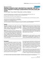
Báo cáo y học: "Antiphospholipid antibody profiles in lupus nephritis with glomerular microthrombosis: a prospective study of 124 cases" pot
Bạn đang xem bản rút gọn của tài liệu. Xem và tải ngay bản đầy đủ của tài liệu tại đây (922.83 KB, 9 trang )
Open Access
Available online />Page 1 of 9
(page number not for citation purposes)
Vol 11 No 3
Research article
Antiphospholipid antibody profiles in lupus nephritis with
glomerular microthrombosis: a prospective study of 124 cases
Hui Zheng
1
*, Yi Chen
1
*, Wen Ao
1
, Yan Shen
1
, Xiao-wei Chen
1
, Min Dai
1
, Xiao-dong Wang
1
, Yu-
cheng Yan
2
and Cheng-de Yang
1
1
Department of Rheumatology, Renji Hospital, Shanghai Jiaotong University School of Medicine, 145 Shan Dong Zhong Road, Shanghai, 200001,
PR China
2
Department of Nephrology, Renji Hospital, Shanghai Jiaotong University School of Medicine, 145 Shan Dong Zhong Road, Shanghai, 200001, PR
China
* Contributed equally
Corresponding author: Cheng-de Yang,
Received: 17 Jan 2009 Revisions requested: 6 Mar 2009 Revisions received: 30 Apr 2009 Accepted: 22 Jun 2009 Published: 22 Jun 2009
Arthritis Research & Therapy 2009, 11:R93 (doi:10.1186/ar2736)
This article is online at: />© 2009 Zheng et al.; licensee BioMed Central Ltd.
This is an open access article distributed under the terms of the Creative Commons Attribution License ( />),
which permits unrestricted use, distribution, and reproduction in any medium, provided the original work is properly cited.
Abstract
Introduction Glomerular microthrombosis (GMT) is a common
vascular change in patients with lupus nephritis (LN). The
mechanism underlying GMT is largely unknown. Although
several studies have reported the association of
antiphospholipid antibodies (aPL) with GMT, the relation
between GMT and aPL remains controversial. Previous studies
have demonstrated that some aPL could bind to several
hemostatic and fibrinolytic proteases that share homologous
enzymatic domains. Of the protease-reactive aPL, some can
inhibit the anticoagulant activity of activated protein C and the
fibrinolytic function of plasmin, and hinder the antithrombin
inactivation of thrombin. The purpose of this study was to
investigate the prevalence of GMT in LN patients and examine
the relation between the aPL profiles (including some protease-
reactive aPL) and GMT.
Methods Renal biopsy specimens were examined for the
presence of glomerular microthrombi. Plasma samples from 25
LN patients with GMT (LN-GMT group) and 99 LN patients
without GMT (LN-non-GMT group) were tested for lupus
anticoagulant and antibodies against cardiolipin, β2
glycoprotein I, plasmin, thrombin, tissue plasminogen activator,
and annexin II.
Results The prevalence of GMT in LN patients was 20.2%.
Compared with the LN-non-GMT group, the LN-GMT group had
an elevated systemic lupus erythematosus disease activity
index; elevated renal tissue injury activity and chronicity indices;
elevated serum creatinine, blood urea nitrogen, and proteinuria
levels; a lower serum C3 level and much intense glomerular C3,
C1q staining; and a higher frequency of hypertension (P < 0.05
for all). Additionally, the detection rate of lupus anticoagulant,
immunoglobulin G (IgG) anti-β2 glycoprotein I and anti-thrombin
antibodies were higher in the LN-GMT group than in the LN-non-
GMT group (P < 0.05 for all). No statistical differences were
found in the detection rates of IgG anti-cardiolipin, plasmin,
tissue plasminogen activator, or annexin II antibodies (P > 0.05
for all). No detectable difference in IgM autoantibodies to the
above antigens was observed between the two groups.
Conclusions GMT occurs in approximately 20.2% of LN
patients. Patients with GMT have severer renal tissue injuries
and poorer renal functions than patients without GMT. The lupus
anticoagulant and antibodies against β2 glycoprotein I and
thrombin may play a role in GMT.
Introduction
Systemic lupus erythematosus (SLE) is a multisystem autoim-
mune disease. Approximately 40 to 85% of SLE patients
develop renal involvement, lupus nephritis (LN), which is char-
A2: annexin II; aCL: anticardiolipin antibody; ANA: antinuclear antibodies; anti-dsDNA: anti-double-stranded DNA antibody; anti-RNP: anti-ribonucle-
oprotein antibody; aPL: antiphospholipid antibodies; APS: antiphospholipid syndrome; APSN: antiphospholipid syndrome nephropathy; β2GPI: β2
glycoprotein I; ELISA: enzyme-linked immunosorbent assay; FITC: fluorescein isothiocyanate conjugate; GMT: glomerular microthrombosis; GPL: G
phospholipid; H&E: hematoxylin and eosin; Ig: immunoglobulin; ISN: International Society of Nephrology; LAC: lupus anticoagulant; LN: lupus nephri-
tis; MPL: M phospholipid; PBS: phosphate-buffered saline; RPS: Renal Pathology Society; SD: standard deviation; SLE: systemic lupus erythemato-
sus; SLEDAI: systemic lupus erythematosus disease activity index; t-PA: tissue plasminogen activator.
Arthritis Research & Therapy Vol 11 No 3 Zheng et al.
Page 2 of 9
(page number not for citation purposes)
acterized by proteinuria, hematuria, and cylindruria, and even
renal failure in some cases during the course of the disease [1-
3]. Glomerular microthrombosis (GMT) is seen in approxi-
mately 30 to 33% of patients with LN and it is especially seen
in those with severe diffuse proliferative glomerulonephritis
[4,5]. Previous studies have indicated that LN patients with
GMT have more severe renal tissue injuries, poorer responses
to routine treatments, and worse renal outcomes than patients
without GMT [5-12]. Thus, apart for the fact that the immune
complex could directly elicit glomerular injuries, GMT may be
another important cause of renal injury and dysfunction in a
subset of LN patients.
Antiphospholipid antibodies (aPL) are a heterogeneous group
of antibodies directed against negatively-charged phospholip-
ids, phospholipid-binding proteins, and phospholipid-protein
complexes. The laboratory criteria of update criteria for definite
antiphospholipid syndrome (APS) includes the lupus antico-
agulant (LAC), the anticardiolipin antibody (aCL), and the anti-
β2 glycoprotein I (β2GPI) antibody [13]. GMT has been asso-
ciated with aPL in LN patients in some studies [4,6,7,9-12,14-
18], but not in others [5,8,19-21]. Recent studies of seven
monoclonal immunoglobulin (Ig) G aCLs from two APS
patients demonstrated that five aCLs reacted with several
hemostatic and fibrinolytic proteases that share homologous
enzymatic domains. Autoantibodies to these proteases,
including thrombin, plasmin, tissue plasminogen activator (t-
PA), prothrombin, protein C, protein S, annexin II (A2), annexin
V, and coagulation factor X, were found in APS patients [22-
28]. Importantly, our previous studies have demonstrated that
some protease-reactive monoclonal IgG aCL can interfere
with the inactivation of thrombin by antithrombin and decrease
the function of plasmin and activated protein C [22,23,29]. In
addition, aPL may bind to A2 and inhibit A2-dependent plas-
min generation [30]. Therefore, aPL may promote various
thrombotic events by interacting with these hemostatic and
fibrinolytic proteases [31]. It is of interest to investigate
whether some protease-reactive aPL are present in LN
patients with GMT. In order to address this, we carried out a
prospective study of 124 LN patients undergoing renal biopsy
to further investigate the prevalence of GMT and examine the
significance of aPL in LN patients with GMT.
Materials and methods
Patients
The study comprised 124 consecutive patients with LN who
had been referred to the Renji Hospital at the Shanghai Jiao-
tong University School of Medicine for renal biopsy between
September 2007 and October 2008. All patients fulfilled the
American College of Rheumatology classification criteria for
the diagnosis of SLE [32]. In addition, all patients had clinical
evidence of LN, which was further proven by pathologic exam-
ination of renal biopsy specimens.
Plasma samples were collected on the day of renal biopsy. The
following demographic, clinical, and serologic data were col-
lected at the time of the renal biopsy: sex; age; duration of SLE
and LN; history of symptomatic thrombosis; levels of blood
urea nitrogen, serum creatinine, serum C3, C4, C1q and pro-
teinuria; prevalence of systemic hypertension; and presence
or absence of antinuclear antibodies (ANA), anti-Sm, anti-ribo-
nucleoprotein (anti-RNP), anti-double-stranded DNA (anti-
dsDNA), anti-histone, and anti-nucleosome antibodies. The
systemic lupus erythematosus disease activity index (SLEDAI)
was used to estimate global disease activity.
In addition, another 100 healthy adults were randomly
recruited to serve as normal controls. All patients were care-
fully examined if they have other potential causes for GMT,
such as systemic sclerosis, thrombotic thrombocytopenic pur-
pura/hemolytic uremic syndrome, malignant hypertension, dia-
betic nephropathy, postpartum renal failure, preeclampsia, HIV
infection, or cyclosporine therapy [6,8].
The patients were informed of the purpose of the study and
gave their informed consent. The institutional review board of
Shanghai Jiaotong University approved this study.
Renal histology
All patients underwent ultrasound-guided renal needle biopsy.
The renal tissues obtained by biopsy were fixed in 10% neutral
buffered formalin, gradually dehydrated, and embedded in par-
affin. Paraffin sections were stained with H&E, periodic acid-
Schiff, Masson's trichrome, and periodic acid-silver methen-
amine. Small portions of fresh renal tissue were snap frozen
and 4 μm cryostat-cut sections were incubated with fluores-
cein isothiocyanate conjugate (FITC)-conjugated rabbit antis-
era against human IgG, IgA, IgM, C1q, or C3 (Dako, Glostrup,
Denmark) and were examined by direct immunofluorescence
[8]. Biopsy specimens were classified using the International
Society of Nephrology/Renal Pathology Society (ISN/RPS)
2003 classification of LN [33]. In addition, particular attention
was paid to GMT. Thrombosis was considered to be present
when thrombi with fibrin-consistent staining properties were
clearly seen by light microscopy occluding the glomerular cap-
illary lumens. In order to confirm the presence of fibrin GMT,
cryostat sections were also incubated with FITC-conjugated
rabbit antiserum against human fibrinogen (Dako, Glostrup,
Denmark). When necessary, laser confocal microscopy was
used to further determine whether the microthrombi were
within the glomerular capillary lumens or not. The patients
were divided into two groups (LN-GMT group and LN-non-
GMT group) based on the presence or absence of GMT.
Activity and chronicity indices of renal tissue injury
Renal tissue injury was evaluated using activity and chronicity
indices as previously reported by Austin and colleagues [34].
The activity index was the sum of the scores (on a scale of 1
to 3) for endocapillary proliferation, karyorrhexis, fibrinoid
Available online />Page 3 of 9
(page number not for citation purposes)
necrosis (with the score for fibrinoid necrosis multiplied by 2),
cellular crescents (with the score multiplied by 2), hyaline
deposits, leukocyte exudation, and interstitial inflammation.
The score on the chronicity index was the sum of the scores
(on a scale of 1 to 3) for glomerular sclerosis, fibrous cres-
cents, tubular atrophy, and interstitial fibrosis.
Immune complex deposits
The intensity of glomerular immunofluorescence staining for
IgG, IgM, IgA, C3, and C1q was semiquantitatively scored on
a scale of 0 to 3, where 0 = no glomerular staining, 1 = mild
glomerular staining, 2 = moderate glomerular staining, and 3
= intense glomerular staining [19].
LAC assay
LAC was detected using a LAC Screen/LAC Confirm Kit
(Instrumentation Laboratory Company, Lexington, MA, USA)
on the IL coagulation system (Instrumentation Laboratory
Company, Lexington, MA, USA) to determine the dilute Rus-
sell's viper venom time according to the manufacturer's
instructions. The results of LAC are expressed as normalized
LAC ratios, and ratios more than 1.2 were considered positive.
aCL assay
The presence of IgG and IgM aCL were detected using quan-
titative ELISA kits (EUROIMMUN Medizinische Labordiagnos-
tika AG, Lübeck, Germany) according to the manufacturer's
instructions. The levels of aCL were expressed as standard
units for either G phospholipid (GPL) units/ml or M phosphol-
ipid (MPL) units/ml and values more than 12 GPL units/ml or
more than 12 MPL units/ml were considered positive.
Anti-β2GPI antibodies
IgG and IgM anti-β2GPI antibodies were measured with
ELISA kits (EUROIMMUN Medizinische Labordiagnostika AG,
Lübeck, Germany). The levels of anti-β2GPI antibodies were
expressed in relative units and values of more than 20 RU/ml
were considered positive for either the IgG or IgM isotype.
Assay for anti-plasmin, thrombin, t-PA, and A2
antibodies
For anti-plasmin, anti-thrombin, and anti-t-PA antibodies
detection, high-binding ELISA plates (Costar, Cambridge, MA,
USA) were coated with 5 μg/ml of human plasmin or α-
thrombin (Haematologic Technologies, Essex Junction, VT,
USA) or 10 μg/ml human t-PA (Merck KGaA, Darmstadt, Ger-
many) in 0.01 M PBS, pH 7.4. Following an overnight incuba-
tion at 4°C, the plates were blocked with PBS containing
0.3% gelatin and incubated for two hours at 37°C. Plasma
samples were diluted with PBS containing 0.1% gelatin,
plated in duplicate, and incubated for one hour at 37°C. After
washing with PBS containing 0.1% Tween-20, the bound
human IgG was detected with affinity-isolated, antigen-spe-
cific, horseradish peroxidase-conjugated goat anti-human IgG
(Fc specific; Sigma-Aldrich, St. Louis, MO, USA). After an
additional incubation for one hour at 37°C, 100 μl of the
tetramethylbenzidine/hydrogen peroxidase substrate solution
(Kirkegard & Perry Labs, Gaithersburg, MD, USA) was added
and the reaction terminated with 50 μl of 0.5 M sulfuric acid.
The results were read at a wavelength of 450 nm in a micro-
plate reader (Bio-Rad Laboratories, Hercules, CA, USA). The
ELISA for the detection of anti-A2 antibodies was similar
except for the following modifications. The wells were coated
with 10 μg/ml (in PBS) human A2 generated in our laboratory
(Ao W et al, unpublished observations). The bound human IgG
or IgM against A2 were all detected with affinity-isolated, anti-
gen-specific, horseradish peroxidase-labeled goat anti-human
IgG or IgM (Fc specific; Sigma-Aldrich, St. Louis, MO, USA).
For each of the above antibodies, the mean absorbance plus
three times the standard deviation (SD) of the normal controls
was used as the cutoff for determining positivity.
Statistical analysis
Categorical data between different groups were compared by
chi-squared test or Fisher's exact test when required. For con-
tinuous variables, the comparisons were carried out using the
student's t-test for two independent samples or the Mann-
Whitney U test for non-normal data. P values less than 0.05
were considered statistically significant.
Results
Demographic, clinical, and laboratory characteristics of
the LN patients
This study examined 124 LN patients (105 women and 19
men) with a mean age (± SD) of 33 ± 14 years. There were 25
patients in the LN-GMT group (22 women and 3 men) and 99
patients in the LN-non-GMT group (83 women and 16 men).
The mean age (± SD) of the two groups was 35 ± 14 and 33
± 14 years, respectively. Among the 100 normal controls, 85
were women and 15 were men. The mean age (± SD) of the
control group was 35 ± 10 years. No significant difference
was seen among the three groups in terms of age or gender
(P > 0.05 for all).
SLEDAI, blood urea nitrogen, serum creatinine, and proteinu-
ria levels were all significantly greater in the LN-GMT group
than in the LN-non-GMT group (P < 0.05 for all). In addition,
patients in the LN-GMT group also had a higher frequency of
systemic hypertension (P < 0.05). The serum C3 level was
significantly lower in the LN-GMT group than in the LN-non-
GMT group (P < 0.05). However, the duration of SLE or LN,
the level of serum C4 or C1q, as well as the antecedent history
of thrombosis, was not statistically different between the two
groups (P > 0.05 for all). Except for a lower frequency of anti-
Sm antibodies in the LN-GMT group (P < 0.05), we failed to
find any association between GMT and ANA, anti-ribonucleo-
protein, anti-dsDNA, anti-histone, or anti-nucleosome antibod-
ies (P > 0.05 for all; Table 1). None of the 124 patients had
other potential causes for GMT as described previously.
Arthritis Research & Therapy Vol 11 No 3 Zheng et al.
Page 4 of 9
(page number not for citation purposes)
Renal biopsy findings
The presence of GMT was detected in 25 of the 124 patients
both by light microscopy and immunofluorescence micros-
copy (Figure 1). The distribution of the ISN/RPS classification
of the 124 patients was as follows: 3 were class I, 2 were
class II, 15 were class III, 39 were class IV, 28 were class V,
21 were class (III + V), 16 were class (IV + V), and no patients
were class VI (Table 2). Class IV LN was the most frequently
observed form in the LN-GMT group (64%), while class V had
a slightly higher frequency (28.3%) than any other class in the
LN-non-GMT group. Among the patients with class IV LN,
41% developed GMT. No GMT was detected in patients with
class I, II, or III LN.
When compared with the LN-non-GMT group, the LN-GMT
group was more likely to be associated with class IV LN (P <
0.05). The activity and chronicity indices were also signifi-
cantly higher in the LN-GMT group than in the LN-non-GMT
group (P < 0.05 for all), with median (25th–75th percentile)
activity index values of 8 (6 to 9.5) and 3 (2 to 5), respectively,
and median (25th–75th percentile) chronicity index values of
3 (2 to 4) and 2 (1 to 3), respectively (Table 2). A significant
relation was found between the presence of GMT and the
intensity of glomerular IgM, C3, or C1q staining (P < 0.05 for
all; Table 3).
aPL profiles in LN patients with or without GMT
LAC was detected in 7 (28.0%) of the 25 LN patients with
GMT and in 8 (8.1%) of the 99 patients without GMT. IgG
anti-β2GPI antibodies were detected in 32.0% of the LN-GMT
group and 11.1% of the LN-non-GMT group (Figure 2a). Nine
patients (36.0%) in the LN-GMT group and 17 patients
(17.2%) in the LN-non-GMT group had significant levels of
IgG anti-thrombin antibodies (Figure 2b). GMT was strongly
associated with LAC, IgG anti-β2GPI, and anti-thrombin anti-
bodies (P < 0.05 for all; Table 4; Figure 2). The detection rate
of IgG aCL, anti-plasmin, anti-t-PA, or anti-A2 antibodies in the
LN-GMT group was not statistically different from the LN-non-
GMT group (P > 0.05 for all; Table 4). IgM aCL, anti-β2GPI,
and anti-A2 antibodies were also detected and no significant
Table 1
Demographic, clinical, and laboratory characteristics of the LN patients*
LN-GMT
(n = 25)
LN-non-GMT
(n = 99)
P value
Sex (male/female) 3/22 16/83 0.837
Age (years) 35 ± 14 33 ± 14 0.485
SLE duration (months) 48 (8 to 126) 19 (4 to 69.5) 0.096
LN duration (months) 19 (1 to 78) 4.5 (1–24) 0.101
SLEDAI 17 ± 6 12 ± 6 0.002
Proteinuria (g/24 hours) 3.24 (2.22 to 4.66) 2.01 (1.19 to 3.66) 0.005
Serum creatinine (μmol/L) 99.7(67.85 to 118.5) 59.6(49.08 to 82.75) <0.001
Blood urea nitrogen (mmol/L) 11.3 (6.05 to 15) 6.4 (4.9 to 10) 0.007
Serum C3 (g/L) 0.44 ± 0.23 0.62 ± 0.26 0.003
Serum C4 (g/L) 0.09 (0.06 to 0.17) 0.10 (0.05 to 0.17) 0.717
Serum C1q (g/L) 0.38 (0.34 to 0.43) 0.33 (0.29 to 0.40) 0.092
ANA (positive/negative)
†
20/3 82/8 0.837
Anti-Sm (positive/negative) 3/20 32/58 0.037
Anti-RNP (positive/negative) 4/19 34/56 0.065
Anti-dsDNA (positive/negative)
‡
20/5 67/29 0.312
Anti-nucleosome(positive/negative)
¶
10/6 21/25 0.246
Anti-histone (positive/negative)
§
10/5 30/19 0.703
*Except where indicated otherwise, values are expressed as mean ± standard deviation or median (25th–75th percentile) as appropriate.
†
In two patients in the LN-GMT group and nine patients in the LN-non-GMT group, ANA, anti-RNP, and anti-Sm antibodies were not detected.
‡
In three patients in the LN-non-GMT group, anti-dsDNA antibodies were not detected.
¶
In nine patients in the LN-GMT group and fifty three patients in the LN-non-GMT group, anti-nucleosome antibodies were not detected.
§
In 10 patients in the LN-GMT group and 50 patients in the LN-non-GMT group anti-histone antibodies were not detected.
ANA = antinuclear antibodies; anti-dsDNA = anti-double-stranded DNA antibody; anti-RNP = anti-ribonucleoprotein antibody; GMT = glomerular
microthrombosis; LN = lupus nephritis; SLE = systemic lupus erythematosus; SLEDAI = systemic lupus erythematosus disease activity index.
Available online />Page 5 of 9
(page number not for citation purposes)
differences were observed in the detection rate of any IgM
antibody between the two groups (P > 0.05 for all; data not
shown).
Discussion
GMT is a common vascular change in patients with LN, espe-
cially in those with severe diffuse proliferative glomerulonephri-
tis. This prospective study of 124 consecutive patients
undergoing renal biopsy demonstrates that GMT occurred in
approximately 20.2% of the LN patients. This prevalence is
lower than has been reported by other groups (approximately
30 to 33%) [4,5], which may be due to differences in patient
selection. Consistent with previous studies [4,5,16], we also
found that GMT was associated with class IV LN (i.e., diffuse
proliferative glomerulonephritis). In our study, 41% of the
patients with class IV LN developed GMT.
Microthrombi could mechanically obstruct glomerular capillar-
ies, diminishing the blood supply to glomeruli and renal
tubules, thereby causing chronic hypoxic/ischemic injuries to
the affected glomeruli and tubules. This would, in turn,
decrease the glomerular filtration rate leading to the loss of
nephrons and impair renal function. Clinical studies have indi-
cated that LN patients with GMT have more severe renal tis-
sue injuries, poorer responses to general treatment, and worse
renal outcomes than patients without GMT [4-12]. Consistent
Figure 1
Glomerular microthrombi in the renal biopsy specimens of a patientGlomerular microthrombi in the renal biopsy specimens of a patient. (a)
Fibrin microthrombi stained red (black arrow; hematoxylin and eosin
stained). (b) Fibrin microthrombi stained red (red arrow; Masson's tri-
chrome stained). (c) Fibrin microthrombi stained purple (black arrow;
periodic acid-Schiff stained). (d) Fibrin microthrombi stained dark
brown (red arrow; periodic acid-silver methenamine stained). (e) Micro-
thrombi containing fibrin/fibrinogen within the glomerular capillary
lumen (white arrow; direct immunofluorescent staining of fibrinogen).
Magnification: ×400.
Table 2
Comparison between histologic parameters of LN-GMT and LN-
non-GMT groups*
LN-GMT
(n = 25)
LN-non-GMT
(n = 99)
P value
ISN/RPS classification <0.001
†
I 0 (0) 3 (3.0)
II 0 (0) 2 (2.0)
III 0 (0) 15 (15.2)
IV 16 (64.0) 23 (23.2)
V 0 (0) 28 (28.3)
VI 0 (0) 0 (0)
III + V 2 (8.0) 19 (19.2)
IV + V 7 (28.0) 9 (9.1)
Activity index 8 (6 to 9.5) 3 (2 to 5) <0.001
Chronicity index 3 (2 to 4) 2 (1 to 3) 0.004
* Except where indicated otherwise, values are the number (%) of
patients or median (25th–75th percentile) as appropriate.
†
P value for the difference in the ISN/RPS classification distribution
between the two groups.
GMT = glomerular microthrombosis; ISN = International Society of
Nephrology; LN = lupus nephritis; RPS = Renal Pathology Society.
Table 3
Relation between immune complex deposits and the presence
of GMT*
LN-GMT
(n = 25)
LN-non-GMT
(n = 99)
P value
IgG 2 (1 to 2) 2 (1 to 2) 0.967
IgA 1 (0 to 2) 1 (1 to 1) 0.253
IgM 1 (1 to 2) 0 (0 to 1) 0.001
C3 2 (1 to 2) 1 (0 to 2) 0.005
C1q 2 (1 to 2) 1 (1 to 2) 0.003
*Except where indicated otherwise, values are expressed as median
(25th to 75th percentile).
GMT = glomerular microthrombosis; Ig = immunoglobulin; LN =
lupus nephritis.
Arthritis Research & Therapy Vol 11 No 3 Zheng et al.
Page 6 of 9
(page number not for citation purposes)
with these findings, we demonstrate in the present study that
LN patients with GMT have higher activity and chronicity indi-
ces than those without GMT. The SLEDAI score and the levels
of blood urea nitrogen, serum creatinine, and proteinuria, as
well as the frequency of systemic hypertension, were all signif-
icantly greater in patients with GMT. Taken together, GMT may
be an important cause of renal injury and renal dysfunction in
a subset of patients with LN. Nevertheless, the mechanisms
underlying GMT in patients with LN remain obscure.
aPL, including LAC, aCL, and anti-β2GPI antibodies, are con-
sidered to be of pathogenic significance in thrombosis in APS
and SLE patients, which makes them the most frequently
examined factors in the investigation of the pathogenesis of
GMT in LN. GMT has been found to be associated with LAC
and/or aCL in LN patients in some studies [4,6,7,9-12,14-18],
but not in others [5,8,19-21]. Kant and colleagues [4] exam-
ined 105 kidney biopsy specimens from LN patients and found
that GMT was detected in 34 cases, among which 7 were
LAC positive. A strong association was observed between the
detection of LAC and GMT. Bhandari and colleagues [7]
reported that the frequency of aCL was 60% in LN patients
with GMT, which was statistically higher than in patients with-
out GMT. They considered aCL as a strong predictor of GMT
in LN. However, Miranda and colleagues [5] investigated the
frequency and distribution of GMT in 108 renal biopsies from
Mexican lupus patients and found that GMT was not associ-
ated with aCL. Antiphospholipid syndrome nephropathy
(APSN), the intrarenal vascular involvement attributable to pri-
mary or secondary APS, has recently aroused increasing
research attention [6,8,35-43]. According to previously pub-
lished reports[8,36], APSN includes acute lesion, that is,
thrombotic microangiopathy, and chronic lesions, that is,
fibrous intimal hyperplasia, organized thrombi with or without
recanalization, fibrous arterial and arteriolar occlusion, and
focal cortical atrophy. APSN occurs in SLE and is independ-
ent of LN. In a retrospective study carried out on 150 cases,
Cheunsuchon and colleagues [43] demonstrated that the
prevalence of APSN in Thai SLE patients who underwent renal
biopsies was 34%. In this widely investigated entity, GMT is
defined as an acute event. As chronic APSN often developed
from acute APSN [6], and titers of aPL may vary in different
Figure 2
Presence of IgG anti-β2GPI and anti-thrombin antibodies in 124 LN patientsPresence of IgG anti-β2GPI and anti-thrombin antibodies in 124 LN
patients. (a) Plasma samples from 25 patients with lupus nephritis and
glomerular microthrombosis (LN-GMT), 99 patients without GMT (LN-
non-GMT), and 100 normal controls were analyzed for IgG anti-β2 glyc-
oprotein I (β2GPI) antibodies at a dilution of 1:201. Horizontal bars
indicate the median values; the dashed line represents the cutoff value
(20 RU/ml). (b) Plasma samples from LN-GMT group, LN-non-GMT
group, and 100 normal controls were analyzed for IgG anti-thrombin
antibodies at a dilution of 1:100. Horizontal bars indicate the median
OD for each group; the dashed line represents the cutoff, which is
mean OD+ three standard deviations of the 100 normal controls.
Results are representative of two experiments.
Table 4
Association between GMT and aPL profiles*
LN-GMT
(n = 25)
LN-non-GMT
(n = 99)
P value
LAC 7 (28.0) 8 (8.1) 0.017
aCL 11 (44.0) 27 (27.3) 0.105
Anti-β2GPI antibodies 8 (32.0) 11 (11.1) 0.023
Anti-thrombin antibodies 9 (36.0) 17 (17.2) 0.039
Anti-plasmin antibodies 8 (32.0) 16 (16.2) 0.132
Anti-t-PA antibodies 3 (12.0) 9 (9.1) 0.951
Anti-A2 antibodies 7 (28.0) 14 (14.4) 0.176
*Except where indicated otherwise, values are the number (%) of
patients.
A2 = annexin II; aCL = anticardiolipin antibody; aPL =
antiphospholipid antibodies; β2GPI = β2 glycoprotein I; GMT =
glomerular microthrombosis; LAC = lupus anticoagulant; LN = lupus
nephritis; t-PA = tissue plasminogen activator.
Available online />Page 7 of 9
(page number not for citation purposes)
stages of disease [13], we only analyzed the association
between acute APSN and aPL in this study. A group of French
investigators evaluated the incidence of APSN in 114 patients
with LN and found that 55% of the patients with acute APSN
were LAC positive. Acute APSN was associated with LAC but
not with aCL [8]. In our study, we also found an association
between LAC and GMT, but failed to find any association
between IgG or IgM aCL and GMT. This probably indicates
that GMT may be associated with aPL that recognize antigens
such as β2GPI and some hemostatic and fibrinolystic pro-
teases instead of cardiolipin.
β2GPI may act as a cofactor of aPL in inhibiting phospholipid-
dependent coagulation. IgG and IgM anti-β2GPI antibodies
assays have been added in the revised criteria (Sydney 2006
International Classification criteria for APS) [13]. Previous
studies have found that the prevalence of anti-β2GPI antibod-
ies in LN patients was much higher than in non-LN patients
[44,45]. Our study is the first to investigate the association
between anti-β2GPI antibodies and GMT in LN. We found that
the titers and the frequency of IgG anti-β2GPI antibodies in
patients with GMT were markedly higher than in patients with-
out GMT, which indicates that anti-β2GPI antibodies may have
a role in GMT.
Increasing evidence revealed by in vivo and in vitro studies
have indicated that aPL may influence the dynamic equilibrium
between hemostasis and fibrinolysis by cross-reacting with
some hemostatic and fibrinolytic proteases (e.g., plasmin,
thrombin, t-PA, protein C, protein S, A2, annexin V, and coag-
ulation factor X) thereby facilitating various kinds of thrombotic
events [22-28]. Our previous studies have demonstrated an
association between some protease-reactive aPL and APS,
thrombotic events, and pulmonary arterial hypertension in SLE
patients [46,47]. To our knowledge, however, there has been
no reports examining the relation between these antibodies
and GMT in patients with LN. We found that anti-thrombin
antibodies can be detected in the plasma of LN patients. The
positive rate of anti-thrombin antibodies in patients with GMT
was 36.0%, which was significantly higher than in patients
without GMT. The titers of anti-thrombin antibodies were also
higher in patients with GMT. Hwang and colleagues sug-
gested that some anti-thrombin antibodies may bind to
thrombin and interfere with the thrombin-antithrombin interac-
tion and thus reduce the antithrombin inactivation of thrombin,
and as a result contribute to thrombosis [22]. Our previous
study has found that one of the cardiolipin-induced autoanti-
bodies, CL15, bound to plasmin and was able to reduce the
plasmin-mediated lysis of fibrin clots in vitro [23]. In this study,
we found that the detection rate of IgG anti-plasmin antibodies
in patients with GMT was slightly, but not statistically, higher
than in patients without GMT, which is probably because anti-
plasmin antibodies are not the major antibodies contributing to
the development of GMT in LN. However, this presumption
should be made with caution, as previous studies have dem-
onstrated a positive association of anti-plasmin antibodies
with thrombosis in both APS and SLE [46,47]. Therefore, it
will be necessary to further investigate the role of anti-plasmin
antibodies in LN with GMT. Additionally, no association
between anti-t-PA antibodies and GMT was observed. How-
ever, this may be due to a conformational change of t-PA under
different conditions [24]. The detection of anti-A2 antibodies
in the sera of APS patients also suggests an important role of
A2 in hemostasis and fibrinolysis [30,48]. For the first time, we
evaluated the levels of IgG and IgM anti-A2 antibodies in LN
patients and found that the anti-A2 antibody prevalence by
IgG or IgM isotype was 16.9% and 10.5%, respectively. How-
ever, no association between IgG or IgM anti-A2 antibody and
GMT in LN was observed. This may be due to the fact that aPL
bind to A2 indirectly, and require assistance from cofactors
such as β2GPI [49].
Many studies have shown that complement activation may play
an important role in thrombotic events. aPL may activate the
complement pathway, generating split products that lead to
fetal loss and thrombosis [31,50,51]. Pierangeli and col-
leagues [52] demonstrated that C3- and C5-deficient mice
were resistant to aPL-induced thrombosis. Nangaku and col-
leagues [53] found that temporarily inhibiting C5b-9 (the mem-
brane attack complex) could prevent renal thrombotic
microangiopathy. We also demonstrated an association
between GMT and complement activation. Patients with GMT
had a lower serum C3 level and much intense glomerular C3,
C1q staining than those without GMT. These findings imply
that complement activation, induced by or coordinated with
aPL, may be essential to GMT.
Conclusions
The prospective study carried out on 124 patients undergoing
renal biopsy demonstrates that GMT occurs in approximately
20.2% of LN patients and that LAC and autoantibodies
against β2GPI and thrombin play a role in GMT in LN. How-
ever, the mechanisms by which these antibodies induce GMT
remain largely unknown. aPL may activate the complement
pathway, generating split products that lead to thrombosis.
Taking into account the important role of GMT in LN progno-
sis, whether those patients with renal biopsy-proven GMT
should be treated with anticoagulants in the absence of other
thrombotic processes has become a problem that urgently
needs to be solved. It will be important in the future to carry out
pertinent long-term prospective studies in a broad spectrum of
the representative population and to test this hypothesis in ani-
mal models.
Competing interests
The authors declare that they have no competing interests.
Authors' contributions
HZ and YC performed most of the experiments and prepared
the manuscript. AW performed the majority of the A2 genera-
Arthritis Research & Therapy Vol 11 No 3 Zheng et al.
Page 8 of 9
(page number not for citation purposes)
tion and participated in the statistical analysis. YS and X-WC
worked on the clinical data presentation. MD and X-DW per-
formed the majority of the renal tissue preparation. Y-CY per-
formed the light microscopy and immunofluorescence
analysis. C-DY was responsible for the main experimental
design, data interpretation, and for finalizing the manuscript.
All authors read and approved the final manuscript.
Acknowledgements
This work was supported by grants from the National Natural Science
Foundation of China (No. 30772009), the Foundation of Shanghai Sci-
ence & Technical Committee (No. 07JC14070), and the Shanghai
Leading Academic Discipline Project (No. T0203). The authors would
like to thank Ms Bei Wu and Ms Jian-Hua Yao for their technical assist-
ance on the immunofluorescence staining.
References
1. Wallace DJ, Hahn BH: Dubois' lupus erythematosus. 7th edi-
tion. Baltimore: Lippincott, Williams & Wilkins; 2007.
2. Mills JA: Systemic lupus erythematosus. N Engl J Med 1994,
330:1871-1879.
3. Lam GK, Petri M: Assessment of systemic lupus erythemato-
sus. Clin Exp Rheumatol 2005, 23:S120-132.
4. Kant KS, Pollak VE, Weiss MA, Glueck HI, Miller AN, Hess EV:
Glomerular thrombosis in systemic lupus erythematosus:
prevalence and significance. Medicine (Baltimore) 1981,
60:71-86.
5. Miranda JM, Garcia-Torres R, Jara LJ, Medina F, Cervera H, Fraga
A: Renal biopsy in systemic lupus erythematosus: significance
of glomerular thrombosis. Analysis of 108 cases. Lupus 1994,
3:25-29.
6. Tektonidou MG, Sotsiou F, Nakopoulou L, Vlachoyiannopoulos
PG, Moutsopoulos HM: Antiphospholipid syndrome nephropa-
thy in patients with systemic lupus erythematosus and
antiphospholipid antibodies: prevalence, clinical associations,
and long-term outcome. Arthritis Rheum 2004, 50:2569-2579.
7. Bhandari S, Harnden P, Brownjohn AM, Turney JH: Association of
anticardiolipin antibodies with intraglomerular thrombi and
renal dysfunction in lupus nephritis. QJM 1998, 91:401-409.
8. Daugas E, Nochy D, Huong DL, Duhaut P, Beaufils H, Caudwell V,
Bariety J, Piette JC, Hill G: Antiphospholipid syndrome neph-
ropathy in systemic lupus erythematosus. J Am Soc Nephrol
2002, 13:42-52.
9. Kincaid-Smith P, Fairley KF, Kloss M: Lupus anticoagulant asso-
ciated with renal thrombotic microangiopathy and pregnancy-
related renal failure. Q J Med 1988, 68:795-815.
10. Farrugia E, Torres VE, Gastineau D, Michet CJ, Holley KE: Lupus
anticoagulant in systemic lupus erythematosus: a clinical and
renal pathological study. Am J Kidney Dis 1992, 20:463-471.
11. Perdiguero M, Boronat M, Marco P, Rivera F: The role of
antiphospholipid antibodies in lupus nephropathy. Nephron
1995, 71:35-39.
12. Bridoux F, Vrtovsnik F, Noel C, Saunier P, Mougenot B, Lemaitre
V, Dracon M, Lelievre G, Vanhille P: Renal thrombotic microan-
giopathy in systemic lupus erythematosus: clinical correla-
tions and long-term renal survival. Nephrol Dial Transplant
1998,
13:298-304.
13. Miyakis S, Lockshin MD, Atsumi T, Branch DW, Brey RL, Cervera
R, Derksen RHWM, DE Groot PG, Koike T, Meroni PL, Reber G,
Shoenfeld Y, Tincani A, Vlachoyiannopoulos PG, Krilis SA: Inter-
national consensus statement on an update of the classifica-
tion criteria for definite antiphospholipid syndrome (APS). J
Thromb Haemost 2006, 4:295-306.
14. Kleinknecht D, Bobrie G, Meyer O, Noel LH, Callard P, Ramdane
M: Recurrent thrombosis and renal vascular disease in
patients with a lupus anticoagulant. Nephrol Dial Transplant
1989, 4:854-858.
15. Frampton G, Hicks J, Cameron JS: Significance of anti-phos-
pholipid antibodies in patients with lupus nephritis. Kidney Int
1991, 39:1225-1231.
16. Wakai S, Matsumura O, Koike T, Maruyama N, Yumura W: Anti-
cardiolipin antibody and renal microthrombi in lupus nephritis.
Ryumachi 1991, 31:481-487.
17. Yamamoto K, Loskutoff DJ: The kidneys of mice with autoim-
mune disease acquire a hypofibrinolytic/procoagulant state
that correlates with the development of glomerulonephritis
and tissue microthrombosis. Am J Pathol 1997, 151:725-734.
18. Naiker IP, Rughubar KN, Duursma J, Pudifin DJ, Seedat YK: Anti-
cardiolipin antibodies in South African patients with lupus
nephritis: a clinical and renal pathological study. Am J Nephrol
2000, 20:351-357.
19. Cohen D, Koopmans M, Kremer Hovinga IC, Berger SP, Roos van
Groningen M, Steup-Beekman GM, de Heer E, Bruijn JA, Bajema
IM: Potential for glomerular C4d as an indicator of thrombotic
microangiopathy in lupus nephritis. Arthritis Rheum 2008,
58:2460-2469.
20. Descombes E, Droz D, Drouet L, Grunfeld JP, Lesavre P: Renal
vascular lesions in lupus nephritis. Medicine (Baltimore) 1997,
76:355-368.
21. Gong R, Liu Z, Li L: Epistatic effect of plasminogen activator
inhibitor 1 and beta-fibrinogen genes on risk of glomerular
microthrombosis in lupus nephritis: interaction with environ-
mental/clinical factors. Arthritis Rheum 2007, 56:1608-1617.
22. Hwang KK, Grossman JM, Visvanathan S, Chukwuocha RU,
Woods VL Jr, Le DT, Hahn BH, Chen PP: Identification of anti-
thrombin antibodies in the antiphospholipid syndrome that
interfere with the inactivation of thrombin by antithrombin. J
Immunol 2001, 167:7192-7198.
23. Yang CD, Hwang KK, Yan W, Gallagher K, FitzGerald J, Grossman
JM, Hahn BH, Chen PP: Identification of anti-plasmin antibod-
ies in the antiphospholipid syndrome that inhibit degradation
of fibrin. J Immunol 2004, 172:5765-5773.
24. Lu CS, Horizon AA, Hwang KK, FitzGerald J, Lin WS, Hahn BH,
Wallace DJ, Metzger AL, Weisman MH, Chen PP: Identification
of polyclonal and monoclonal antibodies against tissue plas-
minogen activator in the antiphospholipid syndrome. Arthritis
Rheum 2005, 52:4018-4027.
25. Ma K, Simantov R, Zhang JC, Silverstein R, Hajjar KA, McCrae KR:
High affinity binding of beta 2-glycoprotein I to human
endothelial cells is mediated by annexin II. J Biol Chem 2000,
275:15541-15548.
26. Oosting JD, Derksen RH, Bobbink IW, Hackeng TM, Bouma BN,
de Groot PG: Antiphospholipid antibodies directed against a
combination of phospholipids with prothrombin, protein C, or
protein S: an explanation for their pathogenic mechanism?
Blood 1993, 81:2618-2625.
27. Nojima J, Kuratsune H, Suehisa E, Futsukaichi Y, Yamanishi H,
Machii T, Iwatani Y, Kanakura Y: Association between the prev-
alence of antibodies to beta(2)-glycoprotein I, prothrombin,
protein C, protein S, and annexin V in patients with systemic
lupus erythematosus and thrombotic and thrombocytopenic
complications. Clin Chem 2001, 47:1008-1015.
28. Yang YH, Hwang KK, FitzGerald J, Grossman JM, Taylor M, Hahn
BH, Chen PP: Antibodies against the activated coagulation fac-
tor X (FXa) in the antiphospholipid syndrome that interfere
with the FXa inactivation by antithrombin. J Immunol 2006,
177:8219-8225.
29. Hwang KK, Yang CD, Yan W, Grossman JM, Hahn BH, Chen PP:
A thrombin-cross-reactive anticardiolipin antibody binds to
and inhibits the anticoagulant function of activated protein C.
Arthritis Rheum 2003, 48:1622-1630.
30. Cesarman-Maus G, Rios-Luna NP, Deora AB, Huang B, Villa R,
Cravioto Mdel C, Alarcon-Segovia D, Sanchez-Guerrero J, Hajjar
KA: Autoantibodies against the fibrinolytic receptor, annexin 2,
in antiphospholipid syndrome. Blood 2006, 107:4375-4382.
31. Chen PP, Yang CD, Ede K, Wu CC, FitzGerald JD, Grossman JM:
Some antiphospholipid antibodies bind to hemostasis and
fibrinolysis proteases and promote thrombosis. Lupus 2008,
17:916-921.
32. Hochberg MC: Updating the American College of Rheumatol-
ogy revised criteria for the classification of systemic lupus ery-
thematosus. Arthritis Rheum 1997, 40:1725.
33. Weening JJ, D'Agati VD, Schwartz MM, Seshan SV, Alpers CE,
Appel GB, Balow JE, Bruijn JA, Cook T, Ferrario F, Fogo AB, Gin-
zler EM, Hebert L, Hill G, Hill P, Jennette JC, Kong NC, Lesavre P,
Lockshin M, Looi LM, Makino H, Moura LA, Nagata M: The classi-
Available online />Page 9 of 9
(page number not for citation purposes)
fication of glomerulonephritis in systemic lupus erythemato-
sus revisited. J Am Soc Nephrol 2004, 15:241-250.
34. Austin HA 3rd, Muenz LR, Joyce KM, Antonovych TT, Balow JE:
Diffuse proliferative lupus nephritis: identification of specific
pathologic features affecting renal outcome. Kidney Int 1984,
25:689-695.
35. Griffiths MH, Papadaki L, Neild GH: The renal pathology of pri-
mary antiphospholipid syndrome: a distinctive form of
endothelial injury. QJM 2000, 93:457-467.
36. Nochy D, Daugas E, Droz D, Beaufils H, Grunfeld JP, Piette JC,
Bariety J, Hill G: The intrarenal vascular lesions associated with
primary antiphospholipid syndrome. J Am Soc Nephrol 1999,
10:507-518.
37. Nzerue CM, Hewan-Lowe K, Pierangeli S, Harris EN: "Black swan
in the kidney": renal involvement in the antiphospholipid anti-
body syndrome. Kidney Int 2002, 62:733-744.
38. Rollino C, Mazzucco G, Boero R, Beltrame G, Quattrocchio G,
Ferro M, Milan M, Berruti S, Quarello F: Is it possible to diagnose
primary anti-phospholipid syndrome (PAPS) on the basis of
renal thrombotic microangiopathy (PAPS nephropathy) in the
absence of other thrombotic process? Ren Fail 2003,
25:1043-1049.
39. Fakhouri F, Noel LH, Zuber J, Beaufils H, Martinez F, Lebon P,
Papo T, Chauveau D, Bletry O, Grunfeld JP, Piette JC, Lesavre P:
The expanding spectrum of renal diseases associated with
antiphospholipid syndrome. Am J Kidney Dis 2003,
41:1205-1211.
40. D'Cruz DP: Renal manifestations of the antiphospholipid syn-
drome. Lupus 2005, 14:45-48.
41. Amigo MC: Kidney disease in antiphospholipid syndrome.
Rheum Dis Clin North Am 2006, 32:509-522.
42. Hill GS, Nochy D: Antiphospholipid syndrome in systemic
lupus erythematosus. J Am Soc Nephrol 2007, 18:2461-2464.
43. Cheunsuchon B, Rungkaew P, Chawanasuntorapoj R, Pattaragarn
A, Parichatikanond P: Prevalence and clinicopathologic findings
of antiphospholipid syndrome nephropathy in Thai systemic
lupus erythematosus patients who underwent renal biopsies.
Nephrology (Carlton) 2007, 12:474-480.
44. Fialova L, Zima T, Tesar V, Mikulikova L, Malbohan IM, Merta M,
Certikova V: Antiphospholipid antibodies in patients with lupus
nephritis. Ren Fail 2003, 25:747-758.
45. Loizou S, Samarkos M, Norsworthy PJ, Cazabon JK, Walport MJ,
Davies KA: Significance of anticardiolipin and anti-beta(2)-
glycoprotein I antibodies in lupus nephritis. Rheumatology
(Oxford) 2000, 39:962-968.
46. Li SJ, Qian J, Wu MF, Zhang W, Chen XX, Guo Q, Gu YY, Yang
CD: The significance of antiphospholipid antibodies against
the target antigens in SLE patients with pulmonary arterial
hypertension. Clin J Rheumatol 2007, 11:520-523.
47. Qian J, Li SJ, Chen XX, Wang Y, Gu YY, Bao CD, Chen SL, Yang
CD: Clinical significance and prevalence of some coagulation-
related factors antibodies in patients with antiphospholipid
syndrome or systemic lupus erythematosus with thrombosis.
Shanghai Med J 2007, 30:161-164.
48. Salle V, Maziere JC, Smail A, Cevallos R, Maziere C, Fuentes V,
Tramier B, Makdassi R, Choukroun G, Vittecoq O, Goeb V, Ducroix
JP: Anti-annexin II antibodies in systemic autoimmune dis-
eases and antiphospholipid syndrome. J Clin Immunol 2008,
28:291-297.
49. Zhang J, McCrae KR: Annexin A2 mediates endothelial cell acti-
vation by antiphospholipid/anti-beta2 glycoprotein I antibod-
ies. Blood 2005, 105:1964-1969.
50. Holers VM, Girardi G, Mo L, Guthridge JM, Molina H, Pierangeli
SS, Espinola R, Xiaowei LE, Mao D, Vialpando CG, Salmon JE:
Complement C3 activation is required for antiphospholipid
antibody-induced fetal loss. J Exp Med 2002, 195:211-220.
51. Pierangeli SS, Vega-Ostertag M, Liu X, Girardi G: Complement
activation: a novel pathogenic mechanism in the antiphos-
pholipid syndrome. Ann N Y Acad Sci 2005, 1051:413-420.
52. Pierangeli SS, Girardi G, Vega-Ostertag M, Liu X, Espinola RG,
Salmon J: Requirement of activation of complement C3 and C5
for antiphospholipid antibody-mediated thrombophilia. Arthri-
tis Rheum 2005, 52:2120-2124.
53. Nangaku M, Alpers CE, Pippin J, Shankland SJ, Kurokawa K, Adler
S, Morgan BP, Johnson RJ, Couser WG: CD59 protects glomer-
ular endothelial cells from immune-mediated thrombotic
microangiopathy in rats.
J Am Soc Nephrol 1998, 9:590-597.