Báo cáo y học: "Increased production of soluble CTLA-4 in patients with spondylarthropathies correlates with disease activity" pps
Bạn đang xem bản rút gọn của tài liệu. Xem và tải ngay bản đầy đủ của tài liệu tại đây (525.26 KB, 11 trang )
Open Access
Available online />Page 1 of 11
(page number not for citation purposes)
Vol 11 No 4
Research article
Increased production of soluble CTLA-4 in patients with
spondylarthropathies correlates with disease activity
Éric Toussirot
1,2,3
*, Philippe Saas
4
*, Marina Deschamps
4
, Fabienne Pouthier
5
, Lucille Perrot
3,4
,
Sylvain Perruche
4
, Jacqueline Chabod
5
, Pierre Tiberghien
4,5
and Daniel Wendling
1,2
1
Department of Rheumatology, University Hospital Jean Minjoz, Bd Fleming 25030 Besançon cedex, France
2
EA 3186 «Agents Pathogènes et Inflammation» University of Franche-Comté, IFR133, Place St Jacques, Besançon, France
3
CIC-Biotherapy 506, University Hospital St-Jacques, Place St Jacques, 25030 Besançon cedex, France
4
INSERM UMR645, University of Franche-Comté, EFS Bourgogne Franche-Comté, Plateforme de Biomonitoring, CIC-Biotherapy 506, IFR133, Bd
Fleming, 25020 Besançon cedex, France
5
EFS Bourgogne Franche-Comté, Bd Fleming, 25020 Besançon cedex, France
* Contributed equally
Corresponding author: Éric Toussirot,
Received: 17 Dec 2008 Revisions requested: 3 Feb 2009 Revisions received: 6 Jun 2009 Accepted: 1 Jul 2009 Published: 1 Jul 2009
Arthritis Research & Therapy 2009, 11:R101 (doi:10.1186/ar2747)
This article is online at: />© 2009 Toussirot et al.; licensee BioMed Central Ltd.
This is an open access article distributed under the terms of the Creative Commons Attribution License ( />),
which permits unrestricted use, distribution, and reproduction in any medium, provided the original work is properly cited.
Abstract
Introduction Spondylarthropathies (SpA) are characterized by
abnormal immune responses including T cell activation.
Cytotoxic T lymphocyte associated molecule-4 (CTLA-4) is
involved in down-regulating immune responses. A soluble form
of CTLA-4 (sCTLA-4), resulting from an alternative splicing, has
been identified and was found increased in several autoimmune
diseases. Here, we evaluated circulating levels of sCTLA-4 as a
marker of immune dysregulation in SpA. Intracellular CTLA-4
and levels of CTLA-4 transcript expression in peripheral blood
lymphocytes (PBL) were also studied.
Methods Sera from 165 patients with SpA were evaluated for
sCTLA-4 measurements. Results were compared with those
from 71 patients with rheumatoid arthritis (RA) and 88 healthy
subjects. In 32 patients with SpA, 22 patients with RA and 15
healthy controls, we analyzed the intracellular CTLA-4
expression in CD4+ T cells, CD8+ T cells, activated (HLA-
DR+Foxp3-) CD4+ T cells, CD4+ regulatory (CD25+Foxp3+)
T cells and in CD3 negative cells by flow cytometry. Expression
of the full length (coding for membrane CTLA-4) and spliced
form (coding for sCTLA-4) of CTLA-4 transcripts in PBL were
analyzed by quantitative real-time polymerase chain reaction
(QRT-PCR).
Results High levels of sCTLA-4 were found in the SpA group
compared to the RA group and healthy controls (P < 0.0001).
Soluble CTLA-4 serum levels strongly correlated with clinical
index of disease activity BASDAI (r = 0.42, P < 0.0001) and C-
reactive protein (CRP) levels (r = 0.17, P = 0.037). In contrast
to RA patients, SpA patients did not exhibit changes in
intracellular CTLA-4 expression in the different PBL subsets
tested. Finally, the SpA group showed a preferential expression
of the spliced CTLA-4 mRNA (P = 0.0014) in PBL.
Conclusions SpA patients exhibit high levels of circulating
sCTLA-4 that may result from an alternative splicing of CTLA-4
transcripts. This may influence immune activation and regulation
in SpA.
Introduction
Seronegative spondylarthropathies (SpA) are chronic inflam-
matory diseases mainly affecting the axial skeleton and leading
to progressive spinal ankylosis. Peripheral arthritis and
enthesopathic lesions are also observed. The prototype of the
disease is ankylosing spondylitis (AS), and other clinical fea-
tures include reactive arthritis, psoriatic arthritis, enteropathic
arthritis and undifferentiated SpA [1]. All these diseases have
ANOVA: analysis of variance; APC: antigen presenting cells; BASDAI: Bath Ankylosing Spondylitis Disease Activity Index; BASFI: Bath Ankylosing
Spondylitis Functional Index; CRP: C-reactive protein; CTLA-4: cytotoxic T lymphocyte antigen-4; EDTA: ethylenediaminetetraacetic acid; ELISA:
enzyme-linked immunosorbent assay; ESR: erythrocyte sedimentation rate; FITC: fluorescein isothiocyanate; HAQ: Health Assessment Question-
naire; HLA: Histocompatibility Leukocyte Antigen; NSAIDs: non-steroidal anti-inflammatory drugs; PBL: peripheral blood lymphocytes; Pe-cy 7: phy-
coerythrin-cyanin 7; RA: rheumatoid arthritis; sCTLA-4: soluble cytotoxic T lymphocyte antigen-4; SEM: standard error of the mean; SLE: systemic
lupus erythematosus; SpA: spondylarthropathies; TNF: tumor necrosis factor; Treg: regulatory T cells.
Arthritis Research & Therapy Vol 11 No 4 Toussirot et al.
Page 2 of 11
(page number not for citation purposes)
a common genetic background, namely the Histocompatibility
Leukocyte Antigen (HLA) Class I B27, which is strongly asso-
ciated to SpA and particularly to AS.
The pathophysiology of these diseases is not completely
understood, but it is believed that the genetic component is
directly involved as demonstrated by transgenic rats express-
ing human HLA-B27 [2]. Environmental factors also play a role
in SpA, particularly bacterial antigens from the gut or the gen-
itourinary tract [3]. Different cellular subsets are observed in
the joints or entheseal structures of SpA including macro-
phages and neutrophils, and also CD8+ and CD4+ T lym-
phocytes [4,5]. It has been suggested that aberrant immune
response occurs in SpA or that SpA results from molecular
mimicry. It is thought that unregulated CD8+ and/or CD4+ T
cell responses to bacterial or self antigens participate in SpA
pathophysiology and that both responses involve the HLA-
B27 molecule [6]. Indeed, CD4+T cells are required for the
development of the disease in the animal model of transgenic
rat for human HLA-B27.
CD28 expressed on T cells and its ligands, CD80 and CD86,
present on the antigen presenting cells (APC) are the proto-
typical costimulatory molecules [7]. Cytotoxic T lymphocyte
associated molecule-4 (CTLA-4 or CD152) is expressed on T
cells after activation, but it is constitutively expressed by
CD4+CD25+ regulatory cells (Treg) [8]. This molecule is
homologous to CD28 and binds to CD80 and CD86 with a
higher affinity. Membrane bound CTLA-4 acts by interfering
with CD28/CD80-CD86 interactions by competitive binding
and also by mediating a negative signal interfering with T cell
receptor stimulation [9]. Thus, CTLA-4 triggering leads to a
down regulation of T cell response, induction of apoptosis
and/or immunological anergy [7]. On the other hand, CTLA-4
is absolutely required for the suppressive function of naturally
occurring FoxP3+ Treg, at least in mice [10]. In addition to
CD4+ and CD8+ T cells, B cells also express CTLA-4 [8].
A soluble form of CTLA-4 (sCTLA-4) has also been described
[11,12]. This soluble molecule results from an alternative tran-
script of the CTLA-4 gene, which lacks the third exon encod-
ing the transmembrane domain of the CTLA-4 molecule
[11,12]. This soluble form, detected in CD4+ and CD8+ T
cells, B cells and lymphoid organs [8], is able to bind CD80
and CD86. Recent data showed the increase of sCTLA-4 in
the serum of patients with various autoimmune diseases, such
as thyroiditis [13], myasthenia gravis [14], systemic lupus ery-
thematosus (SLE) [15,16], and systemic sclerosis [17]. It has
been suggested that sCTLA-4 may interfere with normal
immune response, favoring autoreactivity in these conditions
[11]. In addition, genetic studies revealed that there is an asso-
ciation between CTLA-4 gene polymorphism and susceptibil-
ity to autoimmune diseases [18].
Abnormal T cell activation is observed in SpA, suggesting a
possible defect of lymphocyte inhibitory molecules. Thus,
here, we investigated the serum levels of sCTLA-4 in a large
cohort of patients with SpA. Then, we analyzed the cellular
expression of CTLA-4 on different peripheral blood lym-
phocyte (PBL) subsets, as well as CTLA-4 transcripts in PBL.
The results obtained in SpA patients were compared with
those obtained in patients with rheumatoid arthritis (RA).
Materials and methods
Patients with spondylarthropathy
We enrolled 165 consecutive patients who met the European
Spondylarthropathy Study group criteria for SpA [19] and
were receiving follow-up at our department (Rheumatology
Department, Besançon, France). Clinical assessment
included the following demographic data: age, sex, disease
duration, and extra-articular manifestations (uveitis). The clini-
cal activity was evaluated using the Bath Ankylosing Spondyli-
tis Disease Activity Index (BASDAI) [20] and the functional
score Bath Ankylosing Spondylitis Functional Index (BASFI)
[21]. Erythrocyte sedimentation rate (ESR), and C reactive
protein (CRP) levels were used as laboratory parameters to
assess inflammation. Biologic assessment also included HLA-
B27 determination. All the patients received non-steroidal anti-
inflammatory drugs (NSAIDs) and some of them had a second-
line treatment such as sulfasalazine (n = 23) or methotrexate
(n = 10). A limited number of patients had corticosteroids at a
low dosage (daily prednisolone 10 mg; n = 8). In order to
avoid anti-TNF- agent as a confounding factor, patients
under or who had previously received anti-TNF- agent were
excluded from this study.
Patients with rheumatoid arthritis
Seventy-one patients with RA meeting 1987 American Col-
lege of Rheumatology criteria [22] were included for purposes
of comparison. For each of these patients, the following data
were recorded: age; sex; disease duration; extra-articular man-
ifestations (subcutaneous nodules, vasculitis, sicca syndrome,
pulmonary, or cardiac involvement); tender and swollen joint
counts; the Health Assessment Questionnaire (HAQ) score;
the ESR and CRP levels; and whether tests were positive for
rheumatoid factors.
Patients with systemic lupus erythematosus
In order to have another comparative group (with previously
determined elevated serum sCTLA-4 levels) [15,16], 14
patients with SLE (13 females, 1 male, mean age 44.1 ± 5.5
years) responding to the 1982 revised American Rheumatism
Association criteria [23] were also recruited.
Control subjects
The control group consisted of 88 healthy subjects without
inflammatory conditions (platelets donors). HLA class I antigen
determination was available in this group. As most of the
patients with SpA were HLA-B27 positive and the frequency
Available online />Page 3 of 11
(page number not for citation purposes)
of this antigen in the normal population is around 8%, we
selected 24 HLA-B27-positive subjects from the platelet
donors for this control group.
All the patients and control subjects gave their informed con-
sent to participate in the study according to the Helsinki dec-
laration and our study protocol was approved by our local
ethics committee (comité d'éthique clinique du CHU de
Besançon).
Determination of circulating soluble CTLA-4
Serum concentrations of sCTLA-4 in patients with SpA, RA, or
SLE and control subjects were determined by ELISA using
reagent kits for human sCTLA-4 (AbCys, Paris, France)
according to the manufacturer's instructions.
Flow cytometric analysis of intracellular expression of
CTLA-4 on lymphocyte subsets
Absolute numbers of blood T cells, CD4+ and CD8+ T cells
were determined by single platform flow cytometry using the
TetraCXP
®
method, Flow-Count fluorospheres, and FC500
®
cytometer (Beckman Coulter, Villepinte, France) according to
the manufacturer's recommendations [24]. CTLA-4 is not
expressed on human naive resting T cells and is only
expressed after activation on the surface at low levels (less
than 10% of total CTLA-4) [8], so we decided to evaluate
intracellular CTLA-4 expression in fixed and permeabilized lym-
phocytes to appreciate CTLA-4 expression in different lym-
phocyte subsets. Staining was performed on
ethylenediaminetetraacetic acid (EDTA) venous blood sam-
ples after red blood cell lysis. Cells were stained with allophy-
cocyanin-conjugated CD3, phycoerythrin-cyanin 7 (PE-Cy7)-
conjugated CD4, fluorescein isothiocyanate (FITC)-conju-
gated CD8 antibodies (Beckman Coulter, Villepinte, France),
then fixed, permeabilized and stained again using PE-conju-
gated anti-CTLA-4 (anti-CD152) antibody (Beckman Coulter,
Villepinte, France) before being analyzed using FACS Canto
II
®
cytometer (BD Biosciences, Le Pont de Claix, France). The
same experiments were also performed using conjugated anti-
HLA-DR, FITC-conjugated anti-CD25, PE Texas Red (ECD)
conjugated anti-CD69 (Beckman Coulter, Villepinte, France)
or conjugated anti-FoxP3 (clone PCH101, eBioscience, San
Diego, CA, USA) [25] antibodies. Intracellular expression of
CTLA-4 was examined in CD3+ T cells, in activated (HLA-
DR+) CD4+ T cells, in early activated (CD69+) CD4+ T cells,
and in (CD25+ FoxP3+) CD4 + Treg. Non-specific staining
was determined using labeled irrelevant control antibodies
from the same antibody providers. Intracellular CTLA-4 analy-
sis was performed in 32 SpA patients, 22 RA patients, and 15
healthy controls. These patients and healthy donors were ran-
domly selected and corresponded to consecutive patients
(samples were collected between August 2006 and April
2007).
Quantification of full length and spliced CTLA-4
transcripts in peripheral blood mononuclear cells
The human CTLA-4 gene contains four exons. The full length
CTLA-4 mRNA contains these four exons, encoding the
CTLA-4 molecule expressed at the cell membrane [9]. sCTLA-
4 is generated by alternatively spliced mRNA. This splicing
event deletes the entire transmembrane region of the CTLA-4
molecule (corresponding to exon 3). Thus, the spliced CTLA-
4 mRNA contains only three exons, encoding for the soluble
form of CTLA-4 [11]. Total RNA was extracted from 3 × 10
6
of
cells using the RNeasy
®
Blood mini kit (QIAGEN, Court-
aboeuf, France) according to the manufacturer's instructions,
and reverse transcribed using random hexamers and M-MLV
reverse transcriptase (Life Technologies, Rockville, MD, USA)
to use as a template for quantitative real-time polymerase
chain reaction (QRT-PCR). QRT-PCR reactions were per-
formed in duplicate using gene-specific probes and a universal
master mix (Applied Biosystems, Courtaboeuf, France) on an
iCycler iQ thermocycler (Bio-Rad Laboratories, Marnes-la-
Coquette, France). For full length CTLA-4 transcript analysis,
gene-specific primers and bi-fluorescent hydrolysis probes
were obtained from Assays-on-DemandTM gene expression
products (#Hs99999101_m1; Applied Biosystems, Court-
aboeuf, France). Expression of spliced CTLA-4 transcripts
was analyzed using the following primer pair designed by our-
selves (sense, anti-sense and probe respectively):
5'-CATCTGCAAGGTGGAGCTCAT-3',
5'-GGCTTCTTTTCTTTAGCAATTACATAAATC-3', and
5'-FAM-ACCGCCATACTACCTGGGCATAGGCA-TAMRA-
3'. Analysis was carried out using the Ct method [26] with
the pool of 15 healthy donor PBL samples as a calibrator and
18S as a housekeeping reference gene. Target gene expres-
sion was expressed as a fold change compared with healthy
donor PBL.
Statistical analysis
Results were expressed as mean ± standard error of the mean
(SEM; range). Only non-parametric tests were used. Statisti-
cal analysis between the three groups (healthy controls, SpA,
and RA) involved non-parametric analysis of variance
(ANOVA) using the Kruskal-Wallis test. This test was used to
compare age, circulating sCTLA-4 levels, T cell subsets, and
intracellular CTLA-4 expression. When a significant difference
was found between the three groups, a two-group analysis
was performed a second time (SpA vs controls, RA vs con-
trols and SpA vs RA) using the Mann-Whitney U-test. This test
was also used to compare variables (disease duration, ESR,
CRP) between SpA and RA patients and between SLE
patients and healthy controls (sCTLA-4). Qualitative data (sex)
were analyzed using the chi-squared test. Wilcoxon test was
used to compare the relative expression of the full length and
the spliced form of CTLA-4 transcripts in patients with SpA
and in patients with RA. Spearman's r-test was used to calcu-
late correlations between sCTLA-4 and indices of disease
activity of SpA or RA, and between CTLA-4 transcripts and
Arthritis Research & Therapy Vol 11 No 4 Toussirot et al.
Page 4 of 11
(page number not for citation purposes)
sCTLA-4 in patients with SpA. Values of P less than 0.05 were
considered significant.
Results
Study population
The demographic, clinical characteristics, and laboratory fea-
tures of the patients with SpA, patients with RA and healthy
controls (platelet donors) are reported in Table 1. Of the 165
patients with SpA, 117 had AS, 7 had reactive arthritis, 5 had
psoriatic arthritis, 5 had enteropathic arthritis, and 31 had
undifferentiated SpA. At the time of evaluation, extra-articular
manifestations were present in 21% and peripheral arthritis in
27.8% of patients with SpA. The HLA-B27 antigen was
detected in 87.8% of patients. Among RA patients, 36.6%
had extra-articular organ involvement.
The three groups (SpA, RA, healthy controls) differed regard-
ing age (P < 0.0001) and pairwise tests showed that the
patients with RA were older compared with the patients with
SpA or healthy controls (P < 0.0001). However, age was sim-
ilar in the SpA group and the control group (P = 0.28). The
proportion of female patients was higher in the RA group than
in the SpA and control groups (P < 0.0001), whereas the sex
ratio was the same in the SpA and control subjects (P = 0.9).
No differences were found between SpA and RA for disease
duration, ESR, or CRP levels (all P > 0.05).
Analysis of circulating sCTLA-4
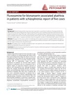
Serum sCTLA-4 levels were significantly higher in the SpA
group (3.66 ± 0.3 ng/ml, 1 to 22.7) compared with the RA
group (2.25 ± 0.4 ng/ml, 0 to 15.7) or the control group (0.25
± 0.1 ng/ml, 0 to 3.4; Kruskal-Wallis: P < 0.0001). Serum
sCTLA-4 levels were also found higher in patients with SLE
(19.58 ± 2.7 ng/ml, 5.9 to 41) compared with healthy controls
(Mann-Whitney P < 0.0001) or compared to patients with
SpA or RA (Mann-Whitney P < 0.0001). Both SpA and RA
patients had higher sCTLA-4 values compared with controls
(Mann-Whitney P < 0.0001 and P = 0.001, respectively),
whereas sCTLA-4 levels remained higher in the SpA group
compared with RA (P < 0.0001; Figure 1).
Table 1
Clinical characteristics and laboratory features of patients with SpA or RA and healthy controls
HC
(n = 88)
SpA
(n = 165)
RA
(n = 71)
P
Age (years) 44.4 ± 0.8
(20 to 62)
42.9 ± 1.1
(18 to 75)
59.1 ± 1.4
(19 to 80)
< 0.0001 (KW)
SpA vs HC (MW): P = 0.28
RA vs SpA and RA vs HC (MW): P <
0.0001
Sex 56 M/16 F 121 M/44 F 27 M/44 F SpA vs HC vs RA: P < 0.0001 (
2
)
SpA vs HC: P = 0.9
RA vs SpA and RA vs HC: P < 0.0001 (
2
)
Disease duration (years) 9.2 ± 0.6
(0.5 to 35)
10.2 ± 1.1 (0.5 to 40) 0.5 (MW)
Extra-articular manifestations (%) 35/165 (21) 26/71 (36.6)
Peripheral arthritis (%) 46/165 (27.8)
BASDAI (0 to 100) 35.9 ± 2.2
(0.4 to 94)
BASFI (0 to 100) 41.2 ± 2.9
(0 to 92)
Swollen joint count (0 to 28) 7.2 ± 0.8 (0 to 22)
Tender joint count (0 to 28) 4.7 ± 0.8 (0 to 28)
HAQ (0 to 3) 1.5 ± 0.1 (0 to 3)
HLA-B27 (%) 145 (87.8)
Rheumatoid factors (%) 64/71 (90)
ESR (mm/h) 28.2 ± 2.1 (1 to 163) 30.3 ± 3.1 (1 to 125) 0.3 (MW)
CRP (mg/l) 25.1 ± 3.2 (0 to 350) 22.3 ± 3.5 (1 to 132) 0.8 (MW)
Results are expressed as mean ± standard error of the mean (range). The Kruskal-Wallis test was used to compare age, disease duration, ESR,
and CRP in the three groups. Then, Mann-Whitney U-test was used to calculate the exact P values between two groups.
BASDAI = Bath Ankylosing Spondylitis Disease Activity Index; BASFI = Bath Ankylosing Spondylitis Functional Index; CRP = C-reactive protein;
ESR = erythrocyte sedimentation rate; HAQ = Health Assessment Questionnaire; HC = healthy controls; HLA = Histocompatibility Leukocyte
Antigen; KW = Kruskal-Wallis; MW = Mann-Whitney; NS = not significant; RA = rheumatoid arthritis; SpA = spondylarthropathie;
2
= chi-
squared test.
Available online />Page 5 of 11
(page number not for citation purposes)
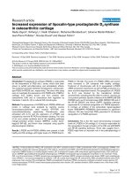
In the SpA group, serum sCTLA-4 correlated with BASDAI (r
= 0.42, P < 0.0001; Figure 2) and CRP (r = 0.17, P = 0.037)
but not with ESR, BASFI, age, or disease duration (all P >
0.05). Serum sCTLA-4 levels were not influenced by the
expression of the HLA-B27 antigen, the presence of extra-
articular disease, or peripheral arthritis (all P > 0.05), whereas
male patients had higher sCTLA-4 than female patients (Mann-
Whitney: 3.97 ± 0.3, 1 to 22.7; vs 2.84 ± 0.39, 1 to 17.3 ng/
ml; P = 0.02). In addition, age was not correlated with serum
sCTLA-4 levels in the different groups of subjects (all P >
0.05).
In the RA group, sCTLA-4 levels were correlated with swollen
joint count (r = -0.25, P = 0.04) but not with laboratory param-
eters exploring inflammation (ESR and CRP), HAQ score, and
tender joint score (all P > 0.05).
Finally, in normal subjects, we did not observe any significant
differences in serum sCTLA-4 levels between male and female
subjects or between HLA-B27-positive and HLA-B27-nega-
tive subjects (all P > 0.05).
To better understand this increase in circulating sCTLA-4 in
patients with SpA, we then evaluated intracellular CTLA-4
expression. This analysis was performed in a representative
number of patients and controls (i.e., 32 patients with SpA, 22
patients with RA, and 15 healthy controls).
Analysis of blood lymphocyte subsets
Before analysis of intracellular CTLA-4 expression in lym-
phocyte subsets, we first compared absolute numbers of cir-
culating CD3+, CD4+, and CD8+ T cells in the SpA group, in
the RA group, and in healthy volunteers involved in this analy-
sis. The three groups differed significantly regarding the
number of CD3+ and CD8+ T cells (P 0.005; Table 2).
Indeed, RA patients had lower absolute numbers of CD3+ and
CD8+ T cells compared with healthy controls or patients with
SpA (P < 0.01 for each test). No difference in CD3+ T cells
was observed between SpA patients and healthy controls,
while CD8+ T cells were found to be lower in SpA, a result
that was just near the significant level (P = 0.051).
We then examined the percentage of circulating activated
CD4+ T cells and Treg in the three studied groups. No differ-
ence was observed between these three groups for activated
CD4+ T cells or for Treg (all P > 0.05; Table 2).
Analysis of intracellular CTLA-4 expression in T cell
subsets
As shown in Table 2 and Figure 3, a significant difference was
observed in the percentage of cells positive for intracellular
CTLA-4 in CD4+ T cells, Treg, non T cells (CD3-negative cells
in the lymphocyte gate, as assessed by flow cytometry; this
presumably corresponds to B cells rather than natural killer
cells [8]) and in the total lymphocyte gate between the three
groups (P < 0.01 for each test), while no similar difference
was found for activated CD4+ T cells or CD8+ T cells. How-
ever, SpA patients and healthy controls did not differ with
regard to intracellular CTLA-4 in all these distinct lymphocyte
subsets (all P > 0.05). Conversely, RA patients had lower
intracellular CTLA-4 expression in CD4+ T cells and Treg
compared with healthy controls or patients with SpA (all P <
0.05). The expression of intracellular CTLA-4 in CD3 negative
cells (presumably B cells) was found to be higher in RA
Figure 1
Serum concentrations of soluble Cytotoxic T Lymphocyte Antigen- 4 (sCTLA-4) in patients with spondylarthropathies (SpA, n = 165), with rheumatoid arthritis (RA, n = 71) and healthy controls (HC, n = 88)Serum concentrations of soluble Cytotoxic T Lymphocyte Antigen- 4
(sCTLA-4) in patients with spondylarthropathies (SpA, n = 165), with
rheumatoid arthritis (RA, n = 71) and healthy controls (HC, n = 88). Cir-
culating sCTLA-4 was measured by ELISA, as described in the Material
& Methods section. Horizontal lines represent means (***: P < 0.005;
statistical tests and exact p values are given in the Result section).
Figure 2
Correlation between soluble Cytotoxic T Lymphocyte Antigen- 4 (sCTLA-4) and clinical index of disease activity Bath Ankylosing Spond-ylitis Disease Activity Index in 165 patients with spondylarthropathiesCorrelation between soluble Cytotoxic T Lymphocyte Antigen- 4
(sCTLA-4) and clinical index of disease activity Bath Ankylosing Spond-
ylitis Disease Activity Index in 165 patients with spondylarthropathies.
The Bath Ankylosing Spondylitis Disease Activity Index (BASDAI) was
evaluated as described in [19]. The statistical test used was Spear-
man's r test. When the eight extreme values (white circles) are not con-
sidered for correlation analysis, a significant correlation between
sCTLA-4 and BASDAI score is still observed: (Spearman's test: r =
0.382, P < 0.0001).
Arthritis Research & Therapy Vol 11 No 4 Toussirot et al.
Page 6 of 11
(page number not for citation purposes)
patients compared with healthy controls or SpA (P < 0.01).
This increase of intracellular CTLA-4 in CD3-negative cells of
RA patients leads to an increase of total intracellular CTLA-4
when considering all the lymphocytes (P = 0.004), while no
difference was observed between SpA patients and healthy
controls in these cells. Thus, increase of circulating sCTLA-4
in patients with SpA did not reflect an increase of intracellular
CTLA-4 (independent on the examined T cell population). This
prompted us to complete our analysis by evaluating the differ-
ent CTLA-4 transcripts.
Analysis of CTLA-4 full length and spliced mRNA
transcripts
The pool of PBL from the 15 healthy donors was used as a ref-
erence. In patients with SpA, we observed an increase expres-
sion of the spliced CTLA-4 mRNA transcript (mean ± SEM,
range, 1.608 ± 0.2, 0.09 to 4.14) relative to the full form (Fig-
ure 4; mean ± SEM, range, 0.942 ± 0.11, 0.03 to 2.3, P =
0.0014). Conversely, there was no difference in the expres-
sion of the full length form versus the spliced form in patients
with RA (mean ± SEM, range, 1.117 ± 0.28, 0.1 to 2.7 for the
spliced form transcript vs. 1.157 ± 0.4, 0.13 to 4.59 for the full
length transcript expression, P = 0.7). We also found a ten-
dency for a correlation between spliced CTLA-4 mRNA tran-
Table 2
Absolute number of circulating CD3+ T cells, CD4+ T cells, CD8+ T cells, percentage ofactivated CD4+ T cells, regulatory CD4+ T
cells (Treg) and percentage of cells expressing intracellular CTLA-4 in healthy controls, patients with Spondylarthropathies and
rheumatoid arthritis
Healthy controls
(n = 15)
Spondylarthropathies
(n = 32)
Rheumatoid arthritis
(n = 22)
P
CD3+ T cells (/mm
3
)
1397 ± 79
(925 to 1806)
1360 ± 80.8
(622 to 2681)
1129 ± 140
(336 to 3024)
0.005 (KW)
RA vs HC 0.004 (MW)
SpA vs HC NS
SpA vs RA 0.007 (MW)
CD4+ T cells (/mm
3
)
817 ± 45
(449 to 1024)
875 ± 49.5
(391 to 1699)
804 ± 102
(239 to 2306)
NS
CD8+ T cells (/mm
3
)
573 ± 47
(379 to 860)
457 ± 39.6
(130 to 979)
326 ± 52
(72 to 1260)
0.0006 (KW)
RA vs HC 0.0007 (MW)
SpA vs HC 0.051
RA vs SpA 0.006 (MW)
HLA-DR+ Foxp3-
CD4+ T cells
(% of CD4+ T cells)
16.6 ± 5.53
(5 to 70)
22.03 ± 4.56
(4 to 100)
12.14 ± 1.29
(5 to 26)
NS
CD25+ Foxp3+
CD4+ T cells
(% of CD4+T cells)
7.94 ± 1.04
(3.6 to 16)
8.2 ± 0.61
(4 to 14.4)
8.72 ± 0.7
(2.9 to 13.5)
NS
Intracellular CTLA-4 in CD4+ T cells (%) 4.35 ± 1.10
(1 to 13.3)
2.51 ± 0.40
(0 to 5.9)
1.35 ± 0.48
*,§
(0 to 7.5)
0.005 (KW)
RA vs HC 0.003
SpA vs HC NS
RA vs SpA 0.02
Intracellular CTLA-4 in CD4+ Treg (%) 2.76 ± 0.44
(0.5 to 6.3)
1.88 ± 0.34
(0 to 5)
0.90 ± 0.32
*,§
(0 to 3.9)
0.007 (KW)
RA vs HC 0.0045
SpA vs HC NS
SpA vs RA 0.027
Intracellular CTLA-4 in Activated CD4+ T cells
(%)
1.59 ± 0.72
(0 to 7)
0.63 ± 0.26
(0 to 5)
0.5 ± 0.29
(0 to 5)
NS
Intracellular CTLA-4 in CD8+ T cells (%) 2.03 ± 0.57
(0.4 to 7)
2.64 ± 0.32
(0 to 6.3)
3.49 ± 0.88
(0.1 to 13.7)
NS
Intracellular CTLA-4 in CD3- lymphocytes (%) 0.89 ± 0.72
(0 to 8)
0.67 ± 0.21
(0 to 3.3)
3.50 ± 0.77
*,§
(0 to 11)
0.0025 (KW)
RA vs HC 0.0077
SpA vs HC NS
SpA vs RA: 0.0017
Intracellular CTLA-4 in the lymphocyte gate
(total lymphocytes) (%)
7.27 ± 1.27
(1.4 to 15)
3 ± 0.48
(0 to 7.6)
6.89 ± 1.3
§
(0.1 to 19.3)
0.004 (KW)
RA vs HC 0.0045
SpA vs HC NS
SpA vs RA 0.007
Percentage of intracellular CTLA-4 was determined by flow cytometry. Results are expressed as mean ± standard error of the mean, (range). The
Kruskal-Wallis (KW) test was used to compare T cell subsets and intracellular CTLA-4 expression in the three groups. Then, Mann-Whitney U-test
was used to calculate the exact P values between two groups.
HC = healthy controls; HLA = Histocompatibility Leukocyte Antigen; KW = Kruskal-Wallis; MW = Mann-Whitney; NS = not significant; RA =
rheumatoid arthritis; SpA = spondylarthropathie.
Available online />Page 7 of 11
(page number not for citation purposes)
script levels and serum sCTLA-4 in patients with SpA (r =
0.43, P = 0.07; Figure 5). No similar correlation was observed
in the RA group. Altogether, these data suggest that the
increase of sCTLA-4 found in SpA patient serum could be
related to an increase of spliced CTLA-4 mRNA transcripts.
Discussion
Here, we evaluated circulating sCTLA-4 as a marker of dis-
ease activity for SpA. The role of sCTLA-4 is still not com-
pletely understood in contrast to membrane-bound CTLA-4, a
negative regulator of T cell functions [8,9]. sCTLA-4 – result-
ing from an alternative transcript of the CTLA-4 gene [11,12]
– is also able to bind CD80 and CD86 and may prevent mem-
brane-bound CTLA-4 or CD28 interactions with their ligands
[12]. Thus, sCTLA-4 may interfere with both (CD28-mediated)
activating and (CTLA-4-mediated) inhibitory signaling path-
ways. The role of sCTLA-4 in T cell responses and inflamma-
tory diseases have been recently highlighted by the
description of the association of CTLA-4 gene polymorphisms
(49 G/G and CT60 G/G) with the risk for common autoim-
mune diseases, such as Graves' disease, autoimmune
hypothyroidism, and type I diabetes [18]. The CTLA-4
CT60G/G genotype was associated with lower sCTLA-4 tran-
script abundance in CD4+ T cells [17]. Despite the absence
of sCTLA-4 protein analysis, these data by Ueda and col-
leagues [18] suggest that reduced sCTLA-4 (at least in CD4+
T cells) is associated with increased T cell activation and
thereby autoreactivity.
On the contrary, further studies on circulating sCTLA-4-ana-
lyzed by ELISA- in several autoimmune diseases showed high
levels of serum sCTLA-4 in patients with Graves' disease [13],
Figure 3
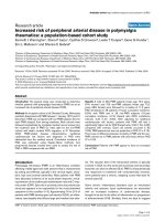
Representative analysis of intracellular Cytotoxic T Lymphocyte Antigen- 4 (CTLA-4) expression in peripheral blood lymphocyte subsetsRepresentative analysis of intracellular Cytotoxic T Lymphocyte Antigen- 4 (CTLA-4) expression in peripheral blood lymphocyte subsets. This analy-
sis was performed by flow cytometry as described in the Material & Methods section. (a) CD4+ T cells, CD8+ T cells and CD3- cells were identified
in the PE-CY7 fluorescence vs allophycocyanin fluorescence gate based on the expression of CD3 and CD4 (CD4+ T cells, gate R6), of only CD3
(CD8+ T cells, gate R7), of neither CD3 nor CD4 (CD3- cells, gate R4). Then, intracellular expression of CTLA-4 was determined on these cell pop-
ulations by comparing PE-conjugated isotype control antibody staining (open curves) with PE-conjugated anti-CD152 monoclonal antibody staining
(gray filled curves). (b) Using the same gating strategy (CD3 vs CD4 dot plot), Treg were analyzed using FoxP3 vs CD25 dot plot. Intracellular
CTLA-4 expression was also determined on CD3+ CD4+ CD25+ FoxP3+ cells by comparing PE-conjugated isotype control antibody staining
(open curves) with PE-conjugated anti-CD152 (CTLA-4) monoclonal antibody staining (gray filled curves). The same analysis was performed for acti-
vated (CD3+ CD4+ HLA-DR+) T cells (data not shown). An example of intracellular CTLA-4 expression in CD4+ T cells, CD8+ T cells (defined as
CD3+ CD4-), CD3- cells and Treg obtained with such gating strategy for a patient with spondylarthropathy is shown.
Arthritis Research & Therapy Vol 11 No 4 Toussirot et al.
Page 8 of 11
(page number not for citation purposes)
Hashimoto thyroiditis [13], myasthenia gravis [14], SLE
[15,16], and systemic sclerosis [17]. In SLE, elevated serum
sCTLA-4 levels were correlated with the clinical index of dis-
ease activity SLEDAI in one study [16], but not in a second
[15]. In systemic sclerosis, patients exhibited high levels of
serum sCTLA-4 that correlated with disease activity [16]. In
addition, sCTLA-4 was associated with cutaneous extension
of fibrosis and tended to decrease in parallel to skin sclerosis
improvement in a longitudinal analysis of some patients [17].
Taken together, these results suggest that high levels of circu-
lating sCTLA-4 are observed in several autoimmune diseases
and can be associated with disease severity and activity. This
argues for a role of sCTLA-4 in enhancing T cell activation or
preventing T cell regulation, leading finally to exacerbation of
the disease [16,17].
The main finding of our study is that there are increased levels
of serum sCTLA-4 in patients with SpA, compared with levels
observed in patients with RA or in healthy volunteers. Circulat-
ing sCTLA-4 correlated with clinical index of disease activity
BASDAI and CRP levels. As previously reported [12-15],
serum sCTLA-4 was very low in our group of healthy controls,
even in the 24 positive HLA-B27 subjects. We also confirm
that patients with SLE had high circulating sCTLA-4 levels
(around 70-fold more than control subjects). Our cohort of
patients included mainly AS and undifferentiated SpA, but we
did not observed significant differences between SpA sub-
groups (data not shown). As expected, the majority of patients
with SpA expressed the HLA-B27 antigen, but again, this had
no influence on the levels of sCTLA-4. In addition, no correla-
tion was found between sCTLA-4, age, or the clinical charac-
teristics of the patients. However, we found that male patients
had higher sCTLA-4 levels than female patients but this may
be explained by a predominance of male patients with SpA
(73.3% in our series).
Our patients had conventional treatments for SpA (i.e. mainly
NSAIDs) and a limited number of patients had a second-line
treatment. However, it is considered that these treatments had
no influence on T cell activation and costimulation pathways
and therefore on the level of sCTLA-4. Eight SpA patients
among the 165 analyzed here received oral prednisolone. Cor-
ticosteroids have been shown to decrease membrane CTLA-
4 expression after human T cell activation [27]. Moreover, oral
prednisolone in asthmatic patients has been shown to reduce
serum sCTLA-4 concentrations after two weeks of treatment
[28]. However, this can not be evaluated in our cohort
because the number of patients on prednisolone is very low
and concentrations are different (<10 mg/day in our study vs
30 mg/day [28]). The impact of therapy on circulating sCTLA-
4 is an interesting question to address.
The increase of sCTLA-4 in the serum of our patients with SpA
contrasted with a normal percentage of cells positive for intra-
cellular CTLA-4 (in comparison with the percentage found in
healthy controls), suggesting a preferential expression of the
spliced form of the CTLA-4 gene. Indeed, CTLA-4 mRNA
analysis confirms this hypothesis because the spliced CTLA-
4 RNA transcripts were found to be expressed at a higher level
compared with the full length form. These results indicate that
SpA patients had a high production of sCTLA-4 which was
related to a preferential transcription of the CTLA-4 mRNA
Figure 4
Relative expression of the 2 Cytotoxic T lymphocyte Antigen-4 (CTLA-4) transcripts, the full length form and the spliced form in peripheral blood lymphocytes of patients with spondylarthropathies (SpA) and patients with rheumatoid arthritis (RA)Relative expression of the 2 Cytotoxic T lymphocyte Antigen-4 (CTLA-4) transcripts, the full length form and the spliced form in peripheral blood
lymphocytes of patients with spondylarthropathies (SpA) and patients with rheumatoid arthritis (RA). The full length form (gray bars) contains four
exons and codes for the CTLA-4 molecule expressed at the cell membrane while the spliced form (open bars) contains three exons, lacks the trans-
membrane region and codes for the soluble CTLA-4 molecule [8]. Results (mean ± standard error of the mean) are obtained using the Ct method
and expressed as a fold change of each CTLA-4 transcript comparative with the reference obtained with the pool of 15 healthy donor PBL. Wil-
coxon test was used to compare the relative production of the full length and the spliced form in patients with SpA and in patients with RA (*** P <
0.005). NS = not significant.
Available online />Page 9 of 11
(page number not for citation purposes)
spliced form. The reason for the main expression of these
CTLA-4 transcripts in SpA is not known but one may hypoth-
esize that genetic background could play a role [18]. However,
our cohort to date did not include a sufficient number of
patients to appreciate the relation between circulating sCTLA-
4, CTLA-4 mRNA spliced form, and CTLA-4 polymorphism.
SpAs are characterized by an infiltration of the sacroiliac joints,
the synovium and entheseal structures by inflammatory cells.
Different cells are found in these lesions [4,5]. Both CD4+ and
CD8+ T lymphocytes are described at the pathological sites
as well as B cells and macrophages [4,5]. These infiltrated T
cells are activated and require the active participation of cos-
timulation pathways. Thus, a high production of sCTLA-4 in
SpA may potentially influence immune responses. As stated
above, sCTLA-4 may block the down regulation of activated T
cells mediated by CTLA-4-CD80/CD86 interactions, enhanc-
ing immune response in situ. Costimulation pathways have not
been previously evaluated in SpA. However, in the HLA-B27
transgenic rat model of SpA, a decrease in formation of conju-
gates between dendritic cells expressing HLA-B27 and T cells
was observed [29]. This decrease in dendritic cell-T cell con-
jugates was not related to a decrease of CD80 or CD86
expression, but resulted from a defective CD86 costimulatory
pathway [29]. One may speculate that this defective APC
function is due to the blockade of CD86 by sCTLA-4.
Treg are also implicated in the inflammatory response in SpA.
Treg were found to be enriched in synovial tissues from
patients with SpA, contrasting with a low percentage of Treg
in the blood compartment [30]. In our study, we did not
observe a significant reduction in the percentage of circulating
Treg in our SpA patients as compared with healthy controls
and RA patients. Treg constitutively expressed CTLA-4 [8]
and suppressive functions of some Treg subsets are depend-
ent on CTLA-4 [10,31]. Thus, one may speculate that a high
production of sCTLA-4 in SpA may interfere with Treg func-
tions, leading to a sustained and/or enhanced immune
response. As stated above, the relation between sCTLA-4 and
disease activity in different autoimmune diseases argues for a
blockade of the inhibitory signal resulting from CTLA-4/CD80-
CD86 binding.
In this study, serum sCTLA-4 was found mildly elevated in
patients with RA compared with healthy controls. In addition,
the percentage of cells positive for intracellular CTLA-4 was
found decreased in CD4+ T cells and also in Treg. These
results were not explained by the relative lymphopenia that
characterized our RA patients, which involved CD3+ T cells
and CD8+ T cells, while CD4+ T cells and Treg did not differ
between patients and controls. On the other hand, intracellular
CTLA-4 was found increased in the CD3-negative cell popu-
lation, presumably B cells. Finally, there was no preferential
expression of CTLA-4 transcripts in RA. Altogether, these
results suggest that patients with RA had mild elevation of
sCTLA-4 that may result from an increased expression of
CTLA-4 in 'non T' cell subsets. RA is a chronic joint disease
characterized by a major infiltration of the synovium by T cells.
These cells are activated with the main implication of the
CD28/CTLA-4 – CD80/CD86 ligand pairs. Altered cell sur-
face expression of CD28, CTLA-4, CD80, and CD86 has
been described in RA [32]. Thus, high levels of sCTLA-4
(whatever the producing cell was) may sustain T cell activation
leading to inflammation and 'autoaggression', as observed in
other autoimmune diseases. Reduced CTLA-4 expression in
Treg can be related to defective function previously reported
in RA [33,34]. A recent study reports that diminished CTLA-4
expression by Treg from RA patients was related to an
increase rate of CTLA-4 internalization after activation [34].
Conclusions
Similar to other autoimmune diseases [16,17], SpA are char-
acterized by high serum sCTLA-4 levels with a significant and
positive correlation with disease activity. These high levels of
circulating sCTLA-4 in SpA were not explained by altered
intracellular expression of CTLA-4, but by a preferential
expression of the spliced form of CTLA-4 transcripts, a result
that was not observed in RA. This suggests a potential immu-
nologic role for sCTLA-4 in SpA. These results also indicate
that sCTLA-4 could potentially be used as a surrogate marker
for measuring disease activity in SpA.
Finally, the implication of sCTLA-4 in SpA also suggests the
potential use of agents blocking costimulation pathways in this
group of disorders. Indeed, a recent manuscript reports the
use of abatacept – the fusion protein associating the CTLA-4
extracellular domain and the constant fragment region of
Figure 5
Correlation between serum concentrations of soluble Cytotoxic T Lym-phocyte Antigen-4 (sCTLA-4, expressed in ng/ml) and the spliced form of CTLA-4 transcript expression (spliced form of the CTLA-4 mRNA, analyzed in PBL and expressed as a fold change comparative with the reference obtained with the pool of 15 healthy donor PBL)Correlation between serum concentrations of soluble Cytotoxic T Lym-
phocyte Antigen-4 (sCTLA-4, expressed in ng/ml) and the spliced form
of CTLA-4 transcript expression (spliced form of the CTLA-4 mRNA,
analyzed in PBL and expressed as a fold change comparative with the
reference obtained with the pool of 15 healthy donor PBL). This spliced
form of the CTLA-4 transcript encodes for the soluble CTLA-4 mole-
cule. The statistical test used was Spearman's r test.
Arthritis Research & Therapy Vol 11 No 4 Toussirot et al.
Page 10 of 11
(page number not for citation purposes)
human IgG1 and blocking CD28/CD80-CD86 costimulatory
pathway- in a patient with undifferentiated SpA. After 12
months of treatment with abatacept, the patient was in com-
plete remission [35].
Competing interests
The authors declare that they have no competing interests.
Authors' contributions
ET is the main investigator who conceived and designed the
study and drafted the manuscript. He contributed to the
recruitment of the patients involved in the study, analyzed the
Results, and performed the statistical analysis. PS is responsi-
ble for laboratory parameter assessment. He contributed to
the preparation of the manuscript by writing the Methods sec-
tion. He analyzed and contributed to the discussion of the
results. MD performed CTLA-4 transcript analysis, supervised
sample collection, ELISA, and cytometry analysis. LP collected
samples and performed flow cytometry experiments, ELISA.
SP supervised cytometry and analyzed cytometry results. FP
participated to the study by recruiting healthy controls (plate-
lets donors) including HLA-B27 positive subjects. JC selected
the HLA-B27 positive healthy donors among platelet donors.
PT is the director of EFS Bourgogne Franche-Comté and of
INSERM UMR645 University of Franche-Comté. He contrib-
uted to the discussion of the results. DW is the head of the
Department of Rheumatology and contributed to the work as
a clinical investigator. All authors read and approved the final
manuscript.
Acknowledgements
This work was supported by a grant from «La Société Française de Rhu-
matologie» (E. Toussirot), «l'Association Franc-Comtoise pour la recher-
che, l'enseignement en rhumatologie» (E. Toussirot) and le CIC-
Biotherapy 506, University Hospital Besançon (L. Perrot). We thank Idir
Idirène, Maryse Billot, Christine Colombain, Céline Pagneux Eléonore
Gravelin, and Amélie Verdot for expert technical assistance.
References
1. Calin A: Terminology, introduction, diagnostic criteria, and
overview. In The spondylarthritides Edited by: Calin A, Taurog JD.
Oxford: Oxford University Press; 1998:1-15.
2. Hammer RE, Maika SD, Richardson JA, Tang JP, Taurog JD: Spon-
taneous inflammatory disease in transgenic rats expressing
HLA-B27 and human 2 microglobulin: an animal model of
HLA-B27 associated disorder. Cell 1990, 63:1099-1112.
3. Pachebo-Tena C, Zhang X, Stone M, Burgos-Vargas R, Inman R:
Innate immunity in host-microbial interactions: beyond B27 in
the spondylarthropathies. Curr Opin Rheumatol 2002,
14:373-382.
4. Baeten D, Demetter P, Cuvelier C, Van Den Bosch, Kruithof E, Van
Damme N, Verbuggen G, Mielants H, Veys EM, De Keyser F: Com-
parative study of the synovial histology in rheumatoid arthritis,
spondylarthropathy, and osteoarthritis: influence of disease
duration and activity. Ann Rheum Dis 2000, 59:945-953.
5. Laloux L, Voisin MC, Allain J, Martin N, Kerboull L, Chevalier X,
Claudepierre P: Immunohistological study of entheses in
spondylarthropathies: comparison in rheumatoid arthritis and
osteoarthritis. Ann Rheum Dis 2001, 60:316-321.
6. Toussirot E, Wendling D: The immunogenetics of ankylosing
spondylitis. Rev Med Interne 2006, 27:762-771.
7. Lenschow DJ, Walunas TL, Bluestone JA: CD28/B7 system of T
cell costimulation. Annu Rev Immunol 1996, 14:233-258.
8. Teft WA, Kirchhof MG, Madrenas J: A molecular perspective of
CTLA-4 function. Annu Rev Immunol 2006, 24:65-97.
9. Carreno BM, Bennett F, Chau TA, Ling V, Luxenberg D, Jussif J,
Baroja ML, Madrenas J: CTLA-4 (CD152) can inhibit T cell acti-
vation by two different mechanisms depending on its level of
cell surface expression. J Immunol 2000, 165:1352-1356.
10. Wing K, Onishi Y, Prieto-Martin P, Yamaguchi T, Miyara M,
Fehervari Z, Nomura T, Sakaguchi S: CTLA-4 control over
Foxp3+ regulatory T cell function. Science 2008, 322:271-275.
11. Oaks M, Hallett K, Penwell RT, Stauber EC, Warren SJ, Tector AJ:
A native soluble form of CTLA-4. Cell Immunol 2000,
201:
144-153.
12. Pawlak E, Kochanowska IE, Frydecka I, Kielbinski M, Potoczek S,
Bilinska M: The soluble CTLA-4 receptor: a new marker in
autoimmunes diseases. Arch Immunol Ther Exp (Warsz) 2005,
53:336-341.
13. Oaks MK, Hallett KM: Cutting edge: a soluble form of CTLA-4 in
patients with autoimmune thyroid disease. J Immunol 2000,
164:5015-5018.
14. Wang XB, Kakuolidou M, Giscombe R, Qiu Q, Huang DR, Pir-
skanen R, Lefvert AK: Abnormal expression of CTLA-4 by T cells
from patients with myasthenia gravis: effect of an AT-rich
sequence. J Neuroimmunol 2002, 130:224-232.
15. Liu MF, Wang CR, Chen PC, Fung LL: Increased expression of
soluble cytotoxic T lymphocyte associated antigen-4 molecule
in patients with systemic lupus erythematosus. Scand J
Immunol 2003, 57:568-572.
16. Wong CK, Lit LCW, Tam LS, Li EK, Lam CWK: Aberrant produc-
tion of soluble costimulatory molecules CTLA-4, CD28, CD80
and CD86 in patients with systemic lupus erythematosus.
Rheumatology 2005, 44:989-994.
17. Sato S, Fujimoto M, Hasegawa M, Komura K, Yanaba K, Hayakawa
I, Matsushita T, Takehara K: Serum soluble CTLA-4 levels are
increased in diffuse cutaneous systemic sclerosis. Rheumatol-
ogy 2004, 43:1261-1266.
18. Ueda H, Howson JM, Esposito L, Heward J, Snook H, Chamberlain
G, Rainbow DB, Hunter KM, Smith AN, Di Genova G, Herr MH,
Dahlman I, Payne F, Smyth D, Lowe C, Twells RC, Howlett S,
Healy B, Nutland S, Rance HE, Everett V, Smink LJ, Lam AC, Cord-
ell HJ, Walker NM, Bordin C, Hulme J, Motzo C, Cucca F, Hess JF,
et al.: Association of T cell regulatory gene CTLA-4 with sus-
ceptibility to autoimmune disease. Nature 2003, 423:506-511.
19. Dougados M, Linden S van der, Juhlin R, Huitfeldt B, Amor B, Calin
A, Cats A, Dijkmans B, Olivieri I, Pasero G: The European Spond-
ylarthropathy Study Group preliminary criteria for the classifi-
cation of spondylarthropathy. Arthritis Rheum 1991,
34:1218-1227.
20. Garrett P, Jenkinsson T, Kennedy LG, Whitelock H, Gaisford P,
Calin A: A new approach to defining disease status in ankylos-
ing spondylitis: the Bath Ankylosing Spondylitis Disease Activ-
ity Index. J Rheumatol 1994, 21:2286-2291.
21. Calin A, Garrett S, Whitelock H, Kennedy LG, O'Hea J, Mallorie P,
Jenkinson T: A new approach to defining functional ability in
ankylosing spondylitis: the development of the Bath Ankylos-
ing Spondylitis Functional Index (BASFI). J Rheumatol 1994,
21:2281-2285.
22. Arnett FC, Edworthy SM, Bloch DA, McShane DJ, Fries JF, Cooper
NS, Healey LA, Kaplan SR, Liang MH, Luthra HS: The American
Rheumatism Association 1987 revised criteria for the classifi-
cation of rheumatoid arthritis. Arthritis Rheum 1988,
31:315-324.
23. Tan EM, Cohen AS, Fries JF, Masi AT, McShane DJ, Rothfield NF,
Schaller JG, Talal N, Winchester RJ: The 1982 revised criteria for
the classification of systemic lupus erythematosus. Arthritis
Rheum 1982, 25:1271-1277.
24. Reimann KA, O'Gorman MR, Spritzler J, Wilkening CL, Sabath DE,
Helm K, Campbell DE: Multisite comparison of CD4 and CD8 T-
lymphocyte counting by single- versus multiple-platform
methodologies: evaluation of Beckman Coulter flow-count
fluorospheres and the tetraONE system. The NIAID DAIDS
New Technologies Evaluation Group. Clin Diagn Lab Immunol
2000, 7:344-351.
25. Tran DQ, Ramsey H, Shevach EM: Induction of FOXP3 expres-
sion in naive human CD4+FOXP3 T cells by T-cell receptor
stimulation is transforming growth factor-beta dependent but
does not confer a regulatory phenotype. Blood 2007,
110:2983-2990.
Available online />Page 11 of 11
(page number not for citation purposes)
26. Pfaffl MW: A new mathematical model for relative quantifica-
tion in real-time RT-PCR. Nucleic Acids Res 2001, 29:e45.
27. Agarwal SK, Marshall GD Jr: Role of CD28/B7 costimulation in
the dexamethasone-induced suppression of IFN-gamma. J
Interferon Cytokine Res 2000, 20:927-934.
28. Qin XJ, Shi HZ, Qin SM, Kang LF, Huang CP, Zhong XN: Effects
of allergen inhalation and oral glucocorticoid on serum solu-
ble CTLA-4 in allergic asthmatics. Allergy 2005, 60:774-779.
29. Hacquard-Bouder C, Flagarone G, Bosquet A, Smaoui F, Monnet
D, Ittah M, Breban M: Defective costimulatory function is a strik-
ing feature of antigen presenting cells in an HLA-B27 trans-
genic rat model of spondylarthropathy. Arthritis Rheum 2004,
50:1624-1635.
30. Cao D, Van Vollenhoven R, Klareskog L, Trollmo C, Malmstrom V:
CD25 bright CD4+ regulatory T cells are enriched in inflamed
joints of patients with chronic rheumatic disease. Arthritis Res
Ther 2004, 6:R335-346.
31. Manzotti CN, Tipping H, Perry LC, Mead KI, Blair PJ, Zheng Y, San-
som DM: Inhibition of human T cell proliferation by CTLA-4 uti-
lizes CD80 and requires CD25+ regulatory cells. Eur J Immunol
2002, 32:2888-2896.
32. Liu MF, Kohsaka H, Sakurai H, Azuma M, Okumura K, Saito I, Miya-
saka N: The presence of costimulatory molecules CD86 and
CD28 in rheumatoid arthritis synovium. Arthritis Rheum 1996,
39:110-114.
33. Ehrenstein MR, Evans JG, Singh A, Moore S, Warnes G, Isenberg
DA, Mauri C: Compromised function of regulatory T cells in
rheumatoid arthritis and reversal by anti-TNF alpha therapy. J
Exp Med 2004, 200:277-285.
34. Flores-Borja F, Jury EC, Mauri C, Ehrenstein MR: Defects in
CTLA-4 are associated with abnormal regulatory T cell func-
tion in rheumatoid arthritis. Proc Natl Acad Sci USA 2008,
105:19396-19401.
35. Olivieri I, D'Angelo S, Mennillo GA, Pistone G, Scarano E, Padula
A: Abatacept in spondyloarthritis refractory to tumour necrosis
factor alpha inhibition. Ann Rheum Dis 2009, 68:151-152.