Báo cáo y học: "Cytokines in chronic rheumatic diseases: is everything lack of homeostatic balance" pps
Bạn đang xem bản rút gọn của tài liệu. Xem và tải ngay bản đầy đủ của tài liệu tại đây (1 MB, 11 trang )
Available online />Page 1 of 11
(page number not for citation purposes)
Abstract
Biological systems have powerful inbuilt mechanisms of control
intended to maintain homeostasis. Cytokines are no exception to
this rule, and imbalance in cytokine activities may lead to inflam-
mation with subsequent tissue and organ damage, altered function,
and death. Balance is achieved through multiple, not mutually
exclusive, mechanisms including the simultaneous production of
agonist and antagonistic cytokines, expression of soluble receptors
or membrane-bound nonsignaling receptors, priming and/or re-
programming of signaling, and uncoupling of ligand/receptor pairing
from signal transduction. Insight into cytokine balance is leading to
novel therapeutic approaches particularly in autoimmune conditions,
which are intimately linked to a dysregulated cytokine production.
Introduction
To explore the complex regulation of cytokine activities it may
be of help to bear in mind the example of rheumatoid arthritis
(RA). A major step forward in RA treatment was achieved
when it became possible to control disease manifestations
such as joint destruction by blocking TNF. This could indicate
that a single cytokine, in this case TNF, drives unopposed a
series of events that lead to inflammation and destruction.
The situation is less simple inside the joint, however, where
proinflammatory cytokines co-exist alongside their endoge-
nous inhibitors. This is a consequence of ongoing processes
in which proinflammatory stimuli induce their anti-inflam-
matory counterparts and the imbalance between the two
results in disease.
The cytokine network is a homeostatic system that may be
comparable with the acid/base equilibrium. The biological
activity of any cytokine in biological fluids can be interpreted
correctly only by taking into account the activities of other
synergistic or antagonistic cytokines, of their respective
inhibitors, and the extent to which each cytokine receptor is
expressed. Interactions between intracellular signals modu-
late further cytokine activities. In addition, cell types with
polarized patterns of cytokine production contribute to the
balance. Owing to their potent activities in many different
processes – including cell growth and differentiation, organ
development, inflammation, immune response, and repair
processes aiming at homeostasis – cytokine activities have to
be tightly controlled. Since one of the main functions of cyto-
kines is to mediate interactions between the immune and
inflammatory responses, it is thought that chronic immuno-
inflammatory diseases might be caused in part by the uncon-
trolled production of cytokines. Furthermore, depending on
the stage of inflammation or the biological effect under
scrutiny, the same cytokine may have proinflammatory or anti-
inflammatory activities. Many different mechanisms of regu-
lation have been identified affecting both cells and soluble
mediators (Table 1).
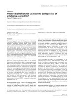
The present review describes the key levels of imbalance that
have been associated with chronic inflammation and tissue
destruction. This has to be integrated in general processes of
disease initiation through the innate and adaptive immune
responses ending in tissue and organ damage (Figure 1).
Balance in cytokines
Balance between IL-1 and IL-1 natural antagonists
Amongst the most powerful proinflammatory cytokines, IL-1
stands out as a paradigmatic example of fine-tuned regulation
of biological activities through a complex system of ligands
with agonist and antagonist functions, as well as signaling
Review
Cytokines in chronic rheumatic diseases: is everything lack of
homeostatic balance?
Carlo Chizzolini
1
, Jean-Michel Dayer
2
and Pierre Miossec
3
1
Department of Immunology and Allergy, University Hospital and School of Medicine, Geneva University Hospital, 1211 Geneva 14, Switzerland
2
School of Medicine, University of Geneva, rue Michel Servet 1, 1211 Geneva 14, Switzerland
3
Department of Immunology and Rheumatology, Hospital Edouard Herriot, University of Lyon, 69437 Lyon, France
Corresponding author: Carlo Chizzolini,
Published: 14 October 2009 Arthritis Research & Therapy 2009, 11:246 (doi:10.1186/ar2767)
This article is online at />© 2009 BioMed Central Ltd
CCR = CC-family chemokine receptor; DARC = Duffy antigen receptor for chemokines; EAE = experimental allergic encephalomyelitis; Foxp3 =
forkhead box p3; IFN = interferon; IL = interleukin; IL-1R = IL-1 receptor; IL-6Rα = IL-6 receptor alpha; IL-1Ra = IL-1 receptor antagonist; NF =
nuclear factor; RA = rheumatoid arthritis; RANTES = regulated on activation, normal T-cell expressed and secreted; SIGIRR = single immunoglobu-
lin IL-1-related receptor; sIL-6Rα = soluble IL-6Rα; SOCS = suppressors of cytokine signaling; STAT = signal transducer and activator of transcrip-
tion; TGFβ = transforming growth factor beta; Th = T-helper type; TNF = tumor necrosis factor; Treg = T cell with regulatory function; Wnt =
wingless integration site.
Arthritis Research & Therapy Vol 11 No 5 Chizzolini et al.
Page 2 of 11
(page number not for citation purposes)
and nonsignaling receptors (Figure 2). First of all, a natural
ligand of IL-1 receptors – IL-1 receptor antagonist (IL-1Ra) –
prevents recruitment of the accessory protein needed to
signal, thus acting as a competitor to IL-1 [1]. Interestingly,
IL-1Ra is preferentially produced by monocytes/macrophages
stimulated by anti-inflammatory cytokines (see below).
Second, two IL-1 receptors (Il-1RI and IL-1RII) are expressed
at the surface of many cell types. An important functional
difference, however, exists between the two receptors.
Indeed, in contrast to IL-1RI, which transduces the signal,
IL-1RII does not transduce and acts as a decoy receptor.
Furthermore, both receptors may be shed from the cell
surface by matrix metalloproteinases, and by binding to IL-1
or IL-1Ra soluble receptors may modulate their bioavailability,
ultimately affecting cell responses. One of the many members
of the IL-1 family, IL-1F5, also has inhibitory activities [2].
Some patients have autoantibodies to IL-1α and these may
also play a role by blocking IL-1 biological activity. Regulation
is also provided by single immunoglobulin IL-1-related
receptor (SIGIRR), also known as Toll–IL-1 receptor 8, which
is a member of the Toll-like receptor/IL-1R family. Its small
single extracellular immunoglobulin domain does not support
ligand binding. Besides, the intracellular domain of SIGIRR
cannot activate NFκB because it lacks two essential amino
acids (Ser447 and Tyr536) in its highly conserved Toll–IL-1
receptor domain. SIGIRR rather acts as an endogenous
inhibitor of Toll-like receptor and IL-1 signaling, because
overexpression of SIGIRR in Jurkat or HepG2 cells
substantially reduced lipopolysaccharide-induced or IL-1-
induced activation of NFκB. Furthermore, lupus-prone mice
have an accelerated course of disease when lacking Toll–IL-1
receptor 8 [3,4].
Table 1
Balance in cytokine activities according to biological processes
Process Cytokines
Inflammation IL-1 / IL-1 receptor antagonist, IL-1 receptor II, soluble IL-1 receptor I, soluble IL-1 receptor II
TNF / soluble TNF receptor I, soluble TNF receptor II
IL-6 / soluble gp130
IL-18 / IL-18 binding protein
IL-22 / IL-22 binding protein
IL-13 / IL-13 receptor alpha
CXCL
ELR+
/ CXCL
ELR–
Several proinflammatory chemokines (CXC and CC) / Duffy antigen receptor for chemokines
Several proinflammatory chemokines (CC not CXC) / D6
CCL19, CCL21, CCL25, CXCL13 / CCX-CKR
Chemerin 9 / chemerin 15
Immune cell responses Th1 cells / Th2 cells
Th17 cells /Th2 cells
Th17 cells / T cells with regulatory function
T cells with regulatory function / Th1, Th2, Th17 cells
Tissue repair and remodeling Transforming growth factor beta / TNF
IL-1 / IFNγ
IL-4 / IFNγ
CD4 T-cell differentiation IL-12 / IL-4
Transforming growth factor beta / IL-6 + T-cell growth factor beta
Tissue destruction Osteoprotegerin / RANKL
WNT / Dickkopf-1
Metabolism Adiponectin / leptin, vistatin, resistin
In view of the pleiotropic actions of cytokines, the table presents a far from complete view of possible opposing activities of cytokines and their
ligands. The back slash (/) separates the opposing molecules in respect of a given biological activity. RANKL, receptor activator of NKκB ligand;
WNT, wingless integration site.
The production by monocytes–macrophages of IL-1 and
IL-1Ra is dependent on many distinct stimuli, including T-cell
contact. Of interest, apolipoprotein A1, a negative acute-
phase reactant, may act as negative feedback regulator by
reducing IL-1 but not IL-1Ra production induced by T-cell
contact. IFNβ favors the production of IL-1Ra while
simultaneously inhibiting IL-1. Similar activities are shared by
IL-4, IL-13 and transforming growth factor beta (TGFβ),
which in this context are generally considered anti-inflam-
matory in that they increase IL-1Ra and, to a lesser extent,
decrease IL-1 production (Table 2). A similar type of regula-
tion is provided by leptin, which can modulate the expression
of IL-1Ra and the release of IL-1β by beta cells in human
islets [5].
Phosphatidylinositide 3 kinase is among the most important
signaling pathways involved in the control of the IL-1/IL-1Ra
balance in human monocytes, in so far as inhibition of
phosphatidylinositide 3 kinase delta markedly decreases IL-1
while increasing IL-1Ra [6,7]. A further example of the
plasticity of the IL-1/IL-1Ra balance in human monocytes is
the increase in IL-1Ra but decrease in T-cell-induced IL-1β in
the presence of glatiramer acetate, a therapeutic agent used
in multiple sclerosis [8].
Balance in TNF and IL-6 activities
TNF and IL-6 have become successful targets of biological
therapies in a variety of inflammatory conditions starting with
RA, thus underling their pivotal role in inflammation. Several
excellent reviews have been devoted to these two cytokines
and their relevance in human diseases [9-13]. Therefore we
shall here overview only the basic mechanisms involved in the
regulation of their biological activities, in particular stressing
differences in the activity of their respective soluble receptors.
Trimeric TNF, mostly produced by activated macrophages
and T cells, acts by binding to two distinct TNF receptors:
TNF-RI (p55), which is widely expressed; and TNF-RII (p75),
mostly present on cells of the immune system (Figure 2).
Both receptors can be enzymatically shed from the surface of
the cells and, once in the body fluids, both can bind TNF and
neutralize its biological activity [14]. The receptors therefore
act as natural inhibitors of TNF, and their production is
regulated by several stimuli including TNF itself.
At variance with TNF, IL-6 acts by binding to a heterodimeric
receptor composed of the common gp130 chain, shared with
oncostatin M, IL-11, ciliary neurotrophic factor-1, cardio-
tropin-1, and leukemia inhibitor factor, and to its specific IL-6
receptor alpha (IL-6Rα). The signaling chain is gp130, affinity
of which for IL-6 is increased in the presence of IL-6Rα. Of
interest, IL-6Rα exists as a cell-bound form expressed on few
cell types – particularly hepatocytes, phagocytes, and some
lymphocytes – but also in a soluble form abundantly present
in body fluids. Soluble IL-6Rα (sIL-6Rα) has the capacity of
binding to IL-6 and to increase its affinity for gp130. Since
gp130 is ubiquitously expressed, sIL-6Rα offers the
opportunity to cells that do not express IL-6Rα to become
responsive to IL-6, a phenomenon called trans-signaling. In
transgenic mice sIL-6Rα functions as a carrier protein for its
ligand, thereby markedly prolonging the plasma half-life of IL-
6, indicating that IL-6 signaling is increased by sIL-6Rα [15].
The agonistic properties of sIL-6Rα by enhancing IL-6
signaling are well documented. There are results indicating
also antagonistic properties of sIL-6Rα, however, which may
explain why IL-6 may in some circumstances acts as an anti-
inflammatory mediator [16].
Available online />Page 3 of 11
(page number not for citation purposes)
Figure 1
Conceptual framework for the role of cytokine imbalance in the
pathogenesis of chronic inflammatory diseases. DC, dendritic cells;
HDL-ApoA-1, high-density lipoprotein apolipoprotein A1; MΦ,
macrophage.
Figure 2
Schematic representation of agonists and antagonists determining the
biological activities of IL-1 and TNF. icIL-1Ra, intracellular IL-1 receptor
antagonist; SIGIRR, single immunoglobulin IL-1-related receptor;
sIL-1Ra, soluble IL-1 receptor antagonist; sIL-1R, soluble IL-1
receptor; sTNF, soluble TNF; sTNFR, soluble TNF receptor.
Besides a soluble form of IL-6Rα, a soluble form of gp130
(sgp130) has been detected in healthy human sera with
antagonistic properties. Of interest, the antagonistic activity
of sgp130 is markedly enhanced in the presence of sIL-6Rα
[17]. Cell responses to IL-6 are therefore finely tuned by the
ratios between-cell bound gp130 and IL-6Rα on the one
side, and on the other by available IL-6, sIL-6Rα and
sgp130.
Balance generated by soluble osteoprotegerin
Another cytokine whose biological activities are modulated by
soluble receptors or natural antagonists is osteoprotegerin,
which is a secreted member of the TNF receptor family that
binds OPGL and blocks its activity. Genetic (including gene-
targeting) studies and functional studies in vitro and in vivo
indicate that osteoprotegerin is a pure, soluble decoy
receptor [18]. Osteoprotegerin also binds and neutralizes
TNF-α-related apoptosis-inducing ligand [19].
Additional cytokines whose biological activities are regulated
by the balance of agonist and soluble nonsignaling receptors
include IL-18/IL-18 binding protein, IL-22/IL-22 binding
protein, and IL-13/IL-13 receptor alpha. These will not be
discussed in the present review, however, owing to the
shortage of space.
Balance in chemokine responses
A balance in chemokine responses is generated via several
distinct, but not mutually exclusive, operational mechanisms.
As previously shown for other cytokines, distinct chemokines
may fulfill opposing functions for a given task. A classical
example is the propensity of CXC chemokines sharing the
ELR motif (CXCL1, CXCL3, CXCL5, CXCL6, and CXCL8) to
exert angiogenic properties, while CXC chemokines lacking
the ELR motif (CXCL9, CXCL10, CXCL11) are more angio-
static [20]. Similarly, chemokines may play opposing roles in
proliferation and apoptosis susceptibility. In addition, a
peculiarity of some chemokine receptors is that they bind
chemokines but fail to signal [21]. Chemokines signal
through seven-transmembrane domain, G-protein-coupled
receptors, of which 19 have been molecularly defined. These
receptor families reflect the two major (CC and CXC)
chemokine families and two minor (C and CX
3
C) chemokine
families [22]. In addition, chemokine receptors whose
structural features are inconsistent with signaling functions
have been described. By binding to chemokines, non-
signaling receptors act as a decoy, scavenge receptors, and
regulate inflammatory and immune responses. The family of
silent chemokine receptors comprises Duffy antigen receptor
for chemokines (DARC), D6 (also known as CC chemokine
binding protein 2), and CCX-CKR (also known as CCRL1). It
is noteworthy that the silent chemokine receptors, which lack
the key residues needed for coupling with G-proteins, have
unusual expression patterns and a wide range of chemokine-
binding properties.
DARC is expressed on erythrocytes and endothelial cells of
postcapillary veins in many organs – including, amongst
others, high endothelial venules in lymphoid organs [23].
DARC binds 11 proinflammatory (both CC and CXC) but not
homeostatic chemokines, and preferentially angiogenic but
not angiostatic chemokines [24]. Chemokines injected in
DARC
–/–
mice rapidly disappear from circulation, indicating a
role of erythrocyte DARC as a sink or reservoir. Endothelial
DARC, however, appears to have a downregulating effect on
inflammation. Overexpression of endothelial DARC in animal
models is therefore associated with both decreased
angiogenesis and tumor growth, while a lack in DARC is
associated with increased tumor growth, metastasis forma-
tion and increased concentrations of CXCL1 and CXCL3
[25,26].
D6 binds most inflammatory CC chemokines, but not CXC
and homeostatic CC chemokines. D6 is expressed at high
concentrations on lymphatic and venular endothelium, parti-
cularly in the skin, gut, lung, and placenta [27]. D6 mediates
chemokine degradation, being constitutively internalized
through clathrin-coated pits. D6
–/–
mice are prone to exag-
gerated inflammatory responses induced by phorbol ester
myristate acetate application to the skin or subcutaneous
injections of complete Freund’s adjuvant [28,29]. Lack of D6
expression in syncytiotrophoblast increases the susceptibility
to inflammation-induced fetal loss [30]. In contrast, trans-
genic expression of D6 in keratinocytes dampens cutaneous
inflammation and reduces tumor growth [31].
Arthritis Research & Therapy Vol 11 No 5 Chizzolini et al.
Page 4 of 11
(page number not for citation purposes)
Table 2
Cytokine roles categorized according to their contribution to
inflammation in rheumatoid arthritis
Proinflammatory Ambivalent Anti-inflammatory
TNF IFNγ IL-1 receptor antagonist
IL-1 Transforming growth factor beta IL-4
a
IL-12 IL-6
b
IL-13
IL-15 IL-10
c
IL-17A/IL-17F IL-25
IL-18 IL-27
CXCL8 IL-35
CCL3
CCL2 7ND
7ND, N-terminal natural deletion variant of monocytes chemotactic
protein-1/CCL2.
a
IL-4 is anti-inflammatory in the context of rheumatoid
arthritis synovial inflammation. By impacting on IgE production,
however, IL-4 is a key cytokine in IgE-mediated inflammation. Similar
considerations apply to IL-13.
b
IL-6 may be proinflammatory or anti-
inflammatory according to the circumstances. IL-6 blockade has been
shown to be clinically useful to control rheumatoid arthritis in
randomized trials.
c
IL-10 is usually anti-inflammatory, but upon priming
of monocytes with IFNα it induces proinflammatory responses.
CCX-CKR appears to have a more limited chemokine-binding
repertoire that includes CCL19, CCL21, CCL25, and
CXCL13, and it is expressed exclusively by stromal cells in
the thymus and lymph nodes, by lymph vessels in the
intestine and by the epidermis [32]. In CCX-CKR
–/–
mice,
trafficking of dendritic cells to lymph nodes under steady-
state conditions appears to be decreased, as well as the
recruitment of hematopoietic precursors to the thymus.
Pathogen-encoded decoys also affect chemokine activities.
Indeed, molecular mimicry of chemokines and their receptor
is an important immune-evasion strategy used by pathogens,
of which numerous examples are known. Viral chemokine
binding protein and Schistosoma mansonii chemokine bind-
ing protein have been described.
The receptor functions of some chemokines appear to vary
according to the context in which they operate. For instance,
IL-10 uncouples CCR2 binding from signaling, and therefore
CCR2 functionally becomes a decoy receptor [33]. An
additional example is the high level of CCR5 expressed in
response to lipoxin A4 on apoptotic neutrophils and T cells.
Lipoxin A4 is produced late during the inflammatory response
when significant tissue damage has already occurred. By
increasing the expression of CCR5 on dying cells, lipoxin A4
contributes to scavenging CCR5 ligands, which therefore are
no longer available for recruiting new cells, which in turn
reduces inflammation.
An additional mechanism regulating chemokine activities is
related to modifications of their primary structure. For
instance, the N-terminal natural deletion variant of monocytes
chemotactic protein-1/CCL2 (called 7ND) inhibits chemo-
taxis mediated by monocytes chemotactic protein-1, and the
extension of RANTES/CCL5 by a single methionine
(met-RANTES) creates a potent and selective RANTES
antagonist.
The particular example of chemerin
Chemerin is a plasma protein known for its proinflammatory
properties exerted upon binding to the G-protein coupled
receptor ChemR23/CMKLR1 – expressed on macrophages
and plasmacytoid dendritic cells – where it induces cell
migration. Chemerin is secreted as an inactive precursor and is
processed by proteases before becoming an active mediator.
As for conventional chemokines, the biologically active
chemerin binds to ChemR23 with its COOH-terminal portion.
Of interest, different proteases generate different chemerin
peptides, which possess opposite functions. Serine
proteases mainly produced by activated neutrophils – early
mediators in inflammation – therefore generate chemerin 9
(9 AA peptide), which is an agonist in the nanomolar range.
Cysteine proteases – mainly produced by macrophages –
which arrive later at the inflammatory site, however, generate
chemerin 15 (15 AA peptide). This peptide in the picomolar
range acts as an antagonist, expressing potent anti-inflam-
matory activities and contributing to reduce inflammation [34].
A further layer of complexity has been added recently with the
description of an additional chemerin receptor named
CCRL2, selectively expressed on mouse mast cells. Upon
binding to this receptor, chemerin induces neither cell
migration nor calcium flux. CCRL2 is therefore supposed to
scavenge chemerin. The experimental test of this hypothesis
led to the opposite result, however, indicating enhanced
inflammation in a rodent model of IgE-mediated passive
cutaneous anaphylaxis. A possible explanation could be that
mast cells bind the N-terminal portion of chemerin with
CCRL2 and present the COOH-terminal portion to cells
expressing ChemR23, which are thus potently activated [35].
The Th1/Th2 balance
In the late 1980s Mosmann and colleagues described the
Th1/Th2 balance when studying a large series of mouse
CD4
+
T-cell clones [36]. They observed that some clones
would produce IFNγ but not IL-4, while others would do the
opposite. Therefore, based on the dicotomic production of
two key cytokines, it was possible to classify T-cell clones
into two groups, which were named Th1 and Th2. The same
concepts were verified by studying human T-cell clones [37].
Naïve T cells could be induced to become Th1 or Th2 simply
by modifying the cytokine present in the milieu during priming,
although the dose of antigen, the amount of co-stimulation,
and the age of antigen-presenting cells could also affect
polarization.
Of major importance, Th1 cytokines were shown to inhibit
Th2 cytokine production and function, and vice versa. This
observation included cytokines important for priming: IL-12
and IFNγ for Th1 cells, and IL-4 for Th2 cells. Starting investi-
gations with mouse models of human diseases, it was found
that models of multiple sclerosis – such as the antigen-
induced experimental acute encephalomyelitis (EAE) – or of
RA – such as type II collagen arthritis – were associated with
the overexpression of IFNγ but not of IL-4. In sharp contrast,
models of allergic diseases such as asthma were associated
with IL-4 without IFNγ expression. In these models, forced
expression of counteracting T-helper cytokines could in many
instances abrogate disease expression [38,39].
Addition of the Th17 pattern
In 2005 the above classification was amended when it was
shown in the mouse that IL-17 was produced by a particular T-
helper cell, named Th17 [40,41] (Figure 3). As early as 1999,
however, it was shown that some T-cell clones obtained from
the synovium of RA patients were producing IL-17 and differed
from the classical Th1/Th2 clones [42]. Indeed, they did not
produce IL-4 and produced little, if any, IFNγ.
The Th1/Th2 paradigm was then revisited; key observations
were made based on the murine EAE model [43]. This model
Available online />Page 5 of 11
(page number not for citation purposes)
was previously associated with Th1 responses. Th1 cells are
induced by IL-12 produced by monocytes and dendritic cells.
IL-12 is a heterodimer composed of p35 and p40 subunits.
Protection from EAE was afforded when IL-12 was blocked
with anti-IL-12p40. IL-23 is also a heterodimer, however,
composed of the IL-12/IL-23 common p40 subunit and the
specific p19 subunit. When inhibitors specific to IL-23 or
p19-deficient mice were used, it was recognized that IL-23
and not IL-12 was responsible for EAE induction by assisting
the expansion of Th17 cells. Many chronic inflammatory
diseases previously thought to be associated with Th1 have
therefore been reclassified as Th17 diseases [44]. The
opposing roles of Th2 and Th17 responses are now clear,
since IL-4 strongly inhibits IL-17 differentiation. For Th1 and
Th17 cells, a more balanced view is now accepted [45]. In
both human and murine conditions, a large proportion of T cells
can express simultaneously IFNγ and IL-17. This is clearly seen
with T-cell clones from peripheral blood. The simultaneous
production of the two cytokines appears uncommon, however,
in inflammatory tissues where T cells producing cytokines take
on a plasma cell-like appearance, possibly indicating full
differentiation with a fixed phenotype [46].
In addition to the production of IL-17 (now referred to as
IL-17A), Th17 cells can produce other cytokines – including
IL-17F (a close member of the IL-17 family), IL-21, and IL-22.
IL-21 acts as an endogenous amplifier of the Th17 lineage
[41]. IL-22 appears more specifically associated with skin
defense [47]. IL-17A and IL-17F share a large number of
functions, with a strong correlation between the genes
induced in RA synoviocytes by the two cytokines, IL-17F
being less potent [48]. In addition, synergistic activities are
seen when combining TNF with IL-17A or IL-17F. IL-17A and
IL-17F may, however, have different roles in mouse models of
inflammation and host defense [49].
IL-17E (also termed IL-25) is a very different member of the
IL-17 family. IL-17E is more a Th2 cytokine, involved in
allergic reactions and inhibiting the Th17 pathway [50].
Consequently, there is another balance between the effects
of IL-17A and IL-17F and those of IL-17E/IL-25.
Balance between Th17 and T cells with regulatory
function
Th1, Th2, and Th17 cells are effector cells contributing to key
functions of the immune response. An additional hetero-
geneous subset of T cells with regulatory function (Tregs) has
recently been identified. Some Tregs occur naturally, whereas
others are induced in response to antigens. Charac-
teristically, Tregs express the transcription factor Foxp3, as
well as CD4 and CD25. The immunomodulating effects of
Tregs are mediated by membrane molecules (for example,
cytotoxic T-lymphocyte-associated protein 4, glucocorticoid-
induced TNF receptor, and OX40) and by cytokines including
IL-10 and TGFβ.
TGFβ is key to the induction of Foxp3-positive regulatory
T cells. Indeed, mice defective in TFGβ die quickly from a
Arthritis Research & Therapy Vol 11 No 5 Chizzolini et al.
Page 6 of 11
(page number not for citation purposes)
Figure 3
Cytokines, hormones, and other soluble mediators controlling biology of Th17 cells leading to tissue destruction. Summary of some of the many
mediators involved in Th17 differentiation, expansion, acquisition of effector function and their relationship with macrophages, which may then
mediate tissue destruction. Orange arrows, enhancement; blunted black heads, inhibition; black arrows, production. AHR, aryl-hydrocarbon
receptor; APO-A-1, apolipoprotein A1; MMP, matrix metalloproteinase; MΦ, macrophage; PGE2, prostaglandin E
2
; RORγt, retinoic acid-related
orphan receptor γt; STAT, signal transducer and activator of transcription; TGFβ, T-cell growth factor beta; Treg = T cell with regulatory function.
massive uncontrolled inflammatory disease [51]. Contrasting
with the effect of TGFβ alone, the simultaneous presence of
TGFβ and IL-6 favors the emergence of Th17 cells alongside
the inhibition of the Tregs [52]. IL-6 – a cytokine with
pleiotropic inflammatory effects – therefore plays a pivotal
part, at least in the mouse, in directing the differentiation of
T cells toward the Th17 or Treg pathways. TNF, IL-1, and IL-
17 interact together to induce massive amounts of IL-6.
Increased inflammation therefore has a positive effect on the
Th17 pathway and a negative effect on its regulation.
The inhibitory functions of IL-27 and IL-35
Some recently identified cytokines such as IL-27 and IL-35
appear to be more involved in dampening the immune
response. IL-27 belongs to the IL-12 cytokine family that also
comprises IL-23 and IL-35, all involved in the regulation of
T-helper cell differentiation. IL-27 is unique in that it induces
Th1 differentiation while simultaneously suppressing immune
responses. The immunosuppressive effects of IL-27 depend
on inhibition of the development of Th17 cells and induction
of IL-10 production [53]. IL-27 exerts potent anti-inflammatory
effects in several infectious and experimental autoimmune
models. In particular, suppressive effects on helper T cells –
which are implicated in the pathogenesis of multiple sclerosis –
suggest that IL-27 may be therapeutically relevant in multiple
sclerosis. While exciting discoveries have been made,
however, these are still at an early stage and further studies
are required to understand the pathophysiological roles of IL-
27 and its therapeutic potential in humans [54].
The inhibitory cytokine IL-35 contributes to regulatory T-cell
function, being specifically produced by Tregs and required
for maximal suppressive activity [55]. Ectopic expression of
IL-35 confers regulatory activity on naive T cells, whereas
recombinant IL-35 suppresses T-cell proliferation. The role of
Tregs in RA has been established in both patients and animal
models. The Tregs increase in patients who are responding
to anti-TNFα therapy. Of the current hypotheses, Treg
expansion or transfer may hold promise for the treatment of
RA [56].
Cytokines, hormones, vitamins, arachidonic
acid metabolites and lipoproteins
A further layer of control at the level of expression of
cytokines, cytokine inhibitors and acute-phase proteins is
provided by hormones. Estrogens as well as androgens
inhibit the production of IL-1β and TNFα by monocytes–
macrophages. Androgens antagonize stimulatory effects of
estrogens. Some studies suggest that estradiol is more
inhibitory to Thl cytokines (for example, IFNγ, IL-2) while
testosterone is inhibitory to Th2 cytokines (for example, IL-4).
On the other hand, cytokines control the hypothalamic–
hypophyseal–adrenal gland axis as well as the sex hormones
[57]. Vitamins may also affect cytokine production by
influencing the polarization of effector CD4
+
T cells. For
instance, retinoic acid enhances Treg expansion while simul-
taneously inhibiting Th17 cells [58]. Conversely, vitamin D
favors Th2 polarization and diverts Tregs from their regulatory
function [59,60]. Finally, prostaglandin E
2
– a metabolite of
arachidonic acid – may also affect cytokine production by
favoring the expansion of Th17 cells [61].
Destruction/repair balance
Chronic inflammatory diseases such as RA are so severe
because the disease process affects matrix metabolism.
Although RA is seen as a destructive disease, it is not well
appreciated that the main problem is in fact the inhibition of
repair activity. Any type of chronic joint inflammation, whether
infectious, inflammatory, or autoimmune, will result in joint
destruction within months or, at best, within a few years, but it
will take decades to observe some kind of joint repair – even
in conditions like osteoarthritis where repair activity is main-
tained. In a model of cell interaction between synoviocytes
and T-cell clones, it was found that Th1 and Th17 clones
induced defects in collagen synthesis in vitro, indicating an
inhibition of their repair activity (Figure 1). In sharp contrast,
Th2 cells induce collagen synthesis, indicating their beneficial
role in repair activity [62]. Very similar conclusions were
obtained when monocytes were incubated with Th1 or Th2
clones. The interaction with a Th1 clone led to the production
of IL-1, a key marker of destructive inflammation, whereas the
use of a Th2 clone led to production of IL-1Ra along with its
anti-inflammatory and anti-destructive properties [63].
Wingless integration site (Wnt) proteins make up a family of
secreted growth factors, identified in virtually every organism;
they regulate key aspects of cellular functions such as
growth, differentiation, and death. Several members of the
Wnt pathway play an important part in bone remodeling.
Dickkopf-1, a soluble inhibitor of the Wnt pathway, controls
bone remodeling. Increased Dickkopf-1 levels are linked to
bone resorption, and decreased levels are linked to new bone
formation. Low-density lipoprotein receptor-related protein 5,
the main receptor that mediates Wnt signaling, plays a critical
role in bone mass regulation. Gain-of-function mutations of
lipoprotein receptor-related protein 5 cause high bone mass
phenotypes, whereas loss-of-function mutations are linked to
severe osteoporosis [64].
Adipose tissue in inflammation: a protective
role via IL-1 receptor antagonist?
Adipokines are beginning to emerge as mediators of inflam-
mation. Knowledge of their precise activities remains in its
infancy, however, and is still controversial [65]. Many of the
adipokines appear to have proinflammatory properties. In
general, adiponectin is considered anti-inflammatory, and
leptin, vistatin and resistin are considered proinflammatory.
The formation of adipose tissue could be due to abnormal
metabolic processes and, at the local level, due to chronic
inflammatory processes such as those occurring in the
synovium in RA or osteoarthritis, or in the peritoneal cavity in
various inflammatory processes of the digestive system.
Available online />Page 7 of 11
(page number not for citation purposes)
Arthritis Research & Therapy Vol 11 No 5 Chizzolini et al.
Page 8 of 11
(page number not for citation purposes)
Figure 4
Schematic examples of cytokine signal modulation. (a) Priming: upon exposure to suboptimal levels of type I interferon or IL-6, no signal is
generated; but if later the cell (macrophage) sees suboptimal levels of IFNγ, then gene transcription initiates and a signal is generated [67,68]. IDO,
indoleamine-2,3-dioxygenase; IFNAR, interferon alpha receptor IL-6Ra, IL-6 receptor alpha; IRF1, interferon regulatory factor 1; STAT, signal
transducer and activator of transcription. (b) Uncoupling of signaling: monocytes chemotactic protein-1 (MCP-1)/CCL2 signal upon CCR2
binding. In the presence of IL-10, binding of MCP-1/CCL2 to CCR2 is preserved but signal is abolished [33]. IL-10R, IL-10 receptor. (c)
Reprogramming of signaling: in macrophages, Toll-like receptor (TLR) 2 activation induces TNF, production of which is reduced by simultaneously
induced homeostatic IL-10 (negative feedback). If the cell has been primed with type I interferon, however, then IL-10 fails to negatively regulate
TLR signaling. In turn, IL-10 becomes a proinflammatory cytokine favoring the production of TNF and other cytokines. The signaling cascade
induced by IL-10 shifts form anti-inflammatory STAT 3 to proinflammatory STAT 1 [70]. Figures in circles indicate sequences of events. AP-1,
activator protein 1.
Adipocytes are said to produce many hormones and pro-
inflammatory mediators. White adipose tissue in humans,
however, is assumed to be the main source of IL-1Ra, and
also contains IL-10. Furthermore, IFNβ was found to be the
principal cytokine inducing IL-1Ra in various white adipose
tissues, such as that present in the synovium. It is possible
that, in addition to other functions, adipose tissue may be part
of a mechanism limiting local inflammation and that fibro-
blasts in the vicinity may further induce IL-1Ra in adipocytes
via the production of IFNβ [66].
Influence of signal transduction in cytokine
balance
Cytokines may have opposing effects on the same cell
depending on the circumstances in which they hit their target.
The timing and the previous activation status are major
determinants of responses that cytokines elicit (Figure 4).
Differential outcomes could be sensitization or amplification
of proinflammatory signals (that is, priming), reprogramming
of signaling resulting in proinflammatory activity of pleiotropic
or anti-inflammatory cytokines, and attenuation of anti-
inflammatory signals and homeostatic mechanisms. Signal
transducer and activator of transcription (STAT) 1 has been
shown in vitro and in vivo to be involved in some of these
effects. For instance, transient exposure to subactivating
concentrations of IFNα or IL-6 primes primary human mono-
cytes for subsequent exposure to IFNγ, resulting in enhanced
interferon regulatory factor 1 and indoleamine-2,3-dioxygenase
gene expression in a STAT-1-dependent manner [67,68].
This may explain robust IFN signatures in RA synovium, not-
withstanding very low amounts of IFNγ. Enhanced expression
of STAT-1-dependent genes upon IFNγ priming of monocytes
is a finely tuned process involving Fcγ receptor/DNAX
activation protein 12, as demonstrated in Fcγ receptor/DNAX
activation protein 12
–/–
mice in which the priming effect is
lost.
IL-10 contributes to homeostatic responses in proinflam-
matory conditions. For instance, in human monocytes, Toll-
like receptor 2 ligation results in NFκB-dependent TNF
production and simultaneously in activator protein-1-depen-
dent IL-10 production [69]. Upon binding to its receptor,
IL-10 decreases TNF production in a STAT-3-dependent
manner, thus exerting a negative feedback. Pre-exposure of
monocytes to IFNα, however, results in IL-10 gaining pro-
inflammatory functions. Of interest, this process is STAT 1
dependent. It has therefore been shown in human monocytes
primed with IFNα that IL-10 not only fails to reduce the
subsequent production of TNF in response to lipopoly-
saccharide, which may simply indicate a loss of function of
the anti-inflammatory activity of IL-10, but in addition primes
monocytes to transcribe genes in response to IL-10 usually
induced by IFN. It appears that, due to the effect of type I
interferons, the balance of IL-10 signaling shifts from STAT 3
(anti-inflammatory) to STAT 1 (proinflammatory) signals.
Furthermore, IL-10 induces chemokine production in IFNα-
primed macrophages, resulting in recruitment of activated
T cells; aberrant IL-10 signaling may therefore contribute to
inflammation in conditions with high interferon levels
(systemic lupus erythematosus) [70].
The suppressors of cytokine signaling (SOCS) family of
intracellular proteins – which encompasses eight members,
sharing a central Src homology domain 2 and a C-terminus
SOCS box – act as negative regulators of intracellular
signaling of the Jak–STAT pathway used by several cyto-
kines. They act by inhibiting the kinase activity, by competing
with substrates needed for signal transduction, and by
targeting associated proteins to proteasome degradation.
Beside negative regulation, SOCS proteins can also affect
the quality of signaling. For instance, in the absence of
SOCS 3, IL-6 induces a wider transcriptional response,
which includes interferon-like gene expression owing to
increased STAT 1 phosphorylation. SOCS proteins therefore
impact on a number of important mechanisms regulating
inflammation and the immune response [71].
Conclusions
Cytokine activities affect most, if not all, biological processes
involved in homeostasis as well as in host defense and auto-
aggression. A continuous, finely tuned, crosstalk between
cytokines, receptors, agonist and antagonist ligands, as well
as with mediators belonging to other families of molecules,
regulates cytokine biological activities. Furthermore, the
context in which cytokines are available, including the
temporal sequence of events preceding the availability of a
given cytokine, very much impact on their capacity to favor or
inhibit inflammation and other biological processes. During
the past three decades we have learned that an imbalance in
cytokine activities is associated with autoimmune and
autoinflammatory disorders. More important, our knowledge
of the many levels of cytokine balance has led to the
generation of important tools to control inflammatory and
destructive diseases. The future will no doubt witness
additional major achievements in this area of medicine.
Available online />Page 9 of 11
(page number not for citation purposes)
This article is part of a special collection of reviews, The
Scientific Basis of Rheumatology: A Decade of
Progress, published to mark Arthritis Research &
Therapy’s 10th anniversary.
Other articles in this series can be found at:
/>The Scientific Basis
of Rheumatology:
A Decade of Progress
Competing interests
The authors declare that they have no competing interests.
Acknowledgements
The field of cytokine balance is very large and imprecisely defined. The
authors would like to apologize to the many authors having contributed
to this fascinating field whose work has not been quoted in the present
review. CC was supported in part by grant No 31003A_124941/1
from the Swiss National Science Foundation.
References
1. Arend WP, Dayer JM: Cytokines and cytokine inhibitors or
antagonists in rheumatoid arthritis. Arthritis Rheum 1990, 33:
305-315.
2. O’Neill LA: The interleukin-1 receptor/Toll-like receptor super-
family: 10 years of progress. Immunol Rev 2008, 226:10-18.
3. Garlanda C, Riva F, Polentarutti N, Buracchi C, Sironi M, De
Bortoli M, Muzio M, Bergottini R, Scanziani E, Vecchi A, Hirsch E,
Mantovani A: Intestinal inflammation in mice deficient in Tir8,
an inhibitory member of the IL-1 receptor family. Proc Natl
Acad Sci U S A 2004, 101:3522-3526.
4. Lech M, Kulkarni OP, Pfeiffer S, Savarese E, Krug A, Garlanda C,
Mantovani A, Anders HJ: Tir8/Sigirr prevents murine lupus by
suppressing the immunostimulatory effects of lupus autoanti-
gens. J Exp Med 2008, 205:1879-1888.
5. Maedler K, Sergeev P, Ehses JA, Mathe Z, Bosco D, Berney T,
Dayer JM, Reinecke M, Halban PA, Donath MY: Leptin modu-
lates beta cell expression of IL-1 receptor antagonist and
release of IL-1
ββ
in human islets. Proc Natl Acad Sci U S A
2004, 101:8138-8143.
6. Molnarfi N, Gruaz L, Dayer JM, Burger D: Opposite regulation of
IL-1
ββ
and secreted IL-1 receptor antagonist production by
phosphatidylinositide-3 kinases in human monocytes acti-
vated by lipopolysaccharides or contact with T cells.
J Immunol 2007, 178:446-454.
7. Molnarfi N, Brandt KJ, Gruaz L, Dayer JM, Burger D: Differential
regulation of cytokine production by PI3K
δδ
in human mono-
cytes upon acute and chronic inflammatory conditions. Mol
Immunol 2008, 45:3419-3427.
8. Burger D, Molnarfi N, Weber MS, Brandt KJ, Benkhoucha M,
Gruaz L, Chofflon M, Zamvil SS, Lalive PH: Glatiramer acetate
increases IL-1 receptor antagonist but decreases T cell-
induced IL-1
ββ
in human monocytes and multiple sclerosis.
Proc Natl Acad Sci U S A 2009, 106:4355-4359.
9. Feldmann M, Maini RN: Anti-TNF alpha therapy of rheumatoid
arthritis: what have we learned? Annu Rev Immunol 2001, 19:
163-196.
10. Feldmann M, Maini SR: Role of cytokines in rheumatoid arthri-
tis: an education in pathophysiology and therapeutics. Immu-
nol Rev 2008, 223:7-19.
11. Brennan FM, McInnes IB: Evidence that cytokines play a role in
rheumatoid arthritis. J Clin Invest 2008, 118:3537-3545.
12. Kishimoto T: Interleukin-6: from basic science to medicine –
40 years in immunology. Annu Rev Immunol
2005, 23:1-21.
13. Smolen JS, Aletaha D, Koeller M, Weisman MH, Emery P: New
therapies for treatment of rheumatoid arthritis. Lancet 2007,
370:1861-1874.
14. Seckinger P, Isaaz S, Dayer JM: Purification and biologic char-
acterization of a specific tumor necrosis factor alpha inhibitor.
J Biol Chem 1989, 264:11966-11973.
15. Peters M, Jacobs S, Ehlers M, Vollmer P, Mullberg J, Wolf E, Brem
G, Meyer zum Buschenfelde KH, Rose-John S: The function of
the soluble interleukin 6 (IL-6) receptor in vivo: sensitization
of human soluble IL-6 receptor transgenic mice towards IL-6
and prolongation of the plasma half-life of IL-6. J Exp Med
1996, 183:1399-1406.
16. Knupfer H, Preiss R: sIL-6R: more than an agonist? Immunol
Cell Biol 2008, 86:87-91.
17. Muller-Newen G, Kuster A, Hemmann U, Keul R, Horsten U,
Martens A, Graeve L, Wijdenes J, Heinrich PC: Soluble IL-6
receptor potentiates the antagonistic activity of soluble gp130
on IL-6 responses. J Immunol 1998, 161:6347-6355.
18. Simonet WS, Lacey DL, Dunstan CR, Kelley M, Chang MS, Luthy
R, Nguyen HQ, Wooden S, Bennett L, Boone T, Shimamoto G,
DeRose M, Elliott R, Colombero A, Tan HL, Trail G, Sullivan J,
Davy E, Bucay N, Renshaw-Gegg L, Hughes TM, Hill D, Pattison
W, Campbell P, Sander S, Van G, Tarpley J, Derby P, Lee R,
Boyle WJ: Osteoprotegerin: a novel secreted protein involved
in the regulation of bone density. Cell 1997, 89:309-319.
19. Sheridan JP, Marsters SA, Pitti RM, Gurney A, Skubatch M,
Baldwin D, Ramakrishnan L, Gray CL, Baker K, Wood WI,
Goddard AD, Godowski P, Ashkenazi A: Control of TRAIL-
induced apoptosis by a family of signaling and decoy recep-
tors. Science 1997, 277:818-821.
20. Mehrad B, Keane MP, Strieter RM: Chemokines as mediators of
angiogenesis. Thromb Haemost 2007, 97:755-762.
21. Mantovani A, Bonecchi R, Locati M: Tuning inflammation and
immunity by chemokine sequestration: decoys and more. Nat
Rev Immunol 2006, 6:907-918.
22. Charo IF, Ransohoff RM: The many roles of chemokines and
chemokine receptors in inflammation. N Engl J Med 2006,
354:610-621.
23. Peiper SC, Wang ZX, Neote K, Martin AW, Showell HJ, Conklyn
MJ, Ogborne K, Hadley TJ, Lu ZH, Hesselgesser J, Horuk R: The
Duffy antigen/receptor for chemokines (DARC) is expressed
in endothelial cells of Duffy negative individuals who lack the
erythrocyte receptor. J Exp Med 1995, 181:1311-1317.
24. Gardner L, Patterson AM, Ashton BA, Stone MA, Middleton J:
The human Duffy antigen binds selected inflammatory but not
homeostatic chemokines. Biochem Biophys Res Commun
2004, 321:306-312.
25. Du J, Luan J, Liu H, Daniel TO, Peiper S, Chen TS, Yu Y, Horton
LW, Nanney LB, Strieter RM, Richmond A: Potential role for
Duffy antigen chemokine-binding protein in angiogenesis and
maintenance of homeostasis in response to stress. J Leukoc
Biol 2002, 71:
141-153.
26. Addison CL, Belperio JA, Burdick MD, Strieter RM: Overexpres-
sion of the duffy antigen receptor for chemokines (DARC) by
NSCLC tumor cells results in increased tumor necrosis. BMC
Cancer 2004, 4:28.
27. Nibbs RJ, Kriehuber E, Ponath PD, Parent D, Qin S, Campbell JD,
Henderson A, Kerjaschki D, Maurer D, Graham GJ, Rot A: The
beta-chemokine receptor D6 is expressed by lymphatic
endothelium and a subset of vascular tumors. Am J Pathol
2001, 158:867-877.
28. Jamieson T, Cook DN, Nibbs RJ, Rot A, Nixon C, McLean P,
Alcami A, Lira SA, Wiekowski M, Graham GJ: The chemokine
receptor D6 limits the inflammatory response in vivo. Nat
Immunol 2005, 6:403-411.
29. Martinez de la Torre Y, Locati M, Buracchi C, Dupor J, Cook DN,
Bonecchi R, Nebuloni M, Rukavina D, Vago L, Vecchi A, Lira SA,
Mantovani A: Increased inflammation in mice deficient for the
chemokine decoy receptor D6. Eur J Immunol 2005, 35:1342-
1346.
30. Martinez de la Torre Y, Buracchi C, Borroni EM, Dupor J, Bonec-
chi R, Nebuloni M, Pasqualini F, Doni A, Lauri E, Agostinis C, Bulla
R, Cook DN, Haribabu B, Meroni P, Rukavina D, Vago L, Tedesco
F, Vecchi A, Lira SA, Locati M, Mantovani A: Protection against
inflammation- and autoantibody-caused fetal loss by the
chemokine decoy receptor D6. Proc Natl Acad Sci U S A 2007,
104:2319-2324.
31. Nibbs RJ, Gilchrist DS, King V, Ferra A, Forrow S, Hunter KD,
Graham GJ: The atypical chemokine receptor D6 suppresses
the development of chemically induced skin tumors. J Clin
Invest 2007, 117:1884-1892.
32. Heinzel K, Benz C, Bleul CC: A silent chemokine receptor regu-
lates steady-state leukocyte homing in vivo. Proc Natl Acad
Sci U S A 2007, 104:8421-8426.
33. D’Amico G, Frascaroli G, Bianchi G, Transidico P, Doni A, Vecchi
A, Sozzani S, Allavena P, Mantovani A: Uncoupling of inflamma-
tory chemokine receptors by IL-10: generation of functional
decoys. Nat Immunol 2000, 1:387-391.
34. Cash JL, Hart R, Russ A, Dixon JP, Colledge WH, Doran J, Hen-
drick AG, Carlton MB, Greaves DR: Synthetic chemerin-derived
peptides suppress inflammation through ChemR23. J Exp
Med 2008, 205:767-775.
35. Zabel BA, Nakae S, Zuniga L, Kim JY, Ohyama T, Alt C, Pan J,
Suto H, Soler D, Allen SJ, Handel TM, Song CH, Galli SJ, Butcher
EC: Mast cell-expressed orphan receptor CCRL2 binds
chemerin and is required for optimal induction of IgE-medi-
ated passive cutaneous anaphylaxis. J Exp Med 2008, 205:
2207-2220.
Arthritis Research & Therapy Vol 11 No 5 Chizzolini et al.
Page 10 of 11
(page number not for citation purposes)
36. Mosmann TR, Schumacher JH, Street NF, Budd R, O’Garra A,
Fong TA, Bond MW, Moore KW, Sher A, Fiorentino DF: Diversity
of cytokine synthesis and function of mouse CD4
+
T cells.
Immunol Rev 1991, 123:209-229.
37. Romagnani S: Human TH1 and TH2 subsets: doubt no more.
Immunol Today 1991, 12:256-257.
38. Miossec P, van den Berg W: Th1/Th2 cytokine balance in
arthritis. Arthritis Rheum 1997, 40:2105-2115.
39. Lubberts E, Joosten LA, Chabaud M, van Den Bersselaar L,
Oppers B, Coenen-De Roo CJ, Richards CD, Miossec P, van Den
Berg WB: IL-4 gene therapy for collagen arthritis suppresses
synovial IL-17 and osteoprotegerin ligand and prevents bone
erosion. J Clin Invest 2000, 105:1697-1710.
40. Harrington LE, Hatton RD, Mangan PR, Turner H, Murphy TL,
Murphy KM, Weaver CT: Interleukin 17-producing CD4
+
effec-
tor T cells develop via a lineage distinct from the T helper type
1 and 2 lineages. Nat Immunol 2005, 6:1123-1132.
41. Bettelli E, Korn T, Kuchroo VK: Th17: the third member of the
effector T cell trilogy. Curr Opin Immunol 2007, 19:652-657.
42. Aarvak T, Chabaud M, Miossec P, Natvig JB: IL-17 is produced
by some proinflammatory Th1/Th0 cells but not by Th2 cells.
J Immunol 1999, 162:1246-1251.
43. Cua DJ, Sherlock J, Chen Y, Murphy CA, Joyce B, Seymour B,
Lucian L, To W, Kwan S, Churakova T, Zurawski S, Wiekowski M,
Lira SA, Gorman D, Kastelein RA, Sedgwick JD: Interleukin-23
rather than interleukin-12 is the critical cytokine for autoim-
mune inflammation of the brain. Nature 2003, 421:744-748.
44. Miossec P: Diseases that may benefit from manipulating the
Th17 pathway. Eur J Immunol 2009, 39:667-669.
45. Steinman L: A rush to judgment on Th17. J Exp Med 2008, 205:
1517-1522.
46. Page G, Sattler A, Kersten S, Thiel A, Radbruch A, Miossec P:
Plasma cell-like morphology of Th1-cytokine-producing cells
associated with the loss of CD3 expression. Am J Pathol
2004, 164:409-417.
47. Zheng Y, Danilenko DM, Valdez P, Kasman I, Eastham-Anderson
J, Wu J, Ouyang W: Interleukin-22, a T(H)17 cytokine, medi-
ates IL-23-induced dermal inflammation and acanthosis.
Nature 2007, 445:648-651.
48. Zrioual S, Ecochard R, Tournadre A, Lenief V, Cazalis MA,
Miossec P: Genome-wide comparison between IL-17A- and
IL-17F-induced effects in human rheumatoid arthritis synovio-
cytes. J Immunol 2009, 182:3112-3120.
49. Ishigame H, Kakuta S, Nagai T, Kadoki M, Nambu A, Komiyama Y,
Fujikado N, Tanahashi Y, Akitsu A, Kotaki H, Sudo K, Nakae S,
Sasakawa C, Iwakura Y: Differential roles of interleukin-17A
and -17F in host defense against mucoepithelial bacterial
infection and allergic responses. Immunity 2009, 30:108-119.
50. Wang YH, Angkasekwinai P, Lu N, Voo KS, Arima K, Hanabuchi
S, Hippe A, Corrigan CJ, Dong C, Homey B, Yao Z, Ying S,
Huston DP, Liu YJ: IL-25 augments type 2 immune responses
by enhancing the expansion and functions of TSLP-DC-acti-
vated Th2 memory cells. J Exp Med 2007, 204:1837-1847.
51. Shull MM, Ormsby I, Kier AB, Pawlowski S, Diebold RJ, Yin M,
Allen R, Sidman C, Proetzel G, Calvin D, et al.: Targeted disrup-
tion of the mouse transforming growth factor-beta 1 gene
results in multifocal inflammatory disease. Nature 1992,
359:693-699.
52. Bettelli E, Carrier Y, Gao W, Korn T, Strom TB, Oukka M, Weiner
HL, Kuchroo VK: Reciprocal developmental pathways for the
generation of pathogenic effector TH17 and regulatory T cells.
Nature 2006, 441:235-238.
53. Yoshida H, Yoshiyuki M: Regulation of immune responses by
interleukin-27. Immunol Rev 2008, 226:234-247.
54. Fitzgerald DC, Rostami A: Therapeutic potential of IL-27 in mul-
tiple sclerosis? Expert Opin Biol Ther 2009, 9:149-160.
55. Collison LW, Workman CJ, Kuo TT, Boyd K, Wang Y, Vignali KM,
Cross R, Sehy D, Blumberg RS, Vignali DA: The inhibitory
cytokine IL-35 contributes to regulatory T-cell function. Nature
2007, 450:566-569.
56. Boissier MC, Assier E, Biton J, Denys A, Falgarone G, Bessis N:
Regulatory T cells (Treg) in rheumatoid arthritis. Joint Bone
Spine 2009, 76:10-14.
57. Burger D, Dayer JM: Cytokines, acute-phase proteins, and hor-
mones: IL-1 and TNF-alpha production in contact-mediated
activation of monocytes by T lymphocytes. Ann N Y Acad Sci
2002, 966:464-473.
58. Xiao S, Jin H, Korn T, Liu SM, Oukka M, Lim B, Kuchroo VK:
Retinoic acid increases Foxp3
+
regulatory T cells and inhibits
development of Th17 cells by enhancing TGF-
ββ
-driven Smad3
signaling and inhibiting IL-6 and IL-23 receptor expression.
J Immunol 2008, 181:2277-2284.
59. Boonstra A, Barrat FJ, Crain C, Heath VL, Savelkoul HF, O’Garra
A: 1
αα
,25-Dihydroxyvitamin D
3
has a direct effect on naive
CD4(+) T cells to enhance the development of Th2 cells.
J Immunol 2001, 167:4974-4980.
60. Urry Z, Xystrakis E, Richards DF, McDonald J, Sattar Z, Cousins
DJ, Corrigan CJ, Hickman E, Brown Z, Hawrylowicz CM: Ligation
of TLR9 induced on human IL-10-secreting Tregs by 1
αα
,25-
dihydroxyvitamin D
3
abrogates regulatory function. J Clin
Invest 2009, 119:387-398.
61. Chizzolini C, Chicheportiche R, Alvarez M, de Rham C, Roux-
Lombard P, Ferrari-Lacraz S, Dayer JM: Prostaglandin E
2
syner-
gistically with interleukin-23 favors human Th17 expansion.
Blood 2008, 112:3696-3703.
62. Chabaud M, Aarvak T, Garnero P, Natvig JB, Miossec P: Potential
contribution of IL-17-producing Th(1)cells to defective repair
activity in joint inflammation: partial correction with Th(2)-pro-
moting conditions. Cytokine 2001, 13:113-118.
63. Chizzolini C, Chicheportiche R, Burger D, Dayer JM: Human Th1
cells preferentially induce interleukin (IL)-1
αα
while Th2 cells
induce IL-1 receptor antagonist production upon cell/cell
contact with monocytes. Eur J Immunol 1997, 27:171-177.
64. Goldring SR, Goldring MB: Eating bone or adding it: the Wnt
pathway decides. Nat Med 2007, 13:133-134.
65. Fantuzzi G: Adiponectin and inflammation: consensus and
controversy. J Allergy Clin Immunol 2008, 121:326-330.
66. Dayer JM, Chicheportiche R, Juge-Aubry C, Meier C: Adipose
tissue has anti-inflammatory properties: focus on IL-1 recep-
tor antagonist (IL-1Ra). Ann N Y Acad Sci 2006, 1069:444-
453.
67. Hu X, Herrero C, Li WP, Antoniv TT, Falck-Pedersen E, Koch AE,
Woods JM, Haines GK, Ivashkiv LB: Sensitization of IFN-
γγ
Jak–
STAT signaling during macrophage activation. Nat Immunol
2002, 3:859-866.
68. Tassiulas I, Hu X, Ho H, Kashyap Y, Paik P, Hu Y, Lowell CA,
Ivashkiv LB: Amplification of IFN-
αα
-induced STAT1 activation
and inflammatory function by Syk and ITAM-containing adap-
tors. Nat Immunol 2004, 5:1181-1189.
69. Hu X, Paik PK, Chen J, Yarilina A, Kockeritz L, Lu TT, Woodgett
JR, Ivashkiv LB: IFN-
γγ
suppresses IL-10 production and syner-
gizes with TLR2 by regulating GSK3 and CREB/AP-1 proteins.
Immunity 2006, 24:563-574.
70. Sharif MN, Tassiulas I, Hu Y, Mecklenbrauker I, Tarakhovsky A,
Ivashkiv LB: IFN-
αα
priming results in a gain of proinflammatory
function by IL-10: implications for systemic lupus erythemato-
sus pathogenesis. J Immunol 2004, 172:6476-6481.
71. Yoshimura A, Naka T, Kubo M: SOCS proteins, cytokine sig-
nalling and immune regulation. Nat Rev Immunol 2007, 7:454-
465.
Available online />Page 11 of 11
(page number not for citation purposes)