Báo cáo y học: "Tumor necrosis factor α-induced adipose-related protein expression in experimental arthritis and in rheumatoid arthritis" pptx
Bạn đang xem bản rút gọn của tài liệu. Xem và tải ngay bản đầy đủ của tài liệu tại đây (1.26 MB, 11 trang )
Open Access
Available online />Page 1 of 11
(page number not for citation purposes)
Vol 11 No 4
Research article
Tumor necrosis factor α-induced adipose-related protein
expression in experimental arthritis and in rheumatoid arthritis
Asuka Inoue
1
, Isao Matsumoto
1,2
, Yoko Tanaka
1
, Keiichi Iwanami
1
, Akihiro Kanamori
3
,
Naoyuki Ochiai
3
, Daisuke Goto
1
, Satoshi Ito
1
and Takayuki Sumida
1
1
Division of Clinical Immunology, Advanced Biomedical Applications, Graduate School of Comprehensive Human Sciences, University of Tsukuba,
1-1-1 Tennodai, Tsukuba 305-8575, Japan
2
PRESTO, Japan Science and Technology Agency, 4-1-8 Honcho Kawaguchi, Saitama 332-0012, Japan
3
Department of Orthopedic Surgery, Advanced Biomedical Applications, Graduate School of Comprehensive Human Sciences, University of
Tsukuba, 1-1-1 Tennodai, Tsukuba 305-8575, Japan
Corresponding author: Isao Matsumoto,
Received: 6 Feb 2009 Revisions requested: 11 Mar 2009 Revisions received: 1 Aug 2009 Accepted: 6 Aug 2009 Published: 6 Aug 2009
Arthritis Research & Therapy 2009, 11:R118 (doi:10.1186/ar2779)
This article is online at: />© 2009 Inoue et al.; licensee BioMed Central Ltd.
This is an open access article distributed under the terms of the Creative Commons Attribution License ( />),
which permits unrestricted use, distribution, and reproduction in any medium, provided the original work is properly cited.
Abstract
Introduction Tumor necrosis factor-alpha (TNFα) plays a pivotal
role in rheumatoid arthritis (RA); however, the mechanism of
action of TNFα antagonists in RA is poorly defined.
Immunization of DBA/1 mice with glucose-6-phosphate
isomerase (GPI) induces severe acute arthritis. This arthritis can
be controlled by TNFα antagonists, suggesting similar etiology
to RA. In this study, we explored TNFα-related mechanisms of
arthritis.
Methods First, we performed GeneChip analysis using
splenocytes of mice with GPI-induced arthritis. Expression of
TNFα-induced adipose-related protein (TIARP) mRNA and
protein in spleens, joints and lymph nodes was evaluated, and
fluctuation of TIARP mRNA was analyzed after administration of
anti-TNFα monoclonal antibody (mAb). Localization of TIARP in
spleen and joints was also explored. Six-transmembrane
epithelial antigen of the prostate (STEAP) families of proteins,
the human ortholog of TIARP gene, were also evaluated in
human peripheral blood mononucleocytes and synovium.
Results Among the arrayed TNFα-related genes, the expression
of TIARP mRNA was the highest (more than 20 times the
control). TIARP mRNA was detected specifically in joints and
spleens of arthritic mice, and their levels in the synovia
correlated with severity of joint swelling. Treatment with anti-
TNF mAb significantly reduced TIARP mRNA expression in
splenocytes. Among the splenocytes, CD11b
+
cells were the
main source of TIARP mRNA. Immunohistochemistry showed
that TIARP protein was mainly localized in hyperplastic
synovium. Among the STEAP family of proteins, STEAP4 was
highly upregulated in joints of patients with RA and especially
co-localized with CD68
+
macrophages.
Conclusions The results shed light on the new mechanism of
action of TNFα antagonists in autoimmune arthritis, suggesting
that TIARP plays an important role in inflammatory arthritis,
through the regulation of inflammatory cytokines.
Introduction
Rheumatoid arthritis (RA) is a chronic inflammatory disorder
with a variable disease outcome and is characterized by
inflammation of multiple joints. The prognosis of RA patients
has improved significantly in recent years after the introduction
of tumor necrosis factor-alpha (TNFα)-based therapy [1].
Despite the wide use of these biologics, their precise mecha-
nisms of action in RA remain unclear.
Several animal models of RA have been described; however,
the therapeutic benefits of TNF antagonists have been con-
firmed in only a few of these models. Schubert and colleagues
CFA: complete Freund's adjuvant; ELISA: enzyme-linked immunosorbent assay; GAPDH: glyceraldehydes-3-phosphate dehydrogenase; GEO: Gene
Expression Omnibus; GPI: glucose-6-phosphate isomerase; GST: glutathione S-transferase; HRP: horseradish peroxidase; IL-6: interleukin-6; mAb:
monoclonal antibody; MACS: magnetic-activated cell sorting; MW: molecular weight; OA: osteoarthritis; PBMC: peripheral blood mononuclear cell;
PBS: phosphate-buffered saline; PCR: polymerase chain reaction; RA: rheumatoid arthritis; STEAP: six-transmembrane epithelial antigen of the pros-
tate; TIARP: tumor necrosis factor alpha-induced adipose-related protein; TNF: tumor necrosis factor; TNFR: tumor necrosis factor receptor.
Arthritis Research & Therapy Vol 11 No 4 Inoue et al.
Page 2 of 11
(page number not for citation purposes)
[2] reported that continuous injections of human TNF receptor
(TNFR) p75-IgG-Fc fusion protein (Etanercept) from days 0 to
9 completely protected against the development of arthritis in
glucose-6-phosphate isomerase (GPI)-induced arthritis. In this
regard, we recently demonstrated a clear therapeutic effect of
anti-TNF monoclonal antibody (mAb) in mice with GPI-
induced arthritis, and the therapeutic response correlated with
the in vitro regulation of TNF production [3]. We also identified
that anti-interleukin-6 (IL-6) receptor mAb blocks the develop-
ment of GPI-induced arthritis [3,4]. These results indicate that
the GPI-induced arthritis model is suitable for studying the
mechanisms of action of TNFα antagonists as well as IL-6
antagonists in RA patients.
Using such a TNFα-dependent arthritis model, we investi-
gated TNFα-related molecules by GeneChip analysis. The
expression of TNFα-induced adipose-related protein (TIARP)
was the highest in GeneChip study. TIARP was identified as a
transmembrane protein that is highly regulated by TNFα in adi-
pocytes [5]. Not only TNFα but also IL-6 regulated the expres-
sion of TIARP [6], suggesting the involvement of the
inflammatory cascade in RA. To our knowledge, however, no
information on its role in arthritis or its localization in joints has
been published.
To explore the role of TIARP in arthritis, we conducted the
present study in GPI-induced arthritis. TIARP mRNA and pro-
teins were upregulated in joints and spleens in mice with GPI-
induced arthritis. Administration of anti-TNFα mAb reduced
TIARP mRNA in splenocytes. In arthritic mice, TIARP mRNA
was expressed mainly in CD11b
+
cells in the spleen, and
TIARP mRNA level was increased in the joints (accompanied
by joint swelling), especially in hyperplastic synovium. Overex-
pression of the human TIARP counterpart, such as six-trans-
membrane epithelial antigen of the prostate-4 (STEAP4), was
noted in the synovia of patients with RA. The results provide
the first characterization of the role of TIARP in inflammatory
arthritis.
Materials and methods
Glucose-6-phosphate isomerase-induced arthritis
Male DBA/1 mice (6 to 8 weeks old) were obtained from
Charles River Laboratories (Yokohama, Japan). Recombinant
human GPI was prepared as described previously [7]. Mice
were immunized by intradermal injection of 300 μg of recom-
binant human GPI-GST (glutathione S-transferase) (hGPI) in
emulsified complete Freund's adjuvant (CFA) (Difco Laborato-
ries Inc., now part of Becton Dickinson and Company, Franklin
Lakes, NJ, USA). Control mice were immunized with 100 μg of
GST in CFA. Arthritic animals were assessed visually, and
changes in each paw were scored on a scale of 0 to 3. A score
of 0 indicates no evidence of inflammation, 1 indicates subtle
inflammation or localized edema, 2 indicates swelling that is
easily identified but localized to the dorsal or ventral surface of
paws, and 3 indicates swelling on all aspects of paws, and the
maximum possible score was 12 per mouse. The experimental
protocol was approved by the Ethics Review Committee for
Animal Experimentation of the University of Tsukuba (Japan).
GeneChip analysis of splenocytes from glucose-6-
phosphate isomerase-induced arthritis
The spleens of three GPI-GST (molecular weight [MW] = 89
kDa) (300 μg)-immunized DBA/1 mice were harvested on day
10. As a control, the spleens of three GST (MW = 26 kDa)
(100 μg)-immunized DBA/1 mice were used. Total RNA was
extracted from the splenocytes using ISOGEN (Nippon Gene
Co., Ltd., Toyama, Japan), and then 15 μg of RNA was used
for cDNA synthesis by reverse transcription followed by syn-
thesis of biotinylated cRNA through in vitro transcription. After
cRNA fragmentation, hybridization with mouse 430A2.0
GeneChip (Affymetrix, Santa Clara, CA, USA) with probes for
43,000 mouse gene ESTs (expressed sequence tags) was
performed in accordance with the protocol provided by the
manufacturer. Analysis was performed by gene expression
software.
Analysis of TIARP and tumor necrosis factor-alpha gene
expression
Spleens and lymph nodes were isolated, cut into small pieces,
and passed through cell strainers (BD Biosciences, Erembod-
egem, Belgium) to obtain single-cell suspensions. The remain-
ing cells were washed twice with phosphate-buffered saline
(PBS). Synovial tissues from the ankle joints were isolated and
minced by scissors. Total RNA was extracted with ISOGEN in
accordance with the instructions provided by the manufac-
turer. cDNA was obtained by reverse transcription with a com-
mercially available kit (Fermentas, Glen Burnie, MD, USA).
Primers sequenced were as follows: TIARP sense 5'-
AGCCCACGTGGTCAAAGCAT-3' and antisense 5'-CCTT-
GGTCCAGTGGGGTGA-3' and glyceraldehydes-3-phos-
phate dehydrogenase (GAPDH) sense 5'-
CGTCCCGTAGACAAAATGGT-3' and antisense 5'-
GAATTTGCCGTGAGTGGAGT-3'.
All polymerase chain reactions (PCRs) were performed in a
Takara PCR Thermal Cycler (Takara Bio Inc., Shiga, Japan).
After denaturation at 95°C for 5 minutes, cycles were set at 10
seconds at 94°C, 10 seconds at 60°C, and 30 seconds at
72°C. Cycling was followed by 10 minutes of elongation at
72°C. PCR products were subjected to electrophoresis in 1%
agarose gels in Tris-borate-EDTA (ethylenediaminetetraacetic
acid) electrophoresis buffer, stained with ethidium bromide,
and detected by ultraviolet transillumination. cDNA samples
were normalized for the housekeeping gene GAPDH.
For real-time PCR, we used a TaqMan Assay-on-Demand
gene expression product (Applied Biosystems, Foster City,
CA, USA). The expression levels of TIARP, TNFα, and GAPDH
(assay ID Mm00475402_m1, Mm00443258_m1, and
Mm99999915_g1, respectively; Applied Biosystems) were
Available online />Page 3 of 11
(page number not for citation purposes)
normalized relative to the expression of GAPDH. Analysis was
performed with an ABI Prism 7500 apparatus (Applied Biosys-
tems) under the following conditions: inactivation of possible
contaminating amplicons with AmpErase UNG for 2 minutes
at 50°C, initial denaturation for 10 minutes at 95°C, followed
by 45 thermal cycles of 15 seconds at 95°C and 60 seconds
at 60°C. The serum TNFα level was measured by an enzyme-
linked immunosorbent assay (ELISA) kit (eBioscience, Inc.,
San Diego, CA, USA). After conditioning, the detection limit of
TNFα concentration was 2 μg/mL.
Preparation of anti-TIARP and anti-STEAP4 antibodies
One rabbit was immunized subcutaneously by TIARP
peptide
5–19
(HADEFPLTTDSSEKQ, amino-terminal peptide
coupled to keyhole limpet hemocyanin) or human ortholog
STEAP4 peptide
3–15
(KTCIDALPLTMNS) [8] with CFA four
times, on days 0, 14, 28, and 42. The rabbit was sacrificed on
day 52, and serum was collected. Serum was first purified by
protein A column and then affinity-purified by TIARP-peptide
5–
19
or STEAP4 peptide
3–15
column. The purified fraction was
confirmed by TIARP peptide
5–19
or STEAP4 peptide
3–15
ELISA.
Western blotting
The cells were washed with PBS and incubated with lysis
buffer (pH 7.4, 50 mM Tris-HCl, 5 mM MgCl
2
, 2 mM phenyl-
methylsulfonyl fluoride [PMSF], and 0.5% NP-40). Where indi-
cated, protein concentrations were quantified using the
bicinchoninic acid reagent (Pierce, Rockford, IL, USA). Sam-
ples (10 μg of total protein) were separated by SDS-PAGE (4/
20% acrylamide; Daiichi Pure Chemicals Co., Ltd., Tokyo,
Japan) and transferred to polyvinylidene fluoride membranes
(Bio-Rad Laboratories, Inc., Hercules, CA, USA). All subse-
quent wash buffers contained 0.05% Tween-20 in PBS. Four
percent Block Ace (Dainippon Pharmaceutical, Osaka, Japan)
was used to block the membranes and to dilute antibodies.
Rabbit polyclonal anti-TIARP antibodies and rabbit anti-actin
antibodies (Sigma-Aldrich, Munich, Germany) were used at
1:3,000 dilution. Horseradish peroxidase (HRP)-conjugated
anti-rabbit secondary antibodies (1:6,000 dilution; Bio-Rad
Laboratories, Inc.) were used to visualize bound anti-TIARP
antibodies or anti-actin antibodies with the ECL [enhanced
chemiluminescence] Western blot detection kit (Amersham,
now part of GE Healthcare, Little Chalfont, Buckinghamshire,
UK).
Treatment with anti-tumor necrosis factor-alpha
monoclonalantibody
We used commercially available anti-TNFα mAb (eBio-
science, Inc.). For a control antibody, we used similar amounts
of rat IgG1 isotype control (R&D Systems, Inc., Minneapolis,
MN, USA). Just after the onset of arthritis (on day 8), a single
dose of 100 μg of anti-TNFα mAb or control antibody was
injected. Spleen was harvested at the indicated time points
and analyzed for TIARP expression. Three independent exper-
iments were performed.
Identification of TIARP-positive cells in splenocytes of
mice with glucose-6-phosphate isomerase-induced
arthritis
The spleens were harvested on day 12 after GPI immunization
and single-splenocyte cell suspensions were prepared as
described above. CD4
+
, CD19
+
, CD11b
+
, and CD11c
+
cells
from splenocytes were isolated by magnetic beads using the
MACS™ [magnetic-activated cell sorting] system (Miltenyi
Biotec, Bergisch Gladbach, Germany). The cells contained
more than 97% CD4
+
, CD19
+
, CD11b
+
, and CD11c
+
cells as
confirmed by fluorescence-activated cell sorting analysis. The
cells were dispensed at 1 × 10
6
cells to analyze the expression
of TIARP mRNA.
Immunohistochemical staining for TIARP/STEAP4
At the indicated time points, the ankles of the mice were
removed, fixed, decalcified, and paraffin-embedded. Sections
(5-μm thick) were stained with hematoxylin and eosin and
were evaluated for histological changes. For immunohisto-
chemical study, endogenous peroxidase activity was inhibited
using 3% hydrogen peroxidase in methanol. Sections were
blocked by 5% bovine serum albumin in PBS for 10 minutes
and then incubated with rabbit anti-TIARP antibody (1:100
dilution) or normal rabbit Ig (1:100 dilution; Dako, Tokyo,
Japan). Isotype-matched HRP-conjugated anti-rabbit IgG anti-
body (Bio-Rad Laboratories, Inc.) was added for 30 minutes.
HRP activity was detected using 3,3-diaminobendine (DAB)
(Nichirei Corporation, Tokyo, Japan) as a substrate. The
stained sections were counterstained with Mayer's hematoxy-
lin for 10 seconds and mounted with aqueous mounting
medium.
For human STEAP4 staining, synovial tissues were obtained
after informed consent was given by RA patients at the time of
joint replacement. All RA patients satisfied the classification
criteria of the American College of Rheumatology (1987) [9].
The synovium was embedded in optimal cutting temperature
compound and frozen in dry ice isopentane, and 5-μm-thick
sections were mounted at -25°C. Anti-human STEAP4 poly-
clonal antibody conjugated with fluorescein isothiocyanate
(FITC protein labeling kit; Pierce) and purified anti-human
CD68 (BD Pharmingen, San Diego, CA, USA) conjugated
with rhodamine (1:100 dilution, Rhodamine protein labeling
kit; Pierce) were used. Nuclei were counterstained with 4'-6'-
diamidine-2-phenylindole dihydrochloride (DAPI) (Molecular
Probes, Inc., now part of Invitrogen Corporation, Carlsbad,
CA, USA). The stained sections were examined under a fluo-
rescent microscope (model FW4000; Leica Microsystems,
Tokyo, Japan).
Arthritis Research & Therapy Vol 11 No 4 Inoue et al.
Page 4 of 11
(page number not for citation purposes)
Patients and analysis of human peripheral blood
mononuclear cells and synovium for STEAP proteins
Peripheral blood mononuclear cells (PBMCs) from three
female patients with RA and three healthy control subjects
were obtained. All RA patients satisfied the classification crite-
ria of the American College of Rheumatology (1987) [9]. Syn-
ovial tissues from 36 RA and 19 osteoarthritis (OA) patients
were obtained at the time of total knee replacement. Written
informed consent was obtained from all subjects, and the
study was approved by the ethics review committee. Total
RNA was extracted with ISOGEN in accordance with the pro-
tocol provided by the manufacturer. cDNA was obtained by
reverse transcription with a commercially available kit. The fol-
lowing primers were used: STEAP2 sense 5'-CCTA-
CAGCCTCTGCTTACCG-3' and antisense 5'-
GAGGGCAAAACAAGAGCAAG-3', STEAP3 sense 5'-
GCCAGAAGAGATGGACAAGC-3' and antisense 5'-GGT-
GCTCTTGCTCTGTAGGG-3', STEAP4 sense 5'-GCTCTC-
CAGTCAGGAGCACT-3' and antisense 5'-
CACACAGCACAGCAGACAAA-3', and GAPDH sense 5'-
GAAGGTGAAGGTCGGAGTC-3' and antisense 5'-GAA-
GATGGTGATGGGATTTC-3'. For real-time PCR, we used a
TaqMan Assay-on-Demand gene expression product (Applied
Biosystems). The expression level of STEAP4 was normalized
relative to the expression of GAPDH. Methods were described
above.
Statistical analysis
All data were expressed as mean ± standard error of the mean.
Differences between groups were examined for statistical sig-
nificance using the Mann-Whitney U test. A P value of less
than 0.05 denoted the presence of a statistically significant
difference.
Results
Induction of glucose-6-phosphate isomerase-induced
arthritis
DBA/1 mice were immunized using the human recombinant
GPI as reported previously [3,4]. All mice developed arthritis
after immunization with 300 μg of GPI. Arthritis was docu-
mented at day 8, and severe arthritis was recorded at day 14,
with ankle swelling reaching a maximum at day 14 but subsid-
ing gradually on follow-up.
Overexpression of tumor necrosis factor-induced
adipose-related protein in splenocytes of arthritic mice
To explore TNF-related genes in GPI-induced arthritis, we per-
formed GeneChip analysis using arthritic splenocytes and
control-immunized splenocytes. Among the arrayed TNFα-
related genes, TIARP mRNA was highly expressed in arthritic
splenocytes, with levels exceeding more than 20 times those
of the control splenocytes (Figure 1). This finding suggests
that TIARP protein is an important molecule in TNFα-depend-
ent arthritis. The data discussed in this publication have been
deposited in the Gene Expression Omnibus (GEO) of the
National Center for Biotechnology Information (Bethesda, MD,
USA) and are accessible through GEO Series accession
number [GEO:GSE17272] [10].
Tumor necrosis factor-alpha and TIARP expression in
glucose-6-phosphate isomerase-induced arthritis
To determine the correlation between TNFα and TIARP in
GPI-induced arthritis, the time course of TIARP expression
was analyzed. Serum TNFα levels were elevated at day 7
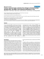
Figure 1
Upregulation of tumor necrosis factor-alpha (TNFα)-related genes in splenocytes of mice with glucose-6-phosphate isomerase (GPI)-induced arthritisUpregulation of tumor necrosis factor-alpha (TNFα)-related genes in
splenocytes of mice with glucose-6-phosphate isomerase (GPI)-
induced arthritis. The mRNA expression levels of TNF-related genes in
splenocytes of mice with GPI-induced arthritic (at day 10) relative to
control splenocytes are shown. TNFα-induced adipose-related protein
(TIARP) was specifically and strongly induced in splenocytes. Gene-
Chip analysis was performed by gene expression software. ADAM17, a
disintegrin and metallopeptidase domain 17; FADD, Fas (tumor necro-
sis factor receptor superfamily 6)-associated via death domain; GITR,
glucocorticoid-induced tumor necrosis factor-related protein-D mRNA;
LITAF, lipopolysaccharide-induced tumor necrosis factor-alpha factor;
NFKB1, nuclear factor kappa B subunit p105; NFKB2, nuclear factor
kappa B subunit p100; RIPK, receptor (tumor necrosis factor receptor
superfamily)-interacting serine-threonine kinase 1 and 2; TNFAIP, tumor
necrosis factor alpha-induced protein; TNFR, tumor necrosis factor
receptor; TNFRSF, tumor necrosis factor receptor superfamily;
TNFRSF12, WSL-1-like protein; TNFRSF22, tumor necrosis factor
receptor family member SOBa mRNA; TNFSF, tumor necrosis factor
(ligand) superfamily; TRAF, tumor necrosis factor receptor-associated
factor; TTRAP, tumor necrosis factor receptor-associated factor and
tumor necrosis factor receptor-associated protein.
Available online />Page 5 of 11
(page number not for citation purposes)
(onset of arthritis, P < 0.05), were at the same elevated levels
at day 14 (peak of arthritis), and then subsided to the basal
level at day 28 (Figure 2a). In contrast, the TNFα mRNA
expression level in arthritic joints tended to increase at day 7,
though insignificantly, in mice with GPI-induced arthritis. The
expression level decreased later to basal levels (Figure 2b).
Both real-time PCR and Western blotting showed upregula-
tion of TIARP mRNA and protein expression at day 7 in splen-
ocytes of mice with GPI-induced arthritis (Figure 2c, left
panel). In the joints of the same mice, upregulation of TIARP
mRNA and protein was noted at days 14 and 28, and the
expression correlated with joint swelling (Figure 2c, right
Figure 2
Serial changes in expression levels of tumor necrosis factor-alpha (TNFα) and TIARP in glucose-6-phosphate isomerase (GPI)-induced arthritisSerial changes in expression levels of tumor necrosis factor-alpha (TNFα) and TIARP in glucose-6-phosphate isomerase (GPI)-induced arthritis.
Serial changes in TNFα concentrations in (a) serum and (b) arthritic joints and (c) TIARP mRNA and protein expression in spleens (left and middle
panels) and arthritic joints (right panel) by real-time polymerase chain reaction (PCR) and Western blotting in mice with GPI-induced arthritis. As
shown in the bottom panel of (c), TIARP mRNA in lymph nodes was also analyzed. Arthritis appeared on days 7 and 8, peaked in severity on day 14,
and then gradually subsided. High expression levels of TIARP mRNA and proteins were detected in splenocytes on day 7 (the onset of arthritis). In
joints, the expression of TIARP mRNA and protein was correlated with joint swelling (days 14 and 28). Data are mean ± standard error of the mean
of five mice per group. *P < 0.05 (Mann-Whitney U test). GAPDH, glyceraldehydes-3-phosphate dehydrogenase; mTNFα, murine tumor necrosis
factor-alpha; TIARP, tumor necrosis factor alpha-induced adipose-related protein.
Arthritis Research & Therapy Vol 11 No 4 Inoue et al.
Page 6 of 11
(page number not for citation purposes)
panel). Moreover, in lymph nodes, TIARP mRNA was upregu-
lated at day 28. But the expression of TIARP mRNA in lymph
nodes was very weak compared with the other tissues (Figure
2c, bottom panel). We also confirmed that the mRNA expres-
sion of TIARP in joints was upregulated at day 28, but not at
day 14, in mice with collagen-induced arthritis and that expres-
sion correlated with joint swelling (data not shown). These
findings suggest that the systemic upregulation of TNFα and
TIARP is involved in the early phase of the disease and that
TIARP expression in arthritic joints seems to correlate with
joint swelling.
Treatment with anti-tumor necrosis factor-alpha
monoclonal antibody suppresses TIARP expression
To test the therapeutic efficacy of anti-TNFα mAb, we injected
anti-TNFα mAb after clinical onset of arthritis at day 8. A single
injection of 100 μg of anti-TNFα mAb at day 8 ameliorated the
disease, as indicated by a rapid fall in the semiquantitative
score of arthritis (Figure 3a) [3]. To explore the relevance of
the therapeutic effect of anti-TNFα mAb on TIARP expression,
we evaluated TIARP expression after injection of anti-TNFα
mAb in mice with GPI-induced arthritis. Treatment of mice with
anti-TNFα mAb resulted in downregulation of TIARP expres-
sion in spleen relative to control Ig injection, although no treat-
ment-related change in TIARP expression was noted at day 14
(P = 0.03) (Figure 3b, top panel). However, in joints, expres-
sion of TIARP mRNA was almost comparable between the
treatment with anti-TNFα mAb and control Ig. These results
suggest that TNF antagonism induces TIARP downregulation
and results in the amelioration of arthritis.
CD11b
+
cells are the main source of TIARP mRNA in
splenocytes of arthritic mice
In the next set of experiments, splenocytes of arthritic mice
were separated into CD4
+
, CD19
+
, CD11b
+
, and CD11c
+
cells by MACS. In naïve mice, CD19
+
, CD11b
+
, and CD11c
+
cells expressed TIARP, and induction of arthritis was associ-
ated with upregulation of TIARP mRNA in CD11b
+
cells, as
demonstrated by quantitative PCR (P < 0.05 at day 7) (Figure
4a). These findings suggest the induction of TIARP in CD11b
+
cells in splenocytes of arthritic mice, especially during the early
phase of the disease.
Localization of TIARP protein in proliferative synovium
Next, immunohistochemical analysis was conducted to deter-
mine the distribution of TIARP in the arthritic joints. For this
purpose, we generated polyclonal anti-TIARP antibodies using
rats, as described previously [5]. TIARP protein was clearly
identified in the proliferative synovium of arthritic joints of mice
(at day 14) (Figure 4b, top panels), whereas almost no signal
was detected in naïve mice (Figure 4b, bottom panels). While
these findings indicate TIARP protein expression in the syn-
ovium, the results do not link such expression with an amelio-
rative or damaging effect on the synovium.
Overexpression of STEAP4 in joints of rheumatoid
arthritis patients and its localization in CD68
+
cells
To determine the role of STEAP4 (the human ortholog of
mouse TIARP)in human RA, we analyzed PBMCs from RA
patients and healthy subjects and synovia from RA patients.
For comparison, we also screened other STEAP family mem-
bers such as STEAP2 and STEAP3 using the same method.
For PBMCs, STEAP4 mRNA was detected in only one RA
patient (1/3). Importantly, STEAP4 mRNA was highly
expressed in all four RA synovia whereas only faint bands were
noted for other STEAP families (Figure 5a). Next, using several
numbers of synovial tissues from patients with RA and OA, we
investigated the expression of STEAP4 mRNA in synovium of
patients with RA and OA. Relative expression of STEAP4 was
almost comparable between RA and OA, although expression
variation tended to be enhanced in RA synovium (Figure 5b).
Moreover, immunohistochemical analysis of synovia of RA
patients showed co-localization of STEAP4 protein with
CD68, a marker for human macrophages (Figure 5c). These
findings suggest that STEAP4 is specifically expressed in
joints and is localized with CD68
+
cells.
Discussion
Although the therapeutic effect of TNF antagonists is con-
firmed in RA [1], only a few animal models of arthritis have
been used to confirm the beneficial effects of TNF antagonists.
For example, a recent study reported the therapeutic effect of
anti-TNF mAb in DNaseII, type I interferon receptor (IFN-IR)
double-knockout mice [11], although this was not a genetically
unaltered mouse. Furthermore, Schubert and colleagues
reported the protective effect of TNF antagonist in GPI-
induced arthritis [2] and arthritis was clearly B cell-dependent
[12]. We recently demonstrated the therapeutic effect of TNF
antagonist in GPI-induced mice. Thus, it is important to explore
TNF-regulated genes in the latter model to understand the
mechanisms of action of TNFα antagonists in RA patients.
When the GeneChip analysis was used, the present results
showed upregulation of TIARP mRNA in the spleen of arthritic
mice. TIARP was first identified as TNFα-induced cell surface
protein in adipose tissues and is also known to be localized in
the liver, kidney, heart, and skeletal muscle [5]. This protein
was detected in the course of adipocyte differentiation and
conversion and is also induced by IL-6 [6]. In this study, we
confirmed its induction in CD11b
+
splenocytes in arthritis and
we confirmed that it is upregulated in the arthritic synovium of
murine GPI-induced arthritis. These findings suggest the
involvement of TIARP in the process of proliferation or differ-
entiation state induced by inflammation. In fact, previous stud-
ies indicated that TIARP is induced by TNFα and IL-6 in
adipocytes [5,6]. TNFα and IL-6 are pleiotropic cytokines
known to play crucial roles in human RA, and significant ther-
apeutic effects of their antagonists have been confirmed in
recent years [1,13]. In GPI-induced arthritis, both TNFα and
IL-6 antagonists have protective effects [3,4], and these
cytokines play important roles in the induction of arthritis in col-
Available online />Page 7 of 11
(page number not for citation purposes)
Figure 3
Suppression of TIARP mRNA by treatment with anti-tumor necrosis factor-alpha monoclonal antibody (anti-TNFα mAb)Suppression of TIARP mRNA by treatment with anti-tumor necrosis factor-alpha monoclonal antibody (anti-TNFα mAb). (a) The development of
arthritis was blocked by administration of anti-TNFα mAb in mice immunized with glucose-6-phosphate isomerase. Data represent arthritis scores.
(b) In spleen, administration of anti-TNFα mAb suppressed the rise in TIARP mRNA (on day 10) (solid bars), but not control Ig (open bars). However,
in joints, expression of TIARP mRNA was almost comparable after the administration of anti-TNFα mAb or control Ig. Data are mean ± standard error
of the mean of five mice per group. *P < 0.05 (Mann-Whitney U test). GAPDH, glyceraldehydes-3-phosphate dehydrogenase; TIARP, tumor necro-
sis factor alpha-induced adipose-related protein.
Arthritis Research & Therapy Vol 11 No 4 Inoue et al.
Page 8 of 11
(page number not for citation purposes)
Figure 4
Identification of TIARP-expressing cells in splenocytes and joints of arthritic miceIdentification of TIARP-expressing cells in splenocytes and joints of arthritic mice. (a) Splenocytes were isolated from naïve (day 0) mice and mice
with glucose-6-phosphate isomerase (GPI)-induced arthritis and then were separated into four groups (CD4
+
, CD19
+
, CD11b
+
, and CD11c
+
) by
magnetic-activated cell sorting. The expression of TIARP mRNA was analyzed by quantitative real-time polymerase chain reaction at days 0 and 7.
TIARP mRNA was expressed mainly on CD11b
+
cells in arthritic mice. Data are mean ± standard error of the mean of five mice per group. *P < 0.05
(Mann-Whitney U test). (b) Joints were obtained from mice with GPI-induced arthritis on day 14 and stained with anti-TIARP antibodies (top panels)
and control antibodies (bottom panels). Inflamed synovial tissue of arthritic mice was stained with anti-TIARP antibodies. GAPDH, glyceraldehydes-
3-phosphate dehydrogenase; TIARP, tumor necrosis factor alpha-induced adipose-related protein.
Available online />Page 9 of 11
(page number not for citation purposes)
Figure 5
Analysis of STEAP mRNA expression by reverse transcription-polymerase chain reaction (RT-PCR) in peripheral blood mononuclear cells (PBMCs) and synovia of rheumatoid arthritis (RA) patients and healthy subjects (HC) and immunohistochemistry for STEAP4 in RA synoviumAnalysis of STEAP mRNA expression by reverse transcription-polymerase chain reaction (RT-PCR) in peripheral blood mononuclear cells (PBMCs)
and synovia of rheumatoid arthritis (RA) patients and healthy subjects (HC) and immunohistochemistry for STEAP4 in RA synovium. (a) The expres-
sion of STEAP4 mRNA and other family members (STEAP2 and STEAP3 mRNAs) was analyzed in PBMCs (RA and HC) and RA synovium using
RT-PCR. In PBMCs, STEAP4 mRNA was detected in a patient with RA (1/3). Surprisingly, STEAP4 mRNA was highly expressed in all four RA syn-
ovia whereas only faint staining was noted for other members of the STEAP family. (b) The expression of STEAP4 mRNA in synovium with RA and
osteoarthritis (OA) patients. STEAP4 mRNA expression was not statistically different between the RA and OA groups. (c) Co-localization of
STEAP4 and CD68 in RA synovium. Images of immunohistochemistry using 4'-6-diamidino-2-phenylindole (DAPI), fluorescein isothiocyanate (FITC)-
anti-STEAP4, and rhodamine-anti-CD68 and a merged image are shown in the middle panels, and images with conjugated control Ig are shown in
the bottom panels. Consecutive hematoxylin-and-eosin staining is shown in the top panel. GAPDH, glyceraldehydes-3-phosphate dehydrogenase;
n.s., not significant; STEAP, six-transmembrane epithelial antigen of the prostate.
Arthritis Research & Therapy Vol 11 No 4 Inoue et al.
Page 10 of 11
(page number not for citation purposes)
laboration with autoantibodies (anti-GPI antibodies) [14].
However, there is no clear scenario of balance between IL-6
and TNFα in arthritis. In TIARP knockdown animals, exposure
to TNFα induced a greater amount of IL-6, suggesting a cru-
cial role of TIARP in the balance between TNFα and IL-6 [15].
It is possible that TIARP expression plays a downregulatory
role in the inflammatory cascade.
At this stage, there is no information on whether TIARP act in
an antagonistic or agonistic manner with arthritis. However,
one report on STAMP2 (a homolog of TIARP protein) [15]
confirmed (a) upregulation of inflammatory cytokines such as
TNFα and IL-6 in STAMP2-deficient mice, (b) upregulation of
macrophage-specific antigens such as CD68 and CD11b, (c)
infiltration of CD68
+
cells in adipose tissues, and (d) STAMP2-
induced suppression of IL-6 expression upon stimulation by
TNFα. These findings suggest that STAMP2 (TIARP) sup-
presses inflammatory cytokines such as TNFα and IL-6 and
also blocks the activation of macrophages/monocytes.
Is this scenario applicable to patients with RA? In humans, the
STEAP protein family was identified in prostate tumors [16,17]
and is also known to be involved in cell apoptosis [18]. Among
this family of genes, STEAP4 is highly expressed in the bone
marrow, followed by placenta and fetal liver [19]. The STEAP4
expression was induced by TNFα in human adipose tissue
[20] and also by TNFα in human synovial cells (our preliminary
result). However, there is no report regarding the expression
of this molecule in articular joints. The present study identified
the expression of human ortholog STEAP4 in the synovium,
especially in CD68
+
macrophages of patients with RA. In
addition, our preliminary data using human synovial cell lines
provide evidence that TNFα stimulation enhances the expres-
sion of STEAP4 protein and that a stably expressed form of
STEAP4 is partially co-localized with endosomes (Tanaka and
colleagues, manuscript in preparation). Further large-scale
studies are required to assess the expression of STEAP4 in
the joints and PBMCs of RA patients before and after treat-
ment with TNF antagonists.
Conclusions
The results of the present study highlighted the important role
of TIARP/STEAP4, a relatively new TNF-induced protein, in
autoimmune arthritis in both mice and humans.
Competing interests
The authors declare that they have no competing interests.
Authors' contributions
AI helped to write the manuscript, conceive of the study, per-
form all experiments, and coordinate statistical study. IM wrote
the manuscript and conceived of the study. YT helped to per-
form all experiments and coordinate statistical study. KI partic-
ipated in the clinical assessment. AK and NO collected the
synovial samples. DG and SI participated in discussion. TS
participated in the full design and coordination of the study. All
authors read and approved the final manuscript.
Acknowledgements
This work was supported in part by a grant from the Japanese Ministry
of Science and Culture (IM and TS).
References
1. Feldmann M, Maini SR: Role of cytokines in rheumatoid
arthritis. Immunol Rev 2008, 223:7-19.
2. Schubert D, Maier B, Morawietz L, Krenn V, Kamradt T: Immuni-
zation with glucose-6-phosphate isomerase induces T cell-
dependent peripheral polyarthritis in genetically unaltered
mice. J Immunol 2004, 172:4503-4509.
3. Matsumoto I, Zhang H, Yasukochi T, Iwanami K, Tanaka Y, Inoue
A, Goto D, Ito S, Tsutsumi A, Sumida T: Therapeutic effects of
antibodies to tumor necrosis factor-alpha, interleukin-6 and
cytotoxic T-lymphocyte antigen 4 immunoglobulin in mice with
glucose-6-phosphate isomerase induced arthritis. Arthritis
Res Ther 2008, 10:R66.
4. Iwanami K, Matsumoto I, Tanaka-Watanabe Y, Inoue A, Mihara M,
Ohsugi Y, Mamura M, Goto D, Ito S, Tsutsumi A, Kishimoto T,
Sumida T: Crucial role of the interleukin-6/interleukin-17
cytokine axis in the induction of arthritis by glucose-6-phos-
phate isomerase. Arthritis Rheum 2008, 58:754-763.
5. Moldes M, Lasnier F, Gauthereau X, Klein C, Pairault J, Fève B,
Chambaut-Guérin AM: Tumor necrosis factor-alpha-induced
adipose-related protein (TIARP), a cell-surface protein that is
highly induced by tumor necrosis factor-alpha and adipose
conversion. J Biol Chem 2001, 276:33938-33946.
6. Fasshauer M, Kralisch S, Klier M, Lossner U, Bluher M, Chambaut-
Guérin AM, Klein J, Paschke R: Interleukin-6 is a positive regu-
lator of tumor necrosis factor alpha-induced adipose-related
protein in 3T3-L1 adipocytes. FEBS Lett 2004, 560:153-157.
7. Matsumoto I, Lee DM, Mansky RG, Sumida T, Hitchon CA, Schur
PH, Anderson RJ, Coblyn JS, Weinblatt ME, Brenner M, Duclos B,
Pasquali JL, El-Gabalawy H, Mathis D, Benoist C: Low prevalence
of antibodies to glucose-6-phosphate isomerase in patients
with rheumatoid arthritis and spectrum of other chronic
autoimmune disorders. Arthritis Rheum 2003, 48:944-954.
8. Korkmaz CG, Korkmaz KS, Kurys P, Elbi C, Wang L, Klokk TI, Ham-
marstrom C, Troen G, Svindland A, Hager GL, Saatcioglu F:
Molecular cloning and characterization of STAMP2, an andro-
gen-regulated six transmembrane protein that is overex-
pressed in prostate cancer. Oncogene 2005, 24:4934-4945.
9. Arnett FC, Edworthy SM, Bloch DA, McShane DJ, Fries JF, Cooper
NS, Healey LA, Kaplan SR, Liang MH, Luthra HS, et al.: The Amer-
ican Rheumatism Association 1987 revised criteria for the
classification of rheumatoid arthritis. Arthritis Rheum 1988,
31:315-324.
10. National Center for Biotechnology Information's Gene Expres-
sion Omnibus [ />acc.cgi?acc=GSE17272]
11. Kawane K, Ohtani M, Miwa K, Kizawa T, Kanbara Y, Yoshioka Y,
Yoshikawa H, Nagata S: Chronic polyarthritis caused by mam-
malian DNA that escapes from degradation in macrophages.
Nature 2006, 443:998-1002.
12. Bockermann R, Schubert D, Kamradt T, Holmdahl R: Induction of
a B-cell-dependent chronic arthritis with glucose-6-phosphate
isomerase. Arthritis Res Ther 2005, 7:R1316-R1324.
13. Nishimoto N, Yoshizaki K, Miyasaka N, Yamamoto K, Kawai S,
Takeuchi T, Hashimoto J, Azuma J, Kishimoto T: Treatment of
rheumatoid arthritis with humanized anti-interleukin-6 recep-
tor antibody: a multicenter, double-blind, placebo-controlled
trial. Arthritis Rheum 2004, 50:1761-1769.
14. Tanaka-Watanabe Y, Matsumoto I, Iwanami K, Inoue A, Goto D, Ito
S, Tsutsumi A, Sumida T: B cell play crucial role as antigen pre-
senting cells and collaborating with inflammatory cytokines in
glucose-6-phosphate isomerase-induced arthritis. Clin Exp
Immunol 2009, 155:285-294.
15. Wellen KE, Fucho R, Gregor MF, Furuhashi M, Morgan C, Lindstad
T, Vaillancourt E, Gorgun CZ, Saatcioglu F, Hotamisligil GS: Coor-
dinated regulation of nutrient and inflammatory response by
Available online />Page 11 of 11
(page number not for citation purposes)
STAMP2 is essential for metabolic homeostasis. Cell 2007,
129:537-548.
16. Hubert RS, Vavanco I, Chen E, Rastegar S, Leong K, Mitchell SC,
Madraswala R, Zhou Y, Kuo J, Raitano AB, Jakobovits A, Saffran
DC, Afar DE: STEAP: a prostate-specific cell-surface antigen
highly expressed in human prostate tumors. Proc Natl Acad
Sci USA 1999, 96:14523-14528.
17. Porkka KP, Helenius MA, Visakorpi T: Cloning and characteriza-
tion of a novel six-transmembrane protein STEAP2, expressed
in normal and malignant prostate. Lab Invest 2002,
82:1573-1582.
18. Sanchez-Pulido L, Rojas AM, Valencia A, Martinez AC, Andrade
MA: ACRATA: a novel electron transfer domain associated to
apoptosis and cancer. BMC Cancer 2004, 4:98.
19. Ohgami RS, Campagna DR, McDonald A, Fleming MD: The Steap
proteins are meralloreductases. Blood 2006, 108:1388-1394.
20. Zhang CM, Chi X, Wang B, Zhang B, Ni YH, Chen RH, Li XN, Guo
XR: Downregulation of STEAP4, a highly-expressed TNF-
alpha-inducible gene in adipose tissue, is associated with
obesity in humans. Acta Pharmacol Sin 2008, 29:587-592.