Báo cáo khoa học: Tumor necrosis factor-a converting enzyme is processed by proprotein-convertases to its mature form which is degraded upon phorbol ester stimulation pptx
Bạn đang xem bản rút gọn của tài liệu. Xem và tải ngay bản đầy đủ của tài liệu tại đây (204.61 KB, 8 trang )
Tumor necrosis factor-a converting enzyme is processed
by proprotein-convertases to its mature form which is degraded
upon phorbol ester stimulation
Kristina Endres, Andreas Anders, Elzbieta Kojro, Sandra Gilbert, Falk Fahrenholz and Rolf Postina
Institute of Biochemistry, Johannes Gutenberg-University, Mainz, Germany
Tumor necrosis factor-a converting enzyme (TACE or
ADAM17) is a member of the ADAM (a disintegrin and
metalloproteinase) family of type I membrane proteins and
mediates the ectodomain shedding of various membrane-
anchored signaling and adhesion proteins. TACE is syn-
thesized as an inactive zymogen, which is subsequently
proteolytically processed to the catalytically active form. We
have identified the proprotein-convertases PC7 and furin
to be involved in maturation of TACE. This maturation
is negatively influenced by the phorbol ester phorbol-12-
myristate-13-acetate (PMA), which decreases the cellular
amount of the mature form of TACE in PMA-treated
HEK293 and SH-SY5Y cells. Furthermore, we found that
stimulation of protein kinase C or protein kinase A signaling
pathways did not influence long-term degradation of mature
TACE. Interestingly, PMA treatment of furin-deficient
LoVo cells did not affect the degradation of mature TACE.
By examination of furin reconstituted LoVo cells we were
able to exclude the possibility that PMA modulates furin
activity. Moreover, the PMA dependent decrease of the
mature enzyme form is specific for TACE, as the amount of
mature ADAM10 was unaffected in PMA-treated HEK293
and SH-SY5Y cells. Our results indicate that the activation
of TACE by the proprotein-convertases PC7 and furin is
very similar to the maturation of ADAM10 although there is
a significant difference in the cellular stability of the mature
enzyme forms after phorbol ester treatment.
Keywords:ADAM10;Alzheimer’sdisease;furin;PC7;
TACE.
ADAMs (a disintegrin and metalloproteinases) are a family
of integral type I membrane glycoproteins which play an
important role in egg-sperm binding and fusion [1,2], muscle
cell fusion [3,4] and the development of neuronal and
epithelial cells [5,6]. ADAM members are characterized by a
well defined domain structure, consisting of a N-terminal
prodomain followed by a metalloproteinase domain, a
disintegrin domain, a cysteine rich domain, which usually
contains an epithelial growth factor repeat, a transmem-
brane and a cytoplasmic domain [7,8]. Approximately half
of the presently known ADAMs have a catalytic site
consensus sequence for zinc-dependent metalloproteinases
(HEXGHXXGXXHD) and are therefore predicted to be
catalytically active [9]. ADAMs are involved in the release of
the extracellular domains of different membrane-anchored
signal proteins such as cytokines, growth factors, growth
factor receptors and adhesion proteins [10]. The cellular
mechanisms and signaling pathways that regulate this
ectodomain shedding are gradually being elucidated
[11–13]. The most intensively studied inducer of the shedding
process is phorbol-12-myristate-13-acetate (PMA), a syn-
thetic activator of protein kinase C (PKC).
For some of the ADAM proteinases it has been shown
that the catalytic site is maintained inactive via a so called
cysteine switch mechanism performed by the N-terminal
prodomain [14,15]. The essential step for zymogen activa-
tion is the proteolytic processing by proprotein-convertases
at a characteristic motif, which is located between the
prodomain and the metalloproteinase domain. Proprotein-
convertases form a family of calcium-dependent endopro-
teinases, which presently comprises seven distinct members,
including furin, PC2, PC1/PC3, PACE4, PC4, PC5/PC6
and PC7/PC8/LPC [16,17]. A large number of proproteins
with various specificities are processed by these subtilisin-
like convertases. Typically, cleavage occurs C-terminal to
the common consensus sequence RX(K/R)R. Proteolytic
activation of substrates, mediated by PC7 or furin, takes
place in the trans-Golgi network, in endosomes and at the
cell surface [18,19]. Both convertases share an overlapping
substrate specificity and therefore the selectivity of substrate
proteolysis depends on each ones exact cellular localization.
As intracellular trafficking is regulated by their cytosolic
domains, which contain different sorting motifs, it is
possible that the localization of PC7 is distinct from that
of furin [19]. Recently, we have demonstrated that over-
expression of the proprotein-convertases PC7 and furin in
Correspondence to R. Postina or F. Fahrenholz, Institute of
Biochemistry, Johannes Gutenberg-University, Becherweg 30,
D-55099 Mainz, Germany.
Fax: + 49 6131 3925348, Tel.: + 49 6131 3925833,
E-mail:
Abbreviations:Ab, amyloid b peptide; ADAM, a disintegrin and
metalloproteinase; APP, amyloid precursor protein; APPsa, a-secre-
tase cleaved soluble APP; APPsb, b-secretase cleaved soluble APP;
DMEM, Dulbecco’s modified Eagle’s medium; HEK293, human
embryonic kidney cells; PC, proprotein-convertase; PMA, phorbol-
12-myristate-13-acetate; TACE, tumor necrosis factor-a converting
enzyme; PVDF, poly(vinylidene difluoride).
(Received 20 February 2003, revised 31 March 2003,
accepted 4 April 2003)
Eur. J. Biochem. 270, 2386–2393 (2003) Ó FEBS 2003 doi:10.1046/j.1432-1033.2003.03606.x
HEK293 cells leads to an increased maturation of
ADAM10, which further results in an enhanced cleavage
of the Alzheimer’s amyloid precursor protein (APP) at the
a-secretase specific site [20].
Three distinct shedding processes are implicated in the
emergence of Alzheimer’s disease [21,22]. The transmem-
brane protein APP is processed at first either by the
a-secretase or the b-secretase leading to the release of two
distinguishable extracellular fragments of APP (APPsa
and APPsb, respectively). The generated membrane
remaining APP stubs are subsequently cleaved by the
c-secretase. Depending on the exact cleavage site of
the c-secretase at the APP stubs, which were generated by
the b-secretase, amyloid b (Ab) peptides comprising 39–42
amino acids are generated [22]. Ab peptides are the major
component of amyloid plaques, which are found in brains
of patients suffering from Alzheimer’s disease. As
a-secretase mediated processing of APP precludes the
formation of the Ab peptides the a-secretase can be
considered as a protective factor against the generation of
these neurotoxic peptides [23]. Protein kinase C activation
by phorbol esters increases the APPsa release and
simultaneously reduces the production of Ab peptides
[24,25].
Three members of the ADAM family have been shown
to act as a-secretases [26–28]. ADAM9 overexpression
has been reported to increase the basal and protein kinase C-
dependent APPsa release [28], but the purified enzyme failed
to cleave a synthetic peptide at the major a-secretase
cleavage-site [29]. In contrast, ADAM10 has been found to
have constitutive and regulated a-secretase activity as well as
many other properties expected for an a-secretase [27].
Additionally, in situ hybridization analysis in human
cortical neurons provided evidence for the coexpression of
APP with ADAM10 and b-site APP-cleaving enzyme
(BACE)suggestingthatADAM10ismostlikelythe
physiologically relevant a-secretase [30]. Finally, experi-
ments performed with TACE-deficient cells pointed to a
participation of TACE in only the regulated, protein
kinase C-stimulated a-secretase pathway [26,31]. In another
cellular context a constitutive a-secretase activity of TACE
was demonstrated [32].
As PC7 and furin act as pro-a-secretase converting
enzymes [20], we investigated the proteolytic processing of
TACE by overexpression of these convertases in HEK293
cells. We were able to show, that both proprotein-conver-
tases contribute to TACE maturation. Moreover, we
examined the effect of PMA on the processing of endo-
genous TACE and ADAM10 in various mammalian cells
and discovered a reduction in the amount of mature TACE
compared to ADAM10 after PMA treatment.
Experimental procedures
Primary antibodies
The following antibodies were used: anti-ADAM10, a
polyclonal rabbit antibody against endogenous ADAM10
and anti-TACE, and a polyclonal rabbit antibody against
endogenous TACE (Chemikon International, Temecula,
CA). Both antibodies are directed against the C-terminal
part of the proteins and therefore recognize both the
full-length as well as the mature enzymes. For detection of
secreted APPsa the monoclonal antibody 6E10 (Signet
Laboratories) was used. As secondary antibodies alkaline
phosphatase-coupled antibodies (Tropix) were used.
Cell culture and transfections
HEK293 cells were cultured in Dulbecco’s modified Eagle’s
medium (DMEM) supplemented with 10% fetal calf serum,
2m
M
glutamine, 100 UÆmL
)1
penicillin and 100 mgÆmL
)1
streptomycin.
LoVoandSH-SY5YcellsweregrowninDMEM
nutrient mixture F-12 supplemented with 10% fetal bovine
serum, 2 m
M
glutamine, 100 UÆmL
)1
penicillin and
100 mgÆmL
)1
streptomycin. Transfection of LoVo cells
was performed using the calcium phosphate method.
Inhibition of TACE processing by decanoyl-RVKR-
chloromethylketone
HEK293 cells were cultured in the presence of 30 l
M
decanoyl-RVKR-chloromethylketone (Bachem AG, Swit-
zerland) in DMEM containing 25 m
M
Hepes, pH 7.0 at
37 °C. Inhibitor-containing medium was changed every
6–8 h. After two days of incubation the cells were lyzed and
analyzed by Western blotting.
Construction of expression vectors
Blunt end cDNAs of either bovine furin or rat PC7
were cloned into pIRES1hyg (Clontech), leading to the
expression vectors pIRES1hyg-furin or pIRES1hyg-PC7,
respectively [20].
Cloning of the furin nucleotide sequence
from LoVo cells
Total RNA from LoVo cells was isolated using the RNeasy
kit (Qiagen, Hilden, Germany). The two-step RT-PCR was
carried out using Superscript II (Lifetechnologies) and Taq
DNA polymerase (Promega) with the specific primers
Fur1_for (5¢-GTGGGCCGGAAAGTGAGCCA-3¢)and
Fur2_rev (5¢-CCCTTGTAGGAGATGAGGCC-3¢). The
resulting 1058 bp amplificate was isolated, subcloned in
pUC57 (MBI Fermentas) and sequenced.
Western blot analysis of TACE and ADAM10
Cells were washed and collected with NaCl/P
i
then cells
were suspended in cracking buffer [(5 m
M
Hepes pH 7,4
containing 2 m
M
dithiothreitol, 2 m
M
1,10-Phenanthroline
and a proteinase inhibitor cocktail (complete mini, Roche)].
Cells were disrupted by shock-freezing in liquid nitrogen
and after thawing centrifuged in a table top centrifuge to
sediment cellular membrane proteins (20 min, 4 °C,
20 000 g). Each pellet was suspended in cracking buffer
and lyzed by addition of an equal volume of 2· Laemmli
buffer containing 100 m
M
dithiothreitol, heated to 95 °C
for 20 min, separated by SDS/PAGE on 7.5% gels and
transferred by electroblotting to poly(vinylidene difluoride)
(PVDF) membranes. After blocking with NaCl/P
i
contain-
ing 0.2% I-Block (Tropix, Bedford) and 0.1% Tween 20 for
Ó FEBS 2003 TACE maturation and vanishing of its mature form (Eur. J. Biochem. 270) 2387
1 h at room temperature, the primary antibodies against
ADAM10 or TACE (anti-TACE, 1 : 2500 or anti-
ADAM10, 1 : 1000) were added for 1 h at room tempera-
ture. Bound antibodies were detected with an alkaline
phosphatase-coupled secondary antibody (Tropix) using the
chemiluminescence substrate CDPstar (Tropix). Emitted
light was densitometrically analyzed by using a digital
camera and the software
AIDA
2.0 (Raytest, Straubenhardt,
Germany).
Isolation and detection of APPsa by Western blot
analysis
Depending on each cell line an appropriate number of cells
was seeded on poly
L
-lysine coated 10-cm dishes and
grown for 20 h close to confluency. Then, cells were
washed twice with serum-free culture medium and incuba-
ted for 4.5 h in serum-free culture medium containing
2m
M
glutamine, 100 UÆmL
)1
penicillin, 100 mgÆmL
)1
streptomycin and 10 lgÆmL
)1
fatty acid-free BSA either
in the absence or presence of 1 l
M
PMA. After collection
of the cell culture supernatant, proteins were precipitated
with a final concentration of 10% trichloroacetic acid by
centrifugation. The pellets were washed twice with ice-cold
acetone, dried and dissolved in 2· Laemmli buffer
containing 100 m
M
dithiothreitol. The samples were heated
to 95 °C for 10 min, separated by SDS/PAGE on 7.5%
gels and blotted onto PVDF membranes. The membranes
were blocked as described above and then were incubated
with antibody 6E10 (1 : 2500) for 1 h at room tempera-
ture. Detection of bound antibodies was performed as
described above.
Results
Proteolytic processing of TACE by PC7 and furin
TACE has been shown to be synthesized as a zymogen,
which is constitutively processed in the secretory pathway.
Removal of the prodomain occurs after the protein exits the
medial Golgi, but before its arrival on the cell surface [33].
TACE possesses the putative proprotein-convertase recog-
nition sequence (RVKR), which is thought to be used to
generate the mature enzyme [34,35].
To test the possibility whether proprotein-convertases are
involved in the maturation of TACE, the synthetic inhibitor
decanoyl-RVKR-chloromethylketone was used. This inhi-
bitor prevents the proteolytic activity of proprotein-conver-
tases by covalently binding at their catalytic site [36].
Immunoblot analysis of endogenous TACE revealed a
clearly lowered amount of the mature enzyme in inhibitor-
treated HEK293 cells compared to untreated cells (Fig. 1A).
This result confirms that proprotein-convertases are
involved in prodomain removal.
Next we examined whether PC7 or furin might be
proprotein-convertases which are able to cleave the TACE
zymogen. Therefore, HEK293 cells were stably transfected
with expression vectors containing either the PC7 or the
furin cDNA. As a control, HEK293 cells were transfected
with the empty expression vector. Cellular membrane
proteins were subjected to Western blot analysis and
immunologically detected proteins corresponding to imma-
ture and mature TACE were densitometrically quantified
(Fig. 1B,C). Whereas the ratio of mature TACE relative to
the immature form in HEK control cells was 131 ± 24%
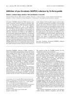
Fig. 1. Proteolytic processing of TACE by proprotein-convertases. In every case TACE was detected with a rabbit polyclonal antibody.
(A) Inhibition of TACE processing by the inhibitor decanoyl-RVKR-chloromethylketone. The inhibitor was added to a final concentration of
30 l
M
to HEK293 cells. After 48 h the cells were lyzed and membrane proteins were immunoblotted. A representative example of three experiments
is shown. (B) Western blot of endogenous TACE in HEK293 cells stably transfected with vector pIRES1hyg alone (HEK293 control), PC7
(HEK293 + PC7) or furin (HEK293 + furin). Blotted cellular membrane proteins were analyzed. (C) Densitometric analysis of TACE pro-
cessing. The proform of TACE in each cell line was set to 100%. The mature form is expressed as percentage of the proform and as mean ± SD of
three independent experiments. Significance was determined by the t-test (w, P < 0.05). (D) Proteolytic processing of TACE in furin-deficient
LoVo cells. Cells were grown in DMEM nutrient mixture F-12 almost to confluency, then lyzed and membrane proteins were analyzed by Western
blotting.
2388 K. Endres et al.(Eur. J. Biochem. 270) Ó FEBS 2003
the ratio in HEK cells overexpressing either PC7 or furin
was 187 ± 24% and 216 ± 25%, respectively. Thus,
increased amounts of the proteolytically processed mature
form were detected in PC7 as well as in furin overexpressing
cells suggesting that both proprotein-convertases are able to
process TACE. As higher amounts of the mature form were
detected in furin overexpressing cells, it appears that TACE
is a better substrate for furin than for PC7.
On basis of this result we investigated the effect of furin-
deficiency on TACE maturation. As the human carcinoma
cell line LoVo expresses only the proprotein-convertases
PACE4 and PC7, but no functionally active furin [37,38],
these cells enabled us to study the role of other proprotein-
convertases in the processing of TACE. Western blot
analysis performed with cellular membrane proteins
revealed both the immature and the mature form of
endogenous TACE indicating that missing furin activity
can be compensated by PC7 and/or PACE4 (Fig. 1D).
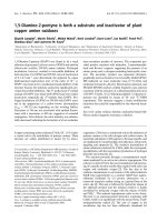
PMA treatment of HEK293 cells causes the loss
of mature TACE but does not affect mature ADAM10
TACE maturation seems to be very similar to the matur-
ation of ADAM10, which is also processed by furin and
PC7 [20]. For TACE it is known that specifically its mature
form disappears from the surface of Jurkat cells after
treatment with the phorbol ester PMA [39]. On the basis of
this result we examined if the mature form of TACE and
that of its closest homologue ADAM10 are also disappear-
ing after PMA stimulation in HEK293 cells. Furthermore,
we were interested in the time course of the disappearance.
HEK293 cells were treated with either 1 l
M
PMA or
dimethylsulfoxide. After 1.5–6 h the cells were harvested
and cellular membranes were isolated. Mature and imma-
ture forms of endogenous TACE and ADAM10 were
detected by Western blot analysis and quantified as
described under Experimental procedures. PMA treatment
induced a time dependent disappearance of the mature form
of TACE, which could clearly be seen 1.5 h after PMA
addition and was evident after 3 h (Fig. 2A,B). In contrast
to TACE, the mature form of ADAM10 was not degraded
within 6 h of PMA treatment (Fig. 2A). This result
implicates a different susceptibility of TACE and ADAM10
turnover to PMA induced signal transduction processes.
Moreover, the amount of mature TACE apparently
decreases linearly with the time of PMA treatment with a
halftime of approximately 6 h (Fig. 2B).
Stimulation of protein kinases C and A do not affect
the amount of mature TACE
As the nonphysiological PKC stimulator PMA decreased
the amount of mature TACE, we were interested in whether
a more physiological pathway for PKC activation causes
similar effects.
HEK293 cells express G protein-coupled muscarinic
receptors and agonist binding results in an intracellular
increase of the second messengers inositol 1,4,5-trisphos-
phate and diacylglycerol. The latter like PMA binds to the
C1b domain of most PKC isoenzymes and activates them.
HEK293 cells were treated with 100 l
M
acetylcholine and
harvested after 4 h as described. However, we did not find
diminished amounts of mature TACE (Fig. 3) although the
used cells responded to the applied ligand with an intracel-
lular calcium-efflux (proved by fura-2/Ca
2+
fluorescence
measurements; data not shown). Therefore, we conclude
Fig. 2. Effect of the phorbol ester PMA on the processing of endogenous
TACE and ADAM10 in HEK293 cells. Cells were incubated for
1.5–6 h in DMEM containing either 1 l
M
PMA dissolved in
dimethylsulfoxide or an equivalent volume of dimethylsulfoxide as
indicated and further handled as described under Experimental pro-
cedures. Cell membrane proteins were separated by SDS/PAGE and
blotted onto a PVDF membrane. Detection of TACE and ADAM10
was performed as described under Experimental procedures. (A) A
typical Western blot is shown. Open arrows mark the immature
enzyme forms and black arrows the mature forms. (B) Quantitative
analysis of mature TACE degradation by Western blot. The ratio of
mature TACE to immature TACE was determined in the absence (s)
or presence of 1 l
M
PMA (d) for the indicated incubation times (1.5–
6 h). Values are the means of a representative experiment performed in
duplicate. An example of three independent experiments is shown.
Fig. 3. Stimulation of PKC and PKA signaling pathways. HEK293
cells were treated for 4 h with either 1 l
M
PMA, 100 l
M
acethylcholine
(Ach) or 0.2 m
M
dibutyryl-cAMP (dB-cAMP) then cellular membrane
proteins were subjected to Western blot analysis. Mature and full-
length forms of TACE were detected with an anti-TACE Ig and
quantified by using an alkaline phosphatase-coupled secondary anti-
body as described under Experimental procedures. Results obtained
with unstimulated cells were set to 100%. Values represent
mean ± SD from a characteristic experiment using triplicates. A
representative example of two experiments is shown.
Ó FEBS 2003 TACE maturation and vanishing of its mature form (Eur. J. Biochem. 270) 2389
that the stimulatory effect of acetylcholine was not main-
tained long enough by the cells to induce the long-term
effect on TACE degradation.
As intracellular signaling pathways act as networks and
mutually influence each other we investigated whether the
PKA signaling pathway might be involved in the reduction
of mature TACE. For this purpose we tested the effect of
the cAMP-analogon dibutyryl-cAMP (dB-cAMP), which is
a strong and long-lasting effector of PKA. HEK293 cells
were incubated in medium supplemented with 0.2 m
M
dB-cAMP for 4 h and membrane proteins were analyzed
by immunoblotting. As shown in Fig. 3 dB-cAMP
displayed no effect on the expression and on the amount
of mature TACE. Similar results were obtained for
ADAM10, where also neither dB-cAMP nor acetylcholine
affected its maturation (not shown).
The effect of PMA on mature TACE and ADAM10
in SH-SY5Y and LoVo cells
To demonstrate that the reduction of catalytically active
TACE following phorbol ester stimulation is not restricted
to HEK293, we tested two other cell lines in respect to
APPsa production and TACE as well as ADAM10
maturation: The human SH-SY5Y cell line is of neuronal
origin; the other line LoVo (colon carcinoma) was chosen
because it was described to be insensitive to PMA in the
context of a-secretase activity and APPsa secretion [40].
Each cell line was incubated for 4 h either with 1 l
M
PMA or dimethylsulfoxide and proteins in the cell culture
supernatants as well as cell membrane proteins were
analyzed by immunoblotting.
In accordance with the result in HEK293 cells, ADAM10
maturation was not affected in LoVo and undifferentiated
SH-SY5Y cells after PMA treatment (Fig. 4A). In contrast,
a PMA mediated disappearance of the mature form of
TACE could be detected in HEK293 and in SH-SY5Y cells
but was completely absent in LoVo cells (Fig. 4A).
As shown in Fig. 4B, PMA treatment induced the release
of APPsa in all tested cell lines. This indicates that at least
the common a-secretase stimulatory properties of PMA are
retained by all cell lines tested. In the cell culture supernatant
of SH-SY5Y cells two forms of APPsa can be detected as
these cells express two isoforms of APP, APP751 and the
neuronal isoform APP695.
PMA-induced release of APPsa by LoVo cells
Recently, it has been reported that the furin-deficient cell
line LoVo is devoid of PKC-dependent APPsa secretion
which was interpreted that furin is involved in regulated
APP shedding [40]. In contrast to this result, our experi-
ments clearly demonstrate that LoVo cells exhibit an
augmented release of endogenous APPsa after treatment
with PMA (Fig. 4B). Because of our contradictory finding
we considered it necessary to verify the identity of the LoVo
cells, which were used in our experiments. The loss of furin
activity in LoVo cells is caused by two mutant furin alleles.
One mutation is a single nucleotide deletion, leading to an
aberrant termination of the furin polypeptide [37], the other
is a nucleotide exchange, which leads to the amino acid
exchange W547R in the homo B domain of furin [41].
To confirm these mutations, furin mRNA of LoVo cells
was amplified by RT-PCR with suitable primers. The
obtained nucleotide sequence contained the expected nuc-
leotide exchange in the furin mRNA (not shown) confirm-
ing the integrity of the LoVo cell line used in our
experiments. In conclusion, our results indicate that furin
is not necessarily needed for the PMA-induced APP
shedding in LoVo cells.
The lack of furin is not the key for the persistence
of mature TACE in LoVo cells after PMA stimulation
In contrast to the other tested cell lines, a PMA mediated
decrease of mature TACE was not observed in furin-
deficient LoVo cells. To test the possibility that furin
participates in mature TACE degradation, we examined
whether overexpression of functionally active furin in LoVo
cells restores the effect of PMA on the degradation of
mature TACE. Therefore, LoVo cells were reconstituted
with furin by a transient transfection. After 48 h transfected
cells were stimulated with 1 l
M
PMA and cellular mem-
brane proteins were analyzed by immunoblotting. When
compared to mock transfected cells (LoVo Hyg) the amount
of mature TACE was increased in cells that were transfected
with the furin expression vector (LoVo Furin, Fig. 5). This
Fig. 4. Effect of PMA on the processing of endogenous TACE and
ADAM10 and on the APPsa release from HEK293 LoVo and SHY-5Y
cells. (A) Proteolytic processing of endogenous TACE and ADAM10
in PMA-treated cells. Cells were treated for 4 h with either 1 l
M
PMA
or dimethylsulfoxide as control. Then cellular membrane proteins were
subjected to Western blot analysis. Mature and full-length forms of
TACE and ADAM10 were detected with suitable antibodies. Open
arrows mark the immature enzyme forms and black arrows the mature
forms in representative experiments. (B) PMA induced APPsa release
from cells. Cells were incubated for 4.5 h in fetal bovine serum-free
DMEM supplemented with 10 lgÆmL
)1
fatty acid-free BSA and either
with 1 l
M
PMA dissolved in dimethylsulfoxide or the equivalent vol-
ume of dimethylsulfoxide as control. The cell culture supernatants
were collected and proteins were precipitated with trichloroacetic acid.
Afterwards, the samples were subjected to Western blot analysis with
the primary antibody 6E10 and an alkaline phosphatase-conjugated
secondary mouse antibody. A representative example of two experi-
ments is shown.
2390 K. Endres et al.(Eur. J. Biochem. 270) Ó FEBS 2003
indicates an effective transfection and confirms that matur-
ation of TACE is mediated by furin.
Nevertheless, in furin reconstituted LoVo cells no loss of
mature TACE occurred after PMA treatment suggesting
that furin is not involved in a mechanism which decreases
the amount of mature TACE (Fig. 5).
Discussion
The prodomain of the catalytically active members of the
ADAM family is thought to act as an inhibitor of the
proteinase via a cysteine switch mechanism [42,43]. There-
fore removal of the prodomain is required to obtain the
proteolytically active enzyme [14,33,44]. Recently, we have
shown that ADAM10 is proteolytically processed by both
furin and PC7 and that the removal of the prodomain is
accompanied by an enhanced proteolytic activity [20]. For
TACE it has been shown that the maturation occurs during
the transit of the protein through the late Golgi compart-
ment suggesting that prodomain removal is performed by a
furin-type proprotein-convertase [33]. Consistent with this
model, TACE contains a putative proprotein-convertase
cleavage site, which might be used to generate the mature
enzyme [34,35]. Here we demonstrate that proprotein-
convertases are indeed involved in the maturation of TACE
and that pro-TACE is proteolytically processed by both
furin and PC7 to its mature form, most likely to increase its
proteolytical activity. Because higher amounts of mature
TACE could be detected in furin overexpressing cells, it
might be that pro-TACE is a better substrate for furin than
for PC7. However, this observation may also be due to
different expression levels of the proprotein-convertases and
is therefore difficult to substantiate. The examination of
TACE processing in LoVo cells indicates that there is
redundancy in the proteolytic maturation of TACE as other
members of the PC family can compensate a lacking furin
activity. Therefore, we cannot exclude that additional
members of the PC family also contribute to TACE
activation.
Our results further demonstrate that long-term treatment
of HEK293 and SH-SY5Y cells with the phorbol ester
PMA negatively regulates the amount of the mature form of
TACE. This is in accordance to results obtained with Jurkat
cells where the phorbol ester effect on mature TACE
reduction was attributed to protein degradation [39]. In
contrast to HEK293, SH-SY5Y and Jurkat cells TACE
maturation is unaffected by PMA in LoVo cells.
Interestingly, the amount of mature ADAM10 is not
significantly affected in spite of PMA stimulation in the
tested cell lines. Thus, the mature forms of TACE and
ADAM10 differ in their cellular stability. While mature
TACE is degraded during long-term PMA treatment
ADAM10 resists degradation. As TACE possesses an
internalization motive (YESL) in its cytoplasmic domain
and the effect of PMA on the amount of mature TACE
was inhibited by blocking endocytosis [39] it is possible that
the effect of phorbol esters on TACE maturation depends
on vesicle formation and endocytosis.
PMA is known to bind to the C1b domain of PKC and to
activate its activity. To elucidate whether activation of PKC
indeed mediates mature TACE disappearance we stimula-
ted PKC via the G protein-coupled muscarinic acetylcholine
receptor. However, long-term treatment of cells with a
receptor-saturating concentration of acetylcholine did not
influence mature TACE degradation although the cells used
in our study responded on ligand application with a fast
Ca
2+
efflux. The calcium efflux is mediated by the second
messenger inositol 1,4,5-trisphoshate, which is generated
together with diacylglycerol from phosphatidyl inositol
4,5-bisphosphate by PLCb. Obviously, receptor mediated
increase of diacylglycerol and activation of PKC does not
affect the degradation of mature TACE.
An agonist-induced activation of cellular signaling
pathways is a short-term effect. G protein-coupled
receptors are desensitized upon permanent agonist avail-
ability and therefore do not respond any longer to
effector protein activation. As mature TACE degradation
is a long-term effect, the short-term activation of PKC
by diacylglycerol might not cause a similar effect.
Alternatively, our results with acetylcholine stimulation
of cells which had no effect on TACE maturation
indicates that the effect of PMA may be independent of
PKC and may include other PMA binding molecules
such as the Munc proteins, which are involved in vesicle
formation [45].
Intracellular signaling pathways act as networks and are
mutually influenced. Therefore we investigated the effect of
a long-term PKA activation on mature TACE disappear-
ance. Activation of PKA by dibutyryl-cAMP, a more stable
cAMP analogue, did not influence the degradation of
mature TACE indicating that this effect is not dependent
on PKA.
A PMA mediated decrease in the amount of mature
TACE did not occur in the furin-deficient cell line LoVo.
Therefore, we investigated the role of furin in the PMA
mediated decrease of mature TACE.
Furin cycles between the trans-Golgi network and the cell
surface and its localization depends on phosphorylation of
its C-terminus. Whereas casein kinase II mediated furin
phosphorylation is important for its localization to the
trans-Golgi network, unphosphorylated furin is found in
Fig. 5. Reconstitution of furin activity in LoVo cells. LoVo cells were
transiently transfected with a furin cDNA containing expression vector
(LoVo Furin) or with the empty vector as control (LoVo Hyg).
Treatment with 1 l
M
PMA or dimethylsulfoxide as control was per-
formed for 4 h. Subsequently, TACE proteins were detected and
quantified in cell membrane fractions as described in Experimental
procedures. The proform of TACE in each cell line was set to 100%.
The mature form is expressed as percentage of the proform and as
mean ± SD of three independent experiments.
Ó FEBS 2003 TACE maturation and vanishing of its mature form (Eur. J. Biochem. 270) 2391
secretory granules [46]. Furthermore, the activity of the
furin phosphorylating casein kinase II can be increased by
PKC [47]. Thus, decreased amounts of mature TACE after
PMA treatment might be the result of a PKC-induced
colocalization of TACE and furin in a cellular compartment
where TACE is degraded. There furin probably acts as a
cofactor which activates the TACE degrading cascade.
Reconstitution of furin activity in LoVo cells, however,
did not rescue the PMA induced degradation of mature
TACE although the cells were able to respond on PMA
treatment with APPsa secretion. This indicates that the
enzymatic activity of furin may not be required for the PMA
induced disappearance of mature TACE. Nevertheless, we
cannot exclude the possibility, that the increased maturation
of TACE in furin transfected LoVo cells compensates to
some extent the effect of a PMA-induced degradation of
mature TACE. As LoVo cells are of carcinoma origin,
another mutation or a chromosomal rearrangement event
could be responsible for the inactivation of the mature
TACE degrading machinery, which is sensitive to phorbol
esters.
Taken together, both TACE and ADAM10 possess
a-secretase activity and are proteolytically activated by PC7
and furin. Furthermore, a furin-independent and PMA
induced disappearance of mature TACE takes place which
is not evident for mature ADAM10.
Thus, mature forms of TACE and ADAM10 differ in
their cellular stability, which may affect their a-secretase
activity in vivo.
Acknowledgements
This work was supported by grants from the Hirnliga e.V.,
the Deutsche Forschungsgemeinschaft (FA-122/4: DFG Priority
Program – Cellular mechanisms of Alzheimer’s disease) and Fonds
der Chemischen Industrie.
References
1. Blobel, C.P., Wolfsberg, T.G., Turck, C.W., Myles, D.G.,
Primakoff, P. & White, J.M. (1992) A potential fusion peptide
andanintegrinliganddomaininaproteinactiveinsperm-egg
fusion. Nature 356, 248–252.
2. Cho, C., O’Dell Bunch, D., Faure, J E., Goulding, E.H., Eddy,
E.M., Primakoff, P. & Myles, D.G. (1998) Fertilization defects in
sperm from mice lacking fertilin b. Science 281, 1857–1859.
3. Yagami-Hiromasa, T., Sato, T., Kurisaki, T., Kamijo, K.,
Nabeshima, Y I. & Fujisawa-Sehara, T. (1995) A metallopro-
tease-disintegrin participating in myoblast fusion. Nature 377,
652–656.
4. Galliano, M F., Huet, C., Frygelius, J., Polgren, A., Wewer,
U.M. & Engvall, E. (2000) Binding of ADAM12, a marker of
skeletal muscle regeneration, to the muscle-specific actin-binding
protein, a-actinin-2, is required for myoblast fusion. J. Biol. Chem.
275, 13933–13939.
5. Rooke, J., Pan, D., Xu, T. & Rubin, G.M. (1996) KUZ, a
conserved metalloprotease-disintegrin protein with two roles in
Drosophila neurogenesis. Science 273, 1227–1231.
6. Peschon, J.J., Slack, J.L., Reddy, P., Stocking, K.L., Sunnarborg,
S.W., Lee, D.C., Russell, W.E., Castner, B.J., Johnson, R.S.,
Fitzner, J.N., Boyce, R.W., Nelson, N., Kozlosky, C.J., Wolfson,
M.F.,Rauch,C.T.,Cerretti,D.P.,Paxton,R.J.,March,C.J.&
Black, R.A. (1998) An essential role for ectodomain shedding in
mammalian development. Science 282, 1281–1284.
7. Weskamp, G. & Blobel, C.P. (1994) A family of cellular proteins
related to snake venom disintegrins. Proc. Natl Acad. Sci. 91,
2748–2751.
8. Wolfsberg, T.G., Primakoff, P., Myles, D.G. & White, J.M. (1995)
ADAM, a novel family of membrane proteins containing a dis-
integrin and metalloprotease domain: multipotential functions in
cell-cell and cell–matrix interactions. J. Cell Biol. 131, 275–278.
9. Black, R.A. & White, J.M. (1998) ADAMs: focus on the protease
domain. Curr. Opin. Cell Biol. 10, 654–659.
10. Schlo
¨
ndorff, J. & Blobel, C.P. (1999) Metalloprotease-disintegrins:
modular proteins capable of promoting cell–cell interactions and
triggering signals by protein-ectodomain shedding. J. Cell Sci. 112,
3603–3617.
11. Fan, H. & Derynck, R. (1999) Ectodomain shedding of TGF-
alpha and other transmembrane proteins is induced by receptor
tyrosine kinase activation and MAP kinase signaling cascades.
EMBO J. 18, 6962–6972.
12. Prenzel, N., Zwick, E., Daub, H., Leserer, M., Abraham, R.,
Wallasch, C. & Ullrich, A. (1999) EGF receptor transactivation by
G-protein-coupled receptors requires metalloproteinase cleavage
of proHB-EGF. Nature 402, 884–888.
13. Nath, D., Williamson, N.J., Jarvis, R. & Murphy, G. (2001)
Shedding of c-Met is regulated by crosstalk between a G-protein
coupled receptor and the EGF receptor and is mediated by a
TIMP-3 sensitive metalloproteinase. J. Cell Sci. 114, 1213–1220.
14. Loechel, F., Overgaard, M.T., Oxvig, C., Albrechtsen, R. &
Wewer, U.M. (1999) Regulation of human ADAM 12 protease by
the prodomain. J. Biol. Chem. 274, 13427–13433.
15. Milla, M.E., Leesnitzer, M.A., Moss, M.L., Clay, W.C., Carter,
H.L., Miller, A.B., Su, J.L., Lambert, M.H., Willard, D.H.,
Sheeley, D.M., Kost, T.A., Burkhart, W., Moyer, M., Blackburn,
R.K., Pahel, G.L., Mitchell, J.L., Hoffman, C.R. & Becherer, J.D.
(1999) Specific sequence elements are required for the expression
of functional tumor necrosis factor-a-converting enzyme (TACE).
J. Biol. Chem. 274, 30563–30570.
16. Steiner, D.F. (1998) The proprotein-convertases. Curr. Opin.
Chem. Biol. 2, 31–39.
17. Seidah, N.G. & Chretien, M. (1999) Proprotein and prohormone
convertases: a family of subtilases generating diverse bioactive
polypeptides. Brain Res. 848, 45–62.
18. Scha
¨
fer, W., Stroh, A., Bergho
¨
fer, S., Seiler, J., Vey, M., Kruse,
M.L.,Kern,H.F.,Klenk,H D.&Garten,W.(1995)Two
independent targeting signals in the cytoplasmatic domain
determine trans-Golgi network localization and endosomal
trafficking of the proprotein-convertase furin. EMBO J. 14,
2424–2435.
19. Wouters, S., Leruth, M., Decroly, E., Vandenbranden, M.,
Creemers, J.W.M., van de Loo, J W.H.P., Ruysschaert, J M. &
Courtoy, P.J. (1998) Furin and proprotein-convertase 7 (PC7)/
lymphoma PC endogenously expressed in rat liver can be resolved
into distinct post-Golgi compartments. Biochem. J. 336, 311–316.
20. Anders, A., Gilbert, S., Garten, W., Postina, R. & Fahrenholz, F.
(2001) Regulation of the a-secretase ADAM10 by its prodomain
and proprotein-convertases. FASEB J. 15, 1837–1839.
21. Sisodia, S.S. (1992) b-Amyloid precursor protein cleavage by
a membrane-bound protease. Proc.NatlAcad.Sci.USA89,
6075–6079.
22. Haass, C. & Selkoe, D.J. (1993) Cellular processing of b-amyloid
precursor protein and the genesis of amyloid b–peptide. Cell 75,
1039–1042.
23. Mills, J. & Reiner, P.B. (1999) Regulation of amyloid precursor
protein cleavage. J. Neurochem. 72, 443–460.
24. Hung, A.Y., Haass, C., Nitsch, R.M., Qiu, W.Q., Citron, M.,
Wurtman,R.J.,Growdon,J.H.&Selkoe,D.J.(1993)Activation
of protein kinase C inhibits cellular production of the amyloid
beta-protein. J. Biol. Chem. 268, 22959–22962.
2392 K. Endres et al.(Eur. J. Biochem. 270) Ó FEBS 2003
25. Felsenstein, K.M., Ingalls, K.M., Hunihan, L.W. & Roberts, S.B.
(1994) Reversal of the Swedish familial Alzheimer’s disease
mutant phenotype in cultured cells treated with phorbol 12,13-
dibutyrate. Neurosci. Lett. 174, 173–176.
26. Buxbaum, J.D., Liu, K N., Luo, Y., Slack, J.L., Stocking, K.L.,
Peschon, J.J., Johnson, R.S., Castner, B.J., Cerretti, D.P. & Black,
R.A. (1998) Evidence that tumor necrosis factor a converting
enzymeisinvolvedinregulateda-secretasecleavage of theAlzheimer
amyloid protein precursor. J. Biol. Chem. 273, 27765–27767.
27. Lammich, S., Kojro, E., Postina, R., Gilbert, S., Pfeiffer, R.,
Jasionowski, M., Haass, C. & Fahrenholz, F. (1999) Constitutive
and regulated a-secretase cleavage of Alzheimer’s amyloid pre-
cursor protein by a disintegrin metalloprotease. Proc.NatlAcad.
Sci. USA 96, 3922–3927.
28. Koike, H., Tomioka, S., Sorimachi, H., Saido, T.C., Maruyama,
K.,Okuyama,A.,Fujisawa-Sehara,A.,Ohno,S.,Suzuki,K.&
Ishiura, S. (1999) Membrane-anchored metalloprotease MDC9
has an alpha-secretase activity responsible for processing the
amyloid precursor protein. Biochem. J. 343, 371–375.
29. Roghani, M., Becherer, J.D., Moss, M.L., Atherton, R.E.,
Erdjument-Bromage, H., Arribas, J., Blackburn, R.K., Weskamp,
G., Tempst, P. & Blobel, C.P. (1999) Metalloprotease-disintegrin
MDC9: intracellular maturation and catalytic activity. J. Biol.
Chem. 274, 3531–3540.
30. Marcinkiewicz, M. & Seidah, N.G. (2000) Coordinated expression
of b-amyloid precursor protein and the putative b-secretase BACE
and a-secretase ADAM10 in mouse and human brain. J. Neuro-
chem. 75, 2133–2143.
31. Merlos-Sua
´
rez, A., Ferna
´
ndez-Larrea, J., Reddy, P., Baselga, J. &
Arribas, A. (1998) Pro-tumor necrosis factor-a processing activity
is tightly controlled by a component that does not affect Notch
processing. J. Biol. Chem. 273, 24955–24962.
32. Slack, B.E., Ma, L.K. & Seah, C.C. (2000) Constitutive shedding
of the amyloid precursor protein ectodomain is up-regulated
by tumor necrosis factor-a converting enzyme. Biochem. J. 357,
787–794.
33. Schlo
¨
ndorff, J., Becherer, J.D. & Blobel, C.P. (2000) Intracellular
maturation and localization of the tumour necrosis factor alpha
convertase (TACE). Biochem. J. 347, 131–138.
34. Black, R.A., Rauch, C.T., Kozlosky, C.J., Peschon, J.J., Slack,
J.L., Wolfson, M.F., Castner, B.J., Stocking, K.L., Reddy, P.,
Srinivasan, S., Nelson, N., Bolani, N., Schooley, K.A., Gerhart,
M.,Davies,R.,Fitzner,J.N.,Johnson,R.S.,Paxton,R.J.,March,
C.J. & Cerretti, D.P. (1997) A metalloproteinase disintegrin that
releases tumor-necrosis factor-a from cells. Nature 385, 729–733.
35. Moss, M.L., Jin, S L.C., Milla, M.E., Burkhart, W., Carter, H.L.,
Chen,W J.,Clay,W.C.,Didsbury,J.R.,Hassler,D.,Hoffman,
C.R.,Kost,T.A.,Lambert,M.H.,Leesnitzer,M.A.,McCauley,
P., McGeehan, G., Mitchell, J., Moyer, M., Pahel, G., Rocquel,
W., Overton, L.K., Schoenen, F., Seaton, T., Su, J L., Warner, J.,
Willard, D. & Becherer, J.D. (1997) Cloning of a disintegrin
metalloproteinase that processes precursor tumor-necrosis
factor-a. Nature 385, 733–736.
36. Garten, W., Stieneke, A., Shaw, E., Wikstrom, P. & Klenk, H D.
(1989) Inhibition of proteolytic activation of influenza virus
hemagglutinin by specific peptidyl chloroalkyl ketones. Virology
172, 25–31.
37. Takahashi, S., Kasai, K., Hatsuzawa, K., Kitamura, N., Misumi,
Y., Ikehara, Y., Murakami, K. & Nakayama, K. (1993) A
mutation of furin causes the lack of precursor-processing activity
in human colon carcinoma LoVo cells. Biochem. Biophys. Res.
Commun. 15, 1019–1026.
38. Seidah, N.G., Hamelin, H., Mamarbachi, M., Dong, W., Tadros,
H., Mbikay, M., Chretien, M. & Day, R. (1996) cDNA structure,
tissue distribution, and chromosomal localization of rat PC7, a
novel mammalian proprotein-convertase closest to yeast kexin-
like proteinases. Proc. Natl Acad. Sci. 93, 3388–3393.
39. Doedens, J.R. & Black, R.A. (2000) Stimulation-induced down-
regulation of tumor necrosis factor-a converting enzyme. J. Biol.
Chem. 275, 14598–14607.
40. Lopez-Perez, E., Zhang, Y., Frank, S.J., Creemers, J., Seidah, N.
& Checler, F. (2001) Constitutive a-secretase cleavage of the
b-amyloid precursor protein in the furin-deficient LoVo cell line:
involvement of the pro-hormone convertase 7 and the disintegrin
metalloprotease ADAM10. J. Neurochem. 76, 1532–1539.
41. Takahashi, S., Nakagawa, T., Kasai, K., Banno, T., Duguay, S.J.,
VandeVen,W.J.M.,Murakami,K.&Nakayama,K.(1995)
A second mutant allele of furin in the processing-incompetent cell
line, LoVo. J. Biol. Chem. 270, 26565–26569.
42. van Wart, H.E. & Birkedal-Hansen, H. (1990) The cysteine switch:
a principle of regulation of metalloproteinase activity with
potential applicability to the entire matrix metalloproteinase gene
family. Proc. Natl Acad. Sci. USA 87, 5578–5582.
43.Grams,F.,Huber,R.,Kress,L.F.,Moroder,L.&Bode,W.
(1993) Activation of snake venom metalloproteinases by a cysteine
switch-like mechanism. FEBS Lett. 335, 76–80.
44. Lum, L., Reid, M.S. & Blobel, C.P. (1998) Intracellular matura-
tion of the mouse metalloprotease disintegrin MDC15. J. Biol.
Chem. 273, 26236–26247.
45. Duncan, R.R., Betz, A., Shipston, M.J., Brose, N. & Chow, R.H.
(1999) Transient, phorbol ester-induced DOC2–Munc13 inter-
actions in vivo. J. Biol. Chem. 274, 27347–27350.
46. Dittie
´
, A.S., Thomas, L., Thomas, G. & Tooze, S.A. (1997)
Interaction of furin in immature secretory granules from neuro-
endocrine cells with the AP-1 adaptor complex is modulated by
casein kinase II phosphorylation. EMBO J. 16, 4859–4870.
47. Sanghera, J.S., Charlton, L.A., Paddon, H.B. & Pelech, S.L.
(1992) Purification and characterization of echinoderm casein
kinase II. Regulation by protein kinase C. Biochem. J. 283,
829–837.
Ó FEBS 2003 TACE maturation and vanishing of its mature form (Eur. J. Biochem. 270) 2393