Báo cáo y học: "Tumor necrosis factor alpha-dependent aggrecan cleavage and release of glycosaminoglycans in the meniscus is mediated by nitrous oxide-independent aggrecanase activity in vitro" pptx
Bạn đang xem bản rút gọn của tài liệu. Xem và tải ngay bản đầy đủ của tài liệu tại đây (1.57 MB, 9 trang )
Open Access
Available online />Page 1 of 9
(page number not for citation purposes)
Vol 11 No 5
Research article
Tumor necrosis factor alpha-dependent aggrecan cleavage and
release of glycosaminoglycans in the meniscus is mediated by
nitrous oxide-independent aggrecanase activity in vitro
Henning Voigt, Angelika K Lemke, Rolf Mentlein, Michael Schünke and Bodo Kurz
Institute of Anatomy, Christian-Albrechts-University Kiel, Olshausenstr. 40, Kiel, 24098, Germany
Corresponding author: Bodo Kurz,
Received: 16 Jun 2008 Revisions requested: 13 Aug 2008 Revisions received: 1 Sep 2009 Accepted: 24 Sep 2009 Published: 24 Sep 2009
Arthritis Research & Therapy 2009, 11:R141 (doi:10.1186/ar2813)
This article is online at: />© 2009 Voigt et al.; licensee BioMed Central Ltd.
This is an open access article distributed under the terms of the Creative Commons Attribution License ( />),
which permits unrestricted use, distribution, and reproduction in any medium, provided the original work is properly cited.
Abstract
Introduction Little is known about factors that induce meniscus
damage. Since joint inflammation appears to be a causative
factor for meniscal destruction, we investigated the influence of
tumor necrosis factor (TNFα) on glycosaminoglycan (GAG)
release and aggrecan cleavage in an in vitro model.
Methods Meniscal explant disks (3 mm diameter × 1 mm
thickness) were isolated from 2-year-old cattle. After 3 days of
TNFα-treatment GAG release (DMMB assay), biosynthetic
activity (sulfate incorporation), nitric oxide (NO) production
(Griess assay), gene expression of matrix-degrading enzymes
(quantitative RT-PCR, zymography), and immunostaining of the
aggrecan fragment NITEGE were determined.
Results TNFα induced release of GAG as well as production of
NO in a dose-dependent manner, while sulfate incorporation
was decreased. TNFα increased matrix metalloproteinase
(MMP)-3 and a disintegrin and metalloproteinase with
thrombospondin motifs (ADAMTS)-4 mRNA expression,
whereas collagen type I was decreased, and aggrecan, collagen
type II as well as MMP-1, -2, -13 and ADAMTS-5 were variably
affected. Zymography also showed a TNFα-dependent increase
in MMP-3 expression, but pre-dominantly in the pro-form. TNFα-
dependent formation of the aggrecanase-specific aggrecan
neoepitope NITEGE was induced. Tissue inhibitor of
metalloproteinases (TIMP)-3, but not TIMP-1 or -2 inhibited
TNFα-dependent GAG release and NITEGE production,
whereas inhibition of TNFα-dependent NO generation with the
NO-synthetase inhibitor L-NMMA failed to inhibit GAG release
and NITEGE production.
Conclusions Our study shows that aggrecanase activity (a) is
responsible for early TNFα-dependent aggrecan cleavage and
GAG release in the meniscus and (b) might be involved in
meniscal degeneration. Additionally, the meniscus is a TNFα-
dependent source for MMP-3. However, the TNFα-dependent
NO production seems not to be involved in release of
proteoglycans under the given circumstances.
Introduction
Meniscal function and integrity are crucial for a healthy knee
joint, because damage to the tissue subsequently leads to
articular cartilage destruction and further degenerative dis-
eases such as osteoarthritis (OA) [1-3]. In order to restore the
meniscal function it is important to understand the pathomech-
anisms of meniscal destruction.
Increased levels of nitric oxide (NO) and pro-inflammatory
cytokines, such as TNFα and IL-1, have been found in the syn-
ovial fluid and tissues of inflamed joints [4,5]. It is also well
established that cytokines can be involved in cartilage tissue
or proteoglycan degradation [6]. It has recently been shown in
a serum-containing porcine in vitro model that these cytokines
are able to inhibit the intrinsic meniscal repair response [7,8],
and part of this effect has been found to be mediated by the
ADAMTS: a disintegrin and metalloproteinase with thrombospondin motifs; ANOVA: analysis of variance; APMA: p-aminophenyl mercuric acetate;
BSA: bovine serum albumin; C
T
: cycle of threshold; DMEM: Dulbecco's Modified Eagle's medium; GAPDH: glyceraldehyde-3-phosphate dehydro-
genase; GAG: glycosaminoglycan; IL: interleukin; L-NMMA: NG-monomethyl-L-arginine.monoacetate; MMP: matrix metalloproteinase; NO: nitric
oxide; OA: osteoarthritis; PBS: phosphate-buffered saline; RA: rheumatoid arthritis; RT-PCR: reverse transcription polymerase chain reaction; TIMP:
tissue inhibitor of metalloproteinases; TNF: tumor necrosis factor.
Arthritis Research & Therapy Vol 11 No 5 Voigt et al.
Page 2 of 9
(page number not for citation purposes)
activation of matrix metalloproteinases (MMPs) [9,10]. The
patterns of enzyme expression during experimental OA sug-
gest that there are similarities in the involvement of MMPs and
aggrecanases in the degradation of menisci and articular car-
tilage [11]. It is therefore suggested that members of the
MMPs as well as the a disintegrin and metalloproteinase with
thrombospondin motifs (ADAMTS) family, such as ADAMTS-
4 (aggrecanase-1) and ADAMTS-5 (aggrecanase-2), must
also be involved in cytokine-dependent degradation of prote-
oglycans in the meniscus. Meniscal expression and biome-
chanical regulation of all these enzymes has recently been
shown in a porcine tissue explant model [12]. Aggrecanases
are known to be responsible for aggrecan degradation in artic-
ular cartilage in diseases such as OA and rheumatoid arthritis
(RA) [13], and cleave the aggrecan core protein at several
specific sites; one is between Glu
373
and Ala
374
which gener-
ates the G1-NITEGE fragment [14,15].
It has been shown in many studies that meniscal tissue can
produce NO during experimental OA [4], or after partial menis-
cectomy [16], mechanical stimulation [17-19], or cytokine
treatment with IL-1 or TNFα [20-22]. However, the mecha-
nisms of endogenous NO involvement in meniscal degenera-
tion still remain unclear. It is associated with cartilage tissue
destruction [19,23], but was also found to protect from IL-1-
mediated proteoglycan degradation [21].
In order to investigate the influence of TNFα on the meniscus
we present a bovine in vitro model that allows the isolation of
meniscal tissue explants of defined geometry and anatomical
location. Using this model we study the effect of TNFα on gly-
cosaminoglycan (GAG) release, biosynthetic activity, NO pro-
duction, aggrecan fragmentation (because aggrecan has been
described as one of the major proteoglycans in the meniscus
[24]), and gene expression of matrix molecules, MMPs and
aggrecanases in the meniscus. We demonstrate that within
three days of incubation there is a TNFα-dependent up-regu-
lation of MMP-3 and ADAMTS-4 expression, as well as aggre-
canase activity. The latter induces GAG release, cleaves
aggrecan at the NITEGE site and is independent of the TNFα-
induced NO production.
Materials and methods
Isolation and culturing of meniscal explant disks
Meniscal explant disks were isolated from bovine menisci
(from 16 to 24 month old cattle), procured from a local abattoir
with authorization from the relevant meat inspectors. This
study does not involve human subjects, human tissue or exper-
imentation of animals. Up to four full thickness tissue cylinders
(10 mm in diameter) per meniscus were punched perpendicu-
lar to the meniscus bottom surface. Tissue disks 1 mm in thick-
ness were sliced including the original meniscal surface using
a sterile scalpel blade, and four to five smaller explant disks (3
mm in diameter × 1 mm thick) were isolated using a biopsy
punch (HEBUmedical, Tuttlingen, Germany) and cultured in
DMEM (supplemented with 100 U/ml penicillin G, 100 μg/ml
streptomycin, and 0.25 μg/ml amphotericin B; Sigma-Aldrich,
St. Louis, MO, USA) in a 37°C, 5% CO
2
environment after
measurement of wet weight. The total of up to 60 explants per
animal (2 knee joints including medial/lateral menisci) were
randomised among the different experimental groups matched
by their anatomical location for every single experiment and
cultured in the absence or presence of varying concentrations
of recombinant human TNFα (R & D Systems, Minneapolis,
MN, USA). In most of the experiments a concentration of 100
ng TNFα/ml was used. Three explant disks per well of a 24-
well plate were cultured in 1 ml medium. After three days of
culture the medium and explants were used for measure-
ments. For inhibitory studies different tissue inhibitor of metal-
loproteinases (TIMPs; R & D Systems, Minneapolis, MN, USA)
and the NO synthetase inhibitor L-NMMA were used. For
these investigations only one meniscal explant per well was
cultured for three days in 200 μl medium in 96-well plates.
Immunohistochemistry
The meniscal explants were fixed overnight in 4% paraformal-
dehyde and embedded in paraffin. Serial sections (7 μm) were
cut sagittally through the entire thickness of the explant disks,
immobilised on glass slides, and deparaffinised. After incuba-
tion for 2.5 minutes in a digester at 100°C (in 0.01 M citric
acid, pH 6.0), they were incubated overnight at 4°C with the
primary antibody (anti-NITEGE; 1:50 dilution in 1% BSA; ABR
Affinity BioReagents, Golden, CO, USA), rinsed in Tris-NaCl
three times for five minutes and incubated with the secondary
antibody AlexaFluor 488 goat anti-rabbit IgG (1:500; Invitro-
gen, Carlsbad, CA, USA) for one hour at room temperature.
After further washing, the sections were labeled for nuclear
staining with bisbenzimide (Sigma, St. Louis, MO, USA),
mounted with fluorescence mounting medium (Dako, Glos-
trup, Denmark), and visualised using the Apotome (ZEISS,
Jena, Germany) fluorescence microscope.
Measurement of biosynthetic activity,
glycosaminoglycans and nitric oxide production
For radiolabel incorporation the meniscal explants were
placed in fresh culture medium containing 10 μCi/ml [
35
SO
4
]-
sulfate (Amersham Pharmacia, GE Healthcare Europe GmbH,
Munich, Germany) for six to eight hours at 37°C under free-
swelling conditions right after cytokine treatment. Afterwards,
the explants were washed in PBS containing 0.5 mM proline
and digested overnight in 1 ml of papain solution (0.125 mg/
ml (2.125 U/ml, Sigma, St. Louis, MO, USA), 0.1 M Na
2
HPO
4
,
0.01 M Na-EDTA, 0.01 M L-cysteine, pH 6.5) at 65°C. A 200
μl aliquot of each sample were added to 2 ml scintillation fluid
(Opti Phase Hi Safe 3, Perkin Elmer, Waltham, MA, USA) and
measured using a Beckmann scintillation counter (Wallac
1904. Turku, Finland). Counts were expressed in cpm/mg wet
weight and normalised to the radiolabel incorporation of
untreated control tissue, which was set to 100%.
Available online />Page 3 of 9
(page number not for citation purposes)
For measurement of GAG release or content the media were
collected after cytokine treatment or the papain-digested
explants were used (see above), and GAG content was deter-
mined by DMMB dye assay photometrically at a wavelength of
520 nm (Photometer Ultraspec II, Biochrom, Cambridge, UK)
using shark chondroitin-sulfate as standard. Values were pre-
sented as μg GAG per mg wet weight of the explants.
Generation of NO was determined by measuring nitrite accu-
mulation in culture supernatants using Griess reagent (1% sul-
fanilamide and 0.1% N-(1-naphtyl)-ethylene diamine-dihydro-
chloride in 5% H
3
PO
4
, Sigma-Aldrich, St. Louis, MO, USA). A
100 μl aliquot of each sample and 100 μl Griess reagent were
mixed and incubated for five minutes, and the absorption was
determined in an automated plate reader (SLT Reader 340
ATTC, SLT-Labinstruments, Achterwehr, Germany) at 540
nm. Sodium nitrite (NaNO
2
, Merck, Darmstadt, Germany) was
used to generate a standard curve for quantification.
Quantitative RT-PCR
After three days of incubation, quantitative real-time RT-PCR
was performed using glyceraldehyde-3-phosphate dehydro-
genase (GAPDH) as reference gene to determine gene
expression levels. Meniscal explants (approximately 100 mg
from each group) were frozen immediately in liquid nitrogen.
Total RNA was extracted after pulverisation of the tissue using
the TRIZOL reagent (1 ml/100 mg wet weight tissue; Invitro-
gen, Carlsbad, CA, USA) followed by extraction with chloro-
form and isopropanol precipitation. The concentration of
extracted RNA was quantified spectro-photometrically at
OD
260
/OD
280
nm. Before real-time RT-PCR was performed
using the Qiagen QuantiTect SYBR
®
Green RT-PCR Kit (Qia-
gen, Hilden, Germany) according to the manufacturer's
instructions the extracted RNA was digested with DNase
(65°C for 10 minutes; Promega, Madison, WI, USA) to remove
any traces of DNA. Bovine primers were designed using
Primer3 Software [25] and used at a concentration of 0.5 μM
(Table 1). Conditions for real-time RT-PCR were as specified
Table 1
List of primers used for real time RT-PCR
Target Sequence (5' to 3') Product size
GAPDH S ATC AAG AAG GTG GTG AAG CAG G 101 bp
GAPDH AS TGA GTG TCG CTG TTG AAG TCG
18sRNA S TCG AGG CCC TGT AAT TGG AA 104 bp
18sRNA AS GCT ATT GGA GCT GGA ATT ACC G
Aggrecan S CCT GAA CGA CAA GAC CAT CGA 101 bp
Aggrecan AS TGG CAA AGA AGT TGT CAG GCT
Collagen type I S AAT TCC AAG GCC AAG AAG CAT G 102 bp
Collagen type I AS GGT AGC CAT TTC CTT GGT GGT T
Collagen type II S AAG AAG GCT CTG CTC ATC CAG G 124 bp
Collagen type II AS TAG TCT TGC CCC ACT TAC CGG T
MMP-1 S GGA CTG TCC GGA ATG AGG ATC T 91 bp
MMP-1 AS TTG GAA TGC TCA AGG CCC A
MMP-2 S GTA CGG GAA TGC TGA CGG GGA ATA 93 bp
MMP-2 AS CCA TCG CTG CGG CCT GTG TCT GT
MMP-3 S CAC TCA ACC GAA CGT GAA GCT 109 bp
MMP-3 AS CGT ACA GGA ACT GAA TGC CGT
MMP-13 S TCT TGT TGC TGC CCA TGA GT 101 bp
MMP-13 AS GGC TTT TGC CAG TGT AGG TGT A
ADAMTS-4 S GCG CCC GCT TCA TCA CTG 101 bp
ADAMTS-4 AS TTG CCG GGG AAG GTC ACG
ADAMTS-5 S AAG CTG CCG GCC GTG GAA GGA A 196 bp
ADAMTS-5 AS TGG GTT ATT GCA GTG GCG GTA GG
ADAMTS = a disintegrin and metalloproteinase with thrombospondin motifs; AS = antisense; bp = base pairs; GAPDH = glyceraldehyde-3-
phosphate dehydrogenase; MMP = matrix-metalloproteinase; S = sense.
Arthritis Research & Therapy Vol 11 No 5 Voigt et al.
Page 4 of 9
(page number not for citation purposes)
by manufacturer's description: reverse transcription 30 min-
utes at 50°C; PCR initial activation step 15 minutes at 95°C;
denaturation 15 seconds at 94°C; annealing 30 seconds at
60°C; extension 30 seconds at 72°C; optional: data acquisi-
tion 30 seconds at melting temperature 70 to 78°C. Differ-
ences of mRNA levels between control and stimulated
samples were calculated using the ΔΔC
T
-method. ΔC
T
repre-
sents the difference between the C
T
(cycle of threshold) of a
target gene and the reference gene (GAPDH). The ΔΔC
T
value
is calculated as the difference between ΔC
T
from the stimu-
lated samples and the control.
Zymography
Protein levels of MMPs were assayed in conditioned media by
gelatin and casein zymography. Equal volumes of medium
samples and loading buffer (2 mM EDTA, 2% (w/v) SDS,
0.02% (w/v) bromophenol blue, 20 mM Tris-HCl, pH 8.0)
were mixed, subjected to electrophoresis using 0.1% (w/v)
gelatin and 0.2% (w/v) casein as substrate in 4.5 to 15% gra-
dient SDS-PAGE, washed in 2.5% (v/v) Triton X-100, rinsed
in distilled water and incubated for 16 hours at 37°C in 50 mM
Tris-HCL (pH 8.5) containing 5 mM CaCl
2
. Gels were stained
with 0.1% (w/v) Coomassie brilliant blue R250 (Serva, Heidel-
berg, Germany) and destained with 10% (v/v) acetic/50% (v/
v) methanol and with 10% (v/v) acetic acid/10% (v/v) metha-
nol. MMPs were identified by molecular weight and substrate
specificity as clear bands against a blue background of undi-
gested substrate. Additionally, samples were incubated with 1
mM 4-aminophenylmercuric acetate (APMA; Sigma-Aldrich,
St. Louis, MO, USA) for three hours at 37°C to activate MMP-
pro-forms prior to loading.
Statistics
Quantitative data are presented as mean ± standard error of
the mean, n represents the number of independent experi-
ments. Statistical analysis of data was made using a one-way
analysis of variance (ANOVA) indicating significant differ-
ences, and comparisons among the various experimental
groups were made using the two-tailed Student's t-test. Differ-
ences were considered significant if P ≤ 0.05.
Results
TNFα-dependent GAG release
We have established an in vitro model for the investigation of
bovine meniscal tissue destruction where tissue explant disks
(3 mm in diameter and 1 mm thick) were isolated from the
meniscal bottom surface (facing the tibial articular cartilage).
Mean GAG content of freshly isolated explants was 14.2 ± 0.8
μg/mg wet weight (n = 8). After three days of culture, 4.8 ±
0.3 μg/mg of GAG was released into the media in control
explants (normalised to the mean GAG content of fresh
explants about one-third of explant GAG is being released dur-
ing culture). Stimulation with TNFα induced a dose-dependent
increase in GAG release: using a concentration of 1 ng/ml
caused an additional but non-significant increase in GAG
release of approximately 8.8 ± 3.7% compared with control
release. With 10 ng TNFα/ml, GAG release increased signifi-
cantly by 30 ± 12% (n = 11), and 100 ng TNF α/ml (chosen
for all subsequent experiments; Figure 1a) increased GAG
release significantly by 24 ± 10% (n = 11). In order to distin-
guish between the release of existing GAG or newly synthe-
sised GAG, radiolabeled sulfate was incorporated after
cytokine treatment. TNFα induced a significant reduction in
sulfate uptake (controls: 100 ± 12% vs TNFα: 55 ± 11%; n =
4), suggesting that the TNFα-dependent increase in media
GAG content must be predominantly the result of an
increased matrix degradation, rather than an increased biosyn-
thetic activity.
TNFα-dependent NO production
TNFα induced a dose-dependent (not shown) and signifi-
cantly increased production of NO in meniscal explants which
increased about four-fold in comparison to the un-stimulated
control (Figure 1b). The NO-synthetase inhibitor L-NMMA
reduced the basal NO production of the tissue significantly
and prevented the TNFα-mediated increase in NO completely.
Influence of NO synthetase inhibition and TIMPs on
TNFα-dependent GAG release
It has been described that proteoglycan degradation in carti-
lage tissues can be mediated by both the production of NO
and the involvement of matrix-degrading enzymes. We there-
fore studied the influence of the NO-synthetase inhibitor L-
NMMA on meniscal tissue. L-NMMA had no significant influ-
ence on the basal GAG release and did not reduce the TNFα-
induced effect (Figure 1a). There was a slight, but not signifi-
cant, increase of GAG release instead. In order to support the
hypothesis that aggrecanases are involved in TNFα-depend-
ent GAG release, we studied the influence of TIMP-1, -2 and
-3. TIMPs are known as specific inhibitors of MMPs, but it has
been reported that TIMP-3 has the additional ability to inhibit
the aggrecanases ADAMTS-4 and -5 [26,27]. TIMPs did not
affect the GAG release in control cultures (not shown). How-
ever, the TNFα-induced GAG release was significantly
reduced by TIMP-3 by approximately 52% (Figure 1c),
whereas TIMP-1 and TIMP-2 showed a trend to increase the
TNFα-induced GAG release, although this effect was not sig-
nificant.
Expression of matrix molecules and matrix degrading
enzymes
To further determine the mechanisms of TNFα-dependent
GAG release, the mRNA of meniscal explants was analyzed
after a three-day incubation by quantitative RT-PCR. GAPDH
had been used as a reference gene, and it had been tested
that there is no significant alteration in the C
T
values of
GAPDH expression under the influence of TNFα (control:
27.1 ± 1.7 versus TNFα: 27.3 ± 0.9; n = 4 independent exper-
iments). Additionally, GAPDH expression had been tested in
relation to another housekeeping gene, 18sRNA: the ratio of
Available online />Page 5 of 9
(page number not for citation purposes)
GAPDH expression remained unaffected under the influence
of TNFα (1.03).
The mRNA levels of most of the genes tested were quite vari-
able under the influence of TNFα except for the matrix-degrad-
ing enzymes MMP-3 and ADAMTS-4 (see below). Collagen
type I mRNA was decreased in all cases (0.75 ± 0.15), while
aggrecan and collagen type II as well as MMP-1 and MMP-13
showed both increases and decreases depending on the
experiment. ADAMTS-5 was not detectable in some cases or
not increased by TNFα. MMP-3 and ADAMTS-4 showed a
mean TNFα-dependent 6.9 ± 2.1 and 3.7 ± 0.8-fold increase
of mRNA expression (Figure 1d). Comparing delta-C
T
-values
(C
TGAPDH
- C
Tgene of interest
) of controls and TNFα-stimulated
meniscal explants allows a statistical analysis and showed a
significant mean change of about 2.5 ± 0.58 for MMP-3 and
1.86 ± 0.16 for ADAMTS-4, indicating a clear up-regulation of
these enzymes in all four independent experiments. The TNFα-
dependent MMP-3 expression was also detectable in the
supernatants of the cultures by casein zymography (Figure 2).
There was only one band detectable in the gels, which was
missing or expressed at lower levels in controls, but strong in
TNFα-stimulated cultures. This band was not visible in gelatin
zymograms (not shown), and had a molecular size of about 57
kDa (typical size for MMP-3, [28]). TIMP-3 as well as L-NMMA
had no influence on the band intensity. However, the enzyme
activator substance APMA altered the size of the band, indi-
cating that most of the enzyme was expressed as a pro-form
[28].
Aggrecan degradation
Immunostaining of the aggrecanase activity-specific aggrecan
neoepitope NITEGE showed very low signals in control tissue
with a clear TNFα-dependent increase in staining in all menis-
cal tissue areas that could be characterised as fibrous carti-
lage (Figures 3a and 3d). Co-incubation with the NO-
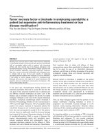
Figure 1
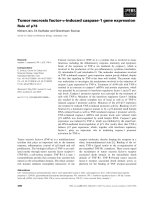
Influence of a three-day incubation with TNFα (100 ng/ml), the NO synthetase inhibitor L-NMMA (1 mM), and the TIMPs (0.1 μM) on the GAG-release, NO production and gene expression level of bovine meniscal tissue explantsInfluence of a three-day incubation with TNFα (100 ng/ml), the NO synthetase inhibitor L-NMMA (1 mM), and the TIMPs (0.1 μM) on the GAG-
release, NO production and gene expression level of bovine meniscal tissue explants. (a) Cumulative glycosaminoglycan (GAG) release (n = 6). (b)
Cumulative nitric oxide (NO) production, measured by photometrical detection of nitrite accumulation (n = 6). (c) Influence of tissue inhibitors of met-
alloproteinases (TIMPs) on TNFα-dependent GAG release (n = 5). (d) TNFα-dependent mRNA levels given as a ratio: the x-fold expression level
compared with un-stimulated control tissue (using the ΔΔCT method with GAPDH as reference gene; control = 1). Each dot represents data from
an independent experiment, bars indicate the mean from four independent experiments. (a to c) All values are mean ± standard error of the mean. *
significantly different from control, P < 0.05. ADAMTS = a disintegrin and metalloproteinase with thrombospondin motifs; Agg = aggrecan; Coll I or
II = collagen type I or II; MMP = matrix metalloproteinase.
Arthritis Research & Therapy Vol 11 No 5 Voigt et al.
Page 6 of 9
(page number not for citation purposes)
synthetase inhibitor L-NMMA failed to influence the TNFα-
dependent NITEGE formation (Figures 3c and 3f), whereas
TIMP-3 clearly inhibited this effect (Figures 3b and 3e).
Discussion
Cartilage catabolism is initiated by proteoglycan degradation
followed by that of collagen fibers. Therefore, our study
focused on the TNFα-dependent depletion of proteoglycans
in a three-day bovine in vitro meniscal model [29]. TNFα
induced a dose-dependent increase in GAG release support-
ing data from other investigations on pro-inflammatory
cytokines in which IL-1 promoted GAG release in lapine and
porcine meniscal tissue [19,21]. TNFα, therefore, appears to
be another key factor in meniscal diseases.
To study the mechanisms of TNFα-dependent proteoglycan
degradation we investigated the transcription of different
matrix-degrading enzymes. One limitation in our study is that
aggrecanases had been detected on the mRNA level only;
there is no measurement of enzyme proteins, which could help
to specify the degradative potencies of enzymes involved in
TNFα-dependent proteoglycan degradation. A reason for the
missing protein detection is that enzyme levels in the tissue are
quite low compared with the large amounts of matrix proteins.
We performed immunostainings in tissue sections (not
shown), but differences in ADAMTS-4 expression were hard
to differentiate, probably due to the fact that immunohisto-
chemistry is not useful for the differentiation of slightly variable
expression levels. We therefore mainly focus on the effect of
inhibitors such as TIMPs or NO synthetase inhibitor (L-
NMMA), and the cleavage products of aggrecan (NITEGE),
which both suggest that aggrecanases must be involved in the
early TNFα-dependent aggrecan degradation and GAG
release in the meniscus (see below).
Increased concentrations of MMPs have been found in animal
models of OA, in osteoarthritic human articular cartilage and in
the synovial fluid of RA and OA patients [11,30-33], but only
little is known about the extent to which the meniscus might be
involved in the production of these enzymes. We demonstrate
that the meniscus can be an additional source for MMP-3 pro-
duction, especially under the influence of TNFα. Wilson and
colleagues [34] emphasise the importance of MMP activity in
meniscal proteoglycan degradation after a 12-day incubation
of bovine meniscal tissue from one to two-weekold calves with
20 ng/ml IL-1 and different enzyme inhibitors, but the authors
do not specify the kind of MMPs. Additionally, Wilusz and col-
leagues [9] found MMPs to be responsible for some of the
repair inhibition by pro-inflammatory cytokines in a serum-con-
taining porcine model. However, in our study most of the
MMP-3 in the culture supernatant was in the pro-form, and it
remains unclear to what extent this enzyme might have been
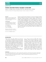
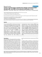
Figure 2
Casein zymograms of culture supernatants after a three day-incubation of meniscal explants under the influence of TNFα, TIMP-3, L-NMMA, or APMACasein zymograms of culture supernatants after a three day-incubation of meniscal explants under the influence of TNFα, TIMP-3, L-NMMA, or
APMA. There are samples from two independent experiments (2 lanes/group) in the upper two zymograms. There is only one major band visible at
about 57 kDa (typical size of MMP-3 pro-form [27]) with lower intensity in control cultures and stronger intensity in TNFα-treated samples. TIMP-3
and L-NMMA have no influence on band intensities. The MMP activator APMA (see lower zymogram) reduces the molecular size of the band (45
kDa) and indicates that the enzyme is pre-dominantly expressed as a pro-form. APMA = p-aminophenyl mercuric acetate; L-NMMA = NG-monome-
thyl-L-arginine.monoacetate; MMP = matrix metalloproteinase; TIMP = tissue inhibitor of metalloproteinases.
Available online />Page 7 of 9
(page number not for citation purposes)
involved in the present GAG release. But it is reasonable to
believe that MMP-3 will be involved in the subsequent TNFα-
dependent matrix degradation, as indicated by Wilson and col-
leagues [34]. TIMP-3, but not the other TIMPs, were able to
inhibit the TNFα-induced GAG release and NITEGE produc-
tion. This suggests that in the early three-day phase of menis-
cal proteoglycan degradation, aggrecanases must be
involved. TIMPs are able to inhibit the active forms of almost all
MMPs by binding to the C-terminal site of these enzymes [35].
However, TIMP-3 additionally inhibits ADAMTS-4 and -5 activ-
ity, whereas TIMP-1 and TIMP-2 have no effect on or even
increase the activity of aggrecanases at concentrations of 1
μM or less [27,36-43]. According to our mRNA study,
ADAMTS-4 might be one of the aggrecanases involved in
TNFα-dependent proteoglycan degradation in bovine menis-
cal tissue, even though final evidence is still missing. This is
supported by the fact that TIMP-3 inhibited, whereas TIMP-1
and -2 increased, the TNFα-dependent GAG release (in con-
trast to TIMP-3, TIMP-1 and -2 are known to stimulate the
activity of ADAMTS-4 under certain conditions [43]).
ADAMTS-4 mRNA has also been found in degenerated
human menisci [44]. Therefore, it is likely that there might be
similar effects in the human meniscus. Other studies showed
that ADAMTS-5 mRNA was expressed next to ADAMTS-4 in
osteoarthritic rabbit menisci [11]. Therefore, it is possible that
both aggrecanases may play a role in the degradation of
meniscal tissue. However, in the present investigation there
was a basal meniscal mRNA expression of ADAMTS-4 in the
bovine meniscus which increased with TNFα-treatment,
whereas ADAMTS-5 mRNA expression was low or not detect-
able.
We were able to localize the NITEGE fragment in meniscal tis-
sue by immunostaining in TNFα-treated explants, while it was
almost non-detectable in control tissue. This is another strong
indicator for aggrecanase involvement, according to many
articular cartilage studies [14,15,45,46]. Additionally, TNFα-
dependent NITEGE-formation could be blocked by TIMP-3,
while TIMP-1 and -2 had no inhibitory effects (not shown).
TIMP-3 is not a specific aggrecanase inhibitor. It has to be
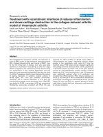
Figure 3
Immunostaining of the aggrecan cleavage product NITEGE in paraffin sections of meniscal explants after three days of incubation with or without TNFα, the protease inhibitor TIMP-3 or the NO synthetase inhibitor L-NMMAImmunostaining of the aggrecan cleavage product NITEGE in paraffin sections of meniscal explants after three days of incubation with or without
TNFα, the protease inhibitor TIMP-3 or the NO synthetase inhibitor L-NMMA. There is an increase in NITEGE-staining (green fluorescence) in (d)
TNFα-treated samples in comparison to (a, b, c) control tissues, and (e) TIMP-3 is able to inhibit formation of NITEGE (f) in contrast to L-NMMA.
Cellular nuclei are counterstained using bisbenzimide (blue fluorescence). L-NMMA = NG-monomethyl-L-arginine.monoacetate; NO = nitric oxide;
TIMP = tissue inhibitor of metalloproteinases.
Arthritis Research & Therapy Vol 11 No 5 Voigt et al.
Page 8 of 9
(page number not for citation purposes)
mentioned that it also regulates the activity of members of the
membrane-bound ADAM-family, sheddases (a disintegrin and
metalloproteinase: ADAM-10, -12 and -17; TACE [47-49]).
The importance of these enzymes should therefore also be
investigated in future studies.
We found a significant TNFα-dependent increase in meniscal
NO production, which could be blocked completely by the
common NO synthetase inhibitor L-NMMA. Although NO has
been described as a meniscal product in several joint diseases
and as an important mediator of meniscal tissue degradation
in several studies [4,16-23], we did not see a stimulating influ-
ence of NO on the TNFα-induced GAG release or aggrecan
cleavage. Our study suggests that NO is not involved in the
early degradation of aggrecan in the meniscus. The slight but
not significant increase in TNFα-induced GAG release after
incubation with L-NMMA might reflect a protective function of
endogenous NO in this context, as it has been shown previ-
ously by others [21].
Conclusions
TNFα-treatment of meniscal tissue causes a reduced biosyn-
thetic activity, release of GAG, degradation of aggrecan, and
up-regulation of MMP-3 expression and aggrecanase activity.
To our knowledge, this is the first report, showing that aggre-
canase activity might be involved in the early TNFα-mediated
aggrecanolysis in the meniscus. Inhibition of aggrecanase
activity or TNFα-activity might therefore help to prevent menis-
cal destruction. TNFα also induces NO production, but it
remains unknown what role NO might play in meniscal prote-
oglycan destruction because there is no evidence for a definite
influence of endogenous NO on GAG release or aggrecan
cleavage at the NITEGE site in this study.
Competing interests
The authors declare that they have no competing interests.
Authors' contributions
HV made the acquisition of data and part of the analysis of the
data, and was also involved in drafting of the manuscript. AKL
carried out the analysis and interpretation of mRNA data. RM
made substantial contributions to conception and design of
the study. MS revised the manuscript critically for important
intellectual content. BK was involved in the conception and
design of the study, analysis and interpretation of the data, and
did most of the drafting of the manuscript. All authors read and
approved the final manuscript.
Acknowledgements
We thank Rita Kirsch, Elsbeth Schulz, and Frank Lichte for their techni-
cal support. We also thank the NFZ Norddeutsche Fleischzentrale
GmbH for the utilization of the knee joints. The study was funded by the
Endo-Stiftung, Stiftung des Gemeinnützigen Vereins ENDO-Klinik e.V.,
Hamburg, Germany.
References
1. DiCarlo EF: Pathology of the meniscus. In Knee meniscus:
Basic and clinical foundations 1st edition. Edited by: Mow VC,
Arnoczky SP, Jackson DW. New York: Raven Press, Ltd;
1992:117-30.
2. Roos H, Lauren M, Adalberth T, Roos EM, Jonsson K, Lohmander
LS: Knee osteoarthritis after meniscectomy: prevalence of
radiographic changes after twenty-one years, compared with
matched controls. Arthritis Rheum 1998, 41:687-93.
3. Carlson CS, Guilak F, Vail TP, Gardin JF, Kraus VB: Synovial fluid
biomarker levels predict articular cartilage damage following
complete medial meniscectomy in the canine knee. J Orthop
Res 2002, 20:92-100.
4. Hashimoto S, Takahashi K, Ochs RL, Coutts RD, Amiel D, Lotz M:
Nitric oxide production and apoptosis in cells of the meniscus
during experimental osteoarthritis. Arthritis Rheum 1999,
42:2123-2131.
5. Schlaak JF, Pfers I, Meyer Zum Buschenfelde KH, Marker-Her-
mann E: Different cytokine profiles in the synovial fluid of
patients with osteoarthritis, rheumatoid arthritis and seroneg-
ative spondylarthropathies. Clin Exp Rheumatol 1996,
14:155-162.
6. Loo FA van de, Joosten LA, van Lent PL, Arntz OJ, Berg WB van
den: Role of interleukin-1, tumor necrosis factor alpha, and
interleukin-6 in cartilage proteoglycan metabolism and
destruction. Effect of in situ blocking in murine antigen- and
zymosan-induced arthritis. Arthritis Rheum 1995, 38:164-172.
7. Hennerbichler A, Moutos FT, Hennerbichler D, Weinberg JB, Gui-
lak F: Interleukin-1 and tumor necrosis alpha inhibit repair of
the porcine meniscus in vitro. Osteoarthritis Cartilage 2007,
15:1053-1060.
8. McNulty AL, Moutos FT, Weinberg JB, Guilak F: Enhanced inte-
grative repair of the porcine meniscus in vitro by inhibition of
interleukin-1 or tumor necrosis factor alpha. Arthritis Rheum
2007, 56:3033-3042.
9. Wilusz RE, Weinberg JB, Guilak F, McNulty AL: Inhibition of inte-
grative repair of the meniscus following acute exposure to
interleukin-1 in vitro. J Orthop Res 2008, 26:504-512.
10. McNulty AL, Weinberg JB, Guilak F: Inhibition of matrix metallo-
proteinases enhances in vitro repair of the meniscus. Clin
Orthop Relat Res 2009, 467:1557-1567.
11. Bluteau G, Conrozier T, Mathieu P, Vignon E, Herbage D, Mallein-
Gerin F: Matrix metalloproteinase-1, -3, -13 and aggrecanase-
1 and -2 are differentially expressed in experimental osteoar-
thritis.
Biochim Biophys Acta 2001, 1526:147-158.
12. Zielinska B, Killian M, Kadmiel M, Nelsen M, Haut Donahue TL:
Meniscal tissue explants response depends on level of
dynamic compressive strain. Osteoarthritis Cartilage 2009,
17:754-760.
13. Tortorella MD, Burn TC, Pratta MA, Abbaszade I, Hollis JM, Liu R,
Rosenfeld SA, Copeland RA, Decicco CP, Wynn R, Rockwell A,
Yang F, Duke JL, Solomon K, George H, Bruckner R, Nagase H,
Itoh Y, Ellis DM, Ross H, Wiswall BH, Murphy K, Hillman MC Jr,
Hollis GF, Newton RC, Magolda RL, Trzaskos JM, Arner EC: Puri-
fication and cloning of aggrecanase-1: a member of the
ADAMTS family of proteins. Science 1999, 284:1664-1666.
14. Sandy JD, Neame PJ, Boynton RE, Flannery CR: Catabolism of
aggrecan in cartilage explants. Identification of a major cleav-
age site within the interglobular domain. J Biol Chem 1991,
266:8683-8685.
15. Arner EC, Pratta MA, Decicco CP, Xue CB, Newton RC, Trzaskos
JM, Magolda RL, Tortorella MD: Aggrecanase. A target for the
design of inhibitors of cartilage degradation. Ann N Y Acad Sci
1999, 878:92-107.
16. Kobayashi K, Mishima H, Hashimoto S, Goomer RS, Harwood FL,
Lotz M, Moriya H, Amiel D: Chondrocyte apoptosis and regional
differential expression of nitric oxide in the medial meniscus
following partial meniscectomy. J Orthop Res 2001,
19:802-808.
17. Gupta T, Zielinska B, McHenry J, Kadmiel M, Haut Donahue TL: IL-
1 and iNOS gene expression and NO synthesis in the superior
region of meniscal explants are dependent on the magnitude
of compressive strains. Osteoarthritis Cartilage 2008,
16:1213-1219.
18. Fermor B, Weinberg JB, Pisetsky DS, Misukonis MA, Banes AJ,
Guilak F: The effects of static and intermittent compression on
Available online />Page 9 of 9
(page number not for citation purposes)
nitric oxide production in articular cartilage explants. J Orthop
Res 2001, 19:729-737.
19. Shin SJ, Fermor B, Weinberg JB, Pisetsky DS, Guilak F: Regula-
tion of matrix turnover in meniscal explants: role of mechani-
cal stress, interleukin-1, and nitric oxide. J Appl Physiol 2003,
95:308-313.
20. LeGrand A, Fermor B, Fink C, Pisetsky DS, Weinberg JB, Vail TP,
Guilak F: Interleukin-1, tumor necrosis factor alpha, and inter-
leukin-17 synergistically up-regulate nitric oxide and prostag-
landin E2 production in explants of human osteoarthritic knee
menisci. Arthritis Rheum 2001, 44:2078-2083.
21. Cao M, Stefanovic-Racic M, Georgescu HI, Miller LA, Evans CH:
Generation of nitric oxide by lapine meniscal cells and its
effect on matrix metabolism: stimulation of collagen produc-
tion by arginine. J Orthop Res 1998, 16:104-111.
22. Murrell GA, Doland MM, Jang D, Szabo C, Warren RF, Hannafin
JA: Nitric oxide: an important articular free radical. J Bone Joint
Surg Am 1996, 78:265-274.
23. Amin AR, Abramson SB: The role of nitric oxide in articular car-
tilage breakdown in osteoarthritis. Curr Opin Rheumatol 1998,
10:263-268.
24. Valiyaveettil M, Mort JS, McDevitt CA: The concentration, gene
expression, and spatial distribution of aggrecan in canine
articular cartilage, meniscus, and anterior and posterior cruci-
ate ligaments: a new molecular distinction between hyaline
cartilage and fibrocartilage in the knee joint. Connect Tissue
Res 2005, 46:83-91.
25. Primer3 Input (version 0.4.0) [ />input.htm]
26. Kashiwagi M, Tortorella M, Nagase H, Brew K: TIMP-3 is a potent
inhibitor of aggrecanase 1 (ADAM-TS4) and aggrecanase 2
(ADAM-TS5). J Biol Chem 2001, 276:12501-12504.
27. Gendron C, Kashiwagi M, Hughes C, Caterson B, Nagase H:
TIMP-3 inhibits aggrecanase-mediated glycosaminoglycan
release from cartilage explants stimulated by catabolic fac-
tors. FEBS Lett 2003, 555:431-436.
28. Manicourt DH, Lefebvre V: An assay for matrix metalloprotein-
ases and other proteases acting on proteoglycans, casein, or
gelatin. Anal Biochem 1993, 215:171-179.
29. Pratta MA, Yao W, Decicco C, Tortorella MD, Liu RQ, Copeland
RA, Magolda R, Newton RC, Trzaskos JM, Arner EC: Aggrecan
protects cartilage collagen from proteolytic cleavage. J Biol
Chem 2003, 278:45539-45545.
30. Billinghurst RC, Dahlberg L, Ionescu M, Reiner A, Bourne R,
Rorabeck C, Mitchell P, Hambor J, Diekmann O, Tschesche H,
Chen J, Van Wart H, Poole AR: Enhanced cleavage of type II col-
lagen by collagenases in osteoarthritic articular cartilage. J
Clin Invest 1997, 99:1534-1545.
31. Yoshihara Y, Nakamura H, Obata K, Yamada H, Hayakawa T,
Fujikawa K, Okada Y: Matrix metalloproteinases and tissue
inhibitors of metalloproteinases in synovial fluids from
patients with rheumatoid arthritis or osteoarthritis. Ann
Rheum Dis 2000, 59:455-461.
32. Lohmander LS, Hoerrner LA, Lark MW: Metalloproteinases, tis-
sue inhibitor, and proteoglycan fragments in knee synovial
fluid in human osteoarthritis. Arthritis Rheum 1993,
36:181-189.
33. Pelletier JP, Faure MP, DiBattista JA, Wilhelm S, Visco D, Martel-
Pelletier J: Coordinate synthesis of stromelysin, interleukin-1,
and oncogene proteins in experimental osteoarthritis. An
immunohistochemical study. Am J Pathol 1993, 142:95-105.
34. Wilson CG, Zuo F, Sandy JD, Levenston ME: Inhibition of MMPs,
but not of ADAMTS-4, reduces IL-1-stimulated fibrocartilage
degradation [abstract]. Transactions of the Orthopaedic
Research Society 2006, 31:31. Chicago, IL, USA, March 19-22,
2006
35. Baker AH, Edwards DR, Murphy G:
Metalloproteinase inhibitors:
biological actions and therapeutic opportunities. J Cell Sci
2002, 115:3719-3727.
36. Apte SS, Olsen BR, Murphy G: The gene structure of tissue
inhibitor of metalloproteinases (TIMP)-3 and its inhibitory
activities define the distinct TIMP gene family. J Biol Chem
1995, 270:14313-14318.
37. Zhao H, Bernardo MM, Osenkowski P, Sohail A, Pei D, Nagase H,
Kashiwagi M, Soloway PD, DeClerck YA, Fridman R: Differential
inhibition of membrane type 3 (MT3)-matrix metalloproteinase
(MMP) and MT1-MMP by tissue inhibitor of metalloproteinase
(TIMP)-2 and TIMP-3 rgulates pro-MMP-2 activation. J Biol
Chem 2004, 279:8592-8601.
38. Butler GS, Will H, Atkinson SJ, Murphy G: Membrane-type-2
matrix metalloproteinase can initiate the processing of proge-
latinase A and is regulated by the tissue inhibitors of metallo-
proteinases. Eur J Biochem 1997, 244:653-657.
39. Butler GS, Apte SS, Willenbrock F, Murphy G: Human tissue
inhibitor of metalloproteinases 3 interacts with both the N-
and C-terminal domains of gelatinases A and B. Regulation by
polyanions. J Biol Chem 1999, 274:10846-10851.
40. Will H, Atkinson SJ, Butler GS, Smith B, Murphy G: The soluble
catalytic domain of membrane type 1 matrix metalloprotein-
ase cleaves the propeptide of progelatinase A and initiates
autoproteolytic activation. Regulation by TIMP-2 and TIMP-3. J
Biol Chem 1996, 271:17119-17123.
41. Knäuper V, Lopez-Otin C, Smith B, Knight G, Murphy G: Bio-
chemical characterization of human collagenase-3. J Biol
Chem 1996, 271:1544-1550.
42. Hashimoto G, Aoki T, Nakamura H, Tanzawa K, Okada Y: Inhibi-
tion of ADAMTS4 (aggrecanase-1) by tissue inhibitors of met-
alloproteinases (TIMP-1, 2, 3 and 4). FEBS Lett 2001,
494:192-195.
43. Westling J, Fosang AJ, Last K, Thompson VP, Tomkinson KN,
Hebert T, McDonagh T, Collins-Racie LA, LaVallie ER, Morris EA,
Sandy JD: ADAMTS4 cleaves at the aggrecanase site (Glu373-
Ala374) and secondarily at the matrix metalloproteinase site
(Asn341-Phe342) in the aggrecan interglobular domain. J Biol
Chem 2002, 277:16059-16066.
44. Ito T, Ishiguro N, Ito H, Shibata H, Oguchi T, Iwata H: mRNA
expression for aggrecanases and ADAMS in degenerated
menisci of the knee (abstract).
Transactions of the Orthopaedic
Research Society 2000, 25:796. Orlando, FL, USA, March 12-15,
2000
45. Pratta MA, Scherle PA, Yang G, Liu RQ, Newton RC: Induction of
aggrecanase 1 (ADAM-TS4) by interleukin-1 occurs through
activation of constitutively produced protein. Arthritis Rheum
2003, 48:119-133.
46. Wilson CG, Palmer AW, Zuo F, Eugui E, Wilson S, Mackenzie R,
Sandy JD, Levenston ME: Selective and non-selective metallo-
proteinase inhibitors reduce IL-1-induced cartilage degrada-
tion and release of mechanical properties. Matrix Biol 2007,
26:259-268.
47. Amour A, Knight CG, Webster A, Slocombe PM, Stephens PE,
Knauper V, Docherty AJ, Murphy G: The in vitro activity of ADAM-
10 is inhibited by TIMP-1 and TIMP-3. FEBS Lett 2000,
473:275-279.
48. Amour A, Slocombe PM, Webster A, Butler M, Knight CG, Smith
BJ, Stephens PE, Shelley C, Hutton M, Knauper V, Docherty AJ,
Murphy G: TNF-alpha converting enzyme (TACE) is inhibited
by TIMP-3. FEBS Lett 1998, 435:39-44.
49. Loechel F, Fox JW, Murphy G, Albrechtsen R, Wewer UM: ADAM
12-S cleaves IGFBP-3 and IGFBP-5 and is inhibited by TIMP-
3. Biochem Biophys Res Commun 2000, 278:511-515.