Báo cáo y học: "Are CD4+CD25-Foxp3+ cells in untreated new-onset lupus patients regulatory T cells" doc
Bạn đang xem bản rút gọn của tài liệu. Xem và tải ngay bản đầy đủ của tài liệu tại đây (1.16 MB, 9 trang )
Open Access
Available online />Page 1 of 9
(page number not for citation purposes)
Vol 11 No 5
Research article
Are CD4
+
CD25
-
Foxp3
+
cells in untreated new-onset lupus patients
regulatory T cells?
Hua-xia Yang
1
*, Wen Zhang
1
*, Li-dan Zhao
1
, Yang Li
1
, Feng-chun Zhang
1
, Fu-lin Tang
1
, Wei He
2
and Xuan Zhang
1
1
Department of Rheumatology, Peking Union Medical College Hospital, Chinese Academy of Medical Sciences and Peking Union Medical College,
#41 Da-Mu-Cang-Hu-Tong Street, Beijing, 100032, China
2
Department of Immunology, School of Basic Medicine, Peking Union Medical College, and Institute of Basic Medical Sciences, Chinese Academy
of Medical Sciences, #5 Dong-Dan-San-Tiao, Beijing, 100005, China
* Contributed equally
Corresponding author: Xuan Zhang,
Received: 3 Apr 2009 Revisions requested: 15 May 2009 Revisions received: 14 Sep 2009 Accepted: 12 Oct 2009 Published: 12 Oct 2009
Arthritis Research & Therapy 2009, 11:R153 (doi:10.1186/ar2829)
This article is online at: />© 2009 Yang et al.; licensee BioMed Central Ltd.
This is an open access article distributed under the terms of the Creative Commons Attribution License ( />),
which permits unrestricted use, distribution, and reproduction in any medium, provided the original work is properly cited.
Abstract
Introduction Our previous study has reported that, in patients
with untreated new-onset lupus (UNOL), there was an abnormal
increase in the number of CD4
+
CD25
-
Foxp3
+
T cells that
correlated with disease activity and significantly decreased after
treatment. However, little is known about the nature of this cell
entity. The aim of this study was to explore the nature of
abnormally increased CD4
+
CD25
-
Foxp3
+
T cells in UNOL
patients.
Methods The expressions of surface (CD4, CD25, CD127,
chemokine receptor 4 [CCR4], glucocorticoid-induced tumor
necrosis factor receptor [GITR], and cytotoxic T lymphocyte-
associated antigen 4 [CTLA-4]) and intracellular (Foxp3)
molecules as well as cytokine synthesis of peripheral blood
mononuclear cells from 22 UNOL patients were analyzed by
flow cytometry. The proliferative and suppressive capacities of
different T-cell subgroups from UNOL patients were also
assessed.
Results In UNOL patients, the percentages of CD127
low/-
in
CD25
high
, CD25
low
, and CD25
-
subpopulations of CD4
+
Foxp3
+
T cells were 93.79% ± 3.48%, 93.66% ± 2.31%, and 91.98%
± 2.14%, respectively (P > 0.05), whereas the expressions of
Foxp3 showed significant differences in CD25
high
(91.38% ±
2.57%), CD25
low
(71.89% ± 3.31%), and CD25
-
(9.02% ±
2.21%) subpopulations of CD4
+
CD127
low/-
T cells (P < 0.01).
The expressions of surface CCR4, GITR, and CTLA-4 on
CD4
+
CD25
-
Foxp3
+
T cells were significantly less than
CD4
+
CD25
+
Foxp3
+
T cells (P < 0.05). Moreover, unlike
CD4
+
CD25
+
Foxp3
+
T cells, CD4
+
CD25
-
Foxp3
+
T cells also
synthesized interferon-gamma, interleukin (IL)-4, IL-2, and IL-17
(P < 0.05), though less than CD4
+
CD25
+
Foxp3
-
T cells. The
suppressive capacity was most prominent in
CD4
+
CD25
high
CD127
low/-
, followed by
CD4
+
CD25
low
CD127
low/-
. CD4
+
CD25
-
CD127
-
T cells showed
the least suppressive capacity, which was similar to the effector
T cells.
Conclusions CD4
+
CD25
-
Foxp3
+
T cells in UNOL patients are
different from regulatory T cells, both phenotypically and
functionally. CD127 is not an appropriate surface marker for
intracellular Foxp3 in CD4
+
CD25
-
T cells.
CCR4: chemokine receptor 4; CTLA-4: cytotoxic T lymphocyte-associated antigen 4; FACS: fluorescence-activated cell sorting; FITC: fluorescein
isothiocyanate; GITR: glucocorticoid-induced tumor necrosis factor receptor; IFN-γ: interferon-gamma; IL: interleukin; nTreg: naturally occurring reg-
ulatory T cell; PBMC: peripheral blood mononuclear cell; PE: phycoerythrin; SLE: systemic lupus erythematosus; Teff: effector T cell; Treg: regulatory
T cell; TSLP: thymic stromal lymphopoietin; UNOL: untreated new-onset lupus.
Arthritis Research & Therapy Vol 11 No 5 Yang et al.
Page 2 of 9
(page number not for citation purposes)
Introduction
Systemic lupus erythematosus (SLE) is a systemic autoim-
mune disease characterized by polyclonal activation of B and
T lymphocytes. It remains controversial whether the frequency
and function of CD4
+
CD25
+
Foxp3
+
regulatory T cells (Tregs)
are altered in SLE patients [1]. In our previous study, we found
that, in patients with untreated new-onset lupus (UNOL), there
was an abnormal increase in the number of CD4
+
CD25
-
Foxp3
+
T cells (instead of CD4
+
CD25
+
Foxp3
+
Tregs) that cor-
related with disease activity and significantly decreased after
glucocorticoid treatment [2]. As Foxp3 is currently thought to
be one of the best markers for naturally occurring Tregs
(nTregs), it is intriguing to explore the nature of this abnormally
increased cell entity in UNOL patients.
To answer this question requires direct functional assay and
indirect phenotypic analysis. The crucial step of function assay
is to find a proper surface substitute for intracellular Foxp3 in
CD4
+
CD25
-
T cells. A study has suggested that low expres-
sion of CD127 (receptor alpha chain of interleukin-7 [IL-7])
could be used as a surface marker for intracellular Foxp3 in
human CD4
+
CD25
+
Tregs [3]. Whether this is still true in
CD4
+
CD25
-
T cells remains to be defined.
Other cell surface molecules, including glucocorticoid-
induced tumor necrosis factor receptor (GITR), cytotoxic T
lymphocyte-associated antigen 4 (CTLA-4), and chemokine
receptor 4 (CCR4), have been investigated in Tregs. GITR has
been found to be increased on CD4
+
CD25
+
Tregs and plays
a key role in dominant immunological self-tolerance [4,5].
CTLA-4 is also predominantly expressed on CD4
+
CD25
+
Tregs from thymus and peripheral blood and participates in the
maintenance of immunologic self-tolerance [6]. Another cell
surface molecule, CCR4, is selectively expressed on Th2-type
cells and Tregs [7-9]. Foxp3-transduced naïve CD4
+
CD25
-
T
cells have increased expression of CCR4 and obtain suppres-
sive function as CD4
+
CD25
+
Tregs [10].
Following our report, a recent study declared that these
CD4
+
CD25
-
Foxp3
+
T cells functionally resembled conven-
tional Tregs by fluorescence-activated cell sorting (FACS)
CD4
+
CD25
-
CD127
-
T cells as a substitute for CD4
+
CD25
-
Foxp3
+
T cells from SLE patients [11]. In our current study,
however, by analyzing the correlation of CD127 and Foxp3 on
CD4
+
CD25
-
, CD4
+
CD25
low
, and CD4
+
CD25
high
T cells, we
found that, unlike in CD4
+
CD25
high
T cells, CD127
low/-
was not
a perfect surface marker for intracellular Foxp3 in CD4
+
CD25
-
T cells; therefore, CD4
+
CD25
-
CD127
low/-
T cells could not be
used as a live substitute for CD4
+
CD25
-
Foxp3
+
T cells to per-
form functional assay. We then set out to examine surface
expressions of GITR, CTLA-4, and CCR4 and (importantly)
cytokine synthesis function of CD4
+
CD25
-
Foxp3
+
,
CD4
+
CD25
+
Foxp3
+
, and CD4
+
CD25
+
Foxp3
-
T cells. We
found that CD4
+
CD25
-
Foxp3
+
T cells in UNOL patients are
different from Tregs, both phenotypically and functionally.
Materials and methods
Patients and healthy controls
Twenty-two UNOL patients of Chinese ethnicity (19 women
and 3 men) were recruited in this study. All patients fulfilled the
SLE classification criteria of the American College of Rheuma-
tology. The mean age was 27.8 ± 9.1 years, and disease dura-
tion was 42 ± 28 days. Systemic lupus erythematosus disease
activity index (SLEDAI) was 9.3 ± 5.2. Twenty-five gender- and
age-matched healthy volunteers were involved as healthy con-
trols. This study was approved by the ethics committee of
Peking Union Medical College Hospital, and informed consent
was obtained from each patient and healthy volunteer.
Antibodies
Except as otherwise indicated, the monoclonal antibodies and
reagents were obtained from eBioscience, Inc. (San Diego,
CA, USA): fluorescein isothiocyanate (FITC)-conjugated anti-
human CD4 (L3T4), PEcy5-conjugated anti-human CD25 (IL-
2R), phycoerythrin (PE)-conjugated anti-human GITR, PE-con-
jugated anti-human CTLA-4, allophycocyanin-conjugated anti-
human Foxp3, and PE-conjugated anti-human IL-17 and their
respective isotype controls. PEcy7-conjugated CCR4, PE-
conjugated anti-human interferon-gamma (IFN-γ), PE-conju-
gated anti-human IL-2, and PE-conjugated anti-human IL-4
and their matched isotype controls were purchased from BD
Pharmingen (San Diego, CA, USA).
Preparation of peripheral blood mononuclear cells and
cell culture
Peripheral blood was collected, and peripheral blood mononu-
clear cells (PBMCs) were prepared by Ficoll-Hypaque density
gradient centrifugation. For intracellular cytokine staining,
freshly isolated PBMCs were cultured in complete RPMI 1640
media (Invitrogen Ltd., Paisley, UK) supplemented with 10%
fetal bovine serum (HyClone, Logan, UT, USA), 100 U/mL
penicillin, and 100 μg/L streptomycin, as well as 20 ng/mL
phorbol 12-myristate 13-acetate (PMA) (Sigma-Aldrich, St.
Louis, MO, USA) and 500 ng/mL ionomycin (Sigma-Aldrich),
in the presence of 10 μg/mL Brefeldin A (BD Pharmingen) in
a humidified CO
2
-containing atmosphere at 37°C for 6 hours.
Flow cytometry analysis
PBMCs were washed in phosphate-buffered saline containing
2% fetal calf serum and 0.09% NaN
3
. Cells (1 × 10
6
) were
incubated with FITC-CD4 (20 μL) and PEcy5-CD25 (20 μL)
and with PEcy7-CCR4 (5 μL), PE-GITR (20 μL), or PE-CTLA4
(20 μL) at 4°C for 30 minutes. Subsequently, cells were per-
forated, and intracellular staining for Foxp3 and for PE-anti-
IFN-γ (20 μL), PE-anti-IL-4 (20 μL), PE-anti-IL-2 (20 μL), or PE-
anti-IL-17 (20 μL) was performed according to the instructions
of the manufacturer. Stained cells were then analyzed by a
FACScanto (BD Biosciences, San Jose, CA, USA).
Available online />Page 3 of 9
(page number not for citation purposes)
Functional assays
For the assessment of T-cell proliferation, FACS-sorted
CD4
+
CD25
-
CD127
+
, CD4
+
CD25
high
CD127
low/-
,
CD4
+
CD25
low
CD127
low/-
, and CD4
+
CD25
-
CD127
-
from
PBMCs of UNOL patients were stimulated by soluble anti-
CD3 monoclonal antibody (200 ng/mL) in U-bottom 96-well
plates. For the assessment of suppressive function of different
T-cell subpopulations, 5 × 10
4
CD4
+
CD25
high
CD127
low/-
,
CD4
+
CD25
low
CD127
low/-
, or CD4
+
CD25
-
CD127
-
T cells
were respectively cultured in the presence of CD4
+
CD25
-
CD127
+
T cells (cell ratio 1:1) and irradiated PBMCs (1 ×
10
5
) in RPMI 1640 plus 10% fetal calf serum at 37°C in a
humidified CO
2
-containing atmosphere for 72 hours. CCK-8
solution was added, and optical density value was measured
4 hours later.
Statistical analysis
All statistical analyses were performed using SPSS 13.0 soft-
ware (SPSS Inc., Chicago, IL, USA). Numbers of CD4
+
sub-
populations were compared using the Student t test. A P value
of less than 0.05 was considered significant.
Results
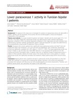
Correlations of CD127 and Foxp3 expressions on
CD4
+
CD25
-
, CD4
+
CD25
low
, and CD4
+
CD25
high
T cells from
UNOL patients
CD4
+
T cells were divided into three subgroups by CD25
expression: CD4
+
CD25
high
, CD4
+
CD25
low
, and CD4
+
CD25
-
T cells. We gated CD127
low/-
expression on Foxp3
+
T cells
and backgated Foxp3 expression on CD127
low/-
T cells,
respectively. We found that all CD4
+
Foxp3
+
T cells had a low
expression level of CD127, regardless of CD25 expression.
Percentages of CD127
low/-
in CD25
high
, CD25
low
, and CD25
-
subpopulations of CD4
+
Foxp3
+
T cells were 93.79% ±
3.48%, 93.66% ± 2.31%, and 91.98% ± 2.14%, respectively
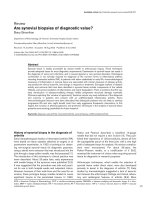
(P > 0.05) (Figure 1). On the other hand, the expressions of
Foxp3 on CD4
+
CD127
low/-
T cells showed significant differ-
ences in CD25
high
(91.38% ± 2.57%), CD25
low
(71.89% ±
3.31%), and CD25
-
(9.02% ± 2.21%) subpopulations (P <
0.01) (Figure 2). Foxp3 expressions in CD4
+
CD127
low/-
T cells
were high in CD25
high
but low in CD25
-
subpopulations. This
result suggested that, unlike in CD4
+
CD25
high
T cells,
CD127
low/-
was not a perfect candidate surface marker for
Figure 1
Expressions of CD127 on CD25
high
, CD25
low
, and CD25
-
subpopulations of CD4
+
Foxp3
+
T cells from patients with untreated new-onset lupus (UNOL)Expressions of CD127 on CD25
high
, CD25
low
, and CD25
-
subpopulations of CD4
+
Foxp3
+
T cells from patients with untreated new-onset lupus
(UNOL).
Arthritis Research & Therapy Vol 11 No 5 Yang et al.
Page 4 of 9
(page number not for citation purposes)
intracellular Foxp3 in CD4
+
CD25
-
T cells and that
CD4
+
CD25
-
CD127
low/-
T cells could not be used as a live
substitute for CD4
+
CD25
-
Foxp3
+
T cells to perform functional
assay.
Expressions of GITR, CTLA-4, CCR4 and effector T cell-
related cytokines on CD4
+
subpopulations from UNOL
patients
Expressions of GITR, CTLA-4, CCR4, and effector T cell-
related cytokines (IFN-γ, IL-4, IL-2, and IL-17) on
CD4
+
CD25
-
Foxp3
+
, CD4
+
CD25
+
Foxp3
-
,
CD4
+
CD25
+
Foxp3
+
, and CD4
+
CD25
-
Foxp3
-
T cells from
UNOL patients and healthy controls
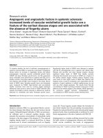
As shown in Table 1 and Figure 3, in UNOL patients, there was
no significant difference between CD4
+
CD25
-
Foxp3
+
and
CD4
+
CD25
+
Foxp3
-
T cells in the expressions of GITR, CTLA-
4, and CCR4 (P > 0.05), whereas they were both less than
CD4
+
CD25
+
Foxp3
+
T cells (P < 0.01). Moreover, the expres-
sions of effector T cell (Teff)-related cytokines, including IFN-
γ, IL-4, IL-2, and IL-17, were analyzed to examine cytokine syn-
thesis capacity of CD4
+
CD25
-
Foxp3
+
T cells. As shown in
Table 2 and Figure 4, in UNOL patients, unlike Tregs
(CD4
+
CD25
+
Foxp3
+
), CD4
+
CD25
-
Foxp3
+
T cells also syn-
thesized IFN-γ, IL-4, IL-2, and IL-17 (P < 0.05), though less
than Teffs (CD4
+
CD25
+
Foxp3
-
).
Functional assays of T-cell subgroups from UNOL
patients
CD4
+
CD25
-
CD127
+
(Teffs), CD4
+
CD25
high
CD127
low/-
(Tregs), CD4
+
CD25
low
CD127
low/-
, and CD4
+
CD25
-
CD127
-
T
cells from UNOL patients were sorted respectively. First, all
Figure 2
Expressions of Foxp3 in CD25
high
, CD25
low
, and CD25
-
subpopulations of CD4
+
CD127
low/-
T cells from patients with untreated new-onset lupus (UNOL)Expressions of Foxp3 in CD25
high
, CD25
low
, and CD25
-
subpopulations of CD4
+
CD127
low/-
T cells from patients with untreated new-onset lupus
(UNOL).
Table 1
Expressions of GITR, CTLA-4, and CCR4 on CD4
+
subpopulations from untreated new-onset lupus patients and healthy controls
Subgroups GITR, % CTLA-4, % CCR4, %
UNOL HC UNOL HC UNOL HC
CD4
+
CD25
-
Foxp3
+
4.41 ± 0.67 5.13 ± 1.23 39.78 ± 1.67 53.12 ± 4.29 35.76 ± 2.53 34.33 ± 2.90
CD4
+
CD25
+
Foxp3
+
22.49 ± 1.75 29.88 ± 3.24 73.89 ± 2.76 81.66 ± 4.85 49.44 ± 2.75 56.91 ± 3.17
CD4
+
CD25
+
Foxp3
-
6.52 ± 0.89 4.89 ± 1.32 33.57 ± 2.98 40.59 ± 5.55 31.99 ± 3.76 32.23 ± 5.54
CD4
+
CD25
-
Foxp3
-
5.35 ± 0.88 11.77 ± 2.75 15.05 ± 2.24 16.06 ± 4.25 13.58 ± 2.57 10.11 ± 3.63
Values are presented as mean ± standard deviation. CCR4, chemokine receptor 4; CTLA-4: Cytotoxic T lymphocyte-associated antigen 4; GITR:
glucocorticoid-induced tumor necrosis factor receptor; HC: healthy controls; UNOL: patients with untreated new-onset lupus.
Available online />Page 5 of 9
(page number not for citation purposes)
four subgroups were stimulated with anti-CD3 and assessed
for their proliferative ability. CD4
+
CD25
high
CD127
low/-
Tregs
(0.205 ± 0.043) were found to be anergic compared with
CD4
+
CD25
-
CD127
+
Teffs (0.421 ± 0.102). Similarly,
CD4
+
CD25
low
CD127
low/-
(0.210 ± 0.062) and CD4
+
CD25
-
CD127
-
(0.272 ± 0.081) T cells showed a reduced prolifera-
tive response (Figure 5).
Then, CD4
+
CD25
high
CD127
low/-
, CD4
+
CD25
low
CD127
low/-
,
and CD4
+
CD25
-
CD127
-
T cells were respectively cocultured
with CD4
+
CD25
-
CD127
+
Teffs. The suppressive capacity as
shown by optical density was most prominent in
CD4
+
CD25
high
CD127
low/-
(0.213 ± 0.032), followed by
CD4
+
CD25
low
CD127
low/-
(0.281 ± 0.061) and CD4
+
CD25
-
CD127
-
(0.387 ± 0.087). CD4
+
CD25
-
CD127
-
T cells showed
the least suppressive capacity, which was similar to the Teffs,
in line with its lesser expression of Foxp3 (9.02% ± 2.21%)
(Figure 5).
Discussion
Foxp3 is currently thought to be one of the best markers for
nTregs. It plays a pivotal role in the development and matura-
tion of Tregs. Foxp3-deficient mice develop systemic autoim-
mune disease, and evidence from adoptive transfer
experiments suggests that this is the direct result of nTreg
defect. Moreover, overexpression of Foxp3 in murine CD4
+
T
cells is sufficient to generate Tregs in vitro. In humans, Foxp3
deficiency also leads to a systemic autoimmune disease
known as IPEX (immune dysregulation, polyendocrinopathy,
enteropathy X-linked syndrome). It has been shown, however,
that the expression of Foxp3 is necessary, but not sufficient, to
confer regulatory function of Tregs. Foxp3 is also expressed
on some activated CD4
+
T cells [12]. Bonelli and colleagues
[13] reported that Foxp3 expression on CD4
+
T cells signifi-
cantly correlated with CD69 expression and that Foxp3 might
be associated with T-cell activation.
In our previous study, we found that a significant increase of
CD4
+
CD25
-
Foxp3
+
T cells in UNOL patients correlated with
disease activity and that the cell number significantly
decreased after glucocorticoid treatment [2]. Whether these
cells are Tregs or activated Teffs remains to be determined.
Functional assay would be a direct way to identify the nature
of CD4
+
CD25
-
Foxp3
+
T cells if only we could find a proper
surface substitute for intracellular Foxp3 in CD4
+
CD25
-
T
cells. A study showed that low expression of CD127 could be
used as a surface marker for intracellular Foxp3 in human
CD4
+
CD25
+
Tregs [3]. CD127 is expressed not only on lym-
phocytes, but also on monocytes and dendritic cells. Its ligand,
IL-7, is a pivotal cytokine involved in the development and sur-
vival of T and B lymphocytes [14]. In addition, thymic stromal
lymphopoietin (TSLP) signals through CD127 in a het-
erodimeric complex with TSLP receptor [15]. TSLP-activated
dendritic cells might participate in the homeostatic mainte-
nance of CD4
+
and development of Tregs in thymus [16].
In this study, we gated and backgated expressions of CD127
and Foxp3 in CD4
+
CD25
-
T cells. We confirmed that
CD4
+
CD25
high
CD127
low/-
could be used as a substitute for
isolating CD4
+
CD25
high
Foxp3
+
Tregs, whereas the expres-
sion of Foxp3 on CD4
+
CD127
low/-
T cells showed significant
differences in CD25
high
(91.38% ± 2.57%), CD25
low
(71.89%
± 3.31%), and CD25
-
(9.02% ± 2.21%) subpopulations.
Foxp3 expression on CD4
+
CD127
low/-
T cells was high in both
CD25
high
and CD25
low
subpopulations but low in CD25
-
sub-
populations. This result suggested that, unlike in
CD4
+
CD25
high
T cells, CD127 was not a perfect surface
marker for intracellular Foxp3 in CD4
+
CD25
-
T cells. It is also
important to note that, although the CD25
low
population lies
adjacent to CD25
-
on a FACS plot (as shown in Figure 2), they
belong to two different cell entities as their Foxp3 expressions
as well as their suppressive capacity and response to in vitro
stimulation were different. If the sorted CD25
-
subgroup was
Table 2
Expressions of IFN-γ, IL-4, IL-2, and IL-17 on CD4
+
subpopulations from untreated new-onset lupus patients and healthy controls
IFN-γ, % IL-4, % IL-2, % IL-17, %
UNOL HC UNOL HC UNOL HC UNOL HC
CD4
+
CD25
-
Foxp3
+
7.56 ± 1.23 5.79 ± 1.05 2.97 ± 0.83 2.02 ± 0.83 3.59 ± 1.95 5.09 ± 1.95 4.61 ± 1.54 1.54 ± 1.02
CD4
+
CD25
+
Foxp3
+
0.72 ± 0.34 1.22 ± 0.58 0.39 ± 0.37 0.88 ± 0.37 0.73 ± 0.49 0.22 ± 0.49 0.38 ± 0.32 0.08 ± 0.06
CD4
+
CD25
+
Foxp3
-
16.43 ± 3.51 16.81 ± 3.97 13.15 ± 2.99 12.94 ± 2.99 20.41 ± 4.91 19.91 ± 4.91 5.58 ± 1.51 2.37 ± 1.51
CD4
+
CD25
-
Foxp3
-
3.54 ± 1.05 5.92 ± 1.57 0.94 ± 0.56 0.66 ± 0.56 2.92 ± 1.42 8.22 ± 1.42 0.49 ± 0.35 0.67 ± 0.42
Values are presented as mean ± standard deviation. HC: healthy controls; IFN-γ: interferon-gamma; IL: interleukin; UNOL: patients with untreated
new-onset lupus.
Arthritis Research & Therapy Vol 11 No 5 Yang et al.
Page 6 of 9
(page number not for citation purposes)
'contaminated' with CD25
low
, it would bias function analysis of
CD4
+
CD25
-
Foxp3
+
T cells from Teffs to Tregs [11].
Another possible explanation of the differences between the
study of Bonelli and colleagues [13] and ours is that there may
be a difference between untreated, newly diagnosed patients
and those more chronically ill who were drawn from an outpa-
tient population. It is possible that, as a consequence of ill-
ness, true CD25
+
Tregs have become CD25
-
, whereas this
has not occurred yet in patients with new-onset disease.
Figure 3
Expressions of glucocorticoid-induced tumor necrosis factor receptor (GITR), cytotoxic T lymphocyte-associated antigen 4 (CTLA-4), and chemok-ine receptor 4 (CCR4) on CD4
+
subpopulations from patients with untreated new-onset lupus (UNOL)Expressions of glucocorticoid-induced tumor necrosis factor receptor (GITR), cytotoxic T lymphocyte-associated antigen 4 (CTLA-4), and chemok-
ine receptor 4 (CCR4) on CD4
+
subpopulations from patients with untreated new-onset lupus (UNOL). (A) CD4
+
CD25
-
Foxp3
+
. (B) CD4
+
CD25
+
Foxp3
+
. (C) CD4
+
CD25
+
Foxp3
-
. (D) CD4
+
CD25
-
Foxp3
-
.
Available online />Page 7 of 9
(page number not for citation purposes)
Figure 4
Expressions of interferon-gamma (IFN-γ), interleukin (IL)-4, IL-2, and IL-17 on CD4
+
subpopulations from patients with untreated new-onset lupus (UNOL)Expressions of interferon-gamma (IFN-γ), interleukin (IL)-4, IL-2, and IL-17 on CD4
+
subpopulations from patients with untreated new-onset lupus
(UNOL). (A) CD4
+
CD25
-
Foxp3
+
. (B) CD4
+
CD25
+
Foxp3
+
. (C) CD4
+
CD25
+
Foxp3
-
. (D) CD4
+
CD25
-
Foxp3
-
.
Arthritis Research & Therapy Vol 11 No 5 Yang et al.
Page 8 of 9
(page number not for citation purposes)
In our study, we found that the expressions of GITA, CTLA-4,
and CCR4 on CD4
+
CD25
-
Foxp3
+
T cells resembled
CD4
+
CD25
+
Foxp3
-
Teffs and were significantly less than
CD4
+
CD25
+
Foxp3
+
Tregs. Moreover, unlike
CD4
+
CD25
+
Foxp3
+
Tregs, CD4
+
CD25
-
Foxp3
+
T cells also
synthesized IFN-γ, IL-4, IL-2, and IL-17, though less than
CD4
+
CD25
+
Foxp3
-
Teffs, suggesting that the abnormally
increased CD4
+
CD25
-
Foxp3
+
T cells in UNOL patients were
not simple and pure Tregs.
Conclusions
CD4
+
CD25
-
Foxp3
+
T cells in UNOL patients are different
from Tregs, both phenotypically and functionally. CD127 is not
an appropriate surface marker for intracellular Foxp3 in
CD4
+
CD25
-
T cells.
Competing interests
The authors declare that they have no competing interests.
Authors' contributions
HY and WZ developed the study, analyzed the data, and
drafted the manuscript. LZ and YL participated in the data col-
lection, performed the data analysis, and helped in the drafting
of the manuscript. XZ and FZ participated in the development
of the study, data analysis, and the drafting of the manuscript.
FT and WH conceived the study and drafted the manuscript.
All authors have read and approved the manuscript.
Acknowledgements
This work was supported by New Century Excellent Talents, Ministry of
Education of China (NCET-04-0191), National Natural Sciences Foun-
dation of China (30972731), Natural Sciences Foundation of Beijing
(7052052), and the National Program for Key Basic Research Project
(2007CB512405 for Immunology), Ministry of Science and Technology,
China.
References
1. Horwitz DA: Regulatory T cells in systemic lupus erythemato-
sus: past, present and future. Arthritis Res Ther 2008, 10:227.
2. Zhang B, Zhang X, Tang FL, Zhu LP, Liu Y, Lipsky PE: Clinical sig-
nificance of increased CD4
+
CD25
-
Foxp3
+
T cells in patients
with new-onset systemic lupus erythematosus. Ann Rheum
Dis 2008, 67:1037-1040.
3. Kramer S, Schimpl A, Hunig T: Immunopathology of interleukin
(IL) 2-deficient mice: thymus dependence and suppression by
thymus-dependent cells with an intact IL-2 gene. J Exp Med
1995, 182:1769-1776.
4. Shimizu J, Yamazaki S, Takahashi T, Ishida Y, Sakaguchi S: Stim-
ulation of CD25(+)CD4(+) regulatory T cells through GITR
breaks immunological self-tolerance. Nat Immunol 2002,
3:135-142.
5. McHugh RS, Whitters MJ, Piccirillo CA, Young DA, Shevach EM,
Collins M, Byrne MC: CD4(+)CD25(+) immunoregulatory T
cells: gene expression analysis reveals a functional role for
the glucocorticoid-induced TNF receptor. Immunity 2002,
16:311-323.
6. Takahashi T, Tagami T, Yamazaki S, Uede T, Shimizu J, Sakaguchi
N, Mak TW, Sakaguchi S: Immunologic self-tolerance main-
tained by CD25(+)CD4(+) regulatory T cells constitutively
expressing cytotoxic T lymphocyte-associated antigen 4. J
Exp Med 2000, 192:303-310.
7. Curiel TJ, Coukos G, Zou L, Alvarez X, Cheng P, Mottram P, Evde-
mon-Hogan M, Conejo-Garcia JR, Zhang L, Burow M, Zhu Y, Wei
S, Kryczek I, Daniel B, Gordon A, Myers L, Lackner A, Disis ML,
Knutson KL, Chen L, Zou W: Specific recruitment of regulatory
T cells in ovarian carcinoma fosters immune privilege and pre-
dicts reduced survival. Nat Med 2004, 10:942-949.
8. Iellem A, Mariani M, Lang R, Recalde H, Panina-Bordignon P, Sini-
gaglia F, D'Ambrosio D: Unique chemotactic response profile
and specific expression of chemokine receptors CCR4 and
CCR8 by CD4(+)CD25(+) regulatory T cells. J Exp Med 2001,
194:847-853.
9. Imai T, Nagira M, Takagi S, Kakizaki M, Nishimura M, Wang J, Gray
PW, Matsushima K, Yoshie O: Selective recruitment of CCR4-
bearing Th2 cells toward antigen-presenting cells by the CC
chemokines thymus and activation-regulated chemokine and
macrophage-derived chemokine. Int Immunol 1999, 11:81-88.
Figure 5
Assessment of proliferative and suppressive capacities of CD4
+
CD25
-
CD127
+
, CD4
+
CD25
high
CD127
low/-
, CD4
+
CD25
low
CD127
low/-
, and CD4
+
CD25
-
CD127
-
T cells from patients with untreated new-onset lupusAssessment of proliferative and suppressive capacities of CD4
+
CD25
-
CD127
+
, CD4
+
CD25
high
CD127
low/-
, CD4
+
CD25
low
CD127
low/-
, and CD4
+
CD25
-
CD127
-
T cells from patients with untreated new-onset lupus. Values are presented as mean ± standard error of the mean (n = 8). OD, opti-
cal density.
Available online />Page 9 of 9
(page number not for citation purposes)
10. Yagi H, Nomura T, Nakamura K, Yamazaki S, Kitawaki T, Hori S,
Maeda M, Onodera M, Uchiyama T, Fujii S, Sakaguchi S: Crucial
role of FOXP3 in the development and function of human
CD25
+
CD4
+
regulatory T cells. Int Immunol 2004,
16:1643-1656.
11. Bonelli M, Savitskaya A, Steiner CW, Rath E, Smolen JS, Schei-
necker C: Phenotypic and functional analysis of CD4
+
CD25
-
Foxp3
+
T cells in patients with systemic lupus erythematosus.
J Immunol 2009, 182:1689-1695.
12. Allan SE, Crome SQ, Crellin NK, Passerini L, Steiner TS, Bac-
chetta R, Roncarolo MG, Levings MK: Activation-induced FOXP3
in human T effector cells does not suppress proliferation or
cytokine production. Int Immunol 2007, 19:345-354.
13. Bonelli M, von Dalwigk K, Savitskaya A, Smolen JS, Scheinecker
C: Foxp3 expression in CD4
+
T cells of patients with systemic
lupus erythematosus: a comparative phenotypic analysis. Ann
Rheum Dis 2008, 67:664-671.
14. Kang J, Der SD: Cytokine functions in the formative stages of a
lymphocyte's life. Curr Opin Immunol 2004, 16:180-190.
15. Palmer MJ, Mahajan VS, Trajman LC, Irvine DJ, Lauffenburger DA,
Chen J: Interleukin-7 receptor signaling network: an integrated
systems perspective. Cell Mol Immunol 2008, 5:79-89.
16. Willerford DM, Chen J, Ferry JA, Davidson L, Ma A, Alt FW: Inter-
leukin-2 receptor alpha chain regulates the size and content of
the peripheral lymphoid compartment. Immunity 1995,
3:521-530.