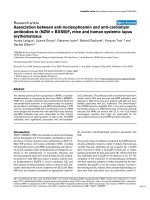
Báo cáo y học: "Relationship between anti-dsDNA, anti-nucleosome and anti-alpha-actinin antibodies and markers of renal disease in patients with lupus nephritis: a prospective longitudinal study" pdf
Bạn đang xem bản rút gọn của tài liệu. Xem và tải ngay bản đầy đủ của tài liệu tại đây (659.36 KB, 9 trang )
Open Access
Available online />Page 1 of 9
(page number not for citation purposes)
Vol 11 No 5
Research article
Relationship between anti-dsDNA, anti-nucleosome and
anti-alpha-actinin antibodies and markers of renal disease in
patients with lupus nephritis: a prospective longitudinal study
Jessica J Manson
1
, Alexander Ma
1
, Pauline Rogers
2
, Lesley J Mason
1
, Jo H Berden
3
, Johan van
der Vlag
3
, David P D'Cruz
4
, David A Isenberg
1
and Anisur Rahman
1
1
Centre for Rheumatology Research, University College London, Windeyer Institute, 46 Cleveland Street, London W1T 4JF, UK
2
Joint University College London Hospital/University College London and Royal Free Biomedical Research Unit, Research and Development (1st
Floor Maple House), Rosenheim Wing, 25 Grafton Way, London WC1E 6DB, UK
3
Nephrology Research Laboratory, Nijmegen Centre for Molecular Life Sciences, Department of Nephrology, Radboud University Nijmegen Medical
Centre, PO Box 9101, 6500 HB Nijmegen, The Netherlands
4
Lupus Research Unit, The Rayne Institute, St Thomas' Hospital, Lambeth Palace Road, London SE1 7EH, UK
Corresponding author: Anisur Rahman,
Received: 12 Aug 2009 Revisions requested: 22 Sep 2009 Revisions received: 3 Oct 2009 Accepted: 14 Oct 2009 Published: 14 Oct 2009
Arthritis Research & Therapy 2009, 11:R154 (doi:10.1186/ar2831)
This article is online at: />© 2009 Manson et al.; licensee BioMed Central Ltd.
This is an open access article distributed under the terms of the Creative Commons Attribution License ( />),
which permits unrestricted use, distribution, and reproduction in any medium, provided the original work is properly cited.
Abstract
Introduction Glomerulonephritis is a major cause of morbidity
and mortality in patients with systemic lupus erythematosus
(SLE). Deposition of autoantibodies in the glomeruli plays a key
role in the development of lupus nephritis (LN). Different groups
have proposed that either anti-nucleosome antibodies or
antibodies that bind the intrinsic renal antigen, α-actinin, are
central to the pathogenesis of LN. These theories have been
based mainly on cross-sectional studies in patients and on
experiments in animal models. No previous longitudinal studies
have compared the relationships between levels of these
antibodies and markers of renal function. We assessed how well
anti-α-actinin, anti-nucleosome and anti-double-stranded DNA
(anti-dsDNA) antibodies reflected renal outcome measures in
patients with new-onset LN followed for up to 2 years.
Methods Renal disease activity was monitored by measuring
urine protein/creatinine ratio (PCR), serum albumin and a
composite outcome of renal remission. At each time point, anti-
nucleosome and anti-α-actinin antibodies were measured by
enzyme-linked immunosorbent assay. High-avidity anti-dsDNA
antibodies were measured using the Farrzyme assay. We
analysed relationships between levels of the three antibodies
and between antibody levels and renal outcome measures over
time.
Results Levels of anti-nucleosome and anti-dsDNA were
positively correlated with each other (r = 0.6, P = 0.0001) but
neither correlated with anti-α-actinin level. At baseline, mean
anti-nucleosome levels were higher in patients with LN than in
healthy controls (0.32 versus 0.01, P < 0.001). The same was
true for anti-dsDNA antibodies (0.50 versus 0.07, P < 0.001)
but not for anti-α-actinin (0.33 versus 0.29). Over the follow-up
period, anti-nucleosome and anti-dsDNA levels associated
positively with urine PCR (P = 0.041 and 0.051, respectively)
and negatively with serum albumin (P = 0.027 and 0.032,
respectively). Both anti-nucleosome and anti-dsDNA levels were
significantly lower during renal remission than when renal
disease was active (P = 0.002 and 0.003, respectively).
However, there was no relationship between anti-α-actinin
levels and urine PCR, serum albumin or remission status.
Conclusions This prospective longitudinal clinical study is the
first to compare levels of anti-nucleosome, anti-dsDNA and anti-
α-actinin antibodies in the same patients with SLE. Our results
support the concept that, in the majority of patients, anti-
nucleosome antibodies play a major role in pathogenesis of LN,
in contrast to anti-α-actinin antibodies.
anti-dsDNA: anti-double-stranded DNA; AR: absorbance ratio; dsDNA: double-stranded DNA; ELISA: enzyme-linked immunosorbent assay; LN:
lupus nephritis; OD: optical density; PBS-T: phosphate-buffered saline/0.05% Tween 20; PCR: protein/creatinine ratio; SD: standard deviation; SLE:
systemic lupus erythematosus.
Arthritis Research & Therapy Vol 11 No 5 Manson et al.
Page 2 of 9
(page number not for citation purposes)
Introduction
Lupus nephritis (LN) occurs in 40% to 60% of patients with
systemic lupus erythematosus (SLE) [1]. Koffler and col-
leagues [2] first demonstrated deposition of autoantibodies in
LN renal tissue. A range of evidence from clinical [3], renal
biopsy [4] and animal [5-7] studies suggested that anti-dou-
ble-stranded DNA (anti-dsDNA) antibodies were the main
autoantibodies involved in the pathogenesis of LN. It has been
argued that high-avidity anti-dsDNA antibodies are particularly
linked to pathogenicity, and some laboratory tests have been
developed specifically to test for these high-avidity antibodies
[8]. However, there are clearly some patients with persistently
high anti-dsDNA levels who never develop LN [9] and there is
no simple relationship between the ability of passively trans-
ferred monoclonal antibodies to bind dsDNA and the ability of
the same antibodies to cause glomerulonephritis [5-7].
In some cases, modification of antibodies by mutagenesis
increased binding to dsDNA but reduced pathogenicity [7]. In
other cases, pathogenic monoclonal antibodies were found
not to bind dsDNA at all after rigorous purification and were
actually anti-nucleosome antibodies [10,11]. Furthermore,
when a rat kidney perfusion system was used, glomerular bind-
ing of monoclonal antibodies was shown to require the pres-
ence of nucleosomes [12]. It has therefore been argued that
binding to nucleosomes is a major determinant of pathogenic-
ity of autoantibodies in LN [13,14].
An alternative theory holds that direct cross-reaction of anti-
dsDNA with intraglomerular antigens is key [13,15]. Although
cross-reactivity with a number of proteins (including laminin
and type IV collagen) has been postulated (reviewed in [13]),
the importance of anti-α-actinin antibodies has been particu-
larly stressed in recent years. This emphasis on the possible
pathogenic role of anti-α-actinin antibodies has arisen as a
result of studies in murine models [6,16] and clinical studies
[17-19], although anti-α-actinin antibodies could not be eluted
from glomerular deposits in mice with LN [20]. However, no
previous study has compared anti-nucleosome and anti-α-
actinin antibody levels in the same patients.
In this study, we identified 16 patients with new-onset LN and
followed them prospectively for up to 2 years. We tested their
blood for both anti-nucleosome and anti-α-actinin antibodies,
allowing (for the first time) direct comparison of both of these
important specificities in the same patients with LN. Further-
more, we examined the associations between levels of both
anti-nucleosome and anti-α-actinin antibodies, levels of high-
avidity anti-dsDNA antibodies and markers of renal disease,
including an assessment of whether the patients entered renal
remission.
Materials and methods
Sixteen patients with new-onset biopsy-proven LN were
recruited prospectively from the Lupus Clinics at University
College London and St Thomas' hospitals (London). All of the
patients fulfilled the American College of Rheumatology
revised criteria for SLE [21,22]. Blood samples were taken at
the time of recruitment and then at routine follow-up appoint-
ments and were spun at 500 g for 10 minutes to produce
serum, which was stored at -20°C. To establish normal values
for the enzyme-linked immunosorbent assays (ELISAs) used,
30 healthy volunteer donors with age and gender distributions
similar to those of the controls were recruited from staff at Uni-
versity College London Hospital and University College Lon-
don. Serum was collected and stored as above. Patients and
healthy controls gave informed consent. This study received
approval from the Thames Valley Multi-Centre Research Eth-
ics Committee (reference number 04/MRE12/58) and was
passed by the Joint University College London/University Col-
lege London Hospitals Committee on the Ethics of Human
Research and the St Thomas' Hospital Research Ethics Com-
mittee.
Renal outcome measures
For each patient at each time point, urine was tested for pro-
tein/creatinine ratio (PCR), and serum was tested for albumin
and creatinine in the routine clinical laboratory. Statistical anal-
ysis was carried out on three outcome measures: absolute val-
ues of urine PCR and serum albumin and a composite score
for renal disease activity. This composite score was defined
using measurements of urine PCR and serum albumin and cre-
atinine. Complete renal remission was defined as follows:
PCR of not more than 30 mg/mmol, normal serum albumin and
normal serum creatinine. Partial remission was defined as fol-
lows: decrease in urine PCR by at least 50%, serum albumin
of at least 30 g/L, and either normal serum creatinine if the
baseline creatinine was less than 260 μmol/L or a 50%
decrease in creatinine if the baseline value was at least 260
μmol/L. Patients who did not fulfil these criteria for either com-
plete or partial remission were considered to have active LN.
Human IgG anti-nucleosome enzyme-linked
immunosorbent assay
Serum samples were diluted 1:800 in phosphate-buffered
saline/0.05% Tween 20 (PBS-T) and tested in duplicate for
binding to nucleosomes prepared from Jurkat cells. The meth-
ods for obtaining nucleosomes and carrying out the ELISA
have been described previously [9,23]. Monoclonal human
IgG antibodies with well-defined anti-nucleosome-binding
properties [23] were used as positive and negative controls.
To standardise results between ELISA plates, readings were
taken when the positive control had reached an optical density
(OD) of approximately 1.2.
Human IgG anti-α-actinin enzyme-linked
immunosorbent assay
Serum samples were diluted 1:100 in PBS-T and tested in an
anti-α-actinin ELISA. The ELISA method was as described
previously [17]. Again, the positive and negative controls were
Available online />Page 3 of 9
(page number not for citation purposes)
human monoclonal antibodies produced in our laboratory with
known anti-α-actinin-binding properties [23]. Plates were read
when the OD of the positive control reached 1.2.
Detection of high-avidity human IgG anti-double-
stranded DNA antibodies
High-avidity anti-dsDNA antibody titre was measured using
the Farrzyme assay (The Binding Site, Birmingham, UK) in
accordance with the instructions of the manufacturer [8].
Expression of results of enzyme-linked immunosorbent
assay tests
ODs from all of the assays were converted to absorbance
ratios (ARs) to standardise the data and minimise interassay
variation. The mean OD for each sample was calculated from
the duplicates. The mean OD was then divided by the stand-
ard positive control on that assay plate to give the AR.
Statistical analysis
The data were analysed in GraphPad Prism (GraphPad Soft-
ware Inc., San Diego, CA, USA) and using the 'xt' commands
for longitudinal data in Stata 9.2 (StataCorp LP, College Sta-
tion, TX, USA). The aim of the analysis was to assess which
antibody level (high-avidity anti-dsDNA, anti-nucleosome or
anti-α-actinin) best reflected the renal outcome measures,
PCR, albumin and remission status. First, the continuous lab-
oratory variables (all three antibodies, albumin and PCR) were
tested for normality and, if necessary, were transformed using
log transformation. PCR measurements and ARs for anti-
nucleosome and high-avidity anti-dsDNA antibodies were so
transformed, and consequently changes in these measure-
ments are presented as percentages rather than as absolute
values. Mean ARs for the baseline binding data in the patient
and control groups were compared using the Student t test
using the Satterthwaite approximation for unequal variances
where appropriate. Possible correlations between the results
of the three antibody assays were investigated by calculating
the Pearson correlation coefficient, using log-transformed data
where appropriate. For the two continuous outcome variables
(albumin and PCR), each explanatory variable was analysed
one at a time and significant variables then were included in a
multivariable sensitivity analysis. Maximum likelihood random
effects models were used to fit linear regression models with
random intercepts. The residuals from all models were
checked for normality using a normal plot. The relationship
between remission status and the three laboratory explanatory
variables was investigated using the regression specification
for a one-way analysis of variance. Anti-nucleosome level, anti-
α-actinin level and high-avidity anti-dsDNA level were each
considered in turn as the outcome with remission as the
explanatory variable. The Wald test was used to test contrasts
between the coefficients for the different levels of renal remis-
sion (active disease, partial remission and complete remis-
sion). Due to the size of the dataset, there was insufficient
statistical power to carry out multivariable analysis, and signif-
icant results from these analyses should be treated with cau-
tion.
Results
Characteristics of patients and control subjects
Sixteen patients were enrolled, and baseline details for all
patients are given in Table 1. There were 15 females and one
male. The mean age was 33.4 years (standard deviation [SD]
10.9, range 18 to 56). Three of the patients were Black, six
were South Asian and seven were White. The mean age of the
30 healthy control subjects was 39 years (SD 11.5, range 24
to 64). There were 24 females and six males. Five were Black,
four were South Asian and 21 were White. Renal biopsies
were classified in accordance with the World Health Organi-
zation criteria [24]. Two patients had class II disease, seven
had class III or IV, three had pure class V and four had class V
with III or IV. At baseline, the mean urine PCR was 285 mg/
mmol (range 34 to 1,017), and the mean serum albumin was
31 g/L (range 17 to 44). Medications at time of enrolment are
shown in Table 1. All patients but one were taking oral pred-
nisolone (daily dose range 5 to 60 mg), and 12 were also
treated with immunosuppressants (mycophenolate, cyclo-
phosphamide or azathioprine). Patient LN11 differed from all
of the others in several important respects. He was the only
man, the only patient with increased serum creatinine at base-
line (156 μmol/L) and the only patient to require renal dialysis.
Data from this patient are nevertheless included in all of the
analyses below except where stated specifically. Anti-nucleo-
some and high-avidity anti-dsDNA but not anti-α-actinin level
were higher in patients with LN than in healthy controls.
Baseline binding data from all patients were compared with
the results from 30 normal controls (Figure 1). The mean (SD)
ARs for anti-nucleosome antibodies were significantly higher
for LN patients than controls (means were calculated on raw
data, but log-transformed data were analysed where appropri-
ate) (0.32 [0.35] versus 0.01 [0.01], P < 0.0001) as were the
high-avidity anti-dsDNA AR (0.50 [0.50] versus 0.07 [0.01], P
< 0.0001) but not anti-α-actinin (0.33 [0.32] versus 0.29
[0.25]). For each assay, the upper limit of normal was taken as
the mean plus three SDs of the results from the 30 normal con-
trols. At baseline, 13 out of 16 patients were above this cutoff
in the anti-nucleosome and high-avidity anti-dsDNA assays,
whereas only two had anti-α-actinin levels that were above the
upper limit of normal.
Changes in antibody levels over the follow-up period
Follow-up dates were determined by the need of the patient to
attend the clinic. The mean number of weeks over which the
patients were followed was 37.3 (range 10 to 85). The mean
interval between test points was 9.3 weeks (range 2 to 26). All
patients had samples collected on at least three occasions,
with a median number of time points per patient of 4.5 (range
3 to 8). On average, all three assays demonstrated significant
downward linear trends over time. Anti-nucleosome antibodies
Arthritis Research & Therapy Vol 11 No 5 Manson et al.
Page 4 of 9
(page number not for citation purposes)
decreased by 1.6% per week, and high-avidity anti-dsDNA
antibodies decreased by 0.8% per week. Anti-α-actinin anti-
bodies decreased by 0.0015 per week, which (given the mean
baseline AR for anti-α-actinin antibodies of 0.25) is the equiv-
alent of a 0.6% decrease per week. In general, if anti-nucleo-
some antibodies were high for any patient at any time point, so
were high-avidity anti-dsDNA antibodies. By contrast, high
anti-α-actinin antibodies (taken as an AR of at least 0.5) were
detected in only three patients (LN3, LN9 and LN15) and were
seen in combination with low AR for the other two assays. Six
patients had low titres (AR <0.25) of anti-nucleosome and
high-avidity anti-dsDNA antibodies throughout the study
period.
Consistent with these findings, analysis of the whole dataset
showed significant positive correlation between anti-nucleo-
some and high-avidity anti-dsDNA levels (r = 0.6, P = 0.0001)
but no correlation between anti-nucleosome and anti-α-actinin
or high-avidity anti-dsDNA and anti-α-actinin (Figure 2). Anti-
nucleosome and high-avidity anti-dsDNA levels associate pos-
itively with urine PCR and negatively with serum albumin over
time. Analysis of the relationship between high-avidity anti-
dsDNA antibody titre and urine PCR revealed a significant
positive linear trend (P = 0.041). The relationship between
anti-nucleosome antibodies and PCR reached borderline sta-
tistical significance (P = 0.051). When we repeated the anal-
ysis excluding patient LN11, both associations were
statistically significant: P = 0.021 for anti-nucleosome and P
= 0.043 for high-avidity anti-dsDNA. On average, urine PCR
increased by 18% for every twofold increase in anti-nucleo-
some level and by 27% for every twofold increase in anti-
dsDNA level.
There were significant negative linear trends between albumin
levels and anti-nucleosome (P = 0.027) and high-avidity anti-
dsDNA (P = 0.032) antibody titre. Repeating the analysis after
exclusion of patient LN11 confirmed the analysis and showed
stronger levels of statistical significance (P = 0.001 and
0.011, respectively). On average, albumin decreased by 1.16
g/L for every twofold increase in anti-nucleosome titre and by
1.59 g/L for every twofold increase in high-avidity anti-dsDNA
antibody titre. There was no relationship between anti-α-
actinin levels and either urine PCR (P = 0.401) or serum albu-
min (P = 0.332).
Remission status
Analysis of the three-level remission score (active disease, par-
tial remission and complete remission) demonstrated signifi-
cant differences between the groups in all three antibody
levels (anti-nucleosome, P = 0.001; high-avidity anti-dsDNA, P
= 0.0063; and anti-α-actinin, P = 0.0368). Application of the
Wald test (which assesses whether there is a true difference
Table 1
Baseline patient data
Patient ID Gender Ethnicity Biopsy result Urine PCR, mg/mmol Serum albumin, g/L Treatment, daily dose in mg
LN1 Female Asian III 58 38 P (20), H (400)
LN2 Female White IV, V 457 30 P (20), MP
a
LN3 Female Black V 123 28 P (30), H (400)
LN4 Female White IV 533 21 P (10), H (400), MMF (1,500)
LN5 Female White V 569 23 P (10), H (400)
LN6 Female Black III 202 30 P (5), C
a
LN7 Female Asian III 96 20 P (15), A (100)
LN8 Female Asian III/V 208 36 P (12.5), H (200)
LN9 Female Asian III 34 28 P (60), C
a
LN10 Female Asian IV 66 44 P (10), MMF (2,000)
LN11 Male Asian IV 1,017 17 P (20), M (1,000), C
a
LN12 Female White II 312 34 H (400)
LN13 Female White II 127 33 H (400), MP
a
LN14 Female White V 366 34 P (20), A (100)
LN15 Female White III/V 88 43 P (5), A (100)
LN16 Female Black IV/V 297 37 P (20)
a
MP and C signify a recent intravenous pulse of methylprednisolone or cyclophosphamide, respectively; thus, there is no value for daily dose.
Values for daily dose of each drug refer to the dose being taken at the time of entry into the study. A, azathioprine; H, hydroxychloroquine; LN,
lupus nephritis; MMF, mycophenolate mofetil; P, prednisolone; PCR, protein/creatinine ratio.
Available online />Page 5 of 9
(page number not for citation purposes)
Figure 1
Baseline binding data for binding to nucleosomes, double-stranded DNA and α-actininBaseline binding data for binding to nucleosomes, double-stranded
DNA and α-actinin. On each graph, absorbance ratios of the 30 normal
controls and the 16 patients with lupus nephritis are plotted. The mean
and standard deviation are also plotted. The groups were compared
using the Student t test. The dotted black line shows the upper limit of
normal for each assay. anti-dsDNA, anti-double-stranded DNA; ns, not
significant.
Figure 2
Correlation between binding to (a) nucleosomes and double-stranded DNA (dsDNA), (b) nucleosomes and α-actinin and (c) dsDNA and α-actininCorrelation between binding to (a) nucleosomes and double-stranded
DNA (dsDNA), (b) nucleosomes and α-actinin and (c) dsDNA and α-
actinin. Absorbance ratios (ARs) for each assay are plotted against
each other. The Pearson correlation coefficient is given for each associ-
ation (r). ns, not significant.
Arthritis Research & Therapy Vol 11 No 5 Manson et al.
Page 6 of 9
(page number not for citation purposes)
between groups) suggested that there was no real difference
in the levels between partial and complete remission. The par-
tial and complete remission categories were combined such
that there were only two possible renal activity levels for each
patient at each time point (active or remission), and the data
were then re-analysed. The association between activity status
and anti-nucleosome or high-avidity anti-dsDNA antibody titre
was maintained (P = 0.002 and 0.003, respectively), but there
was no observed difference in the levels of anti-α-actinin anti-
bodies between active disease and remission. On average,
anti-nucleosome levels were 53.8% lower and high-avidity
anti-dsDNA levels were 34.5% lower when patients were in
remission than when they had active disease.
Longitudinal studies in individual patients
Several different patterns were discernible. In three patients
(LN2, LN12 and LN14), levels of all three antibodies remained
low from baseline throughout the follow-up period and were
not closely related to outcome measures. An example of this
pattern is illustrated in Figure 3a. In three other patients (LN3,
LN9 and LN15), anti-α-actinin levels remained high throughout
the observation period whereas levels of the other two autoan-
tibodies remained low. However, there was no clear relation-
ship between anti-α-actinin levels and serum albumin or urine
PCR in these patients and no particular clinical or demo-
graphic feature distinguished them from the other 13 patients.
An example of this pattern is illustrated in Figure 3b. In the
remaining cases, anti-nucleosome or high-avidity anti-dsDNA
antibodies or both were increased during the period of obser-
vation. In some patients, the decrease in anti-dsDNA antibod-
ies (patient LN8, Figure 3c) or anti-nucleosome antibodies
(patient LN5, Figure 3d) mirrored changes in serum albumin
and urine PCR.
Discussion
This is the first clinical study to achieve a direct comparison of
the associations between anti-nucleosome, high-avidity anti-
dsDNA and anti-α-actinin levels and outcomes of renal dis-
ease in patients with LN followed longitudinally. The results
show that assessment of any renal outcome measure (whether
serum albumin, urine PCR or remission) over time favoured the
theory that renal disease activity was linked to the presence of
anti-nucleosome and anti-dsDNA antibodies and not to anti-α-
actinin antibodies.
Nucleosomes released in apoptotic debris, but not cleared
efficiently from the circulation [25], are critical immunogenic
stimulants for both T cells and B cells in SLE [13,26]. The
mechanism by which anti-nucleosome antibodies bind glomer-
uli has been reviewed extensively elsewhere [13,14]. Recent
electron microscopy data from renal biopsies of both human
and murine LN confirm that autoantibodies in those tissues co-
localise with electron-dense extracellular deposits of chroma-
tin [27,28]. High titres of anti-nucleosome antibodies have
been found in up to 87% of patients with SLE [26,29,30], and
some studies have noted a particular association with renal
disease [31,32]. When highly purified nucleosomes are used
as the antigen, the anti-nucleosome assay is very specific for
patients with SLE [33]. However, these serological studies
either were cross-sectional or did not evaluate quantitative
markers of renal impairment [30].
A Dutch study of 52 patients with proliferative LN [34], fol-
lowed longitudinally for a year as part of a clinical trial, showed
good correlation between these two serological measures (r =
0.63, P < 0.001), as in our study. Data on renal outcomes
across the whole patient group were reported in terms of
relapse or remission, but data on serum albumin and urine
PCR were not given. The investigators did not observe
increases in anti-nucleosome or anti-dsDNA antibody titre
prior to renal relapse. There was no association between lev-
els of anti-nucleosome antibodies at disease entry and occur-
rence of relapse or time to remission. Data on individual
patients were not given.
Alpha-actinin-4 is an actin-binding protein present in both
podocytes and mesangial cells. Two groups showed that the
ability of murine monoclonal anti-dsDNA or anti-nucleosome
antibodies to cause pathogenicity in mice was related to their
ability to cross-react with α-actinin [6,16], and one of the
groups then showed that a cross-reactive human anti-dsDNA/
anti-α-actinin antibody caused glomerulonephritis in these
mice [35]. However, the electron-dense deposits that are the
sites of autoantibody deposition in lupus-prone NZB/W F1
mice do not co-localise with α-actinin. Three previous clinical
studies have looked at anti-α-actinin levels in patients with
SLE [17-19]. One study showed that purified anti-dsDNA anti-
bodies from patients with SLE were more likely to cross-react
with α-actinin if the patients had nephritis [17]. Two subse-
quent studies [18,19] were both cross-sectional. Both groups
showed that anti-α-actinin antibodies can occur in patients
with diseases other than SLE (for example, rheumatoid arthritis
[19] and autoimmune hepatitis [15]) and occur in both
patients with LN and patients with lupus but not nephritis. In
one study, positivity for anti-α-actinin distinguished patients
with nephritis from those without nephritis more clearly than
positivity for anti-dsDNA, although only 10 out of 24 patients
with nephritis were positive for anti-α-actinin [18]. Neither
study looked at anti-nucleosome antibody levels, and neither
showed any correlation between levels of anti-α-actinin anti-
bodies and indicators of renal disease such as proteinuria. To
our knowledge, ours is the first longitudinal study of anti-α-
actinin levels in patients with LN. Although our results do not
favour an important role for cross-reactive anti-
α-actinin anti-
bodies in most patients with LN, they leave open the possibility
that these antibodies may be important in a minority of such
patients. Three of our 16 patients had increased levels of anti-
α-actinin antibodies but not anti-dsDNA or anti-nucleosome
antibodies. This is in contrast to the study of Renaudineau and
colleagues [18], who found that 21 out of 22 patients with
Available online />Page 7 of 9
(page number not for citation purposes)
Figure 3
Patterns of relationship between antibody titre and outcome in individual patientsPatterns of relationship between antibody titre and outcome in individual patients. Graphic display of enzyme-linked immunosorbent assay data (anti-
nucleosome, anti-α-actinin and high-avidity anti-double-stranded DNA [Farrzyme]) with urine protein/creatinine ratio (PCR) or serum albumin (Alb).
dsDNA, double-stranded DNA; LN, lupus nephritis.(a) Patient LN2 - all three antibody levels remained low. (b) Patient LN15 - anti-α-actinin high, oth-
ers low. (c) Patient LN8 - anti-dsDNA levels mirror changes in albumin and PCR. (d) Patient LN5 - anti-nucleosome levels mirror changes in albumin
and PCR.
Arthritis Research & Therapy Vol 11 No 5 Manson et al.
Page 8 of 9
(page number not for citation purposes)
SLE who had increased anti-α-actinin levels also had
increased anti-dsDNA. Fewer than half of these 22 patients
had LN.
Although our results are important in shedding more light on
the pathogenesis of LN, they do not suggest any changes in
clinical practice. Neither anti-nucleosome nor anti-α-actinin
tests seem likely to be better predictors of renal outcome than
anti-dsDNA ELISA, which is already widely used for monitor-
ing patients with SLE. Improved monitoring of renal disease in
patients with LN is more likely to be achieved by combining
anti-dsDNA tests with assays more specific for renal dysfunc-
tion, such as urinary gelatinase B-associated lipocalin (nGAL),
which has been shown to be a marker of renal disease in
cross-sectional studies of both adult [36] and paediatric [37]
SLE.
Conclusions
This is the first prospective longitudinal study of patients with
new-onset biopsy-proven LN to study antibody levels and
renal outcome measures at multiple time points within the first
two years after diagnosis. In particular, we measured both anti-
nucleosome and anti-α-actinin levels, specificities that have
been studied previously in separate groups of patients with LN
but never in the same group. The most important conclusion of
our study is that anti-nucleosome and high-avidity anti-dsDNA
antibodies are much more closely related to renal outcome
measures in the majority of these patients than anti-α-actinin
levels.
Competing interests
The authors declare that they have no competing interests.
Authors' contributions
JJM helped to recruit patients for the study and to obtain sam-
ples, to carry out immunoassays and statistical analyses, to
conceive and design the study and to write the final manu-
script. DPD helped to recruit patients for the study and to
obtain samples. AM and LJM helped to carry out immu-
noassays. PR helped to carry out statistical analyses. JHB and
JV originally developed the anti-nucleosome ELISA and
advised on immunoassays and design of the study. AR helped
to conceive and design the study and to write the final manu-
script. DAI helped to conceive and design the study. All
authors read and approved the final manuscript.
Acknowledgements
JJM and LJM were supported by the Arthritis Research Campaign
(grants 16555 and 17045, respectively). Farrzyme kits were donated by
The Binding Site (Birmingham, UK). This work was undertaken at Uni-
versity College London Hospital/University College London, which
received a proportion of funding from the funding scheme of the
National Institute for Health Research Biomedical Research Centres of
the Department of Health.
References
1. Cervera R, Khamashta MA, Font J, Sebastiani GD, Gil A, Lavilla P,
Mejía JC, Aydintug AO, Chwalinska-Sadowska H, de Ramón E,
Fernández-Nebro A, Galeazzi M, Valen M, Mathieu A, Houssiau F,
Caro N, Alba P, Ramos-Casals M, Ingelmo M, Hughes GR, Euro-
pean Working Party on Systemic Lupus Erythematosus: Morbidity
and mortality in systemic lupus erythematosus during a 10-
year period: a comparison of early and late manifestations in a
cohort of 1,000 patients. Medicine (Baltimore) 2003,
82:299-308.
2. Koffler D, Schur PH, Kunkel HG: Immunological studies con-
cerning the nephritis of systemic lupus erythematosus. J Exp
Med 1967, 126:607-624.
3. ter Borg EJ, Horst G, Hummel EJ, Limburg PC, Kallenberg CG:
Measurement of increases in anti-double-stranded DNA anti-
body levels as a predictor of disease exacerbation in systemic
lupus erythematosus. A long-term, prospective study. Arthritis
Rheum 1990, 33:634-643.
4. Winfield JB, Faiferman I, Koffler D: Avidity of anti-DNA antibodies
in serum and IgG glomerular eluates from patients with sys-
temic lupus erythematosus. Association of high avidity antina-
tive DNA antibody with glomerulonephritis. J Clin Invest 1977,
59:90-96.
5. Ehrenstein MR, Katz DR, Griffiths MH, Papadaki L, Winkler TH,
Kalden JR, Isenberg DA: Human IgG anti-DNA antibodies
deposit in kidneys and induce proteinuria in SCID mice. Kid-
ney Int 1995, 48:705-711.
6. Mostoslavsky G, Fischel R, Yachimovich N, Yarkoni Y, Rosenmann
E, Monestier M, Baniyash M, Eilat D: Lupus anti-DNA autoanti-
bodies cross-react with a glomerular structural protein: a case
for tissue injury by molecular mimicry. Eur J Immunol 2001,
31:1221-1227.
7. Katz JB, Limpanasithikul W, Diamond B: Mutational analysis of
an autoantibody: differential binding and pathogenicity. J Exp
Med 1994, 180:925-932.
8. Jaekell HP, Trabandt A, Grobe N, Werle E: Anti-dsDNA antibody
subtypes and anti-C1q antibodies: toward a more reliable
diagnosis and monitoring of systemic lupus erythematosus
and lupus nephritis. Lupus 2006, 15:335-345.
9. Ng KP, Manson JJ, Rahman A, Isenberg DA: Association of anti-
nucleosome antibodies with disease flare in serologically
active clinically quiescent patients with systemic lupus ery-
thematosus. Arthritis Rheum 2006, 55:
900-904.
10. Guth AM, Zhang X, Smith D, Detanico T, Wysocki LJ: Chromatin
specificity of anti-double-stranded DNA antibodies and a role
for Arg residues in the third complementarity-determining
region of the heavy chain. J Immunol 2003, 171:6260-6266.
11. Mason LJ, Lambrianides A, Haley JD, Manson JJ, Latchman DS,
Isenberg DA, Rahman A: Stable expression of a recombinant
human antinucleosome antibody to investigate relationships
between antibody sequence, binding properties, and patho-
genicity. Arthritis Res Ther 2005, 7:R971-983.
12. Kramers C, Hylkema MN, van Bruggen MC, Lagemaat R van de,
Dijkman HB, Assmann KJ, Smeenk RJ, Berden JH: Anti-nucleo-
some antibodies complexed to nucleosomal antigens show
anti-DNA reactivity and bind to rat glomerular basement mem-
brane in vivo. J Clin Invest 1994, 94:568-577.
13. van Bavel CC, Fenton KA, Rekvig OP, Vlag J van der, Berden JH:
Glomerular targets of nephritogenic autoantibodies in sys-
temic lupus erythematosus. Arthritis Rheum 2008,
58:1892-1899.
14. Berden JH, Licht R, van Bruggen MC, Tax WJ: Role of nucleo-
somes for induction and glomerular binding of autoantibodies
in lupus nephritis. Curr Opin Nephrol Hypertens 1999,
8:299-306.
15. Renaudineau Y, Deocharan B, Jousse S, Renaudineau E, Putter-
man C, Youinou P: Anti-alpha-actinin antibodies: a new marker
of lupus nephritis. Autoimmun Rev 2007, 6:464-468.
16. Deocharan B, Qing X, Lichauco J, Putterman C: Alpha-actinin is
a cross-reactive renal target for pathogenic anti-DNA antibod-
ies. J Immunol 2002, 168:3072-3078.
17. Mason LJ, Ravirajan CT, Rahman A, Putterman C, Isenberg DA: Is
alpha-actinin a target for pathogenic anti-DNA antibodies in
lupus nephritis? Arthritis Rheum 2004, 50:866-870.
18. Renaudineau Y, Croquefer S, Jousse S, Renaudineau E,
Devauchelle V, Gueguen P, Hanrotel C, Gilburd B, Saraux A,
Available online />Page 9 of 9
(page number not for citation purposes)
Shoenfeld Y, Putterman C, Youinou P: Association of alpha-
actinin-binding anti-double-stranded DNA antibodies with
lupus nephritis. Arthritis Rheum 2006, 54:2523-2532.
19. Becker-Merok A, Kalaaji M, Haugbro K, Nikolaisen C, Nilsen K,
Rekvig OP, Nossent JC: Alpha-actinin-binding antibodies in
relation to systemic lupus erythematosus and lupus nephritis.
Arthritis Res Ther 2006, 8:R162.
20. Kalaaji M, Sturfelt G, Mjelle JE, Nossent H, Rekvig OP: Critical
comparative analyses of anti-alpha-actinin and glomerulus-
bound antibodies in human and murine lupus nephritis. Arthri-
tis Rheum 2006, 54:914-926.
21. Tan EM, Cohen AS, Fries JF, Masi AT, McShane DJ, Rothfield NF,
Schaller JG, Talal N, Winchester RJ: The 1982 revised criteria for
the classification of systemic lupus erythematosus. Arthritis
Rheum 1982, 25:1271-1277.
22. Hochberg MC: Updating the American College of Rheumatol-
ogy revised criteria for the classification of systemic lupus ery-
thematosus. Arthritis Rheum 1997, 40:1725.
23. Lambrianides A, Giles I, Ioannou Y, Mason L, Latchman DS, Man-
son JJ, Isenberg DA, Rahman A: Arginine mutation alters binding
of a human monoclonal antibody to antigens linked to sys-
temic lupus erythematosus and the antiphospholipid syn-
drome. Arthritis Rheum 2007, 56:2392-2401.
24. Churg J, Bernstein J, Blassock RJ: Renal Disease: Classification
and Atlas of Glomerular Disease 2nd edition. New York: Igaku-
Shoin; 1995.
25. Munoz LE, Gaipl US, Franz S, Sheriff A, Voll RE, Kalden JR, Her-
rmann M: SLE a disease of clearance deficiency? Rheumatol-
ogy (Oxford) 2005, 44:1101-1107.
26. Bruns A, Blass S, Hausdorf G, Burmester GR, Hiepe F: Nucleo-
somes are major T and B cell autoantigens in systemic lupus
erythematosus. Arthritis Rheum 2000, 43:2307-2315.
27. Kalaaji M, Mortensen E, Jorgensen L, Olsen R, Rekvig OP: Nephri-
togenic lupus antibodies recognize glomerular basement
membrane-associated chromatin fragments released from
apoptotic intraglomerular cells. Am J Pathol 2006,
168:1779-1792.
28. Kalaaji M, Fenton KA, Mortensen ES, Olsen R, Sturfelt G, Alm P,
Rekvig OP: Glomerular apoptotic nucleosomes are central tar-
get structures for nephritogenic antibodies in human SLE
nephritis. Kidney Int 2007, 71:664-672.
29. Amoura Z, Piette JC, Chabre H, Cacoub P, Papo T, Wechsler B,
Bach JF, Koutouzov S: Circulating plasma levels of nucleo-
somes in patients with systemic lupus erythematosus: corre-
lation with serum antinucleosome antibody titers and absence
of clear association with disease activity. Arthritis Rheum
1997, 40:2217-2225.
30. Ghirardello A, Doria A, Zampieri S, Tarricone E, Tozzoli R, Villalta
D, Bizzaro N, Piccoli A, Gambari PF: Antinucleosome antibodies
in SLE: a two-year follow-up study of 101 patients. J Autoim-
mun 2004, 22:235-240.
31. Amoura Z, Koutouzov S, Chabre H, Cacoub P, Amoura I, Musset
L, Bach JF, Piette JC: Presence of antinucleosome autoantibod-
ies in a restricted set of connective tissue diseases: antinucle-
osome antibodies of the IgG3 subclass are markers of renal
pathogenicity in systemic lupus erythematosus. Arthritis
Rheum 2000, 43:76-84.
32. Cervera R, Vinas O, Ramos-Casals M, Font J, Garcia-Carrasco M,
Siso A, Ramirez F, Machuca Y, Vives J, Ingelmo M, Burlingame
RW: Anti-chromatin antibodies in systemic lupus erythemato-
sus: a useful marker for lupus nephropathy. Ann Rheum Dis
2003, 62:431-434.
33. Suer W, Dahnrich C, Schlumberger W, Stocker W: Autoantibod-
ies in SLE but not in scleroderma react with protein-stripped
nucleosomes. J Autoimmun 2004, 22:325-334.
34. Grootscholten C, Dieker JW, McGrath FD, Roos A, Derksen RH,
Vlag J van der, Daha MR, Berden JH: A prospective study of anti-
chromatin and anti-C1q autoantibodies in patients with prolif-
erative lupus nephritis treated with cyclophosphamide pulses
or azathioprine/methylprednisolone. Ann Rheum Dis 2007,
66:693-696.
35. Zhao Z, Weinstein E, Tuzova M, Davidson A, Mundel P, Marambio
P, Putterman C: Cross-reactivity of human lupus anti-DNA anti-
bodies with alpha-actinin and nephritogenic potential. Arthritis
Rheum 2005, 52:522-530.
36. Pitashny M, Schwartz N, Qing X, Hojaili B, Aranow C, Mackay M,
Putterman C: Urinary lipocalin-2 is associated with renal dis-
ease activity in human lupus nephritis. Arthritis Rheum 2007,
56:1894-1903.
37. Brunner HI, Mueller M, Rutherford C, Passo MH, Witte D, Grom A,
Mishra J, Devarajan P:
Urinary neutrophil gelatinase-associated
lipocalin as a biomarker of nephritis in childhood-onset sys-
temic lupus erythematosus. Arthritis Rheum 2006,
54:2577-2584.