Critical care medicine - part 9 doc
Bạn đang xem bản rút gọn của tài liệu. Xem và tải ngay bản đầy đủ của tài liệu tại đây (87.4 KB, 15 trang )
Elevated Intracranial Pressure 123
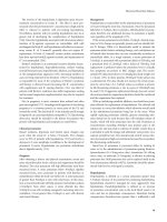
Treatment of Elevated Intracranial Pressure
Treatment Dose Advantages Limitations
Hypocarbia by
hyperventilation
pCO
2
25 to 33
mm Hg respira-
tory rate of 10 to
16/min
Immediate onset, well
tolerated
Hypotension, barotrauma,
duration usually hours or
less
Osmotic Mannitol 0.5 to 1
g/kg IV push
R api d onset,
titratable, predictable
H y pote n s i on, hypo-
kalemia, duration hours or
days
Barbiturates Pentobarbital 25
mg/kg slow IV
infusion over 3-4
hours
Mutes BP and respi-
ratory fluctuations
Hypotension, fixed pupils
(small), duration days
Hemicraniectomy Timing critical Large sustained ICP
reduction
Surgical risk, tissue
herniation through wound
III. Treatment of increased intracranial pressure
A. Positioning the patient in an upright position with the head of the bed at
30 degrees will lower ICP.
B. Hyperventilation is the most rapid and effective means of lowering ICP,
but the effects are short lived because the body quickly compensates.
The pCO
2
should be maintained between 25-33 mm Hg
C. Mannitol can quickly lower ICP, although the effect is not long lasting and
may lead to dehydration or electrolyte imbalance. Dosage is 0.5-1 gm/kg
(37.5-50 gm) IV q6h; keep osmolarity <315; do not give for more than
48h.
D. Corticosteroids are best used to treat increased ICP in the setting of
vasogenic edema caused by brain tumors or abscesses; however, these
agents have little value in the setting of stroke or head trauma. Dosage
is dexamethasone (Decadron) 10 mg IV or IM, followed by 4-6 mg IV, IM
or PO q6h.
E. Barbiturate coma is used for medically intractable ICP elevation when
other medical therapies have failed. There is a reduction in ICP by
decreasing cerebral metabolism. The pentobarbital loading dose is 25
mg/kg body weight over 3-4 hours, followed by 2-3 mg/kg/hr IV infusion.
Blood levels are periodically checked and adjusted to 30-40 mg/dL.
Patients require mechanical ventilation, intracranial pressure monitoring,
and continuous electroencephalographic monitoring.
F. Management of blood pressure. Beta-blockers or mixed beta and alpha
blockers provide the best antihypertensive effects without causing
significant cerebral vasodilatation that can lead to elevated ICP.
124 Status Epilepticus
Status Epilepticus
Status epilepticus (SE) is defined as a continuous seizures lasting at least 5
minutes, or 2 or more discrete seizures between which there is incomplete
recovery of consciousness. Simple seizures are characterized by focal motor or
sensory phenomena, with full preservation of consciousness. Generalized
seizures include generalized tonic-clonic seizures. Complex seizures are
diagnosed when an alteration in consciousness has occurred.
I. Diagnostic evaluation
A. Laboratory evaluation
1. CBC, blood glucose level, serum electrolytes (sodium, magnesium,
calcium), anticonvulsant drug levels, and urinalysis.
2. Lumbar puncture is necessary if meningitis or subarachnoid hemor-
rhage is suspected.
3. Toxicologic screening is indicated in specific situations.
B. CT scan is indicated if tumor, abscess, subarachnoid hemorrhage, or
trauma is suspected, or if the patient has no prior history of seizures.
C. Electroencephalogram. An immediate EEG may be required if the patient
fails to awaken promptly after the seizure.
Etiology of Status Epilepticus
Status epilepticus in a patient with a history of seizure disorder
• Noncompliance with prescribed medical regimen
• Withdrawal seizures from anticonvulsants
• Breakthrough seizures
New onset seizure disorder presenting with status epilepticus
Status epilepticus secondary to medical, toxicologic, or structural
symptoms
• Anoxic brain injury
• Stroke syndromes
• Subarachnoid hemorrhage
• Intracranial tumor
• Trauma
• Theophylline, cocaine, amphetamine or isoniazid overdose; alcohol
withdrawal, gamma hydroxybutyrate
• Hyponatremia, hypernatremia, hypercalcemia, hypomagnesemia,
hepatic encephalopathy
• Meningitis, brain abscess, encephalitis, CNS cysticercosis or
toxoplasmosis
II. Management of generalized convulsive status epilepticus (GCSE)
A. A history should be obtained, and a brief physical examination per-
formed. Initial stabilization consists of airway management, 100%
oxygen by mask, rapid glucose testing, intravenous access, and cardiac
and hemodynamic monitoring.
Status Epilepticus 125
B. Initial pharmacologic therapy
1. Thiamine 100 mg IV push and dextrose 50% water (D5W) 50 mL IV
push.
2. Lorazepam (Ativan) 0.1 mg/kg IV at 2 mg/min. The same dose may
be repeated once. Lorazepam may be given IM if the IV route is
unavailable.
3. Phenytoin maybe used when benzodiazepines are not effective. The
loading dose of phenytoin is 20 mg/kg IV, followed by 4-5 mg/kg/day
(100 mg IV q8h or 200 mg IV q12h); maximum rate for each dose is
50 mg/min in normal saline only. An additional loading dose of
phenytoin 10 mg/kg may be given if necessary.
4. Fosphenytoin (Cerebyx) is a water soluble prodrug of phenytoin. The
advantages of fosphenytoin are faster loading and greater ease of
administration. The dose of fosphenytoin is expressed in phenytoin
equivalents (PE). The loading dose is 20 mg PE/kg IV at 150 mg/min,
followed by 100 mg PE IV q8h. Fosphenytoin may be given IV or IM
in normal saline or D5W.
C. Refractory status epilepticus
1. Intubation should be accomplished and blood pressure support should
be maintained with fluids and pressor agents. EEG monitoring should
be initiated.
2. Midazolam (Versed) should be administered if seizures continue.
Loading dose is 0.2 mg/kg, followed by 0.045 mg/kg/hr. Titrate to 0.6
mg/kg/hr.
3. Propofol (Diprivan) may be used if midazolam (Versed) is ineffective.
Loading dose is 1-2 mg/kg, followed by 2 mg/kg/hr, titrate to 10
mg/kg/hr. Adjust dose to achieve seizure-free status on EEG
monitoring.
4. Phenobarbital may be administered as an alternative to anesthetics
if the patient is not hypoxemic or hyperthermic and seizure activity is
intermittent. The loading dose is 20 mg/kg at 75 mg/min, then 2 mg/kg
IV q12h.
References
Brott T, et al. Treatment of Acute Ischemic Stroke. N Engl J Med 2000; 343:710-722.
Lowenstein DH, et al. Status epilepticus. N Engl J Med 1998; 338:970-976.
126 Status Epilepticus
Diabetic Ketoacidosis 127
Endocrinologic and Nephrologic
Disorders
Michael Krutzik, MD
Guy Foster, MD
Diabetic Ketoacidosis
Diabetic ketoacidosis is defined by hyperglycemia, metabolic acidosis, and
ketosis.
I. Clinical presentation
A. Diabetes is newly diagnosed in 20% of cases of diabetic ketoacidosis. In
patients with known diabetes, precipitating factors include infection,
noncompliance with insulin, myocardial infarction, and gastrointestinal
bleeding.
B. Symptoms of DKA include polyuria, polydipsia, fatigue, nausea, and
vomiting, developing over 1 to 2 days. Abdominal pain is prominent in
25%.
C. Physical examination
1. Patients are typically flushed, tachycardic, tachypneic, and volume
depleted with dry mucous membranes. Kussmaul's respiration (rapid,
deep breathing and air hunger) occurs when the serum pH is between
7.0 and 7.24.
2. A fruity odor on the breath indicates the presence of acetone, a
byproduct of diabetic ketoacidosis.
3. Fever, although seldom present, indicates infection. Eighty percent of
patients with diabetic ketoacidosis have altered mental status. Most
are awake but confused; 10% are comatose.
D. Laboratory findings
1. Serum glucose level >300 mg/dL
2. pH <7.35, pCO
2 <40 mm Hg
3. Bicarbonate level below normal with an elevated anion gap
4. Presence of ketones in the serum
II. Differential diagnosis
A. Differential diagnosis of ketosis-causing conditions
1. Alcoholic ketoacidosis occurs with heavy drinking and vomiting. It
does not cause an elevated glucose.
2. Starvation ketosis occurs after 24 hours without food and is not
usually confused with DKA because glucose and serum pH are
normal.
B. Differential diagnosis of acidosis-causing conditions
1. Metabolic acidoses are divided into increased anion gap (>14
mEq/L) and normal anion gap; anion gap = sodium - (CI
- + HCO
3-
).
2. Anion gap acidoses can be caused by ketoacidoses, lactic acidosis,
uremia, salicylate, methanol, ethanol, or ethylene glycol poisoning.
3. Non-anion gap acidoses are associated with a normal glucose level
and absent serum ketones. Causes of non-anion gap acidoses
include renal or gastrointestinal bicarbonate loss.
128 Diabetic Ketoacidosis
C. Hyperglycemia caused by hyperosmolar nonketotic coma occurs in
patients with type 2 diabetes with severe hyperglycemia. Patients are
usually elderly and have a precipitating illness. Glucose level is markedly
elevated (>600 mg/dL), osmolarity is increased, and ketosis is minimal.
III. Treatment of diabetic ketoacidosis
A. Fluid resuscitation
1. Fluid deficits average 5 liters or 50 mL/kg. Resuscitation consists of
1 liter of normal saline over the first hour and a second liter over the
second and third hours. Thereafter, ½ normal saline should be infused
at 100-120 mL/hr.
2. When the glucose level decreases to 250 mg/dL, 5% dextrose should
be added to the replacement fluids to prevent hypoglycemia. If the
glucose level declines rapidly, 10% dextrose should be infused along
with regular insulin until the anion gap normalizes.
B. Insulin
1. An initial loading dose consists of 0.1 U/kg IV bolus. Insulin is then
infused at 0.1 U/kg per hour. The biologic half-life of IV insulin is less
than 20 minutes. The insulin infusion should be adjusted each hour so
that the glucose decline does not exceed 100 mg/dL per hour.
2. The insulin infusion rate may be decreased when the bicarbonate
level is greater than 20 mEq/L, the anion gap is less than 16 mEq/L,
or the glucose is <250 mg/dL.
C. Potassium
1. The most common preventable cause of death in patients with DKA
is hypokalemia. The typical deficit is between 300 and 500 mEq.
2. Potassium chloride should be started when fluid therapy is started. In
most patients, the initial rate of potassium replacement is 20 mEq/h,
but hypokalemia requires more aggressive replacement (40 mEq/h).
3. All patients should receive potassium replacement, except for those
with renal failure, no urine output, or an initial serum potassium level
greater than 6.0 mEq/L.
D. Sodium. For every 100 mg/dL that glucose is elevated, the sodium level
should be assumed to be higher than the measured value by 1.6 mEq/L.
E. Phosphate. Diabetic ketoacidosis depletes phosphate stores. Serum
phosphate level should be checked after 4 hours of treatment. If it is
below 1.5 mg/dL, potassium phosphate should be added to the IV
solution in place of KCl.
F. Bicarbonate therapy is not required unless the arterial pH value is <7.0.
For a pH of <7.0, add 50 mEq of sodium bicarbonate to the first liter of IV
fluid.
G. Magnesium. The usual magnesium deficit is 2-3 gm. If the patient's
magnesium level is less than 1.8 mEq/L or if tetany is present, magne-
sium sulfate is given as 5g in 500 mL of 0.45% normal saline over 5
hours.
H. Additional therapies
1. A nasogastric tube should be inserted in semiconscious patients to
protect against aspiration.
2. Deep vein thrombosis prophylaxis with subcutaneous heparin
should be provided for patients who are elderly, unconscious, or
severely hyperosmolar (5,000 U every 12 hours).
Diabetic Ketoacidosis 129
IV. Monitoring of therapy
130 Renal Failure
A. Serum bicarbonate level and anion gap should be monitored to
determine the effectiveness of insulin therapy.
B. Glucose levels should be checked at 1-2 hour intervals during IV insulin
administration.
C. Electrolyte levels should be assessed every 2 hours for the first 6-8
hours, and then q8h. Phosphorus and magnesium levels should be
checked after 4 hours of treatment.
D. Plasma and urine ketones are helpful in diagnosing diabetic
ketoacidosis, but are not necessary during therapy.
V. Determining the underlying cause
A. Infection is the underlying cause of diabetic ketoacidosis in 50% of
cases. Infection of the urinary tract, respiratory tract, skin, sinuses, ears,
or teeth should be sought. Fever is unusual in diabetic ketoacidosis and
indicates infection when present. If infection is suspected, antibiotics
should be promptly initiated.
B. Omission of insulin doses is often a precipitating factor. Myocardial
infarction, ischemic stroke, and abdominal catastrophes may precipitate
DKA.
VI. Initiation of subcutaneous insulin
A. When the serum bicarbonate and anion gap levels are normal, subcuta-
neous regular insulin can be started.
B. Intravenous and subcutaneous administration of insulin should overlap
to avoid redevelopment of ketoacidosis. The intravenous infusion may be
stopped 1 hour after the first subcutaneous injection of insulin.
C. Estimation of subcutaneous insulin requirements
1. Multiply the final insulin infusion rate times 24 hours. Two-thirds of the
total dose is given in the morning as two-thirds NPH and one-third
regular insulin. The remaining one-third of the total dose is given
before supper as one-half NPH and one-half regular insulin.
2. Subsequent doses should be adjusted according to the patient's blood
glucose response.
Acute Renal Failure
Acute renal failure is defined as a sudden decrease in renal function sufficient
to increase the concentration of nitrogenous wastes in the blood. It is character-
ized by an increasing BUN and creatinine.
I. Clinical presentation of acute renal failure
A. Oliguria is a common indicator of acute renal failure, and it is marked by
a decrease in urine output to less than 30 mL/h. Acute renal failure may be
oliguric (<500 L/day) or nonoliguric (>30 mL/h). Anuria (<100 mL/day) does
not usually occur in renal failure, and its presence suggests obstruction or
a vascular cause.
B. Acute renal failure may also be manifest by encephalopathy, volume
overload, pericarditis, bleeding, anemia, hyperkalemia, hyperphos-
phatemia, hypocalcemia, and metabolic acidemia.
II. Clinical causes of renal failure
A. Prerenal insult
1. Prerenal insult is the most common cause of acute renal failure,
accounting for 70% of cases. Prerenal failure is usually caused by
Acute Renal Failure 131
reduced renal perfusion secondary to extracellular fluid loss (diarrhea,
diuresis, GI hemorrhage) or secondary to extracellular fluid sequestra-
tion (pancreatitis, sepsis), inadequate cardiac output, renal
vasoconstriction (sepsis, liver disease, drugs), or inadequate fluid
intake or replacement.
2. Most patients with prerenal azotemia have oliguria, a history of large
fluid losses (vomiting, diarrhea, burns), and evidence of intravascular
volume depletion (thirst, weight loss, orthostatic hypotension, tachycar-
dia, flat neck veins, dry mucous membranes). Patients with congestive
heart failure may have total body volume excess (distended neck veins,
pulmonary and pedal edema) but still have compromised renal
perfusion and prerenal azotemia because of diminished cardiac output.
3. Causes of prerenal failure are usually reversible if recognized and
treated early; otherwise, prolonged renal hypoperfusion can lead to
acute tubular necrosis and permanent renal insufficiency.
B. Intrarenal insult
1. Acute tubular necrosis (ATN) is the most common intrinsic renal
disease leading to ARF.
a. Prolonged renal hypoperfusion is the most common cause of
ATN.
b. Nephrotoxic agents (aminoglycosides, heavy metals, radiocontrast
media, ethylene glycol) represent exogenous nephrotoxins. ATN
may also occur as a result of endogenous nephrotoxins, such as
intratubular pigments (hemoglobinuria), intratubular proteins
(myeloma), and intratubular crystals (uric acid).
2. Acute interstitial nephritis (AIN) is an allergic reaction secondary to
drugs (NSAIDs,
$-lactams).
3. Arteriolar injury occurs secondary to hypertension, vasculitis,
microangiopathic disorders.
4. Glomerulonephritis secondary to immunologically mediated inflamma-
tion may cause intrarenal damage.
C. Postrenal insult results from obstruction of urine flow. Postrenal insult is
the least common cause of acute renal failure, accounting for 10%.
Postrenal insult may be caused by obstruction secondary to prostate
cancer, benign prostatic hypertrophy, or renal calculi. Postrenal insult may
be caused by amyloidosis, uric acid crystals, multiple myeloma,
methotrexate, or acyclovir.
III. Clinical evaluation of acute renal failure
A. Initial evaluation of renal failure should determine whether the cause is
decreased renal perfusion, obstructed urine flow, or disorders of the renal
parenchyma. Volume status (orthostatic pulse, blood pressure, fluid intake
and output, daily weights, hemodynamic parameters), nephrotoxic
medications, and pattern of urine output should be assessed.
B. Prerenal azotemia is likely when there is a history of heart failure or
extracellular fluid volume loss or depletion.
C. Postrenal azotemia is suggested by a history of decreased size or force
of the urine stream, anuria, flank pain, hematuria or pyuria, or cancer of the
bladder, prostate or pelvis.
D. Intrarenal insult is suggested by a history of prolonged volume depletion
(often post-surgical), pigmenturia, hemolysis, rhabdomyolysis, or
nephrotoxins. Intrarenal insult is suggested by recent radiocontrast,
132 Acute Renal Failure
aminoglycoside use, or vascular catheterization. Interstitial nephritis may
be implicated by a history of medication rash, fever, or arthralgias.
E. Chronic renal failure is suggested by diabetes mellitus, normochromic
normocytic anemia, hypercalcemia, and hyperphosphatemia.
IV. Physical examination
A. Cardiac output, volume status, bladder size, and systemic disease mani-
festations should be assessed.
B. Prerenal azotemia is suggested by impaired cardiac output (neck vein
distention, pulmonary rales, pedal edema). Volume depletion is suggested
by orthostatic blood pressure changes, weight loss, low urine output, or
diuretic use.
C. Flank, suprapubic, or abdominal masses may indicate an obstructive
cause.
D. Skin rash suggests drug-induced interstitial nephritis; palpable purpura
suggests vasculitis; nonpalpable purpura suggests thrombotic
thrombocytopenic purpura or hemolytic-uremic syndrome.
E. Bladder catheterization is useful to rule out suspected bladder outlet
obstruction. A residual volume of more than 100 mL suggests bladder
outlet obstruction.
F. Central venous monitoring is used to measure cardiac output and left
ventricular filling pressure if prerenal failure is suspected.
V. Laboratory evaluation
A. Spot urine sodium concentration
1. Spot urine sodium can help distinguish between prerenal azotemia and
acute tubular necrosis.
2. Prerenal failure causes increased reabsorption of salt and water and
will manifest as a low spot urine sodium concentration <20 mEq/L and
a low fractional sodium excretion <1%, and a urine/plasma creatinine
ration of >40. Fractional excretion of sodium (%) = ([urine so-
dium/plasma sodium] ÷ [urine creatinine/plasma creatinine] x 100).
3. If tubular necrosis is the cause, the spot urine concentration will be >40
mEq/L, and fractional excretion of sodium will be >1%.
B. Urinalysis
1. Normal urine sediment is a strong indicator of prerenal azotemia or
may be an indicator of obstructive uropathy.
2. Hematuria, pyuria, or crystals may be associated with postrenal
obstructive azotemia.
3. Abundant cells, casts, or protein suggests an intrarenal disorder.
4. Red cells alone may indicate vascular disorders. RBC casts and
abundant protein suggest glomerular disease (glomerulonephritis).
5. White cell casts and eosinophilic casts indicate interstitial nephritis.
6. Renal epithelial cell casts and pigmented granular casts are
associated with acute tubular necrosis.
C. Ultrasound is useful for evaluation of suspected postrenal obstruction
(nephrolithiasis). The presence of small (<10 cm in length), scarred kid-
neys is diagnostic of chronic renal insufficiency.
VI. Management of acute renal failure
A. Reversible disorders, such as obstruction, should be excluded, and
hypovolemia should be corrected with volume replacement. Cardiac output
should be maintained. In critically ill patients, a pulmonary artery catheter
should be used for evaluation and monitoring.
Hyperkalemia 133
B. Extracellular fluid volume expansion. Infusion of a 1-2 liter crystalloid
fluid bolus may confirm suspected volume depletion.
C. If the patient remains oliguric despite euvolemia, IV diuretics may be
administered. A large single dose of furosemide (100-200 mg) may be
administered intravenously to promote diuresis. If urine flow is not
improved, the dose of furosemide may be doubled. Furosemide may be
repeated in 2 hours, or a continuous IV infusion of 10-40 mg/hr (max 1000
mg/day) may be used.
D. The dosage or dosing intervals of renally excreted drugs should be
modified.
E. Hyperkalemia is the most immediately life-threatening complication of
renal failure. Serum potassium values greater than 6.5 mEq/L may lead to
arrhythmias and cardiac arrest. Potassium should be removed from IV
solutions. Hyperkalemia may be treated with sodium polystyrene sulfonate
(Kayexalate), 30-60 gm PO/PR every 4-6 hours.
F. Hyperphosphatemia can be controlled with aluminum hydroxide antacids
(eg, Amphojel or Basaljel), 15-30 ml or one to three capsules PO with
meals, should be used.
G. Fluids. After normal volume has been restored, fluid intake should be
reduced to an amount equal to urinary and other losses plus insensible
losses of 300-500 mL/day. In oliguric patients, daily fluid intake may need
to be restricted to less than 1 L.
H. Nutritional therapy. A renal diet consisting of daily high biologic value
protein intake of 0.5 gm/kg/d, sodium 2 g, potassium 40-60 mg/day, and
at least 35 kcal/kg of nonprotein calories is recommended. Phosphorus
should be restricted to 800 mg/day
I. Dialysis. Indications for dialysis include uremic pericarditis, severe
hyperkalemia, pulmonary edema, persistent severe metabolic acidosis (pH
less than 7.2), and symptomatic uremia.
Hyperkalemia
Body potassium is 98% intracellular. Only 2% of total body potassium, about 70
mEq, is in the extracellular fluid, with the normal concentration of 3.5-5 mEq/L.
I. Pathophysiology of potassium homeostasis
A. The normal upper limit of plasma K is 5-5.5 mEq/L, with a mean K level
of 4.3.
B. External potassium balance. Normal dietary K intake is 1-1.5 mEq/kg
in the form of vegetables and meats. The kidney is the primary organ for
preserving external K balance, excreting 90% of the daily K burden.
C. Internal potassium balance. Potassium transfer to and from tissues, is
affected by insulin, acid-base status, catecholamines, aldosterone,
plasma osmolality, cellular necrosis, and glucagon.
II. Clinical disorders of external potassium balance
A. Chronic renal failure. The kidney is able to excrete the dietary intake of
potassium until the glomerular filtration rate falls below 10 cc/minute or
until urine output falls below 1 L/day. Renal failure is advanced before
hyperkalemia occurs.
134 Hyperkalemia
B. Impaired renal tubular function. Renal diseases may cause
hyperkalemia, and the renal tubular acidosis caused by these conditions
may worsen hyperkalemia.
C. Primary adrenal insufficiency (Addison's disease) is now a rare cause
of hyperkalemia. Diagnosis is indicated by the combination of
hyperkalemia and hyponatremia and is confirmed by a low aldosterone
and a low plasma cortisol level that does not respond to adreno-
corticotropic hormone treatment.
D. Drugs that may cause hyperkalemia include nonsteroidal anti-inflamma-
tory drugs, angiotensin-converting enzyme inhibitors, cyclosporine, and
potassium-sparing diuretics. Hyperkalemia is especially common when
these drugs are given to patients at risk for hyperkalemia (diabetics, renal
failure, advanced age).
E. Excessive potassium intake
1. Long-term potassium supplementation results in hyperkalemia most
often when an underlying impairment in renal excretion already exists.
2. Intravenous administration of 0.5 mEq/kg over 1 hour increases serum
levels by 0.6 mEq/L. Hyperkalemia often results when infusions of
greater than 40 mEq/hour are given.
III. Clinical disorders of internal potassium balance
A. Diabetic patients are at particular risk for severe hyperkalemia because
of renal insufficiency and hyporeninemic hypoaldosteronism.
B. Systemic acidosis reduces renal excretion of potassium and moves
potassium out of cells, resulting in hyperkalemia.
C. Endogenous potassium release from muscle injury, tumor lysis, or
chemotherapy may elevate serum potassium.
IV. Manifestations of hyperkalemia
A. Hyperkalemia, unless severe, is usually asymptomatic. The effect of
hyperkalemia on the heart becomes significant above 6 mEq/L. As levels
increase, the initial ECG change is tall peaked T waves. The QT interval
is normal or diminished.
B. As K levels rise further, the PR interval becomes prolonged, then the P
wave amplitude decreases. The QRS complex eventually widens into a
sine wave pattern, with subsequent cardiac standstill.
C. At serum K is >7 mEq/L, muscle weakness may lead to a flaccid
paralysis. Sensory abnormalities, impaired speech and respiratory arrest
may follow.
V. Pseudohyperkalemia
A. Potassium may be falsely elevated by hemolysis during phlebotomy,
when K is released from ischemic muscle distal to a tourniquet, and
because of erythrocyte fragility disorders.
B. Falsely high laboratory measurement of serum potassium may occur with
markedly elevated platelet counts (>10
6
platelet/mm
3
) or white blood cell
counts (>50,000/mm
3
).
VI. Diagnostic approach to hyperkalemia
A. The serum K level should be repeat tested to rule out laboratory error. If
significant thrombocytosis or leukocytosis is present, a plasma potassium
level should be determined.
B. The 24-hour urine output, urinary K excretion, blood urea nitrogen, and
serum creatinine should be measured. Renal K retention is diagnosed
when urinary K excretion is less than 20 mEq/day.
Hyperkalemia 135
C. High urinary K, excretion of >20 mEq/day, is indicative of excessive K
intake as the cause.
VII. Renal hyperkalemia
A. If urinary K excretion is low and urine output is in the oliguric range, and
creatinine clearance is lower than 20 cc/minute, renal failure is the
probable cause. Prerenal azotemia resulting from volume depletion must
be ruled out because the hyperkalemia will respond to volume restoration.
B. When urinary K excretion is low, yet blood urea nitrogen and creatinine
levels are not elevated and urine volume is at least 1 L daily and renal
sodium excretion is adequate (about 20 mEq/day), then either a defect in
the secretion of renin or aldosterone or tubular resistance to aldosterone
is likely. Low plasma renin and aldosterone levels, will confirm the
diagnosis of hyporeninemic hypoaldosteronism. Addison's disease is
suggested by a low serum cortisol, and the diagnosis is confirmed with a
ACTH (Cortrosyn) stimulation test.
C. When inadequate K excretion is not caused by hypoaldosteronism, a
tubular defect in K clearance is suggested. Urinary tract obstruction, renal
transplant, lupus, or a medication should be considered.
VIII. Extrarenal hyperkalemia
A. When hyperkalemia occurs along with high urinary K excretion of >20
mEq/day, excessive intake of K is the cause. Potassium excess in IV
fluids, diet, or medication should be sought. A concomitant underlying
renal defect in K excretion is also likely to be present.
B. Blood sugar should be measured to rule out insulin deficiency; blood pH
and serum bicarbonate should be measured to rule out acidosis.
C. Endogenous sources of K, such as tissue necrosis, hypercatabolism,
hematoma, gastrointestinal bleeding, or intravascular hemolysis should
be excluded.
IX. Management of hyperkalemia
A. Acute treatment of hyperkalemia
1. Calcium
a. If the electrocardiogram shows loss of P waves or widening of QRS
complexes, calcium should be given IV; calcium reduces the cell
membrane threshold potential.
b. Calcium chloride (10%) 2-3 g should be given over 5 minutes. In
patients with circulatory compromise, 1 g of calcium chloride IV
should be given over 3 minutes.
c. If the serum K level is greater than 7 mEq/L, calcium should be
given. If digitalis intoxication is suspected, calcium must be given
cautiously. Coexisting hyponatremia should be treated with
hypertonic saline.
2. Insulin: If the only ECG abnormalities are peaked T waves and the
serum level is under 7 mEq/L, treatment should begin with insulin
(regular insulin, 5-10 U by IV push) with 50% dextrose water (D50W)
50 mL IV push. Repeated insulin doses of 10 U and glucose can be
given every 15 minutes for maximal effect.
3. Sodium bicarbonate promotes cellular uptake of K. It should be given
as 1-2 vials (50-mEq/vials) IV push.
4. Potassium elimination measures
a. Sodium polystyrene sulfonate (Kayexalate) is a cation exchange
resin which binds to potassium in the lower GI tract. Dosage is 30-
60 gm premixed with sorbitol 20% PO/PR.
136 Hypokalemia
b. Furosemide (Lasix) 100 mg IV should be given to promote
kaliuresis.
c. Emergent hemodialysis for hyperkalemia is rarely necessary except
when refractory metabolic acidosis is present.
Hypokalemia
Hypokalemia is characterized by a serum potassium concentration of less than
3.5 mEq/L. Ninety-eight percent of K is intracellular.
I. Pathophysiology of hypokalemia
A. Cellular redistribution of potassium. Hypokalemia may result from the
intracellular shift of potassium by insulin, beta-2 agonist drugs, stress
induced catecholamine release, thyrotoxic periodic paralysis, and
alkalosis-induced shift (metabolic or respiratory).
B. Nonrenal potassium loss
1. Gastrointestinal loss can be caused by diarrhea, laxative abuse,
villous adenoma, biliary drainage, enteric fistula, clay ingestion,
potassium binding resin ingestion, or nasogastric suction.
2. Sweating, prolonged low-potassium diet, hemodialysis and peritoneal
dialysis may also cause nonrenal potassium loss.
C. Renal potassium loss
1. Hypertensive high renin states. Malignant hypertension, renal artery
stenosis, renin-producing tumors.
2. Hypertensive low renin, high aldosterone states. Primary
hyperaldosteronism (adenoma or hyperplasia).
3. Hypertensive low renin, low aldosterone states. Congenital adrenal
hyperplasia (11 or 17 hydroxylase deficiency), Cushing's syndrome or
disease, exogenous mineralocorticoids (Florinef, licorice, chewing
tobacco), Liddle's syndrome.
4. Normotensive states
a. Metabolic acidosis. Renal tubular acidosis (type I or II)
b. Metabolic alkalosis (urine chloride <10 mEq/day). Vomiting
c. Metabolic alkalosis (urine chloride >10 mEq/day). Bartter's
syndrome, diuretics, magnesium depletion, normotensive hyper-
aldosteronism
5. Drugs associated with potassium loss include amphotericin B,
ticarcillin, piperacillin, and loop diuretics.
II. Clinical effects of hypokalemia
A. Cardiac effects. The most lethal consequence of hypokalemia is cardiac
arrhythmia. Electrocardiographic effects include a depressed ST seg-
ment, decreased T-wave amplitude, U waves, and a prolonged QT-U
interval.
B. Musculoskeletal effects. The initial manifestation of K depletion is
muscle weakness, which can lead to paralysis. In severe cases,
respiratory muscle paralysis may occur.
C. Gastrointestinal effects. Nausea, vomiting, constipation, and paralytic
ileus may develop.
III. Diagnostic evaluation
Hypermagnesemia 137
A. The 24-hour urinary potassium excretion should be measured. If >20
mEq/day, excessive urinary K loss is the cause. If <20 mEq/d, low K
intake, or non-urinary K loss is the cause.
B. In patients with excessive renal K loss and hypertension, plasma renin
and aldosterone should be measured to differentiate adrenal from non-
adrenal causes of hyperaldosteronism.
C. If hypertension is absent and serum pH is acidotic, renal tubular acidosis
should be considered. If hypertension is absent and serum pH is normal
to alkalotic, a high urine chloride (>10 mEq/d) suggests hypokalemia
secondary to diuretics or Bartter's syndrome. A low urine chloride (<10
mEq/d) suggests vomiting.
IV. Emergency treatment of hypokalemia
A. Indications for urgent replacement. Electrocardiographic abnormalities,
myocardial infarction, hypoxia, digitalis intoxication, marked muscle
weakness, or respiratory muscle paralysis.
B. Intravenous potassium therapy
1. Intravenous KCL is usually used unless concomitant hypo-
phosphatemia is present, where potassium phosphate is indicated.
2. The maximal rate of intravenous K replacement is 30 mEq/hour. The
K concentration of IV fluids should be 80 mEq/L or less if given via a
peripheral vein. Frequent monitoring of serum K and constant
electrocardiographic monitoring is recommended when potassium
levels are being replaced.
V. Non-emergent treatment of hypokalemia
A. Attempts should be made to normalize K levels if <3.5 mEq/L.
B. Oral supplementation is significantlysafer than IV. Liquid formulations are
preferred due to rapid oral absorption, compared to sustained release
formulations, which are absorbed over several hours.
1. KCL elixir 20-40 mEq qd-tid PO after meals.
2. Micro-K, 10 mEq tabs, 2-3 tabs tid PO after meals (40-100 mEq/d).
Hypomagnesemia
Magnesium deficiency occurs in up to 11% of hospitalized patients. The normal
range of serum magnesium is 1.5 to 2.0 mEq/L, which is maintained by the
kidney, intestine, and bone.
I. Pathophysiology
A. Decreased magnesium intake. Protein-calorie malnutrition, prolonged
parenteral fluid administration, and catabolic illness are common causes
of hypomagnesemia.
B. Gastrointestinal losses of magnesium may result from prolonged
nasogastric suction, laxative abuse, and pancreatitis.
C. Renal losses of magnesium
1. Renal loss of magnesium may occur secondary to renal tubular
acidosis, glomerulonephritis, interstitial nephritis, or acute tubular
necrosis.
2. Hyperthyroidism, hypercalcemia, and hypophosphatemia may cause
magnesium loss.