Dermatology therapy essentials - part 5 ppsx
Bạn đang xem bản rút gọn của tài liệu. Xem và tải ngay bản đầy đủ của tài liệu tại đây (2.9 MB, 64 trang )
Glanders and melioidosis 257
G
Differential diagnosis
Condyloma acuminata; squamous cell car-
cinoma
Therapy
Surgical excision
ଙ
; interferon
References
Kanik AB, Lee J, Wax F, Bhawan J (1997) Penile
verrucous carcinoma in a 37-year-old circum-
cised man. Journal of the American Academy of
Dermatology 37(2 Pt 2):329–331
Giant condylomata
acuminata of Buschke and
Löwenstein
᭤
Giant condyloma of Buschke and
Löwenstein
Giant follicle
᭤
Dilated pore
Giant hemangioma
syndrome
᭤
Kasabach-Merritt syndrome
Giant malignant condyloma
᭤
Giant condyloma of Buschke and
Löwenstein
Gingivitis, desquamative
᭤
Desquamative gingivitis
Glanders and melioidosis
Synonym(s)
Farcy
;
morve
;
malleus
(glanders);
Whitmore disease
(melioidosis)
Definition
Related diseases produced by bacteria of the
Burkholderia
species, which are gram-nega-
tive rods
Pathogenesis
Causative agent of Glanders: Burkholderia
mallei; primarily a disease of animals such
as horses, mules, and donkeys; once in the
host, synthesis and release of certain toxins
occur; melioidosis: caused by the bacte-
rium Burkholderia pseudomallei; organism
distributed widely in the soil and water of
the tropics and spread to humans through
direct contact with a contaminated source
Clinical manifestation
Similar clinical syndrome in both diseases.
Localized form: bacteria enter the skin
through a laceration or abrasion; local
infection with ulceration and regional lym-
phadenopathy; incubation period 1–5 days;
bacteria that enter the host through
mucous membranes sometimes cause
increased mucus production in the affected
areas
Pulmonary form: occurs when bacteria are
aerosolized and enter respiratory tract via
inhalation or hematogenous spread; with
inhalational melioidosis, cutaneous
abscesses may develop; septicemia: when
bacteria disseminated in the bloodstream
in glanders, usually fatal within 7–10 days
Chronic form: multiple abscesses affecting
the liver, spleen, skin, or muscles
Differential diagnosis
Anthrax; plague; tuberculosis; atypical
mycobacterial infection; brucellosis; North
American blastomycosis; coccidioidomyco-
sis; nocardia infection
PART7.MIF Page 257 Friday, October 31, 2003 10:22 AM
258 Glomangioma
Therapy
Amoxicillin; tetracycline
References
Rosenbloom M, Leikin JB, Vogel SN, Chaudry ZA
(2002) Biological and chemical agents: a brief
synopsis. American Journal of Therapeutics
9(1):5–14
Glomangioma
᭤
Glomus tumor
Glomus tumor
Synonym(s)
Glomangioma
Definition
Benign neoplasm of modified smooth mus-
cle cells (glomus cells)
Pathogenesis
Unknown cause for solitary lesion; multi-
ple glomus tumors, especially those of the
disseminated form, inherited as autosomal-
dominant trait with incomplete pene-
trance; tumors arise from the arterial por-
tion of the glomus body
Clinical manifestation
Solitary glomus tumor: paroxysmal pain,
which can be severe and exacerbated by
pressure or temperature changes, especially
cold; blanchable blue or purple papule,
located most commonly in acral areas,
especially subungual areas of fingers and
toes
Multiple glomus tumors: pain relatively
uncommon
•
Regional variant: blue-to-purple, com-
pressible papules or nodules that are
grouped and limited to a specific area, most
commonly an extremity
•
Disseminated variant: multiple lesions
distributed over the body with no specific
grouping; congenital plaquelike glomus
tumors: grouped papules coalescing into
indurated plaques or clusters of discrete
nodules
Differential diagnosis
Angioleiomyoma; angiolipoma; arteriov-
enous malformation; blue nevus; hemangi-
oma; melanoma; spiradenoma; tufted angi-
oma; Kaposi’s sarcoma; blue rubber bleb
nevus; neurilemmoma
Therapy
Solitary glomus tumor: surgical excision
ଙ
;
multiple glomus tumors: surgical removal
for cosmetic reasons only
References
Alam M, Scher RK (1999) Current topics in nail
surgery. Journal of Cutaneous Medicine & Sur-
gery 3(6):324–335
Parsons ME, Russo G, Fucich L, Millikan LE, Kim
R (1997) Multiple glomus tumors. International
Journal of Dermatology 36(12):894-900
Glossodynia
Definition
Painful sensation in the tongue
References
Marbach JJ (1999) Medically unexplained chronic
orofacial pain. Temporomandibular pain and
dysfunction syndrome, orofacial phantom
pain, burning mouth syndrome, and trigemi-
nal neuralgia. Medical Clinics of North Ameri-
ca 83(3):691–710
Glucagonoma syndrome
Synonym(s)
Necrolytic migratory erythema
Definition
Glucagon-secreting tumor associated with
hyperglucagonemia, necrolytic migratory
PART7.MIF Page 258 Friday, October 31, 2003 10:22 AM
Goltz-Gorlin syndrome 259
G
erythema, and diabetes mellitus; hypoami-
noacidemia; cheilosis; normochromic, nor-
mocytic anemia; venous thrombosis;
weight loss; neuropsychiatric signs and
symptoms; pseudoglucagonoma syndrome:
necrolytic migratory erythema without a
glucogon-secreting tumor, but with another
underlying cause such as cirrhosis, celiac
sprue, or pancreatitis
Pathogenesis
Unclear relation between glucagonoma and
skin findings; levels of glucagon not well
correlated with the episodic course of the
skin manifestations; possible role of rela-
tive zinc deficiency; theories of causation:
related to glucagon-induced hypoalbumine-
mia; zinc-dependent delta-6 desaturation of
linoleic acid; poor hepatic breakdown of
glucagon contributing to an excessive pros-
taglandin-mediated inflammatory response
Clinical manifestation
Presents with nonspecific complaints, such
as weight loss, diabetes mellitus, diarrhea,
and stomatitis; necrolytic migratory ery-
thema: found anywhere on the body, but
most common in the perineum, buttocks,
groin, lower abdomen, and lower extremi-
ties; eruption starts as a pruritic or painful,
erythematous patch that blisters centrally,
erodes, crusts over, and heals with hyper-
pigmentation; annular lesions with conflu-
ence into plaques; confluence in severely
affected areas; associated mucocutaneous
findings, including atrophic glossitis,
cheilosis, dystrophic nails, and buccal
mucosal inflammation
Differential diagnosis
Acrodermatitis enteropathica; candidiasis;
paraneoplastic pemphigus; Hailey-Hailey
disease; Darier disease; pellagra; kwash-
iorkor
Therapy
Surgical resection of the tumor, if local-
ized
ଙ
; in the absence of tumor, treat under-
lying cause
ଙ
References
Chastain MA (2001) The glucagonoma syndrome:
a review of its features and discussion of new
perspectives. American Journal of the Medical
Sciences 321(5):306–320
Glucosyl cerebroside
lipidosis
᭤ Gaucher’s disease
Glucosylceramide lipidosis
᭤ Gaucher’s disease
Glycolic acid
᭤ Alpha hydroxy acid
Glyderm plus
᭤ Alpha hydroxy acids
Goltz syndrome
᭤ Focal dermal hypoplasia
Goltz-Gorlin syndrome
᭤ Focal dermal hypoplasia
PART7.MIF Page 259 Friday, October 31, 2003 10:22 AM
260 Goltz's syndrome
Goltz's syndrome
᭤ Focal dermal hypoplasia
Gonadal dysgenesis
᭤ Turner syndrome
Gonococcal dermatitis-
arthritis syndrome
᭤ Gonococcemia
Gonococcemia
Synonym(s)
Gonococcal dermatitis-arthritis syndrome;
disseminated gonococcal infection
Definition
Sexually transmitted disease caused by the
bacterium Neisseria gonorrhoeae, which
spreads from the initial site of infection
through the bloodstream to other parts of
the body
Pathogenesis
Neisseria gonorrhoeae transmitted through
vaginal, oral, and anal intercourse; infec-
tion also transmitted by a woman to her
newborn during childbirth; dissemination
often occurs during menses
Clinical manifestation
More common in women, often with
asymptomatic infection; disseminated dis-
ease generally follows the primary genital
infection by several days to 2 weeks; fever;
myalgias; tenosynovitis; monoarticular sep-
tic arthritis, affecting large, weight-bearing
joints; acral palpable purpuric papules and
pustules, usually relatively few in number
Differential diagnosis
Meningococcemia or other infectious
causes of septic vasculitis; lupus erythema-
tosus; cryoglobulinemia; Reiter syndrome;
infective endocarditis
Therapy
Ceftriaxone 1 gm intramuscularly or intra-
venously every 24 hours for 3 days or until
24 hours after symptomatic improvement;
complete 7-day course with ciprofloxacin
500 mg PO twice daily or cefixime 400 mg
PO twice daily or azithromycin 500 mg PO
per day
ଙ
; concurrent therapy for presumed
chlamydia with doxycycline 100 mg PO
twice daily for 7 days
References
Brown TJ, Yen-Moore A, Tyring SK (1999) An
overview of sexually transmitted diseases. Part
I. Journal of the American Academy of Derma-
tology 41(4):511–532
Gorlin syndrome
᭤ Basal cell nevus syndrome
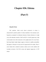
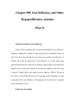
Gonococcemia. Violaceous papule on the toe
PART7.MIF Page 260 Friday, October 31, 2003 10:22 AM
Graft versus host disease 261
G
Gorlin-Goltz syndrome
᭤ Basal cell nevus syndrome
Gottron’s syndrome
᭤ Acrogeria
Gougerot and Blum,
lichenoid pigmented
purpura
᭤ Benign pigmented purpura
Gougerot-Carteaud
papillomatosis
᭤ Confluent and reticulated papillo-
matosis
Gougerot-Houwer-Sjögren
syndrome
᭤ Sjögren syndrome
Gowers’ local panatrophy
᭤ Panatrophy of Gowers
Gowers’ panatrophy
᭤ Panatrophy of Gowers
Graft versus host disease
Synonym(s)
Definition
Immunologic assault and its consequences
when immunologically competent cells are
introduced into an immunoincompetent
host
Pathogenesis
Three criteria for development – (1) graft
containing immunologically competent
cells, (2) host appearing foreign to the graft,
(3) host incapable of reacting sufficiently
against the graft; recognition of epithelial
target tissues as foreign by the immuno-
competent cells, with subsequent induction
of an inflammatory response and eventual
apoptotic death of the target tissue; reac-
tion against the host's keratinocytes, result-
ing in the clinical syndrome
Clinical manifestation
Incidence higher in recipients of allogeneic
hematopoietic cells than in patients receiv-
ing syngeneic or autologous hematopoietic
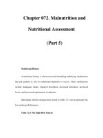
Graft versus host disease. Sclerotic,
hyperpigmented and hypopigmented plaques
on the upper trunk
PART7.MIF Page 261 Friday, October 31, 2003 10:22 AM
262 Granular bacteriosis
cells; greatest incidence in patients in whom
bone marrow is used as the source of
hematopoietic cells
Acute graft versus host disease: observed
10–30 days posttransplant; eruptions usu-
ally begin as faint, tender, erythematous
macules, often centered around hair folli-
cles; as disease progresses, macules some-
times coalesce to form confluent plaques or
papules; subepidermal bullae may occur
Chronic graft versus host disease: evolves
from acute form in 70–90% of patients; risk
increases with the severity of acute reac-
tion; violaceous lichenified papules, often
on the ventral skin surfaces, very similar to
those of lichen planus; lacy white plaques
on the buccal mucosa; scattered scleroder-
matous plaques; widespread disease result-
ing in ulcerations, joint contractures, and
esophageal dysmotility
Differential diagnosis
Acute graft versus host disease: erythema
multiforme; drug eruption; Stevens-John-
son syndrome/toxic epidermal necrolysis;
eruption of lymphocyte recovery
Chronic graft versus host disease: sclero-
derma; lichen planus; lichenoid drug erup-
tion; lupus erythematosus
Therapy
Acute graft versus host disease: pred-
nisone; extracorporeal photochemotherapy
Chronic graft versus host disease: photo-
chemotherapy; methotrexate; extracorpor-
eal photochemotherapy; hydroxychloro-
quine; etretinate
References
Jacobsohn DA, Vogelsang GB (2002) Novel phar-
macotherapeutic approaches to prevention and
treatment of GVHD. Drugs 62(6):879–889
Granular bacteriosis
᭤ Botryomycosis
Granular cell myoblastoma
᭤ Granular cell tumor
Granular cell neurofibroma
᭤ Granular cell tumor
Granular cell neuroma
᭤ Granular cell tumor
Granular cell schwannoma
᭤ Granular cell tumor
Granular cell tumor
Synonym(s)
Granular cell myoblastoma; granular cell
schwannoma; granular cell neuroma;
granular cell neurofibroma; Abrikossof’s
tumor
Definition
Acquired tumor of neural crest origin, char-
acterized by cells with eosinophilic cyto-
plasmic granules
Pathogenesis
Possible tumor derivation from Schwann
cells
Clinical manifestation
Discrete, asymptomatic, firm, flesh-
colored nodule, located within or beneath
the dermis, occurring in the tongue, head,
PART7.MIF Page 262 Friday, October 31, 2003 10:22 AM
Granuloma faciale 263
G
and neck region or dorsal aspect of the
forearms
Differential diagnosis
Fibroma; squamous cell carcinoma; wart;
dermatofibroma; neurofibroma; epider-
moid cyst
Therapy
Surgical excision
ଙ
References
Becelli R, Perugini M, Gasparini G, Cassoni A, Fa-
biani F (2001) Abrikossoff's tumor. Journal of
Craniofacial Surgery 12(1):78–81
Granuloma, actinic
᭤ Actinic granuloma
Granuloma annulare
Synonym(s)
None
Definition
Inflammatory skin disease characterized by
annular plaques consisting of small papules
Pathogenesis
May involve immune mechanisms
Clinical manifestation
Localized variant: flesh-colored to dull red
papules, often in an annular arrangement,
over distal extremities; often occur over
dorsal surfaces of feet, hands and fingers,
and the extensor aspects of arms and legs
Generalized variant: few to thousands of
flesh-colored to dull red papules involving
multiple body regions, often in symmetri-
cal distribution; papules may coalesce into
annular or arcuate plaques; may have large
red patches (vascular granuloma annulare)
Subcutaneous variant: firm, nontender,
flesh-colored-to-pinkish papules or nod-
ules without overlying epidermal altera-
tion, often over the lower extremity
Differential diagnosis
Erythema annulare centrifugum; tinea cor-
poris; lichen planus; lupus erythematosus;
insect bite reaction; sarcoidosis; Lyme dis-
ease; necrobiosis lipoidica; rheumatoid
nodules; acquired perforating disease;
lichen myxedematosus; cutaneous T-cell
lymphoma; erythema multiforme
Therapy
Localized disease: intralesional triamci-
nolone; corticosteroids, topical, superpo-
tent
Generalized disease: photochemotherapy
References
Smith MD, Downie JB, DiCostanzo D (1997)
Granuloma annulare. International Journal of
Dermatology 36(5):326–333
Granuloma faciale
Synonym(s)
Facial granuloma; granuloma faciale eosi-
nophilicum, granuloma faciale with eosi-
nophilia
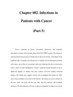
Granuloma annulare. Annular red-brown
plaques on the dorsal aspect of the hand
PART7.MIF Page 263 Friday, October 31, 2003 10:22 AM
264 Granuloma faciale eosinophilicum
Definition
Benign chronic skin disease of unknown
origin, characterized by single or multiple
cutaneous nodules, usually occurring over
the face
Pathogenesis
Sun exposure possible factor in develop-
ment
Clinical manifestation
Solitary or multiple, sharply marginated,
red or violaceous papules or nodules; sur-
face sometimes has telangiectasias and/or
enlarged follicular orifices; usually occurs
on the face, but also on the upper extremi-
ties or trunk
Differential diagnosis
Sarcoidosis, granuloma annulare; discoid
lupus erythematosus; mycosis fungoides;
fixed drug eruption; Jessner’s lymphocytic
infiltrate; lymphoma; leprosy; lupus vul-
garis; foreign body granuloma
Therapy
Triamcinolone 3–4 mg per ml intralesional;
dapsone
References
Inanir I, Alvur Y (2001) Granuloma faciale with
extrafacial lesions. British Journal of Dermatol-
ogy 145(2):360–362
Granuloma faciale
eosinophilicum
᭤ Granuloma faciale
Granuloma faciale with
eosinophilia
᭤ Granuloma faciale
Granuloma fissuratum
᭤ Acanthoma fissuratum
Granuloma gluteale
infantum
Synonym(s)
Kaposi’s sarcoma-like granuloma;
granuloma intertriginosum infantum;
infantile vegetating halogenosis; vegetating
potassium bromide toxic dermatitis;
vegetating bromidism
Definition
Disease characterized by oval, granuloma-
tous nodules on the gluteal surfaces and
groin areas of infants
Pathogenesis
Unclear; unusual cutaneous response to
local inflammation, maceration, and sec-
ondary infection; contact occlusion proba-
bly predisposing factor
Clinical manifestation
Solitary or mulptiple, red-purple to red-
brown, firm-to-hard, discrete dermal nod-
ules with smooth or slightly lichenified sur-
faces; aligned with the long axis parallel to
the skin folds; located on the gluteal sur-
faces, in the groin area, upper thighs, lower
abdomen, or rarely the neck and face
Differential diagnosis
Langerhans cell histiocytosis; candidiasis;
contact dermatitis; lymphoma; mastocyto-
sis; scabies; syphilis; juvenile xanthogranu-
loma; pyogenic granuloma; sarcoma; for-
eign body granuloma
Therapy
Spontaneous resolution; no therapy indi-
cated
PART7.MIF Page 264 Friday, October 31, 2003 10:22 AM
Granulomatosis disciformis chronica et progressiva 265
G
References
Bluestein J, Furner BB, Phillips D (1990) Granulo-
ma gluteale infantum: case report and review of
the literature. Pediatric Dermatology 7(3):196–
198
Granuloma inguinale
Synonym(s)
Donovanosis
Definition
Sexually transmitted disease characterized
by genital lesions presenting as indolent,
progressive ulcerations with a granuloma-
tous appearance
Pathogenesis
Infection caused by a gram-negative pleo-
morphic bacillus, Calymmatobacterium
granulomatis; mode of transmission prima-
rily through sexual contact; mildly conta-
gious
Clinical manifestation
Occurs on glans penis and scrotum in men,
and labia minora, mons veneris, and four-
chette in women; rare cervical involve-
ment; soft, red papules or nodules arising at
the site of inoculation; lesions eventually
ulcerate and produce red, friable, granulo-
matous plaques and nodules; ulcers with
clean, friable bases and distinct, raised,
rolled margins; autoinoculation results in
lesions on adjacent skin; occasional hyper-
trophic or verrucous plaques, with forma-
tion of large, vegetating masses resembling
genital warts; swelling of the external geni-
talia in later-stage lesions
Differential diagnosis
Syphilis; lymphogranuloma venereum;
chronic herpes simplex virus infection;
squamous cell carcinoma; lichen sclerosus
Therapy
Trimethoprim/sulfamethoxazole; doxycy-
cline
References
Brown TJ, Yen-Moore A, Tyring SK (1999) An
overview of sexually transmitted diseases. Part
1. Journal of the American Academy of Derma-
tology 41(4):511–532
Granuloma intertriginosum
infantum
᭤ Granuloma gluteale infantum
Granuloma pyogenicum
᭤ Pyogenic granuloma
Granuloma
telangiectaticum
᭤ Pyogenic granuloma
Granuloma trichophyticum
᭤ Majocchi granuloma
Granuloma tricofitico
᭤ Majocchi granuloma
Granulomatosis disciformis
chronica et progressiva
᭤ Actinic granuloma
PART7.MIF Page 265 Friday, October 31, 2003 10:22 AM
266 Granulomatosis, lymphomatoid
Granulomatosis,
lymphomatoid
᭤ Lymphomatoid granulomatosis
Granulomatosis, Miescher’s
᭤ Miescher's granulomatosis
Granulomatous arteritis
᭤ Temporal arteritis
Granulomatous cheilitis
᭤ Cheilitis granulomatosa
Granulomatous disease of
childhood
᭤ Chronic granulomatous disease
Granulomatous perioral
dermatitis
᭤ Perioral dermatitis
Granulomatous rosacea
᭤ Rosacea
Granulomatous vasculitis
᭤ Wegener’s granulomatosis
Granulomatous vasculitis
with asthma
᭤ Churg-Strauss syndrome
Griscelli syndrome
Synonym(s)
Partial albinism with immunodeficiency
Definition
Disease characterized by partial pigmen-
tary dilution with silvery gray hair, fre-
quent infections, cellular immune defi-
ciency, neurologic abnormalities, and fatal
outcome from an uncontrolled T lym-
phocyte and macrophage activation syn-
drome
Pathogenesis
Caused by two genes: MYA5 and RAB27A;
gene MYA5 produces severe neurological
problems; gene RAB27A causes accelerated
phase sometimes lethal within a short
period of time
Clinical manifestation
Silvery blond hair; occasional subtle pig-
mentary dilution of the skin and iris and
hyperpigmentation in sun-exposed areas;
accelerated phase of the disease with fever,
jaundice, hepatosplenomegaly, lymphaden-
opathy, pancytopenia and generalized lym-
phohistiocytic infiltrates of various organs
including the central nervous system; neu-
rologic manifestations: hyperreflexia, sei-
zures, signs of intracranial hypertension,
regression of developmental milestones,
PART7.MIF Page 266 Friday, October 31, 2003 10:22 AM
Griseofulvin 267
G
hypertonia, nystagmus, and ataxia; variety
of immunological abnormalities, restricted
to the patients with RAB27A defect
Differential diagnosis
Hematophagic lymphohistiocytosis; famil-
ial lymphohistiocytosis; Chediak-Higashi
syndrome; X-linked lymphoproliferative
syndrome
Therapy
Bone marrow transplantation
ଙ
; chemother-
apy for accelerated phase
References
Klein C, Philippe N, Le Deist F, Fraitag S, Prost C,
Durandy A, Fischer A, Griscelli C (1994). Partial
albinism with immunodeficiency (Griscelli
syndrome). Journal of Pediatrics 125(6):886–
895
Griseofulvin
Trade name(s)
Fulvicin P/G; Gris-PEG; Grifulvin V
Generic available
Ye s
Drug class
Oral anti-fungal agent
Mechanism of action
Inhibition of fungal cell wall synthesis
Dosage form
125 mg, 165 mg, 250 mg, 330 mg tablet;
125 mg per 5 ml suspension
Dermatologic indications and dosage
See table
Common side effects
Cutaneous: photosensitivity, vascular reac-
tion
Gastrointestinal: nausea, vomiting,
diarrhea, flatulence
Neurologic: dizziness, paresthesias, confu-
sion
Serious side effects
Bone marrow: granulocytopenia
Gastrointestinal: hepatotoxicity
Drug interactions
Amiodarone; barbiturates; carbamazepine;
clarithromycin; oral contraceptives;
cyclosporine; erythromycin; itraconazole;
ketoconazole; protease inhibitors; tac-
rolimus; warfarin
Griseofulvin. Dermatologic indications and dosage
Disease Adult dosage Child dosage
Onychomycosis 500 mg PO twice daily for
6–12 months
5–10 mg per kg PO daily for
6–12 months
Tinea capitis 250–500 mg PO twice daily for
6–8 weeks
25 mg per kg PO daily for 6–8 weeks
Tinea corporis 250–500 mg PO twice daily for
2–6 weeks
5–10 mg per kg PO daily for
2–4 weeks
Tinea cruris 250–500 mg PO twice daily for
2–6 weeks
5–10 mg per kg PO daily for
2–4 weeks
Tinea faciei 250–500 mg PO twice daily for
2–6 weeks
25 mg per kg PO daily for 6–8 weeks
PART7.MIF Page 267 Friday, October 31, 2003 10:22 AM
268 Groin dermatophytosis
Other interactions
Ethanol
Contraindications/precautions
Hypersensitivity to drug class or compo-
nent; acute intermittent porphyria; preg-
nancy; caution in patients with penicillin
allergy or impaired liver function
References
Bennett ML, Fleischer AB. Loveless JW, Feldman
SR (2000) Oral griseofulvin remains the treat-
ment of choice for tinea capitis in children.
Pediatric Dermatology 17(4):304–309
Groin dermatophytosis
᭤ Tinea cruris
Grönblad-Strandberg
syndrome
᭤ Pseudoxanthoma elasticum
Groove sign
Definition
Enlargement of the nodes above and below
the inguinal ligament in patients with lym-
phogranuloma venereum
References
Brown TJ, Yen-Moore A, Tyring SK (1999) An
overview of sexually transmitted diseases. Part
I. Journal of the American Academy of Derma-
tology 41(4):511–532
Grover disease
᭤ Transient acantholytic dermatosis
Grover’s disease
᭤ Transient acantholytic dermatosis
Gumma
Definition
Soft, tumor-like granulomatous growth
caused by syphilis, appearing during the
late stage, tertiary syphilis, most frequently
in the liver but also occurring in the brain,
testis, heart, skin, and bone
References
Quinn P, Weisberg L (1997) Cerebral syphilitic
gumma. New England Journal of Medicine.
336(14):1027–1028
Günther’s disease
᭤ Erythropoietic porphyria
᭤
Congenital erythropoietic porphyria
Gustatory hyperhidrosis
᭤ Auriculotemporal syndrome
Gustatory sweating
᭤ Auriculotemporal syndrome
PART7.MIF Page 268 Friday, October 31, 2003 10:22 AM
Gym itch 269
G
Guttate parapsoriasis
᭤ Pityriasis lichenoides
᭤ Small plaque parapsoriasis
Guttate psoriasis
᭤ Psoriasis
Gym itch
᭤ Tinea cruris
PART7.MIF Page 269 Friday, October 31, 2003 10:22 AM
PART7.MIF Page 270 Friday, October 31, 2003 10:22 AM
H
Haber’s syndrome
Synonym(s)
None
Definition
Rosacea-like eruption with keratotic
papules and pitted scars
Pathogenesis
Unknown; familial incidence
Clinical manifestation
Permanent flushing of the cheeks, nose,
forehead and chin, with erythema and tel-
angiectasia; keratotic papules; atrophic,
pitted papules; prominent follicles; come-
dones
Differential diagnosis
Rosacea; polymorphous light eruption; seb-
orrheic dermatitis; lupus erythematosus;
tinea faciei; Dowling Degos disease
Therapy
Light hyfrecation or cryotherapy of kera-
totic papules; no effective therapy for ery-
thema
References
McCormack CJ, Cowen P (1997) Haber's syn-
drome. Australasian Journal of Dermatology
38(2):82–84
Hailey-Hailey disease
᭤
Familial benign chronic pemphigus
Hair follicle nevus
᭤
Trichofolliculoma
Hairy leukoplakia
Synonym(s)
Oral hairy leukoplakia
Definition
Oral infection caused by the Epstein-Barr
virus, appearing as white, mildly verrucous
lesions on the lateral surfaces of the tongue
Pathogenesis
Caused by Epstein-Barr virus; unclear
whether a development following superin-
fection with EBV or activation of latent
infection due to reduced immune surveil-
lance
Clinical manifestation
Asymptomatic, white plaque along the lat-
eral tongue borders, with accentuation of
vertical folds; occasionally spreads to the
PART8.MIF Page 271 Friday, October 31, 2003 10:31 AM
272 Hairy tongue
mouth floor, tonsillar pillars, ventral
tongue, and pharynx; occurs almost exclu-
sively in immunocompromised patients,
particularly those infected with HIV
Differential diagnosis
Wart; syphilis; premalignant leukoplakia
(“smoker’s leukoplakia”); traumatic leuko-
plakia; squamous cell carcinoma; candidia-
sis; geographic tongue; lichen planus
Therapy
None
References
Itin PH, Lautenschlager S, Fluckiger R, Rufli T
(1993) Oral manifestations in HIV-infected pa-
tients: diagnosis and management. Journal of
the American Academy of Dermatology 29(5 Pt
1):749–760
Hairy tongue
Synonym(s)
Black hairy tongue
;
lingua nigra
;
lingua vil-
losa
;
lingua villosa nigra
Definition
Condition of defective desquamation of the
filiform papillae of the tongue that results
in an irregular, discolored plaque, with
elongation of filiform papillae and a lack of
normal desquamation
Pathogenesis
Inadequate hygiene or microbial over-
growth stimulates elongation of filiform
papillae; lack of mechanical stimulation
and debridement
Clinical manifestation
Elongation of the filiform papillae on the
dorsal surface of the tongue, which retain
pigments from food, beverages, and
tobacco, resulting in brown, black or red-
dish discoloration
Differential diagnosis
Candidiasis; lichen planus; oral hairy leuko-
plakia
Therapy
Mechanical removal of elongated papillae
by brushing the tongue with a toothbrush
or using a tongue scraper; destruction by
electrodesiccation and curettage or CO2
laser vaporization; tretinoin; acitretin
References
Sarti GM, Haddy RI, Schaffer D, Kihm J (1990)
Black hairy tongue. American Family Physician
41(6):1751–1755
Halcinonide
᭤
Corticosteroids, topical, high
potency
Half-and-half nails
Definition
Distal portion of the nail plate assuming a
reddish-brown color while more proximal
portion remaining white; seen in patients
with renal disease and in many normal peo-
ple
Hairy tongue.
Brown, hypertrophic plaque on
the tongue
PART8.MIF Page 272 Friday, October 31, 2003 10:31 AM
Halo nevus 273
H
References
Mazuryk HA, Brodkin RH (1991) Cutaneous clues
to renal disease. Cutis 47(4):241–248
Hallermann-Streiff
syndrome
Synonym(s)
Francois dyscephaly syndrome
;
Hallermann-Streiff-Francois syndrome
;
oculomandibulodyscephaly with hypotri-
chosis
;
oculomandibulofacial syndrome
Definition
Genetic disorder characterized by malfor-
mations of the skull and facial region,
sparse hair, ocular abnormalities, dental
defects, degenerative skin changes, and
short stature
Pathogenesis
Unknown
Clinical manifestation
Skin findings: sparse hair; atrophy, particu-
larly in the scalp and nasal regions
Craniofacial features: brachycephaly with
frontal and/or parietal bossing; small,
underdeveloped lower jaw; narrow, highly
arched palate; thin, pinched, tapering nose
Ocular findings: congenital cataracts;
microphthalmia; other ocular abnormali-
ties
Dental defects: presence of natal teeth;
hypodontia or partial adontia malforma-
tion; and/or improper alignment of teeth
Skeletal findings: short stature
Differential diagnosis
Progeria; Werner’s syndrome
Therapy
None
References
Cohen MM Jr (1991) Hallermann-Streiff syn-
drome: a review. American Journal of Medical
Genetics 41(4):488–499
Hallermann-Streiff-Francois
syndrome
᭤
Hallermann-Streiff syndrome
Hallopeau, acrodermatitis
continua
᭤
Acrodermatitis continua of Hallo-
peau
Halo nevus
Synonym(s)
Sutton’s nevus
;
nevus of Sutton
;
leukoderma acquisita centrifugum
Definition
Benign skin lesion representing melano-
cytic nevus in which an inflammatory
response produces zone of depigmentation
surrounding the lesion
Pathogenesis
Unclear; apparently an immunologic reac-
tion against melanocyte; cells predomi-
nantly T lymphocytes; precipitating cause
and exact role of lymphocytes unknown
Clinical manifestation
One or more, uniformly colored, evenly
shaped, round or oval pigmented papules
or macule, with regular peripheral hypopig-
mentation; seen most frequently on the
trunk; repigmentation may take place over
months or years, but lesion sometimes
remains white indefinitely
Differential diagnosis
Vitiligo; atypical mole; melanoma; tinea
versicolor; lichen sclerosus; morphea; post-
traumatic hypopigmentation
PART8.MIF Page 273 Friday, October 31, 2003 10:31 AM
274 Halobetasol propionate
Therapy
None indicated for childhood lesions; surgi-
cal excision for adult-onset lesions,
although considered controversial
References
Zeff RA, Freitag A, Grin CM, Grant-Kels JM (1997)
The immune response in halo nevi. Journal of
the American Academy of Dermatology
37(4):620–624
Halobetasol propionate
᭤
Corticosteroids, topical, super
potency
Halodermia
᭤
Knuckle pads
Halogenoderma
Synonym(s)
Bromoderma
;
iododerma
;
fluoroderma
Definition
Skin eruption resulting from exposure to
bromide-containing drugs or substances
such as potassium bromide (bromoderma),
iodide-containing drugs or substances such
as water-soluble contrast media (iodo-
derma), or fluoride-containing drugs or
substances such as fluoride teeth gels
(fluoroderma)
Pathogenesis
May represent a delayed hypersensitivity
allergic response
Clinical manifestation
Bromoderma: multiple, vegetative, ulcerat-
ing and pustular plaques with elevated pap-
illomatous borders, located mainly on the
legs, but also on the face
Iododerma: vesicular, pustular, hemor-
rhagic, suppurative, and/or ulcerative
papules and plaques occurring on the areas
of skin with the highest concentration of
sebaceous glands, such as the face
Fluoroderma: resembles iododerma, with
numerous and scattered papules and nod-
ules
Differential diagnosis
Tuberculosis; sarcoidosis; North American
blastomycosis; rosacea; pyoderma gan-
grenosum; acute febrile neutrophilic der-
matosis; syphilitic gumma; pemphigus veg-
etans
Therapy
Discontinuation of causative agent
ଙ
References
Alagheband M, Engineer L (2000) Lithium and
halogenoderma. Archives of Dermatology
136(1):126–127
Hanhart-Richner syndrome
᭤
Tyrosinemia II
Hansen disease
᭤
Leprosy
Hansen’s disease
᭤
Leprosy
PART8.MIF Page 274 Friday, October 31, 2003 10:31 AM
Hartnup disorder 275
H
Harada syndrome
᭤
Vogt-Koyanagi-Harada syndrome
Harlequin baby
᭤
Ichthyosis fetalis
Harlequin fetus
᭤
Ichthyosis fetalis
Harlequin ichthyosis
᭤
Ichthyosis fetalis
Hartnup aminoaciduria
᭤
Hartnup disease
Hartnup disease
Synonym(s)
Hartnup disorder
,
Hartnup aminoaciduria
,
Hartnup syndrome
Definition
Disorder caused by defective transport of
neutral amino acids in the small intestine
and kidney, resulting in a pellagra-like skin
eruption, cerebellar ataxia, and aminoaci-
duria
Pathogenesis
Failure of the transport of tryptophan and
other neutral alpha-amino acids in the
small intestine and renal tubules; abnor-
mality in tryptophan transport, leading to
niacin deficiency that is responsible for pel-
lagra-like eruption and photosensitivity
Clinical manifestation
Gingivitis, stomatitis, glossitis; photosensi-
tivity; multiple sun exposures leading to
dry, scaly, well-marginated plaques, resem-
bling chronic eczema, affecting preferen-
tially the forehead, cheeks, periorbital
regions, dorsal surface of the hands, and
other light-exposed areas; vesiculobullous
eruption with exudation sometimes occurs;
hypopigmentation and/or hyperpigmenta-
tion that is intensified with further sunlight
exposure; intermittent cerebellar ataxia
with wide-based gait, spasticity, delayed
motor development, and tremulousness, all
reversible with niacin therapy; diarrhea;
attacks sometimes provoked by a febrile ill-
ness, poor nutrition, sulfonamides, and
possibly emotional stress
Differential diagnosis
Polymorphous light eruption; lupus ery-
thematosus; atopic dermatitis; seborrheic
dermatitis; nutritional pellagra; Cockayne
syndrome; carcinoid syndrome; ataxia tel-
angiectasia; xeroderma pigmentosum
Therapy
Niacin 50–100 mg PO 3 times per day
ଙ
;
avoidance of sun exposure; high protein
diet
References
Kahn G (1986) Photosensitivity and photoderma-
titis in childhood. Dermatologic Clinics
4(1):107–116
Hartnup disorder
᭤
Hartnup disease
PART8.MIF Page 275 Friday, October 31, 2003 10:31 AM
276 Hartnup syndrome
Hartnup syndrome
᭤
Hartnup disease
Hashimoto-Pritzker disease
᭤
Congenital self-healing Langerhans
cell histiocytosis
HAT
᭤
African trypanosomiasis
Haverhill fever
᭤
Rat-bite fever
Haxthausen’s disease
᭤
Cold panniculitis
Heat rash
᭤
Miliaria
Hebra’s disease
᭤
Erythema multiforme
Hecht-Beals syndrome
᭤
Beals-Hecht syndrome
Heloma
᭤
Clavus
Hemangiectasia
hypertrophicans
᭤
Klippel-Trenaunay-Weber syndrome
Hemangioendothelioma
Synonym(s)
None
Definition
Varied group of proliferative and neoplas-
tic vascular lesions, with a biological behav-
ior falling somewhere between the benign
hemangioma and malignant angiosarcoma
Pathogenesis
Unknown
Clinical manifestation
Epithelioid hemangioendothelioma: solia-
tary, sometimes painful, soft tissue mass,
sometimes ulcerating, most commonly on
the lower extremities
Spindle cell hemangioendothelioma: firm
blue papules or nodules, often multifocal
within given anatomic sites, occurring over
the distal extremities
Kaposiform hemangioendothelioma: usu-
ally in retroperitoneum, but sometimes
PART8.MIF Page 276 Friday, October 31, 2003 10:31 AM
Hemangiopericytoma 277
H
occurring in the skin; bluish papule or nod-
ule; associated with consumption coagulop-
athy and lymphangiomatosis
Retiform hemangioendothelioma: slow-
growing plaque with ill-defined borders,
usually on the distal extremities
Differential diagnosis
Angiosarcoma; Dabska tumor; Kaposi’s sar-
coma; hemangioma
Therapy
Wide local excision
ଙ
References
Grezard P, Balme B, Ceruse P, Bailly C, Dujardin T,
Perrot H (1999) Ulcerated cutaneous epithelio-
id hemangioendothelioma. European Journal
of Dermatology 9(6):487–490
Hemangioma
Synonym(s)
Angioma
Definition
Dense collections of dilated vessels occur-
ring in the skin or internal organs
References
Dinehart SM, Kincannon J, Geronemus R (2001)
Hemangiomas: evaluation and treatment. Der-
matologic Surgery 27(5):475–485
Hemangioma, capillary
᭤ Capillary hemangioma
Hemangioma, cavernous
᭤ Capillary hemangioma
Hemangioma, cherry
᭤ Cherry hemangioma
Hemangiopericytoma
Synonym(s)
None
Definition
Vascular sarcoma derived from pericytes,
with distinctive histologic features and a
variable course depending on the degree of
cellular atypia
Pathogenesis
Unknown
Clinical manifestation
Rapidly enlarging, asymptomatic, well
demarcated, soft or rubbery, red or bluish
tumor; sessile or somewhat pedunculated;
sometimes has surface lobularity or tel-
angiectasis; located at one of many sites,
including orbit, neck, mediastinum, epicar-
dium, retroperitoneum, and upper and
lower extremity; occurs in all age groups,
but rare prior to the second decade or after
the seventh decade
Differential diagnosis
Fibrous histiocytoma; malignant fibrous
histiocytoma; synovial sarcoma; juxta-
glomerular tumor; vascular leiomyoma;
juvenile hemangioma; myxoid lipoma;
myxoid liposarcoma; mesenchymal chond-
rosarcoma
Therapy
Bland lesions with minimal mitotic activ-
ity: wide local excision
ଙ
; active and dys-
plastic lesions: radical surgical excision,
with or without adjunctive radiotherapy
ଙ
PART8.MIF Page 277 Friday, October 31, 2003 10:31 AM
278 Hematoma
References
Pandey M, Kothari KC, Patel DD (1997) Haeman-
giopericytoma: current status, diagnosis and
management. European Journal of Surgical
Oncology 23(4):282–285
Hematoma
Synonym(s)
None
Definition
Collection of blood within soft tissue that
results in swelling
References
McGillis ST, Ratner D, Clark R, Madani S, et al.
(1998) Atlas of excision and repair. Dermato-
logic Clinics 16(1):181–194
Hemochromatosis
Synonym(s)
Bronze diabetes, iron deposition disease,
hereditary hemochromatosis; genetic
hemochromatosis; primary hemochroma-
tosis
Definition
Abnormal accumulation of iron in paren-
chymal organs, leading to organ toxicity
Pathogenesis
Autosomal recessive trait; associated with
two mutations in the HFE gene; error of
iron metabolism characterized by excess
dietary iron absorption and iron deposi-
tion in tissues; presence of free iron in bio-
logical systems leads to rapid formation of
damaging reactive oxygen metabolites,
which can produce DNA cleavage, impaired
protein synthesis, and impairment of cell
integrity and cell proliferation, resulting in
cell injury and fibrosis
Clinical manifestation
Generalized hyperpigmentation; ichthyo-
sis; skin atrophy; koilonychia; partial alo-
pecia; diabetes mellitus; cirrhosis; conges-
tive heart failure; hepatomegaly; splenome-
galy; arthritis; amenorrhea; loss of libido;
impotence; symptoms of hypothyroidism
Differential diagnosis
Addison’s disease; polymorphous light
eruption; post-inflammatory hyperpigmen-
tation; sun-induced tanning; drug-induced
hyperpigmentation; actinic reticuloid;
poikiloderma of Civatte; argyria; iron over-
load associated with chronic anemia; multi-
ple blood transfusions; hyperplastic eryth-
roid marrow from diseases such as heredi-
tary sideroblastic anemias, severe alpha and
beta thalassemia; myelodysplastic syn-
drome variants
Therapy
Phlebotomy
ଙ
; limiting of alcohol consump-
tion; avoidance of iron supplements and
raw oysters
References
Powell LW (2002) Hereditary hemochromatosis
and iron overload diseases. Journal of Gastro-
enterology & Hepatology 17 Suppl:S191–195
Hemorrhagic jaundice
᭤ Leptospirosis
Henoch-Schönlein purpura
Synonym(s)
Anaphylactoid purpura; Schönlein-Henoch
purpura
Definition
Immunoglobulin (Ig)A-mediated small-ves-
sel vasculitis with involvement of the skin,
PART8.MIF Page 278 Friday, October 31, 2003 10:31 AM
Hereditary angioedema 279
H
gastrointestinal tract, joints, and kidneys,
occurring primarily in children
Pathogenesis
Vascular deposition of IgA immune com-
plexes, which activate complement compo-
nents, which mediate tissue injury
Clinical manifestation
Prodrome of fever, anorexia, and headache;
erythematous macules and papules on but-
tocks and extremities, which become pur-
puric; colic, vomiting, and diarrhea; polyar-
thralgia; proteinuria and hematuria
Differential diagnosis
Urticaria; lupus erythematosus; Churg-
Strauss syndrome; essential mixed cry-
oglobulinemia; polyarteritis nodosa; rheu-
matoid arthritis; benign pigmented pur-
pura; child abuse; bacterial endocarditis;
meningococcemia; Rocky Mountain spot-
ted fever
Therapy
Prednisone; dapsone; azathioprine; intrave-
nous immunoglobulin (IVIG)
References
Saulsbury FT (2001) Henoch-Schonlein purpura.
Current Opinion in Rheumatology 13(1):35–40
Heparin necrosis
Synonym(s)
None
Definition
Necrotic areas of skin, usually at the site of
heparin injection, characterizing a local-
ized hypersensitivity reaction
Pathogenesis
Possible immunologic basis
Clinical manifestation
Begins as localized erythema, typically at
heparin injection sites, usually in women;
burning pain; progression to bulla forma-
tion and necrosis over a few days; more
common in obese or diabetic patients
Differential diagnosis
Pyoderma gangrenosum; calciphylaxis; spi-
der bite reaction; factitial disease; bacterial
pyoderma; herpes simplex virus infection;
fixed drug eruption
Therapy
Discontinuance of heparin therapy
ଙ
;
hydrocolloid dressings to ulcerated area;
ulcer excision and skin grafting if ulcera-
tion persists
References
Levine LE, Bernstein JE, Soltani K, Medenica MM,
Yung CW (1983) Heparin-induced cutaneous
necrosis unrelated to injection sites. Archives
of Dermatology 119(5):400–403
Hepatic porphyria
᭤ Porphyria cutanea tarda
Hepatolenticular
degeneration
᭤ Wilson disease
Hereditary angioedema
Synonym(s)
None
Definition
Hereditary disorder characterized by pain-
less, nonpruritic swelling of the skin
Pathogenesis
Mutations in the C1-INH gene, transmitted
as an autosomal dominant trait; two vari-
PART8.MIF Page 279 Friday, October 31, 2003 10:31 AM
280 Hereditary baldness
ants: type I – low antigenic and functional
plasma levels of C1-INH protein; type II –
presence of normal or elevated antigenic
levels of a dysfunctional mutant protein
together with reduced levels of the func-
tional protein; C1-INH deficiency permits
autoactivation of the first component of
complement (C1) with consumption of C4
and C2
Clinical manifestation
Recurrent, noninflammatory swelling of the
skin and mucous membranes; erythema or
mild urticarial eruption occasionally pre-
ceding edema; sometimes precipitated by
trauma, anxiety, or stress; associated with
lupus erythematosus and other autoim-
mune diseases
Differential diagnosis
Chronic urticaria; pressure-induced urti-
caria; acquired angioedema; ACE inhibitor-
induced angioedema
Therapy
Acute episodes: replacement with C1-INH
concentrates
ଙ
; fresh-frozen plasma; proph-
ylaxis: danazol 400–600 mg PO per day
References
Nzeako UC, Frigas E, Tremaine WJ (2001) Hered-
itary angioedema: a broad review for clini-
cians. Archives of Internal Medicine
161(20):2417–2429
Hereditary baldness
᭤ Androgenetic alopecia
Hereditary coproporphyria
Synonym(s)
None
Definition
One of the porphyrias, characterized by
abdominal pain, neuropsychiatric prob-
lems, constipation, and skin changes
Pathogenesis
Autosomal dominant disease, resulting
from defects in coproporphyrinogen oxi-
dase; related to deposition of formed por-
phyrins in the skin which become photoac-
tive after sunlight exposure
Clinical manifestation
Skin changes: blisters forming in sun-
exposed areas; skin fragility; scarring;
hypertrichosis in sun-exposed areas
Neurologic changes: central nervous sys-
tem signs, including seizures, mental status
changes, cortical blindness, and coma;
peripheral neuropathies predominantly
motor neuropathies; diffuse pain, espe-
cially in the upper body; autonomic neu-
ropathies, including hypertension and tach-
ycardia; psychiatric abnormalities
Differential diagnosis
Porphyria cutanea tarda; acute intermittent
porphyria; adrenal crisis; biliary disease;
fibromyalgia; Addison’s disease; acute abdo-
men from diverse causes; psychosis; lead
intoxication
Therapy
Glucose 400 mg IV per day for mild attacks;
hematin 4 mg per kg per day for 4 days for
acute attacks
ଙ
References
Lim HW, Cohen JL (1999) The cutaneous porphy-
rias. Seminars in Cutaneous Medicine & Sur-
gery 18(4):285–292
Hereditary
hemochromatosis
᭤ Hemochromatosis
PART8.MIF Page 280 Friday, October 31, 2003 10:31 AM
Hermansky-Pudlak syndrome 281
H
Hereditary hemorrhagic
telangiectasia
᭤ Osler-Weber-Rendu syndrome
Hereditary hidrotic
ectodermal dysplasia
᭤ Hidrotic ectodermal dysplasia
Hereditary ichthyosis
vulgaris
᭤ Ichthyosis vulgaris
Hereditary leukokeratosis
᭤ White sponge nevus
Hereditary osteo-
onychodysplasia
᭤ Nail-patella syndrome
Hereditary palmo-plantar
keratoderma
᭤ Unna-Thost palmoplantar kerato-
derma
Hereditary
papulotranslucent
acrokeratoderma
᭤ Acrokeratoelastoidosis
Hereditary symmetrical
aplastic nevi of the temples
᭤ Brauer’s syndrome
Heredofamilial
angiomatosis
᭤ Osler-Weber-Rendu syndrome
Heredopathia atactica
polyneuritiformis
᭤ Refsum disease
Herlitz syndrome
᭤ Epidermolysis bullosa
Hermansky-Pudlak
syndrome
Synonym(s)
None
PART8.MIF Page 281 Friday, October 31, 2003 10:31 AM