General ultrasound In the critically ill - part 3 pptx
Bạn đang xem bản rút gọn của tài liệu. Xem và tải ngay bản đầy đủ của tài liệu tại đây (1.73 MB, 20 trang )
stomach and Duodenum
35
Fig.
6.6.
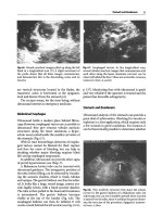
Round, anechoic images, piled up along the left
flank in a longitudinal scan (C).
A
slight movement of
the probe shows that all these images communicate,
and demonstrate this is the descending colon and its
haustra
Fig.
6.7. Esophageal varices. In this longitudinal scan,
several tubular anechoic images that communicate with
each other along the lesser omentum (arrows) can be
observed behind the
liver.
These
are stomachic coronary
varices
(L,
liver;
A,
aorta)
are vertical structures located in the flanks, the
transverse colon is horizontal at the epigastric
level and distinct from the stomach [4].
The rectum seems, for the time being, without
ultrasound interest in emergency medicine.
Abdominal Esophagus
Ultrasound holds a modest place behind fibros-
copy. However, esophageal varices are accessible to
ultrasound: they give sinuous tubular anechoic
structures along the lesser omentum, a hyper-
echoic area located inside the smaller curvature of
the stomach (Fig. 6.7).
With GI tract hemorrhage, detection of esopha-
geal varices cannot be blamed for their rupture
and thus the cause of bleeding, but can help in
deciding whether major bleeding requires blind
life-saving esophageal tamponade.
In addition, ultrasound can provide other signs
of portal hypertension (see Chap. 7).
A Blakemore-Linton tube can be inserted with
ultrasound guidance. The intragastric position of
the tube, before filling, can be detected by visualiz-
ing the acoustic shadow, which is frank, tubular
and unique. The gastric balloon can then be inflat-
ed. It looks like a large, round image, convex out-
side,
highly echoic, with a frank acoustic shadow.
The tube is then pulled to the head until resistance
is encountered. The gastric balloon becomes
visible at the top of the fundus (Fig. 6.8). The
esophageal balloon can then be inflated. It will
create a mark behind the left auricle (see Fig. 19.10,
p 137). Monitoring thus with ultrasound is quick
and very reliable if the operator is trained and the
patient has favorable echogenicity.
Stomach and Duodenum
Ultrasound analysis of the stomach can provide a
great deal of information. Checking for vacuity or
repletion is a first application, which requires only
a few seconds in good conditions. For instance, it
can be theoretically possible to determine whether
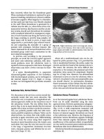
Fig.
6.8.
This arciform structure that stops the echoes
(arrow)
is the gastric balloon of a Blakemore tube. On
echoscopy, one can see it stumble upward when traction
is exerted on the tube, since it outlines the gross tubero-
sity, the very aim of the procedure. Epigastric transver-
sal
scan.
L,
liver
36 Chapter 6 Gastrointestinal Tract
Fig.
6.9.
Major fluid stasis with acute gastric dilatation.
The content is heterogeneous with hyperechoic points
due to aUmentary
particles.
Epigastric transversal scan
this patient can be operated before the traditional
6-h fasting. One can also search for a residue
during enteral feeding or diagnose acute gastric
dilatation in a patient with acute abdominal disor-
der. Acute gastric dilatation is a rare but possible
cause of acute dyspnea, which gastric aspiration
alone can relieve.
Gastric liquid retention gives a massive collec-
tion with multiple echoic particles, like in weight-
lessness,
and sometimes an air-fluid
level
(Fig.
6.9).
This pattern is sometimes impressive and can be
unsettling for the young operator, and should not
lead to diagnoses such as splenic abscess. In our
experience,
very substantial liquid stasis was often
associated with bulbar ulcer, a feature already
described in the literature [5].
The correct positioning of a feeding tube
within the gastric lumen can be assessed, or
alternatively with the mandatory radiograph. Its
tubular structure with frank acoustic shadow is
easily recognized (Fig. 6.4). This application is
very contributive when the end of the tube is at the
antrum level, far less when it remains in the fun-
dus area.
Gastric ulcer can produce a thickened, irregular
wall. The ulcer itself is rarely highlighted. Ultra-
sound will not replace fibroscopy, but represents
an initial approach that should be validated.
The stomach can be used as an acoustic window
for exploring deeper structures such as the pan-
creas.
The stomach should be filled with water,
using the gastric tube that is usually present. A
slight right decubitus will trap the air bubbles in
the vertical portion of the stomach
[6].
Last, a full
stomach can be precisely located in the still hypo-
thetical aim of performing bedside gastrostomy
under sonographic guidance.
A duodenal ulcer will be suspected when a
thickened wall is associated with gastric stasis [5].
A
study based on 20 cases of duodenal ulcer found
an average
7
mm of thickening and reported a sen-
sitivity of
65%
and a specificity of
91%
for ultra-
sound
[2].
In the case of
fluid
collection outside the
duodenum with gas bubbles, or pneumoperi-
toneum (see
Chap.
5),
the
diagnosis of complicated
ulcer (with leakage) is probable [7].
In caustic intoxications, ultrasound can detect
diffuse edema along the GI tract, with a thickened
and hypoechoic
wall.
Search for a left pleural effu-
sion (present if there is esophageal rupture) or
peritoneal effusion is part of the initial examina-
tion and the follow-up of the patient.
Ultrasound's contribution in GI tract hemor-
rhage is detailed in
Chap.
28.
Small and Large Bowel: Introduction
Here again, ultrasound can play a priority role,
when compared to physical examination, plain
radiographs, colonoscopy or even
CT.
In the ICU,
a basic contribution of ultrasound is its ability
to detect the presence or absence of peristalsis
(Fig.
6.10). This information should be considered
crucial. Observations have shown a high correla-
tion between abolished peristalsis and the exis-
tence of an abdominal drama such as mesenteric
infarction or GI tract perforation.
Fig.
6.10. These oblique lines (arrow), which seem to
intersect in time-motion, are typical from a normal
peristalsis. Direct observation in real-time shows the
same pattern.
M,
bowel loop surrounded by effusion
Small and Large
Bowel:
Acute Ischemic Disorders
37
Fig.
6.11.
Three bowel loops are visible in cross-section.
Note the substantial wall
thickening,
which can be accu-
rately measured between a peritoneal effusion and ane-
choic fluid digestive content
Fig. 6.12.
Mesenteric infarction. The entire small bowel
has the same pattern, with moderately thickened wall,
and above all complete absence of
peristalsis.
This
gene-
ral pattern of akinesia is striking in real-time. Note the
fluid content of the bowel
loops.
Pelvic scan
Another accessible item is wall thickness mea-
surement (Fig. 6.11). Parietal thickening is present
in many critical situations. Doppler could find a
place if searching for signs of good perfusion
[8,9],
but this is probably of little relevance and may be
redundant, at least in the ICU setting.
Small and Large Bowel: Acute Ischemic Disorders
We have grouped different disorders such as
mesenteric ischemia, mesenteric infarct or necro-
sis,
colic ischemia and colic necrosis into this sin-
gle section. The problem lies in the difficulty of the
diagnosis, which usually results in delayed treat-
ment and a poor prognosis. Colonoscopy or even
CT are not perfect tools. CT can yield troublesome
false-negative tests.
In this context, ultrasound deserves a top-rank-
ing place according to our experience. Our obser-
vations show a complete and diffuse abolition of
peristalsis in 87% of our cases
[10].
A moderately
thickened wall (5-7 mm) is found in only half our
cases (Fig. 6.12). Peritoneal effusion was present in
half of cases. Portal gas, a quasi-specific sign, was
rarely observed (see Fig. 7.2 and
7.3,
p 42).
We must therefore detail the signs demonstrat-
ing peristalsis. Observation shows that a patient
who is intubated, mechanically ventilated, and
sedated with high-dose morphinomimetics, has
maintained peristalsis. Adding a curare does not
abolish the ultrasound peristalsis. The notion of
sedation or even curarization should therefore
never be retained to explain an akinetic bowel. The
notion of recent laparotomy, even with the proce-
dure touching the bowel, should not be pretext for
a wait-and-see policy, since we have observed peri-
stalsis of the small bowel clearly present 24 h after
colectomy. Last, for still unknown reasons, a small
percentage of ICU patients (12%) without GI tract
impairment show abolition of peristalsis.
In the case of colic ischemia, our observations
often show thickened colic wall (Fig. 6.13). In addi-
tion, small bowel peristalsis is nearly always abol-
ished, a finding that can appear beneficial for an
early diagnosis.
Fig. 6.13. Cross-section of the descending colon. The
lumen is virtual, but the wall can be accurately measu-
red,
here to 7 mm. Colic ischemia
38
Chapter 6 Gastrointestinal Tract
Bowel Dilatation
Fig. 6.14.
The superior mesenteric vein is often clearly
visible
(V),
passing
anterior
to
the abdominal aorta (A).
The two
should
not be
confused.
The
good quality of the
picture makes it possible to study its content, here an-
echoic.
A
local compression maneuver completely col-
lapses the venous
lumen.
Longitudinal
view
The literature is not particularly informative in
this field
[11,12].
It describes dilated loops, aboU-
tion of peristalsis, very thin wall
(1
or
2
mm) in the
arterial causes, and thickened and hypoechoic
wall in the venous causes. In late cases, parietal
microbubbles and flattening of the jejunal valvulae
conniventes, fluid contents without
gas,
peritoneal
effusion, portal gas [13, 14], or even hepatic
abscesses and portal or mesenteric venous throm-
bosis have been described.
The superior mesenteric vein is often accessible
(Fig. 6.14). Since it passes anterior to the rachis, it
is possible to make a compression at this level in
order to assess its patency, and without the help of
the Doppler technique (see Chap. 12).
The diagnosis is classically made using plain radi-
ographs, which raises problems in the supine
patient. CT is increasingly replacing plain radi-
ographs. Yet ultrasound can be highly helpful
when showing the following at the bedside:
• Dilatation of the bowel
[16]. A
dilated jejunum
has a characteristic pattern
(Fig.
6.15), but more
subtiety
is
required to distinguish between dilat-
ed ileum and normal colon.
• Fluid content.
• Complete absence of wall and fluid content
motion in the paralytic ileus, or sometimes to-
and-fro movements only caused by the inertia
of
the
sequestrated liquid.
• Peritoneal effusion is possible.
• An air-hydric level can be detected using the
swirl
sign.
When the patient is supine and when
the probe is applied vertically on the abdomen,
a gas pattern is first observed.
A
slight pressure
is then applied on the abdomen with the probe
and free hand. When this pressure has shifted
the gas collection, a fluid pattern immediately
appears on the screen. At this moment, small
movements made at the side of
the
bed will cre-
ate swirls. The swirls result in sudden appear-
ances and disappearances or an air pattern,
with a complete acoustic barrier. Between the
appearances of air, a fleeting image of fluid is
visible
(Fig.
6.16).
This
very suggestive pattern is
of obvious meaning.
Small
and
Large
Bowel:
Other Acute Disorders
Pseudomembranous Colitis
Studying the ultrasound features of
this
complica-
tion of antibiotics may theoretically select the
requirements for
colonoscopy.
The
ultrasound pat-
tern, insufficiently described in the literature [15],
shows marked thickening of the
colic
wall,
collapse
of the lumen and frequent hemorrhagic ascites.
Our rare observations also showed irregular debris
floating within abundant intraluminal fluid, a pat-
tern evoking parietal dissection.
Fig. 6.15.
Dilated jejunal
loop.
The
wall,
perfectly outU-
ned between peritoneal effusion and fluid content, is
thin.
The
fluid
is
here hypoechoic with
hyperechoic par-
ticles.
The
caliper of this
loop
is 30 mm.
Jejunal villi can
be
recognized (the
fishbone
sign).
Small
intestine occlu-
sion.
Transverse scan of the pelvic area
References
39
cific.
Nonetheless, ultrasound can thus logically
be considered the first test able to detect GI tract
hemorrhage, before the appearance of any clinical
or biological anomaly.
Miscellaneous
Fig. 6.16. Demonstration of the swirl sign using the
time-motion mode. Left, real-time: air barrier at the left,
fluid mass at the right of the screen. Right, time-motion:
the air-fluid level has been gently shaken and the swirl
created is the source of sudden transmissions of the
ultrasonic beam
Let us note here that the presence of peristalsis is
as a rule a reassuring finding. In a series of 20
patients considered for emergency surgery, seven
of them actually surgical cases, the sensitivity of
an abolished peristalsis for the diagnosis of an
abdominal disorder requiring prompt surgery
was
100%,
specificity
77% [10].
Consequently, in a sus-
picion of acute abdomen, the detection of
a
present
peristalsis is a strong argument for ruling out a GI
tract disorder requiring surgery.
Fluid Digestive Sequestration
In a patient with shock, ultrasound detection of
fluid sequestration within the intestines
(Figs.
6.3,
6.5 and 6.9) immediately assumes a hypovolemic
mechanism caused by digestive disorders (this
sign will be associated with other ultrasound signs
of hypovolemia). Briefly scanning the abdomen
makes it possible to roughly evaluate the seques-
trated volume of fluid.
In the same manner, in a patient with hemor-
rhagic shock, ultrasound can identify not yet exte-
riorized melena, which will appear as a fluid in the
bowel
(Fig.
6.17).
This
pattern
is,
of
course,
not spe-
References
Fig.
6.17. Melena. This portion of the small bowel, out-
lined by ascites, is hypoechoic, indicating fluid. As was
the case in this patient, this pattern can be the first sign
of a GI tract hemorrhage
1.
Schmutz GR, Valette JP (1994) Echographie et endo-
sonographie du tube digestif et de la cavite abdomi-
nale.
Vigot, Paris, p 16
2.
Lim JH, Lee DH, Ko YT (1992) Sonographic detec-
tion of duodenal ulcer. J Ultrasound Med 11:
91-94
3.
Weill F (1985) L'ultrasonographie en pathologic
digestive.
Vigot, Paris, pp 455-456
4.
Lim JH, Ko YT, Lee DH, Lim JW, Kim TH (1994)
Sonography of inflammatory bowel disease: findings
and value in differential diagnosis. Am J Roentgenol
163:343-347
5.
Tuncel E (1990) Ultrasonic features of duodenal
ulcer. Gastrointest Radiol 15:207-210
6. Smithius RHM and Op den Orth JO (1989) Gastric
fluid detected by sonography in fasting patients:
relation to duodenal ulcer disease and gastric-outlet
obstruction. Am J Roentgenol 153:731-733
7.
Deutsch
JP,
Aivaleklis A, Taboury J, Martin
B,
Tubia-
na JM (1991) Echotomographie et perforations
d'ulceres gastro-duodenaux. Rev Im Med 3:587-
590
8. Teefey
SA,
Roarke
MC,
Brink
JA,
Middleton
WD,
Bal-
fe DM, Thyssen EP, Hildebolt OF (1996) Bowel wall
thickening: differentiation of inflammation from
ischemia with color Doppler and duplex ultrasono-
graphy Radiology 198:547-551
9. Danse EM, Van Beers BE, Goffette P, Dardenne AN,
Later re PF, Pringot J (1996) Acute intestinal ische-
mia due to occlusion of the superior mesenteric
artery: detection with Doppler sonography. J Ultra-
sound Med 15:323-326
10.
Lichtenstein D, Mirolo C, Meziere G (2001). L'aboli-
tion du peristaltisme
digestif,
un signe echogra-
phique d'infarctus mesenterique. Reanimation 10
[Suppl] 1:203
40 Chapter
6
Gastrointestinal
Tract
11.
Fleischer
AC,
MuhletalerCA,
James AE
(1981) Sono-
graphic assessment of the bowel wall. Am
J
Roentge-
nol 136:887-891
12.
Taboury
J
(1989) Echographie
abdominale.
Masson,
Paris,
pp 253-255
13.
Kennedy
J,
Cathy
L,
Holt RN, Richard R (1987) The
significance of portal vein gas in necrotizing entero-
colitis.
Am
Surg 53:231-234
14.
Porcel
A,
Taboury
J,
Aboulker
CH,
Bernod
JL,
Tubia-
na JM (1985) Aeroportie et infarctus mesenterique:
interet de
Techographie.
Ann Radiol 28:615-617
15.
Downey
DE
and Wilson SR (1991) Pseudomembra-
nous colitis: sonographic features. Radiology 180:
61-64
16.
Mittelstaedt C (1987) Abdominal Ultrasound.
Churchill Livingstone, New York
CHAPTER
7
Liver
The liver is the most voluminous plain organ, but
is rarely a target for emergency therapeutic deci-
sions in the ICU.
Mechanical ventilation, which lowers the
diaphragm, can make its exploration easier. When
the liver is located high, intercostal scans will be
taken, provided the probe is small enough. Liver
analysis is often not exhaustive in such conditions,
but
we
will see that this limitation is relative in the
critically ill patient.
Hepatomegaly
Although some operators can evaluate the weight
of each lobe, the subjective feeling that the liver is
enlarged
is
sufficient for others
[1].
In the critically
ill patient, it is more important to recognize the
cause of this enlargement than the exact dimen-
sions or weight. Usual causes in the ICU are acute
right heart failure and cirrhosis.
The cardiac liver has a homogeneous structure,
with dilatation of hepatic veins and vena cava infe-
rior (Fig. 7.1). This finding will be accessory: the
dilatation of the right heart and the lung disorder
will then be recognized at the same time.
A cirrhotic liver will give numerous signs we
will not detail
here:
a coarse pattern,
a
nodular pat-
tern, atrophy or hypertrophy of one lobe with
resulting global dysmorphia, absence of supple-
ness of the parenchyma, signs of portal hyper-
tension (dilatation of the portal vein, ascites,
reopening of the umbilical vein, splenomegaly and
others). See Fig. 6.7, p 35, for an illustration of
esophageal varices.
As regards tumoral or infectious (abscesses)
enlargements, the cause will immediately appear
on the screen.
Fig. 7.1.
Liver in right heart failure. Dilatation of the
three hepatic veins, which open into an inferior vena
cava
(V)
also
dilated.
Note
that
this
scan
does
not reflect
the site where its caliper should be measured (see
Chap.
13, p 82).
Epigastric subtransverse scan
Portal Gas
This is a situation where ease of diagnosis and effi-
ciency of therapeutic management meet. Portal
gas generally requires prompt surgery [2, 3]. In
a critical scene, portal gas immediately evokes
mesenteric infarction. Ultrasound may give a
chance for the patient to benefit from an earlier
diagnosis. Portal gas is traditionally considered a
pejorative sign [4],but this feeling
is
based on radi-
ographic findings.
Yet
ultrasound is more sensitive
than radiographs [2]. In addition, we have seen
surgical success even when ultrasonic portal gas
was present.
Portal gas yields numerous punctiform hypere-
choic images without acoustic shadow within the
liver parenchyma and usually peripheral
(Fig.
7.2).
In this
case,
we
speak of static portal gas. In some
cases,
one can observe a flux of
gas
particles at the
portal vein
(Fig. 7.3), a
sign
we
called dynamic por-
tal
gas.
In these
cases,
when such particles are seen
coming from the superior mesenteric vein and not
42 Chapter? Liver
Fig. 7.2.
Static portal gas. Numerous hyperechoic punc-
tiform opacities, without acoustic shadow, within the
liver of a patient with mesenteric infarction. Note that
this patient survived, in spite of the classically poor
prognosis of portal gas
Fig. 7.4.
Hepatic abscess
(Klebsiella),
Hypoechoic hete-
rogeneous mass within the hepatic parenchyma
Fig. 7.3.
Dynamic portal
gas. A
visible flow with hyper-
echoic particles
(large arrows)
is observable in the portal
vein. Static portal gas can be seen (small
arrows).
Obli-
que scan of the right hypochondrium, in the axis of the
portal vein
(large
arrows),
in a patient with septic shock
Fig. 7.5. Hepatic abscess
(Streptococcus
milleri). Huge
round hypoechoic mass. In real-time, this mass had a
characteristic internal motion, which indicated a fluid
nature. Percutaneous ultrasound-guided drainage (see
Fig. 26.1,
p 173) has withdrawn 1,150 cc of frank pus
from the splenic vein, they originate logically from
the GI tract.
Volvulus or strangulation, ulcerous colitis, and
intra-abdominal abscesses are other causes de-
scribed in the adult [4].
Hepatic Abscess
Ultrasound is a quick and user-friendly method of
diagnosis, since it spares the highly unpleasant
pain caused by liver shaking. Pain is often absent in
a encephalopathic patient in shock, hence the
interest of a systematic ultrasound examination in
any critically ill new arrival.
Abscess yields an image contrasting with the reg-
ular hepatic echostructure. It is generally hypo-
echoic,
heterogeneous, and roughly round (Fig. 7.4).
A very characteristic sign is sometimes observed:
within the mass, an internal movement is visible, in
rhythm with respiration. This is in fact the inertia of
the pus caused by the movement
(Fig.
73)y the equiv-
alent of the plankton sign discussed in Chap. 5. In
our observations, it proves the fluid nature of the
Diffuse Infectious Disorders
43
Fig.
7.6.
Hydatid cyst of the liver
(arrowheads).
The het-
erogeneous pattern indicates compHcation, here sup-
puration, which was confirmed at the laparotomy of
this patient in septic shock. Longitudinal scan of the
liver.
L,
liver
Fig.
7.8.
Dilatation of intrahepatic bile
ducts.
Vessels
(X)
are visible anterior to portal bifurcation
(V),
producing
a double channel pattern
Diffuse Infectious Disorders
Tuberculous hepatic miliary can be missed by
ultrasound (Fig. 7
J).
In cases where there is strong
clinical suspicion, a prompt liver biopsy should
provide bacteriological confirmation.
Cholestasis
Fig. 7.7. Diffuse tuberculous miliary. In this longitudinal
scan of the liver and the kidney
(JC),
it is hard to detect
frank anomalies. Real-time showed that the liver paren-
chyma pattern was homogeneously granular, but one
can consider it is a subtle sign
collection (regardless of the presence or absence of
posterior enhancement), and above all it indicates
pathological fluid (pus,blood). Highly echoic images
are sometimes seen, indicating microbial gas. Pleu-
ral effusion (generally radiopaque) is possible.
Amebic abscess yields a hypoechoic, well-limit-
ed collection.
Hydatidosis should be evoked before any punc-
ture of fluid hepatic mass. This does not cause a
problem when the cyst is well defined and anech-
oic,
since there is no emergency, but it may in the
suppurative forms, when the cyst becomes echoic
and heterogeneous (Fig. 7,6).
Ultrasound is a quick and simple way to check for
the normal condition of the bile ducts. However,
cholestasis occurring in a ventilated patient is
very frequent. In our observations, the cause of
cholestasis is always medical: sepsis or impairment
of venous return. We are still awaiting a surgical
cause of cholestasis in a patient initially ventilated
for another reason.
This said, in case of an obstacle, ultrasound
will detect bile duct dilatation: the intrahepatic
duct anterior to the portal bifurcation (Fig. 7.8)
or the main duct anterior to the portal vein
(Fig. 7.9). The normal caliper of the main bile
duct is said to be 7 mm (up to 12 mm in the case of
an old cholecystectomy), but some authors have
fixed the upper limit at 4 mm
[5].
When the com-
mon bfle duct is dflated, it acquires a tortuous
route and cannot be visualized in a single view.
The sensitivity of ultrasound is poor for detection
of common bile duct calculi, which rarely produce
posterior shadows, even if massive [6].
44 Chapter? Liver
Fig.7.9.
Anterior
to the portal vein
(V),
the common
bile
duct
(arrow)
is dilated with a 9-mm caliper. Oblique
scan of
epigastric
area.
G,
gallbladder
Fig. 7.10. Hyperechoic structure,
highly dynamic in
real-
time,
visible
at the median hepatic vein
(arrows).
Trap-
ped air
in the hepatic venous
system.
Subtransverse epi-
gastric scan acquired with
an
Ausonics
2000
device
Hepatic Vein Disorders
Ultrasound is an excellent noninvasive method for
examining hepatic vein disorders
[7].
In the Budd-
Chiari syndrome with hepatic veins thrombosis,
these veins are filled with echoic material, are fiU-
form, or are not visible if they have the same
echogenicity
as
the
liver.
Other signs exist but their
description would deviate too far from our initial
objectives. Faithful to a maximal use of two-
dimensional ultrasound, and regarding the rarity
of this disorder (at least in our institutions), we
think that two-dimensional ultrasound should be
done first. Visualization of anechoic hepatic veins,
which can be compressed with the pressure of the
probe, indicate patency of these veins. Obviously,
the operator should search for more frequent
diseases to explain the symptoms bringing suspi-
cion of Budd-Chiari syndrome. If
the
examination
remains noncontributory, then and only then
should a Doppler study
be
indicated.
In critically
ill
patients,
mobile gas is sometimes
observed in the median and left hepatic veins,
which are the non-declive veins (Fig. 7.10). The
most logical explanation is that air accidentally
coming from perfusions (in the
arms,
for instance)
are trapped in these veins. A tricuspid regurgita-
tion, very frequent in the mechanically ventilated
patient, may be the cause.
Hepatic Tumors
Recognition of metastases may give a theoretical
element of prognosis in the acute phase. They are
usually known, but they can be discovered by
ultrasound when no anamnesis is available. The
pattern is usually characteristic: multiple dissemi-
nated images with anarchic distribution, isoechoic,
or hyperechoic with a fine hypoechoic stripe, or
again hypoechoic images (Fig. 7.11). As regards
other tumors, we will be
brief,
since they do not
need particular treatment or reflexion during the
stay in the
ICU.
A
round, regular, anechoic image
is generally a biliary cyst, sometimes also an
uncomplicated hydatid cyst. An echoic heteroge-
Fig.7.11.
Hypoechoic masses, disseminated in the liver
with
a
multicentric
pattern.
Hepatic
metastases.
Perito-
neal effusion surrounding
the
liver
(asterisk)
secondary
to peritoneal metastases
References
45
neous mass within a cirrhotic parenchyma will
be suggestive of hepatocarcinoma. These tumors,
and others (adenoma, focal nodular hyperplasia,
angioma, primitive malignant tumors, heteroge-
neous steatosis, etc.) are extensively described in
excellent textbooks
[1,8,9].
Miscellaneous
In hepatic trauma, identifying hemoperitoneum is
possible, as well as direct patterns of liver contu-
sion in favorable cases (see Fig.
24.1,
p 165).
Aerobilia can be pathological, in ileus by impact-
ed gallstone, or physiological, after biliary surgery.
Numerous air opacities are visible along the biliary
vessels, which converge to the hilum. Thus, the
images are more central than in portal gas.
Interventional Ultrasound
Percutaneous Aspiration
or
Drainage
of
Liver Abscess
We were able to successfully aspirate hepatic
abscesses with the material described in Chap. 26.
Deep locations or locations near the dome can
cause technical problems.
Percutaneous
or
Transjuguiar Liver Biopsy
The presence of permanent ultrasound assistance
means that emergency liver biopsies can be car-
ried out. Three indications can be imagined in the
ICU:
• Documenting diffuse tuberculosis before treat-
ment
• Proving the malignant nature of liver images, if
this finding can modify immediate treatment
• Investigating fulminant hepatitis.
In this last case, hemostasis disorders usually
require a transjuguiar approach, which usually
means transportation of a critically ill patient to a
specialized center. Yet transjuguiar hepatic biopsy
could be performed at the bedside under sono-
graphic guidance, with a double impact. First,
immediate and successful catheterization of the
internal jugular vein (see Chap. 12); second, after
insertion of the material, guidance toward the tar-
get. Let us specify that radioscopy, which gives a
good overview, creates irradiation, and above all,
reduces a three-dimensional shape (the liver) to
a two-dimensional image. Two-dimensional ultra-
sound, in well-trained hands, gives a three-dimen-
sional image of an area. It accurately steers the
material through the inferior vena cava, then the
hepatic
veins.
This visual guidance should decrease
the number of incidents that occur with radio-
scopic guidance.
References
1.
Menu
Y
(1986) Hepatomegalies. In: Nahum H, Menu
Y (eds) Imagerie du foie at des voles biliaires. Flam-
marion,
Paris,
p 86-96
2.
Lee CS, Kuo YC, Peng SM et al (1993) Sonographic
detection of hepatic portal venous gas associated
with suppurative cholangitis. J Clin Ultrasound 21:
331-334
3.
Traverso LW (1981) Is hepatic portal venous gas an
indication for exploratory laparotomy? Arch Surg
116:936-938
4.
Liebman
PR,
Patten
MT,
Manny
J
(1978) Hepatic por-
tal veinous gas in
adults.
Ann Surg 187:281-287
5.
Berk RN, Cooperberg
PL,
Gold
RP,
Rohrmann CA Jr,
Ferrucci
JT
Jr (1982) Radiography of
the
bile
ducts.
A
symposium on the use of new modalities for diagno-
sis and treatment. Radiology 145:1-9
6. Weill F (1985) Uultrasonographie en pathologic
digestive.
Vigot,
Paris
7.
Menu
Y,
AHson D, Lorphelin JM, Valla D, Belghiti J,
Nahum H (1985) Budd-Chiari syndrome, ultrasonic
evaluation. Radiology 157:761-764
8. Taboury
J
(1989) Echographie abdominale. Masson,
Paris
9. Weill F (1985) Lultrasonographie en pathologic
digestive. Vigot,
Paris,
pp 455-456
CHAPTER
8
Gallbladder
Acute acalculous cholecystitis, a classic complica-
tion of the critically ill and a classic indication for
general ultrasound, deserves an entire chapter.
Our experience suggests two comments. First, this
disorder seems to affect mostly the surgical patient
and is exceptional in the medical ICU. Second, if
ultrasound can accurately describe many data, the
very interpretation of these data remains
subtle.
In
fact, the gallbladder can have a vast variety of pat-
terns,
from the normal to the pathological, in pass-
ing by the picturesque (Figs. 8.1, 8.2). A strictly
normal gallbladder in the ICU is an infrequent
finding (see Fig.
4.7, p. 21). The
variations in volume,
wall thickness, content, shape and surroundings
can create infinite combinations. Some are vari-
ants of the normal, some are pathological but
do not require emergency procedures, and others
need prompt surgery, beneficial in shghtly less
than half of the cases in our experience.
Classic Signs
of
Acute Acalculous Cholecystitis
Acute acalculous cholecystitis is found in 5%-15%
of acute cholecystitis and 47% of postoperative
cholecystitis
[1].
The diagnosis is based on infec-
tious syndrome and local signs in an exposed
patient
[2].
Histology alone provides definite diag-
nosis,
a mandatory sign being wall infiltration by
neutrophils. Ultrasound patterns classically asso-
ciate:
• Enlarged gallbladder, with a long axis caliper
over 90 mm and a short axis over 50 mm.
• Wall thickening greater than
3
mm.
• Sludge (echoic, compact, declive sediment).
• Perivesicular fluid collection, valuable in the
absence of ascites.
• Murphy's sign: pain due to the pressure of the
gallbladder. Since ultrasound has the merit of
precisely locating the gallbladder, ultrasound
identification of Murphy's sign is mentioned
when the probe itself appUed in front of the gall-
bladder creates elective pain.
Fig.
8.1.
Elegance
is
not forbidden in an organ
as
critical
as
the gallbladder.
A
simple folding
at the
hepatic
aspect
is enough to confer this discrete charm
Fig. 8.2.
In another gallbladder, a very irregular sludge
seems to represent a crouched coyote in an asymptom-
atic patient
Chronic Subacute Cholecystitis
47
Sensitivity of ultrasound is weak (67%) for some
[3],
high (90%-95%) for others [4,
5].
When dis-
tension, thickening and sludge are combined, sen-
sitivity
falls,
but specificity climbs [2].
Observations
of
Acute Acalculous Cholecystitis
Acute acalculous cholecystitis seems to be specific
to the surgical ICU. It seems to happen especially
after major vascular surgery such as aorta surgery.
Although ultrasound can localize the gallbladder
and can accurately delineate the phenomena
described
above,
we
suspect that these
signs,
taken
one after another or even together, are subject to
a problem of interpretation. Our observations of
histologically proven acute acalculous cholecysti-
tis have led to the following conclusions
(Fig.
8.3).
Size
On average, the gallbladder measured 103 mm on
the long axis (range, 65-150 mm) and 40 mm on
the short axis
(range,
29-55 mm).
The Wall
The wall was always moderately thickened, mea-
suring on average 4.6 mm (minimum observed,
3.0 mm; maximum, 6.2 mm).
Sludge
Sludge was present in
90%
of cases.
Murphy's Sign
in
Ultrasound
We
observed
a
genuine Murphy sign in
8%
of
cases.
Perivesicular Effusion
We observed selective effusion in
12%
of cases.
The problem begins with the existence of a dis-
order very frequently encountered in our histology
reports: chronic subacute cholecystitis. This disor-
der will raise serious diagnostic problems.
Chronic Subacute Cholecystitis
Chronic subacute cholecystitis is
a
histological
def-
inition. In fact, neither ultrasound nor even periop-
erative findings can distinguish it from the acute
acalculous cholecystitis
(Fig. 8.4).
Nearly half of our
Fig.
83a,
b.
Acute acalculous cholecystitis, with histolo-
gical proof A homogeneous thickening of the wall
(4
mm),
a caliper of
30
mm, and dependent sludge are
depicted. There was no pain in this sedated patient.
Above all, this gallbladder is suspect because the pa-
tient developed fever after major aortic surgery,
a
Longi-
tudinal scan, b Transverse scan, in which a moderate
peritoneal effusion
is
visible
(E)
patients operated for suspicion of acute acalculous
cholecystitis in fact had chronic subacute cholecys-
titis.
Chronic subacute cholecystitis does not seem
to require surgery. In our observations, the average
long axis was
105
mm
(range,
84-160
mm),
average
caliper, 37 mm (range, 23-56 mm), average wall
thickness,
4.5
mm (range,
3.0-7.0
mm), sludge was
present in 66% of
cases.
Murphy's sign in 10% and
locaHzed effusion was never present.
However, these data are quite similar to those
seen in acute acalculous cholecystitis (Table 8.1).
One consequence is that this disorder is diag-
nosed, with subsequent surgery, with the same
frequency as acute acalculous cholecystitis. This
probably means useless surgery, in other words,
increased operative risk, and above
all,
this means
that the initial problem remains undiagnosed. A
48 Chapters Gallbladder
Fig. 8.4.
This gallbladder has a homogeneous
5.5-mm
thickened wall, a pattern not really different from
Fig. 8.3. Sludge is also discretely present. Pathological
examination confirmed the diagnosis of chronic sub-
acute cholecystitis
perioperative pattern is sometimes misleading,
and many gallbladders considered acute or even
gangrenous become simple chronic subacute
cholecystitis once under the microscope.
In addition, we have frequently seen ultrasoni-
cally suspect gallbladders that were not operated
and that spontaneously normalized. The problem
is again intricate, as some authors argue that cer-
tain acute acalculous cholecystitis cases can be
cured without surgery, but this should be proven
with a solid methodology.
Common Gallbladder Patterns Seen
In the Intensive Care Unit
It may be timely to specify one point here. In our
experience,
mostly from the medical ICU and over a
systematic observation of our patients since 1989,
acute acalculous cholecystitis
was
rare.
In
11
years
of
practice in the medical ICU and
5
years in the surgi-
cal
ICU
(with major vascular surgery),
we
found one
case of acute acalculous cholecystitis every
500
days
of physician presence in medical patients and
23 days for surgical patients. This means a frequen-
cy 20
times lower for medical patients.
In our critically ill patients with a stable status
and with no superimposed chnical problem, the
majority of gallbladders were enlarged and con-
tained sludge. Wall thickening was extremely fre-
quent; the major form of this thickening will be
dealt with in a later section. Peritoneal effusion
was routine in severely critically ill patients. Let us
examine these signs in detail.
Volume
Volume can vary between complete vacuity to dis-
tension. A completely empty gallbladder can be
hard to detect. One should follow precise land-
marks: the right branch of the portal bifurcation
leads to the fossa vesicae felleae, which always
leads to the gallbladder space (Fig. 8.5). An empty
gallbladder is, in principle, functional, since it is
able to contract. It may also be perforated. A dis-
tended gallbladder (long axis >90 mm, short axis
>50 or 40 mm) is the rule in patients under par-
enteral feeding and taking morphines (Fig. 8.6).
The lumen can be virtual and the wall thickened
(Fig. 8.7). Among other patterns, one can see sep-
tate contents, variations in length, complete calcifi-
cations of the wall, or tumors. Images of these
anomalies are accessible in abdominal ultrasound
textbook
[6,7].
Wall Thickening
The normal wall measures between 1.5 and
3
mm.
A 4-mm cut-off has the advantage of being reli-
able,
but this notion may be obsolete. Modern
units have an improved definition, and wall thick-
Table
8.1.
Acute acalculous versus chronic subacute cholecystitis
Acute acalculous cholecystitis Chronic subacute cholecystitis
Wall thickening
Long axis
Short axis
Sludge
Localized perivesicular effusion
Murphy's ultrasound sign
4.6 mm (3.0-6.2)
103 mm (65-150)
40 mm (29-55)
90%
12%
8%
4.5 mm (3.0-7)
105 mm (84-160)
37 mm (23-56)
66%
0
10%
Extreme
values
are in parentheses.
Common Gallbladder Patterns Seen In the Intensive Care Unit
49
Fig.
8.5.
Example of an empty gallbladder. This discrete
image should be recognized to avoid erroneous diag-
noses
Fig.
8.7.
This gallbladder has virtual lumen, reduced to
an echoic stripe, and an extremely thickened wall, to
12
mm.
Laparotomy and pathology revealed simple gall-
bladder edema in this patient in septic shock with major
lung injury
Fig.
8.6. This enlarged gallbladder (100x40 mm) has a
thickened wall (3.6 mm) and roughly
40%
sludge,
which
is very frequent in the
ICU.
However,
this
gallbladder did
not provoke symptoms in a female patient admitted for
ARDS
(aspiration pneumonia), who eventually recovered
Fig. 8.8. The wall of
this
gallbladder is perfectly outlined
between bile (G) and
ascites.
This wall is perfectly
fine,
a
pattern which easily invalidates the traditional idea that
ascites causes gallbladder wall thickening
ening greater than 3 mm should be considered
with care. The wall can be very distinct when out-
lined between bile and peritoneal effusion
(Fig.
8.8).
It can be impossible to measure precisely. In some
cases,
there is no contrast between the gallbladder
wall and hepatic parenchyma, which makes any
exact measurement illusory.
We routinely find a thickened wall (Fig. 8.6). It
can be split, with two echoic layers surrounding an
hypoechoic
layer.
A
striated pattern is described as
a sign of acute acalculous cholecystitis
[8],
but the
follow-up of our patients does not support this
impression.
Traditionally, a thickened wall is nearly equiva-
lent to acute acalculous cholecystitis. Experience
shows that this sign has very low specificity. The
classic list of causes includes ascites, hepatitis,
hypoalbuminemia, and cardiac failure, a rather
vague term
[9].
Observation shows that, in the case
of ascites, and in spite of the traditional wide-
spread belief to the contrary the wall can be per-
fectly thin (see Fig. 8.8). Cardiac failure is an overly
vague notion. In contrast, acute right heart failure
should certainly be considered a prominent cause,
so much so that we speak of cardiac gallbladder
(see next section).
50 Chapters Gallbladder
Sludge
Sludge is nearly always present in the critically ill
patient, since the gallbladder does not work in a
physiological way. The pattern can vary greatly,
although we could not attribute a particular value
to each. Sludge can be homogeneous (Fig. 8.6) or
heterogeneous, containing hyperechoic dots (could
microlithiases be included in the
mass?).
The
inter-
face between the sludge and the anechoic nonde-
pendent bile can be regular (Fig. 8.6) or ragged
(Fig. 8.2). Sludge can be discrete or massive: in
some
cases,
a 100% sludge yields a pattern isoech-
oic to the liver - a hepatization of the gallblad-
der, so to speak (Fig. 8.9). Excellent knowledge
of anatomy is then required to recognize the gall-
bladder. The sludge can be tumor-shaped. Last,
sludge appears at variable stages: usually occur-
ring during a prolonged stay, it can be present at
admission. Eventually, it can completely vanish.
Murphy's Sign in Ultrasound
Murphy's sign
is
very rarely contributive since crit-
ically ill patients are all sedated or, if not, they are
in shock or encephalopathic. Pain is either absent
or diffuse to the entire body.
Peripheral Peritoneal Effusion
Peritoneal effusion is very frequent in the critical-
ly ill patient. Localized effusion in acute cholecys-
titis is a rare finding. Moreover, the very routine
observation of thin-wall gallbladders surrounded
by extensive peritoneal effusion will prove to any
operator that peritoneal effusion is not in itself a
cause explaining wall thickening.
In Summary
In conclusion, all these changes are routine and of
little relevance, even when integrated in a sugges-
tive context.
A Distinctive Feature: Major Wall Thickening
of the Cardiac Gallbladder
We regularly and frequently observe gallbladders
with the remarkable feature of major wall thicken-
ing,
more than
7 mm,
up to
18
mm
(Fig.
8.10). This
pattern:
• Always occurs in patients with right heart fail-
ure such as acute asthma, pneumonia, adult res-
piratory distress syndrome
(ARDS),
pulmonary
embolism, acute tricuspid regurgitation, exac-
erbation of chronic obstructive pulmonary dis-
ease (COPD), in the most severe forms. This
population is more often seen in medical ICUs,
hence possibly a higher rate of cases observed
here.
• There is no local sign in these generally sedated
patients.
Fig.8.9.
This
gallbladder, which
seems
to be
floating
wit-
hin
massive
peritoneal effusion, contains
a
totally echoic
lumen.
This
shows complete sludge in an asymptomatic
patient
Fig.
8.10.
Cardiac gallbladder. The wall of this gallblad-
der is extremely enlarged, up to 20
mm. A
hypoechoic
layer is surrounded by two echoic
layers.
The lumen is
narrow, probably because of the space taken by the
walls.
This
patient
has
acute
right heart
failure.
Patholo-
gy
confirmed
simple wall
edema
How
to
Establish
the Diagnosis of
Acute
Acalculous Cholecystitis 51
• The gallbladder cavity itself
is
often small, pos-
sibly because the walls enlarge to the detriment
of the cavity.
• In our experience, a dozen observations among
a large number were positively documented,
using laparotomy, for instance. All of these
observations were the result of wall edema,
sometimes chronic subacute cholecystitis, but
never acute acalculous cholecystitis.
• Time allowing, one can observe the complete
regression of this major thickening.
We suggest labeling this frequent observation of
overly thickened wall the cardiac gallbladder, with
analogy
to
cardiac liver or cor pulmonale. It can be
assumed that the cardiac gallbladder:
• Is above all the manifestation of congestive phe-
nomena that are observable at the gallbladder
wall, since this is an accessible area, as retinal
vessels are a privileged site to assess general cir-
culatory function.
• Is frequent.
• Can be occult, because this is a transitory fea-
ture.
Conversely, an ultrasound examination performed
at the climax of the wall thickening can lead to an
erroneous diagnosis of acute acalculous cholecys-
titis,
and result in a certain number of unnecessary
laparotomies.
There
is a
clinical relevance to the recognition of
cardiac gallbladder. Data suggest that the detection
of thickening over 7 mm in a patient with symp-
toms that may
evoke
acute acalculous cholecystitis
should incite the physician to search for another
cause to explain the present symptoms.
A
laparo-
tomy would not only be useless, but also deleteri-
ous if
the
real cause is not recognized.
How to Establish the Diagnosis
of Acute Acalculous Cholecystitis
In conclusion, we believe that if ultrasound is an
excellent method for localizing and measuring
the gallbladder, it cannot distinguish the surgical
emergency from an insignificant variant of the
normal.
Patient Background and Current Situation
It seems wise to evoke acute acalculous cholecysti-
tis only in well-defined patients.
A
major vascular
surgery (of the aorta, for example) is found in half
Fig.
8.11.
The gallbladder of this patient admitted for
exacerbation of chronic respiratory disease had a very
unsettling
pattern:
a
scalloped
wall
with possible debris
detached from
the
left
aspect.
Pathology authenticated
a
simple chronic subacute cholecystitis
of our cases, a major trauma in a quarter of cases.
As
for chronic subacute
cholecystitis,
major vascu-
lar surgery is found in only
16%
of
cases,
trauma in
33%.
Almost all patients with cardiac gallbladder
have
ARDS
or multiple organ failure.
Considering Certain Ultrasound Signs
Let us recall the conclusions of the previous sec-
tion:
a wall
thickening greater than
7
mm in
a
med-
ical ICU patient suspected of having acute acal-
culous cholecystitis should prompt a search for
another cause explaining the symptoms. We still
find this policy valuable after
12
years of observa-
tions.
A subtle study of the signs at the wall showing
ulcerations would be valuable, but our investiga-
tions are at a standstill.
We
sometimes thought we
had visualized shreds detached from the mucosa
(Fig. 8.11), but laparotomy and pathology ruled
out the diagnosis of acute acalculous cholecystitis.
Detachment of the mucosa with shreds floating in
the lumen is described in the literature as a sign of
gangrenous cholecystitis
[10].
In acute acalculous
cholecystitis, there is the notion of
a
very thin wall
in a preperforative
stage.
It seems therefore wise to
study the wall in its entirety, screening for areas of
weakness. However, we are still awaiting our first
case.
Intramural gas should be observed in emphyse-
matous cholecystitis.
We
have not had the privilege
of observing this sign, probably rare. Mural gas
52 Chapters Gallbladder
should
give
hyperechoic punctiform
images,
a
sign
which should not be confused with cholesterol cal-
culi contained in the Rokitansky-Aschoff sinuses
within the delightful setting of gallbladder adeno-
myomatosis, although this is of
little
interest to us
here.
Other Tests
Doppler
If the Doppler could accurately distinguish between
ischemic and edematous wall, it would then be
potentially of interest.
CT
CT does not contribute a great
deal,
since a careful
ultrasound is almost always able to analyze the
gallbladder. Let us note here that measurement of
wall thickening is much more accurate using ultra-
sound rather than CT
[11].
As
a rule, and not only
at the gallbladder
level,
ultrasound has a focal res-
olution superior to that of
CT
(see
Fig.
8.12).
Dynamic Cerulein Test and Scintigraphy
These two tests are of
little
value [10].
Ultrasound-Guided Aspiration of Gallbladder Bile
In our experience, this procedure
is
extremely sim-
ple,
as long as basic rules are respected.
A
simple
21-gauge needle is sufficient. The gallbladder must
be punctured throughout the liver (the hole will
be recovered by the liver). Bile leakage cases
described in the literature result from transperi-
toneal approaches. The dependent bile is aspirat-
ed, since the nondependent area may yield false-
negatives.
Since
pathological
bile is
viscous,
aspira-
tion must be vigorous. The amount of aspired
bile should be sufficient to diminish the possible
hyperpressure and thus limit the risk of leakage.
Conversely, if percutaneous drainage is envisaged,
the volume of the gallbladder should not be
decreased too much. When the tap is in place, the
needle is withdrawn and strong manual compres-
sion is applied at the point of puncture. If strong
compression is not applied, for fear of bile leakage,
hemoperitoneum or subcapsular hematoma of the
liver can result in patients with impaired hemosta-
sis.
Control at
1
and
12
h will search for perivesic-
ular effusion. The vesicular bile of a critically ill
Fig.
8.12a,
b.
It is not difficult to objectify ultrasound's
superiority
(b)
over
CT
(a)
as
regards focal spatial reso-
lution. The gallbladder wall, difficult to view on
CT,
is
sharply visible on ultrasound and can accurately be
measured
patient is usually dark brown or green brown,
mildly sticky, sometimes black like tar, and vis-
cous,
when the sludge itself
has
been aspired.
The risk of vesicular tap is possible though rare.
It should be compared with the risk of allowing
angiocholitis or cholecystitis to develop, which can
be clinically difficult to detect. Of 25 procedures
performed as described, we have encountered no
complications.
This technique is simple and seems
safe.
But is
it
relevant? For some, it is contributive [12] when it
provides proof of infection at the bedside, which
should be present in 66% of the cases [13]. Other
studies [14] question the sensitivity of this proce-
dure,
almost always performed in patients taking
antibiotic therapy. Leukocytes found in the gall-
bladder bile should indicate cholecystitis
[14].
The
most important limit is that acute acalculous
cholecystitis appears more as an ischemic process
than an infectious one [15].
Interventional Ultrasound
53
Fig.
8.13.
Acute purulent cholecystitis. Dependent mas-
ses (lower part of the image) are not typical of sludge
since they are rather echoic, nor do they evoke calculi,
since there is no posterior shadow. Images of membra-
nes seeming to detach from the wall are visible at the
upper part of the image. An ultrasound-guided tap
immediately confirmed the diagnosis (frank pus) and
the patient was immediately sent to the operating room
We have had one case where diagnosis of acute
infectious cholecystitis was immediately made in a
patient admitted for shock, thanks to bedside
ultrasound-guided aspiration of gallbladder
bile.
It
is true that the gallbladder had an atypical pattern,
with particularly echoic sludge, but this pattern
could have been considered as a variant of normal
(one more to add to a long list). Yet the puncture
had withdrawn frank pus, and the patient was
rightly sent to the operating room (Fig. 8.13).
Fig.
8.14. Gallbladder space hematoma. Heterogeneous
echoic pattern, often found in the gallbladder space
after surgical removal
three signs has a negative predictive value of 98%
[16].
Acute calculous cholecystitis rarely raises
diagnostic problems.
Acute Acalculous Cholecystitis
in Calculous Gallbladder
Since calculi are a frequent finding in the general
population, one should find a pertinent term to
label an acute cholecystitis of critically ill patients
occurring in a previously calculous gallbladder.
Interventional Ultrasound
Other Pathological Patterns of the Gallbladder
Cholecystectomy Space
Infection of the cholecystectomy space is frequent-
ly suspected (Fig. 8.14). Ultrasound-guided aspira-
tion appears to be an accessible procedure and can
distinguish pus retention from old sterile blood.
Calculous Acute Cholecystitis
This disorder is rarely of interest to the intensivist.
The calculi give a dependent hyperechoic, round
image with frank posterior shadow (see Fig. 1.4,
p
6).
Calculi are frequently observed and should be
respected if quiet. Obviously, the smaller the cal-
culi,
the more they are able to move and cause
trouble. The association of calculi, thickened wall
and Murphy's sign on ultrasound has a positive
predictive value of 95%, and the absence of these
Diagnostic Aspiration of Bile
This procedure has been discussed in »Ultra-
sound-Guided Aspiration of Gallbladder Bile.«
Percutaneous Cholecystostomy
Some authors underline the easiness of this proce-
dure and the low rate of complications [14, 17].
Technical requirements are the same as those
described for aspiration. Kits are available, with
laterally perforated pigtail catheters. They normal-
ly prevent parietal perforation and dislocation of
material. A series of 322 procedures described a
null mortality rate and a morbidity rate of
2%-5%
[18].
This procedure [19] was advocated as an
alternative to surgery in the critically ill [17,20]. It
provides relief of an obstacle located in the biliary
tract. It was even shown to be effective in sepsis
without obvious causes
[14].
Other teams mistrust
54 Chapters Gallbladder
this apparently attractive technique, since a fragile
wall can easily be perforated [15]. We add two
major arguments against this procedure: first, his-
tological proof is unavailable, and no conclusion
can be drawn from how the situation evolves.
Second, acute acalculous cholecystitis seems more
an ischemic than an infectious disorder, and this
indicates that the gallbladder, and not its content,
should be removed.
From a methodological point of view, it would be
valuable to study a population with clinical and
ultrasound patterns suggestive of acute acalculous
cholecystitis, and to compare the progression of
operated patients and those with a spontaneous
recovery. Such a study will be hard to conduct since
it is ethically difficult to take the risk of allowing
a genuine acute cholecystitis to evolve [21]. Note
simply that this methodological shortcoming
weighs heavily in the published studies [14].
References
1.
Cooperberg PL and Gibney RG (1987) Imaging of
the gallbladder, state of the art. Radiology 163:605-
613
2.
Bodin L and Rouby J J (1995) Diagnostic et traite-
ment des cholecystites aigues alithiasiques en reani-
mation chirurgicale. ACTUAR 27:57-64.
3.
Shuman
WP,
Rogers
JV,
Rudd
TG,
Mack
LA,
Plumley
T, Larson EB (1984) Low sensitivity of sonography
and cholescintigraphy in acalculous cholecystitis.
Am
J
Roentgenol 142:531-537
4.
Mirvis SE, Vainright JR, Nelson AW, Johnston GS,
Shorr
R,
Rodriguez
A,
Whitley
NO
(1986) The diag-
nosis of acute acalculous cholecystitis: a compari-
son of sonography, scintigraphy and CT. Am J
Roentgenol 147:1171-1179
5.
Van Gansbeke D, Matos C, Askenasi R, Braude P,
Tack D, Lalmand B, Avni EF (1989) Echographie
abdominale en urgence, apport et
limites.
Reanima-
tion et Medecine d'Urgence. Expansion Scientifique
Fran^aise,
Paris,
pp 36-53
6. Nahum
H
and Menu
Y
(1986) Imagerie du foie et des
voies biliaires. Flammarion, Paris
7.
Weill F (1985) L'ultrasonographie en pathologie
digestive.
Vigot,
Paris
8. Teefey SA, Baron RL, Bigler SA (1991) Sonography
of the gallbladder: significance of striated thicken-
ing of the gallbladder wall. Am J Roentgenol 156:
945-947
9. Slaer
WJ,
Leopold
GR,
Scheible FW (1981) Sonogra-
phy of the thickened gallbladder
wall:
a
non-specific
finding.
Am J
Roentgenol 136:337-339
10.
Chagnon S,Laugareil P,Blery
M
(1988) Aspect echo-
graphique de la lithiase biliaire et de ses complica-
tions
locales.
Feuillets de Radiologie 28:415-423
11.
Bodin L, Rouby JJ, Langlois P, Bousquet
JC,
You K,
Viars P (1986) Cholecystites aigues alithiasiques en
reanimation.
Etude
randomisee comparant
2
metho-
des therapeutiques: chirurgie et ponction drainage
percutanee sous controle echographique.
In:
Viars
P
(ed) Actualites en Anesthesie-Reanimation. Arnet-
te,
Paris,
pp 157-167
12.
McGahan
JP,
Walter JP (1985) Diagnostic percuta-
neous aspiration of the gallbladder. Radiology 155:
619-622
13.
Sicot C (1992) Les cholestases intra-hepatiques
aigues chez les malades de reanimation. Rean Urg
1:578-583
14.
Lee
MJ,
Saini
S,
Brink
JA,
Hahn
PF,
Simeone
JF,
Mor-
rison MC, Rattner
D,
Mueller RP (1991) Treatment
of critically ill patients with sepsis of unknown
cause:
value of percutaneous cholecystostomy.
Am J
Roentgenol 156:1163-1166
15.
Langlois
P,
Bodin
L,
Bousquet
JC,
Rouby
JJ,
Godet G,
Davy-Mialou C, Wiart
D,
Cortez
A,
Chomette
G,
Gre-
let
J,
Chigot
JP,
Mercadier
M
(1986) Les cholecystites
aigues non lithiasiques post-agressives. Apport de
Techographie au diagnostic et au traitement dans 50
cas.
Gastroenterol Clin Biol 10:238-243
16.
Ralls PW, CoUetti PM, Lapin SA, Chandrasoma P,
Boswell
WD,
Ngo
C,
Radin
DR,
Halls JM
(1985) Real-
time sonography in suspected acute cholecystitis.
Radiology 155:767-771
17.
Vogelzang RL, Nemcek Jr A A (1988) Percutaneous
cholecystostomy: diagnostic and therapeutic effi-
cacy Radiology 168:29-34
18.
Malone DE (1990) Interventional radiologic alter-
natives to cholecystostomy. Radiol Clin North Am
28:1145-1156
19.
Roche A, Cauquil P, HouUe D (1986) Radiologie
interventionnelle des voies
biliaires.
In:
Duvauferri-
er R, Ramee
A,
Guibert
JL
(eds) Radiologie et echo-
graphie interventionnelles,
tome
2.
Axone,
Montpel-
lier,pp 457-494
20.
Picus
D
(1995) Percutaneous gallbladder interventi-
on. Eur Radiol
5
[Suppl]:S180
21.
Johnson
LB
(1987) The importance of early diagno-
sis of acute acalculous cholecystitis. Surg Gynecol
Obstet 164:197-203