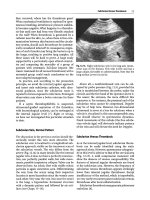
General ultrasound In the critically ill - part 4 ppsx
Bạn đang xem bản rút gọn của tài liệu. Xem và tải ngay bản đầy đủ của tài liệu tại đây (1.67 MB, 20 trang )
CHAPTER
9
Urinary Tract
The study of the urinary tract
is
vast, since mechan-
ical,
infectious and hemodynamic phenomena are
all involved.
The normal pattern of the kidney and bladder is
described in
Chap.
4
(Figs.
4.8
and
4.9,
pp 21-22).
Renal Parenchyma
The diagnosis of acute renal failure is biological,
and the main advantage of ultrasound is first to
rule out the possibility of an obstacle [1].
Arguments suggestive of acute renal failure will
be normal or increased volume (Fig. 9.1). Chronic
renal failure would give small kidneys with thin-
ning of the parenchyma and irregular borders
(Fig. 9.2). Kidneys can show global dedifferentia-
tion. The parenchyma can resemble the sinus
(parenchymocentral dedifferentiation), or, within
the parenchyma, medullary pyramids and cortex
can be hard to detect (corticomeduUary dediffer-
entiation). However, these patterns do not seem
useful in emergency situations.
Acute pyelonephritis is usually barely or not
accessible to two-dimensional ultrasound, but
severe forms can sometimes be diagnosed. Fig-
ure 9.3 shows the routine ultrasound of a 52-year-
old female, admitted for severe sepsis, with mas-
sive bilateral enlargement of the kidneys, with
no differentiation. Diagnosis was hemorrhagic
pyonephritis with diffuse purulent areas.
Parenchymatous candidiasis can sometimes be
diagnosed
(Fig. 9.4).
Emphysematous pyelonephri-
tis,
a rare finding, gives gas bubbles within the
parenchyma. In the case of severe rhabdomyolysis
with acute renal failure, we can observe enlarged
kidneys with complete dedifferentiation. Renal
trauma is presented in
Chap.
24.
A renal cyst is a benign finding. In view of its
great frequency,
we
insert a characteristic example
(Fig. 9.5) and a case of renal polycystic disease
(Fig.
9.6).
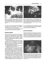
Fig.
9.1.
Acute renal failure. The kidney has a homoge-
neous echoic pattern, i.e., complete dedifferentiation.
Kidney
and liver
(L)
have
the
same
echogenicity,
and the
kidney is barely outlined
(arrows).
This scan, as nearly
all
that
follow,
is
longitudinal
Fig. 9.2. Chronic renal failure. Small size
{arrows)y
thin-
ned parenchyma and irregular borders
56 Chapter 9 Urinary Tract
Fig. 9.3. This kidney is frankly enlarged (long axis,
14 cm) and the peripheral area is extremely thickened
(arrowheads),
without differentiation. It was an acute
pyonephritis responsible for severe septic shock. Each
kidney weighed
500 g
and contained multiple areas with
pus,
necrosis and hemorrhage
Fig.
9.5.
Inferior renal cyst
(asterisk).
The kidney seems
to be interrupted, with a ragged
edge.
This cyst is regu-
lar, anechoic. This pattern, here caricatured, should not
disconcert since it is regularly observed
Fig. 9.4.
Hyperechoic pattern of the pyramids (arrows)
in a patient with patent urinary candidosis
Fig. 9.6.
Renal polycystic disease. Cysts have peripheral
topography and do not communicate with each other,
two features that distinguish it from dilatation of the
urinary cavities
Renal Pelvis
Septic shock, increased creatinemia, and a drop in
diuresis are daily situations. The possibility of a
urinary obstacle will be ruled out in a few seconds
if a small ultrasound unit is readily available.
Dilatation of the renal pelvis is rarely but regu-
larly encountered in our experience. Of 400 con-
secutive critically ill patients, we have had eight
cases,
2%.
This rate was increased if only sepsis or
acute renal failure are considered. Interestingly, the
pain is nearly always absent in septic, encephalo-
pathic patients. Causes encountered in the ICU
were pelvic hematoma, obstruction of the urinary
probe (see Fig. 9.12), bladder distension with over-
flow, blocked calculi or hydronephrosis (Fig. 9.7)
with superimposed pyonephrosis (Fig. 9.8). Detec-
tion of dependent echoic patterns within dilated
cavities of hydronephrosis is characteristic of
pyonephrosis
[2].
Pelvic cancer is a classic cause. In
trauma, a blood clot can again cause obstructive
anuria.
Dilated calices and renal pelvis yield a well-
known pattern. The three calices and the pelvis.
Renal Pelvis
57
Fig. 9.7.
Hydronephrosis. Major dilatation of the renal
pelvis. Note the rounded end, which indicates chronic
obstruction.
This
single scan does not
show
patent signs
of acute infection (see
Fig. 9.8).
Septic shock, transverse
scan of the right kidney
Fig. 9.9. Mild dilatation of the cavities. The pelvis is
slightly more dilated. The end of the calyces is concave
(arrows),
usually a sign of acute obstruction. Longitudi-
nal scan, making it possible to visualize the three calyx
groups
Fig. 9.8.
Sequel of Fig. 9.7. The ultrasound scan of the
kidney now shows heterogeneous echoic masses within
the dilated cavities
(arrows).
These images had a undu-
lating motion in real-time. The diagnosis of superim-
posed infection (pyonephrosis over hydronephrosis)
can now be put forward
Fig. 9.10. In this rarely obtained longitudinal scan of the
right flank, one can observe a dilated ureter
(LO,
inferi-
or vena cava (V) and abdominal aorta
(A).
The ureter is
usually masked by bowel gas
normally virtual or barely visible, are clearly
depicted here, anechoic and communicating with
each other (Fig. 9.9). In polycystic disease, the
numerous cysts do not communicate with each
other (see Fig. 9.6). It should be noted that a mod-
erate dilatation, with persistence of the concavity
of the calyces, indicates acute obstruction. Con-
versely, massive dilatation evokes chronic obstruc-
tion, with the end of the calices rather bulged and
thinned parenchyma (Fig. 9.7).
A dilated ureter is rarely accessible, since intra-
abdominal structures are usually in the way
(Fig. 9.10).
Dilatations of calices and pelvis without
obstruction are possible, and attributable to
chronic infectious episodes, rare causes and the
ampuUary pelvis, a variant of the normal pelvis,
which should affect 8% of the population [3].
For some authors, however, this pattern means
occult obstruction [4], which should be recog-
nized and treated. In our observations, recogni-
tion of a unilateral and moderate dilatation in a
septic patient should not be considered fortu-
itous.
A parapyelitic cyst or hypoechoic fat may, in a
hasty examination, simulate renal dilatation.
58 Chapter 9 Urinary Tract
Fig. 9.11, In this suprapubic transversal scan, probe
pointing toward the rear and the bottom of
the
patient,
one
can
see a
regular round
and medial
structure,
the
in-
flated balloon of
the
urinary
probe.
The
bladder is here
correctly
drained,
thus
virtual (compare with
Fig.
9.12)
Fig. 9.12. Major bladder distension in spite of
a
urinary
probe.
The
balloon and the end of
the
probe are visible.
The probe was obstructed
here.
Longitudinal suprapu-
bic scan
An acute obstruction may not yield dilatation of
the calices if they have lost their compliance (fibro-
sis,
retroperitoneal malignancy), or sometimes
because of major hypovolemia
[5-7].
Only iodine
explorations would make the diagnosis of obstruc-
tion, but we have not yet encountered this situa-
tion.
Bladder
This organ
is
easy to explore and can provide high-
ly contributive information in daily practice. As
a rule, a catheterized bladder is empty. A careful
examination should, if necessary, identify the bal-
loon of the probe, which seems lost in the pelvis,
but always medial
(Fig.
9.11).
A
bladder that is probed but not empty is not a
normal finding
(Fig.
9.12).
In this
case,
the bladder
should be repeatedly examined in order to check
that the trapped volume does not increase. Rarely
but regularly, we observe a genuine distended
bladder. If this occurs in a sedated patient with cir-
culatory support, the physician can make a wrong
diagnosis of anuria and increase drugs or fluid
therapy, before the distension becomes clinically
obvious. Therefore, if anuria occurs in such
patients, the first reflex should be to check if there
is no occult bladder retention.
Similarly, in an obese patient, the clinical diag-
nosis of distended bladder can be difficult. It will
always be immediate with ultrasound. Distended
bladder is maybe one of the most obvious diag-
noses for the beginner in ultrasound (see
Fig.
9.12).
The ultrasound probe, applied just over the pubis,
detects a huge liquid mass which is medial, round,
and near the anterior wall. The pitfalls are easily
avoidable. The most frequent is the peritoneal
effusion that mimics a distended bladder. If a sin-
gle transversal scan is performed at the pelvis,
peritoneal effusion can have a roughly square sec-
tion and look like a moderately distended bladder
(Fig.
9.13).
However,
peritoneal effusion appears to
open when the probe scans more cranially, where-
as a bladder appears to close.
In the female, the association of peritoneal effu-
sion and a full bladder will yield a complex but
characteristic pattern, the Thai dragoon head sign
(Fig.
9.14).
In a recently anuric patient, a daily ultrasound
can detect recovery of diuresis. This procedure
does not last more than 10 s and should prevent a
prolonged and useless insertion of a urinary
probe.
The
bladder content can
be
informative.
A
blood
clot yields an echoic dependent pattern.
A
calculus
gives a dependent image with a frank posterior
shadow.
A
purulent retention can have
a
very char-
acteristic pattern (Fig. 9.15). Last, an enlarged
prostate can be detected
(Fig.
9.16).
Bladder 59
Fig.
9.13.
Suprapubic transversal scan. This medial fluid
image with square section and a tissular image (M)
lif-
ting the floor is highly suggestive of a moderately
distended bladder. It is in fact peritoneal effusion in the
Douglas pouch. The image at M is probably a bowel
loop.
A
dynamic scan of the ultrasound probe upward
and downward will prevent the error: the bladder will
be identified below, and this fluid image will have an
opened shape above
Fig. 9.14. This complex transverse suprapubic scan may
intrigue the operator. One can imagine the head of a
Thai dragon. This is, in fact, a full bladder associated
with peritoneal effusion in
a
young
woman.
The
bladder
is the round shape at the top of the screen. The eyes and
the mouth of the dragoon reflect the peritoneal effusion.
The nose is formed by the uterus and the large liga-
ments.
The teeth are generated by solid structures float-
ing in the effusion - a hemoperitoneum here
Fig.
9.15.
a Two elements can be distinguished in this
bladder, separated by an artifactual line: a dependent
echoic sediment and a nondependent anechoic area.
Pyuria. Transverse scan of the bladder, b Another pat-
tern of pyuria. Multiple hyperechoic elements as in
weightlessness, indicating microbial gas
Fig. 9.16. Medial regular tissular mass protruding in the
bladder lumen, typical from a prostatic adenoma. This
finding is sometimes useful in cases of acute obstructive
renal failure
60 Chapter 9 Urinary Tract
Fig.
9.17.
Empty uterus in a long-axis scan, behind the
bladder. The vacuity line, which indicates absence of
pregnancy,
is
frankly outlined within the uterine muscle
Fig. 9.19. Transverse view of
the
pelvis in
a
young female
in shock.
A
motionless echoic mass indicates a massive
blood clot in a highly probable ectopic pregnancy. The
intensivist should not be asked the precise site of the
pregnancy, since the recognition alone of a peritoneal
effusion
indicates,
here,
immediate lifesaving surgery
Fig.
9.18.
An embryo is visible in the uterus, seemingly
observing the viewer. It is like a cat turned on its back,
head at the right of the image. This should incite the
physician not to overindulge in ionizing radiation pro-
cedures
Uterus and Adnexa
We like to take a look at the uterus before any
emergency radiological examination, in order to
check its vacuity (Fig. 9.17). If a pregnancy is
detected
(Fig.
9.18), the reader should see
Chap.
28,
where all details about management are detailed.
Occurrence of an acute respiratory disease in a
pregnant woman usually raises problems [8]. For
this emergency application, we do not need to
await full repletion of the bladder. In some postop-
erative cases where the suprapubic approach is
impossible, using a perineal approach is autho-
rized, as is an endovaginal investigation with the
small probe covered with a glove. This approach,
although not very academic, is perfectly valuable
when a distended bladder is sought.
The aim of this book is not to describe gyneco-
logical problems such as uterine apoplexy, ectopic
pregnancy or others. It suffices to note that
pyometritis gives a liquid endouterine image.
Hyperechoic punctiform images (gas) are a strong
argument if there is severe pelvic sepsis [9].
Endometritis produces diffuse swallowing of the
parenchyma
[9].
Ectopic pregnancy shows a subtle
direct image for the specialist, and a rough indirect
image for the nonspecialist, the hemoperitoneum.
Note that this hemoperitoneum can be echoic at
the first examination, thus particularly misleading
(Fig. 9.19). The syndromes of defibrination can
provide information on a rapid ultrasound confir-
mation, although history and physical data are
generally sufficient for the decision of a lifesaving
hysterectomy.
Renal Transplantation
A grafted kidney usually lies in the fossa iliaca.
Surgical complications are more accessible to
ultrasound than medical complications. Post-
operative collection can be caused by abscess,
hematoma, lymphocele or urinoma. They can be
References
61
explored with an ultrasound-guided tap. Dilata-
tion of the calices suggests obstruction caused
by edema or anastomotic stenosis of the ureter.
Stenosis of the renal artery should be explored
with Doppler.
Medical compHcations, in spite of numerous
signs of acute or chronic rejection, cyclosporine
toxicity or tubulointerstitial nephritis, are general-
ly diagnosed by renal biopsy [10].
Interventional Ultrasound
Percutaneous nephrostomy makes it possible to
treat a urinary obstruction and to drain infected
urine at the bedside if ultrasound-guided. The kid-
ney is punctured by the posterior or posterolateral
approach. The colon and the pleura are thus avoid-
ed. A needle is inserted in the dilated cavities.
Urine is collected for analysis. A guide is then
introduced through the
needle.
A
drainage catheter
is inserted, sometimes after several dilatations.
We have had the opportunity to use this tech-
nique to treat a patient who could not be moved as
she had severe septic shock. Later, procedures were
performed in the radiology department for local-
izing the obstruction level and in the operating
room for removing the calculus, at this time in a
stabilized patient.
Percutaneous nephrostomy
is
a procedure whose
mortality rate (0.2%) is said to be lower than that
of surgery
[3].
Complications include hemorrhage
or infection and should be balanced with the
advantages.
If suprapubic catheterization is indicated, ultra-
sound guidance provides visual monitoring. A
penetration site more cranial than classically done
should theoretically limit the risk of sepsis of the
prevesical space. Digestive interpositions can then
be ruled out using ultrasound.
References
1.
Resnick MJ and Rifkin MD (1991) Ultrasonography
of the urinary tract. Williams and
Wilkins,
Baltimore
2.
Subramanyan
BR,
Raghavendra
BN,
Bosniak
MA
et
al (1983) Sonography of
pyelonephrosis:
a prospec-
tive study
Am J
Roentgenol 140:991-993
3.
Finas B, Mercatello A, Tognet E, Bret M, Yatim A,
Pinet A, Moskovtchenko JF (1991) Strategies d'ex-
plorations radiologiques dans Tinsuffisance renale
aigue. In: Goulon M (ed) Reanimation et Medecine
d'Urgence. Expansion Scientifique Fran^aise, Paris,
pp 153-174
4.
Laval-Jeantet M (1991) La detection de maladies
graves par echographie systematique chez
le
genera-
liste.
Presse Med 20:979-980
5.
Goldfarb CR, Onseng
F,
Chokshi
V
(1987) Nondila-
ted obstructive uropathy. Radiology 162:879
6. Maillet
PJ,
Pelle-Francoz
D,
Laville
M,
Gay
F,
Pinet
A
(1986) Nondilated obstructive acute renal failure:
diagnostic procedures and therapeutic manage-
ment. Radiology 160:659-662
7.
Charasse
C,
Camus
C,
Darnault
P,
Guille
F,
Le
Tulzo
Y, Zimbacca F, Thomas R (1991) Acute nondilated
anuric obstructive nephropathy
on
echography:
dif-
ficult diagnosis in the intensive care unit. Intensive
Care Med 17:387-391
8. Felten
ML,
Mercier
FJ,
Benhamou
D
(1999) Develop-
ment of acute and chronic respiratory diseases
during pregnancy. Rev Pneumol Clin 55:325-334
9. Ardaens
Y,
Guerin
B,
Coquel Ph (1990) Echographie
pelvienne en gynecologic. Masson, Paris
10.
Cauquil P, Hiesse C, Say C, Vardier JP, Cauquil M,
Brunet AM,Galindo R,Tessier
JP
(1989) Imagerie de
la transplantation renale. Feuillets de Radiologie
29:469-480
CHAPTER
10
The Retroperitoneal Space
The kidneys have been described in
Chap.
9.
Abdominal Aorta
Abdominal aortic analysis should be routine in
any critical situation. The examination should be
done gently, in order to avoid any uncontrolled
pressure.
Bowel
gas
can be
a
source of failure. How-
ever, a left translumbar approach can bypass the
anterior gas obstacles.
Basic signs of abdominal aortic aneurysm are
a loss of parallelism of the aorta walls with a
fusiform or sometimes sacciform shape (Fig. 10.1).
If local conditions are favorable, ultrasound will
provide, like CT, a global overview of the lumen,
thrombosis, wall thickness (increased in the case
of inflammation) and collateral
vessels.
In the case
of leakage, a collection will be found in the left
retroperitoneal space (Fig.
10.2).
In one rare
case,
it
was possible to observe a precise area of whirling
in rhythm with heart frequency, within the hemat-
ic effusion. This dynamic pattern obviously indi-
cated the location of the leakage. This observation
was serendipitous, and indicated extremely urgent
surgery.
Fortuitous discovery of incipient aneurysm is
frequent in the medical ICU and should prompt
further investigations. An atherosclerotic aorta
with irregular borders is a sign indicating that the
patient may have diffuse potential arterial damage.
A dissection of the abdominal aorta yields
enlarged lumen with an intimal flap separating
two channels. When the aorta can
be
followed to its
bifurcation, the progressive disappearance of one
channel can be noted (Fig. 10.3).
Fig. 10.1.
a Transverse scan of the epigastric area. The
aorta
is
recognized
by its
location anterior to the rachis
(R),
at the
left of
the
inferior
vena cava
(V).
A
substantial
enlargement of its caliper is immediately noted. There
is
a
large thrombosis within
the
aneurysm, with
a
tissue-
like peripheral layer and quasi-normal caliper of the
lumen. A simple aortography would obviously unde-
restimate this aneurysm, b Longitudinal scan, specify-
ing the extension of
the
aneurysm
Retroperitoneal Hematoma and Other Disorders
63
Fig.
10.2.
Patient in shock with abdominal pain. Huge
echoic heterogeneous roughly rounded mass with an-
terior contact (transversal scan, left flank approach).
Acute retroperitoneal hematoma, with early clotting
Fig.
10.4.
The caliper of the abdominal aorta in this young
female in shock appears extremely low (9 mm). It may
correspond to major vasoconstriction or hypovolemia.
Epigastric transverse scan.
V,
inferior vena
cava;
R,
rachis
can be altered. However, early findings indicate
that the large-vessel caliper can also be variable
(Fig.
10.4).
The aorta should be supple, not athero-
matous. The measurement is taken at a precise and
therefore reproducible level. We propose crossing
with the left renal vein.
Fig. 10.3. Epigastric transversal view in a patient in
shock with thoracoabdominal pain. Throughout the
liver and at the left of the inferior vena cava (V), the
abdominal aorta is clearly
visible.
It is possible to detect
a flap
{arrowy
which was positioned at the level of the
true channel) separating the aortic lumen into two
parts.
When the probe moves downward, the superior
channel (false channel) progressively vanishes
Other Information Available from Abdominal
Aorta Study
For maximal use of the full potential of noninva-
sive ultrasound, it may be of interest to investigate
the aortic caliper in patients in shock.
One hypothesis is that this caliper diminishes
when there is vasoconstriction. We know that in
case of vasoparalysis, only arteriolar resistances
Retroperitoneal Hematoma and Other Disorders
Ultrasound finds a generally voluminous mass,
heterogeneous, with often a dependent zone that is
rather echoic, corresponding to blood clots, and a
nondependent area that is rather poorly echoic,
corresponding to the serum. This area can be rich
in septations due to fibrin deposits (Fig. 10.2). It is
possible to follow this hematoma up to the inser-
tion of the psoas muscle. However, we must admit
that subtle signs are rarely required in often
plethoric patients. Peritoneal blood effusion can
be associated with contiguity and should not be
misleading.
A posterior translumbar approach is
logical,
but
an extensive hematoma generally comes in contact
with the anterior abdominal wall (clinically
detectable). The differential diagnosis with a pari-
etal hematoma, whose treatment is different, will
be resolved by studying the linking angles.
When a superinfection is suspected, an ex-
ploratory, ultrasound-guided tap is possible.
Pneumoretroperitoneum should theoretically
yield a characteristic image, since air stops the
ultrasound beam.
64 Chapter 10
The
Retroperitoneal Space
Fig. 10.5.
In this transverse epigastric scan, the pancre-
atic parenchyma is perfectly identified, homogeneous,
with a well-defined main pancreatic duct
(arrows),
end
of the common bile duct (M) and confluence of the
portal and mesenteric superior veins
(V).
Normal pan-
creas
Fig. 10.7. Hemorrhagic necrotizing acute pancreatitis,
transverse scan. The pancreas can be identified only
using the vascular landmarks. Numerous hypoechoic
collections along the head (m) and the body (M)
Fig. 10.6, Hemorrhagic necrotizing acute pancreatitis.
The head and body of the pancreas are enlarged (ar-
rows) and heterogeneous. A hypoechoic image can be
distinguished within the head
(M),
and a collection sur-
rounding the pancreatic space, anterior to the body
(asterisk).
A, aorta; a, superior mesenteric artery; V,
inferior vena cava,
v,
splenic
vein.
Transverse scan
Inferior Vena Cava
The inferior vena cava is studied in Chap. 13.
Pancreas
Precisely localized using the vascular landmarks
(see Fig.
4.6,
p
21),
the pancreas can be hard to detect
since there is a frequent reflex ileus [1]. However,
gas collections can be mobilized, and the stomach
can be filled with liquid in order to create an
acoustic window. In favorable cases, the study is
contributive, and the main pancreatic duct and all
the bile ducts can be studied (Fig. 10.5). Maximal
dimensions of a normal pancreas are 35 mm at
the head, 25 mm at the isthmus and 30 mm at the
body [2].
Acute pancreatitis is a familiar field in radiolo-
gy [3]. The organ has increased in size, with a
hypoechoic heterogeneous pattern. Necrotic roads
can be observed in the pancreatic space (Fig. 10.6)
but are also very remote. In some instances, the
pancreas can have a normal pattern [4].
CT is usually indicated in first-line investiga-
tions for the positive diagnosis of acute pancre-
atitis,
since gases are not a hindrance, and a
regional and remote analysis is easy to do. Ultra-
sound is used for monitoring after an initial CT.
Iterative ultrasound scans detect the appearance
of fluid within the pancreas, surrounding it, or
from a distance. Venous thrombosis (splenic or
superior mesenteric veins) is accessible (see p 38,
Chap.
6). The constitution of false aneurysms
(mainly the superior mesenteric artery) can be
monitored.
The appearance of a collection (whose echo-
genicity can be variable) can be caused by simple
necrosis or infectious abscess (Fig. 10.7). Ultra-
sound can answer the question by tapping the
collection, provided there is no bowel or vascular
interposition. One disorder must be ruled out
before any tap: false aneurysm. Doppler is usually
able to answer this question, but if two-dimension-
al ultrasound identifies dynamic changes within
References
65
A pancreatic pseudocyst produces a well-
defined, anechoic image with a thin regular wall.
The size is often substantial. Dependent echoes
suggest superinfection.
Vertebral Disks
Fig.
10,8. This ghostly apparition seemingly observing
the
viewer,
here intended to relax the reader, shows how
well ultrasound can perform. In this transverse scan
passing through an intervertebral disk, the spinal canal
and the intervertebral foramen are well defined, form-
ing the nose and eyes of the creature. Depending on
one's imagination, a gorilla in the mist or one of the
main characters from the »Star Wars« movies may be-
come visible
the collection, it can also answer the question:
slow, nonsystematized particle movements can
be safely tapped. Whirling systolic movements,
when visible, clearly indicate false aneurysm. An
exploratory tap with thin material is easy and dis-
tinguishes abscess from necrosis. An evacuation
procedure requires large, invasive material since
the collection can contain large debris. Some
authors recommend surgery for central collec-
tions,
and percutaneous procedures for peripheral
ones [5].
The rachis, which is the posterior limit of the
retroperitoneum, stops the ultrasound beam.
However, ultrasound can go through interverte-
bral disks. It is then possible to analyze unusual
structures such as the content of the spinal canal
(Fig.
10.8).
We
have not given this analysis
a
partic-
ular relevance (should meningitis yield a particu-
lar pattern?), but Fig. 10.8 is a striking example of
the still untapped features of ultrasound.
References
1.
Silverstein W, Isckoff MB, Hill MC, Barkin J (1981)
Diagnostic imaging of acute pancreatitis: prospec-
tive study using computed tomography and sonogra-
phy
Am J
Roentgenol 137:497
2.
Weill
FS
(1985) Pathologie pancreatique.
In:
Weill FS
(ed) Uultrasonographie en pathologie digestive.
Vigot,
Paris,
pp 345-375
3.
Freeny
P,
Lawson TL (1982) Imaging of the pancreas.
Springer
Verlag,
Berlin Heidelberg
New
York
4.
Lawson TL (1978) Sensitivity of pancreatic ultraso-
nography in the detection of pancreatic disease.
Radiology 128:733
5.
Lee MJ, Rattner DW, Legemate DA, Saini S, Dawson
SL,
Hahn
PF,
Warshaw
AL,
Mueller PR (1992) Acute
complicated
pancreatitis:
redefining the role of inter-
ventional radiology. Radiology 183:171-174
CHAPTER
11
Spleen,
Adrenals,
and Lymph Nodes
These disparate organs are artificially collected in
a single chapter.
Spleen
Ultrasound can diagnose splenomegaly. The probe
must be applied rather posteriorly at the last inter-
costal
spaces.
In a supine patient, the distal part of
the probe should in practice sink into the bed. A
normal spleen can be difficult to
see,
since it can be
surrounded by lung air and bowel
gas.
Conversely,
an enlarged spleen is easily diagnosed. What is
more, the homogeneous or heterogeneous pattern
of the parenchyma can be appraised (Fig.
11.1).
In
an obese patient, for
instance,
ultrasound
will
be of
precious help, even if some think the diagnosis of
splenomegaly remains clinical.
Splenomegaly can also create an acoustic win-
dow making the analysis of the following organs
accessible: adrenals, kidney, tail of the pancreas,
stomach, and aorta.
Splenic abscess in the critically ill is often
occult,
with a paucity of clinical
signs.
In the mini-
mal cases, ultrasound can be normal, showing
only an apparently homogeneous enlarged spleen,
whereas CT shows the abscess perfectly
(Fig.
11.2).
In intermediate cases, the abscess is isoechoic to
the spleen, but is separated from the normal
parenchyma by a thin dark border that clearly
outlines the pathological mass (Fig.
11.3).
Usually,
abscesses yield hypoechoic heterogeneous images
(Fig. 11.4). Hemorrhagic splenic suppuration ac-
companying stercoral peritonitis can yield hy-
poechoic enlarged spleen with liquid-like areas
and hyperechoic elements caused by microbial gas
(Fig. 11.5). Last, the spleen can be discretely het-
erogeneous, not to say normal, in genuine fulmi-
nant tuberculous miliaries (Fig. 11.6).
Perisplenic effusion (see Fig. 5.3), a traumatic
rupture of the spleen (irregular intraparenchy-
matous image, with capsular hematoma), and a
Fig.
11.1.
Splenomegaly (5) covering the entire left kid-
ney.
This
homogeneous spleen
is 16 cm
long.
Longitudi-
nal scan of the left hypochondrium
Fig. 11.2. This spleen was considered homogeneous
using ultrasound, whereas CT revealed an abscess. In
these cases, especially in plethoric, poorly echoic pa-
tients,
the
poor echogenicity of
the
image should
be
re-
cognized,
in
order
to
request other imaging modalities
Interventional Ultrasonography of the Spleen 67
Fig.
11.3.
Splenic abscess isoechoic to the spleen. How-
ever,
a thin stripe is noted. Septic shock in a 68-year-old
female who had had cold abdominal surgery 1 month
before, and without focal cHnical signs at the time of the
examination
Fig.
11.5.
Hypoechoic and heterogeneous splenomegaly
in a septic patient. Surgery revealed stercoral peritonitis
with hemorrhagic suppuration of the spleen
Fig. 11.4. Hypoechoic images (M) within an enlarged
spleen. The tap revealed pus with staphylococcus.
Splenic abscesses complicating endocarditis in
a
48-year-
old male
Fig.
11.6.
This spleen has normal dimensions and quasi-
normal echostructure, except for some mildly hypoech-
oic areas (M). Autopsy of this young man with septic
shock revealed diffuse tuberculous miliary, including
the spleen. The mildly granulose pattern of the spleen
was slightly questionable when subsequently reading
the examination. Longitudinal
scan.
iC,
left kidney
splenic infarct (regular pyramidal hypoechoic
image) can also be diagnosed (Fig. 11.7). Splenic
infarct can become superinfected. Homogeneous
splenomegaly is common in portal hypertension.
On occasion, splenic artery aneurysm can be rec-
ognized. More relevant in daily practice is the
possibility of locating the spleen before any left
thoracentesis (see Fig.
15.7,
p 99).
Interventional Ultrasonography of the Spleen
The spleen, a peripheral organ, is a possible tar-
get for interventional procedures. Percutaneous
drainage of splenic abscesses is an alternative to
surgery
[1-3].
Described complications are hem-
orrhage or infections, but, although spontaneous
mortality of splenic abscess is 100% and 7,S% if
surgically treated
[4],
it is only 2.4% after percuta-
neous procedures [3].
68
Chapter 11
Spleen,
Adrenals,
and
Lymph Nodes
Fig. 11.7.
Splenic infarction. Roughly pyramidal hypo-
echoic image with peripheral base
Fig. 11.9.
If not detecting the adrenal
itself,
ultrasound
can expose the adrenal space perfectly, here between
liver and right kidney. This area is currently being in-
vestigated in our septic patients
Fig.
11.8.
This figure is the sequel to
Fig. 11.4,
after eva-
cuation of
the
abscess.
The
target
is
significantly reduced
The adrenals are usually not visible. They are
surrounded by fat covering the kidney (Fig. 11.9).
Ultrasound signs of the acute adrenals have been
described insufficiently in the literature. In the
case of bilateral hemorrhagic necrosis, an echoic
mass over the kidney has been described [5, 6].
Pheochromocytoma can sometimes yield a volu-
minous mass. Other conceivable applications,
although of limited clinical value, will be the
search for an adrenal tumor in a patient admitted
for severe arterial hypertension, for adrenal metas-
tases,
and last, assessment of acute adrenal failure.
Some authors propose a simple therapeutic
aspiration with a
18-
to 19.5-gauge needle as a first
line of treatment. Antibiotics can possibly be in-
jected in situ
[3].
With a 21-gauge needle,
we
have
diagnosed staphylococcus abscess (see Fig. 11.4)
and subsequently aspirated it (Fig. 11.8), without
hemorrhagic or infectious complications.
Adrenals
Imaging the adrenals in emergency situations is
without doubt of limited value. However, the
potential impact of corticotherapy in septic shock
can be a reason for new interest in this exploration
if it is accepted that accurate detection of adrenal
necrosis will alter management. It is assumed that
CT
will be more accurate than ultrasound, but this
requires transportation of
a
very unstable patient.
Enlarged Lymph Nodes
Voluminous lymph nodes can create obstructions,
for instance of the bile ducts. The diagnosis is
based on one or several masses, round or egg-
shaped, tissular, and above all located along the
vascular axes (see Fig. 12.8, p 73). Detection of
lymph node enlargement allows making certain
diagnoses but, without exception, the definitive
exploration will be made after the critical period.
References
1.
Berkman
WA,
Harris SA Jr, Bernardino ME (1983)
Non-surgical drainage of splenic abscess. Am }
Roentgenol 141:395-397
2.
Lerner
RM,
Spataro RF (1984) Splenic abscess: per-
cutaneous
drainage.
Radiology 153:643-647
References
69
Schwerk
WB,
Gorg
C,
Gorg
K,
Restrepo
I
(1994) Ultra-
sound-guided percutaneous drainage of pyogenic
splenic
abscesses.
J
Clin Ultrasound 22:161-166
Nelken N, Ignatius J, Skinner M, Christensen N
(1987) Changing clinical spectrum of splenic abs-
cess:
a multicenter study and review of
the
literature.
Am
J
Surg 154:27-34
Enriquez G, Lucaya J, Dominguez P, Aso C (1990)
Sonographic diagnosis of adrenal hemorrhage in
patients with fulminant meningococcal septicemia.
Acta Paediatr Scand 79:1255-1258
Mittelstaedt CA, Volberg FM, Merten DF, Brill PW
(1979) The sonographic diagnosis of neonatal adre-
nal hemorrhage. Radiology 131:453-457
CHAPTER
12
Upper Extremity Central Veins
Nearly all of the central venous axes are accessible
to ultrasound (Fig. 12.1). The applications are
numerous in the critically ill:
• Recognizing small or even collapsed veins, the
very ones whose catheterization will be prob-
lematic
• Diagnosing venous thrombosis occurring on
the central catheter
• Recognizing the correct position of a central
venous catheter
• Estimating blood volume in a patient in shock
(see Chap. 13)
• Assisting in rapid central venous catheteriza-
tion, a sometimes thorny situation in the emer-
gency context
We will first discuss the internal jugular vein,
which is familiar to the intensivist, and will then
see in detail the subclavian vein, which we prefer
for central venous access.
Internal Jugular
Vein:
Normal Pattern
The internal jugular vein is recognized outside the
carotid artery (Figs. 12.2,12.3). The carotid artery
is small, with a perfectly round cross-section. The
cross-sectional shape of the vein can be round,
oval,
triangular or even collapsed. In the longitudi-
nal approach, the borders of the vein are never per-
fectly parallel (although in the artery they are).
Dynamic changes in the vein are often vast, where-
as the artery has small, halting systolic expansion.
More than ever, the probe should be held like a
Fig.
12.1.
This figure shows the deep venous axes that are
accessible to ultrasound.
The
vena cava superior and the
brachiocephalica vein, often hard to detect, are indica-
ted with dotted lines
Fig. 12.2.
Normal right internal jugular vein, transverse
scan. The vein is located outside the artery (A), has a
round shape, a caliper of 13x20 mm, and an anechoic
lumen
Internal Jugular
Vein
Thrombosis
71
Fig.
12.3.
Normal internal jugular vein (V) in
a
longitu-
dinal scan. In this scan, the vein lies anterior to the
artery
(A),
not
a
rare finding
Fig. 12.4. A catheter is clearly identified within the
venous
lumen.
This
pattern (two strictly parallel hyper-
echoic lines) is characteristic of
any
catheter.
The
route
through the soft tissues is
also
visible here
pen, as even a slight pressure (not to mention the
weight of the probe alone) can contribute to col-
lapsing the vein.
The ultrasound analysis of this vein, as any
other vein, can be separated into three steps:
1.
Static analysis in static approach: the probe
is applied over the vein, the operator simply
observes. It is already possible to study the
venous area, search for asymmetric caliper
between the right and left vein (see »Ultrasound
and Central Venous Catheterization«), confirm
the presence of a catheter (Fig. 12.4) and detect
thrombosis. The internal jugular vein is differ-
ent from the other veins because it is always
highly accessible to ultrasound and the parasite
echoes are generally absent. Using these fea-
tures,
an anechoic pattern is a basic sign of
absence of thrombosis.
2.
Dynamic analysis in the static approach: this
step provides information on the variations in
venous caliper, as well as the behavior of a
venous thrombosis in the lumen. Any central
vein has respiratory changes: inspiratory flat-
tening (up to the collapse) in spontaneous
ventilation, or conversely inspiratory enlarge-
ment in mechanical ventilation. Heart-rhythm
changes can also occur.
3.
Dynamic approach: the probe's pressure as
applied
by the
operator's hand checks for venous
patency
(Fig.
12.5).
It should
be
emphasized that
this maneuver is not insignificant.
A
reasonable
pressure should be applied. The reasonable lim-
it not to exceed, according to our experience.
is between 0.5 and
1
kg/cm^. This basic notion
will be recalled in Chap. 14. If venous throm-
bosis has previously been recognized using sta-
tic analysis, any compression technique will be
redundant, and could therefore be potentially
dangerous.
Internal Jugular
Vein
Thrombosis
The possibility of diagnosing internal jugular vein
thrombosis with ultrasound has been described
[1].
In our experience, this is a highly accessible
field, using a very simple and feasible technique
(Fig. 12.6).
The venous lumen is no longer anechoic. It con-
tains an irregular echoic mass. This pattern has
remained constant in our observations. With in-
creasing experience, a compression maneuver is
superfluous in typical cases, which are the great
majority: the ultrasound pattern is characteristic.
The main pitfall will worry only beginners:
ghost echoes generated by nearby hyperechoic
surroundings (Fig. 12.7). These parasite echoes
never have the anatomical pattern of a genuine
thrombosis. They have the features of any artifact:
geometrical disposition in the screen, either paral-
lel or meridian. Among other possibly confound-
ing factors, we can cite the sternocleidomastoideus
muscle (a pitfall than can easily be avoided) and
the enlarged lymph node. Both can simulate
venous thromboses, but a simple scan immediate-
ly shows that the suspected structures are not
11 Chapter 12 Upper Extremity Central Veins
Fig.
12.5.
Any vein must normally collapse completely
when pressure is exerted by a probe, and this is the case
in the figure on the right
{arrowhead).
Transverse scan
of the subclavian vein
(V),
with the satellite artery (A)
Fig.
12.6.
On this transverse scan of the cervical vessels
(A, artery), the venous lumen is filled by an echoic
thrombosis. This thrombosis has homogeneous pattern
and is subocclusive: the free lumen is reduced to an an-
echoic moon shape (which should disappear when ap-
plying mild pressure)
Fig. 12.7. This echoic image, in the lumen of the left
internal jugular vein, has regular
shape.
Mild pressure of
the probe completely collapses the
lumen.
This
is
a
ghost
artifact. Left carotid artery at the left of the vein
tubular.
A
lymph node has a beginning and an end,
whereas a vessel has no end (Fig. 12.8). In some
cases,
a very tense patient can contract the cervical
muscles, and the vein can then be hard to com-
press.
Venous thrombosis can have multiple charac-
teristics.
Occlusive Thrombosis
Thrombosis can be totally (Fig. 12.9) or partially
(Fig. 12.6) occlusive. A controlled compression
maneuver does not alter the area of a totally
thrombosed vein. Another sign can be called the
flight sign and is sometimes useful: the compression
maneuver creates a slight movement of the entire
vein. In a normal subject, the compression drives
back the proximal wall toward a distal wall, which
remains in place. If there is a flight sign, the com-
pression maneuver should immediately be inter-
rupted: occlusive venous thrombosis is definitely
diagnosed. In a partial occlusion, the compression
maneuver easily collapses the free lumen. It should
be emphasized that moderate pressure of the
probe is necessary and sufficient to collapse a nor-
mal vein. The same moderate pressure is enough to
Internal Jugular
Vein
Thrombosis
73
Fig.
12.8.
Transverse scan of the
neck.
A
tissue-like mass
(M) is detected outside the artery. This is an enlarged
lymph node, an egg-shaped structure when scanned. In
a single scan, venous thrombosis may have the same
pattern. The
arrow
designates the shifted and flattened
internal jugular vein
Fig. 12.10. Internal jugular vein (V) in a longitudinal
axis,
probe applied in the supraclavicular fossa. A
thrombosis is detected. The
arrow
designates its caudal
end, just at the Pirogoff confluent. In real-time, this
thrombosis has worrying halting dynamics in rhythm
with the heart cycle. Negative progression. A, arterial
vessels
Fig. 12.9. Completely occlusive thrombosis of the left
internal jugular vein in a long
axis.
We
can measure at
least
6
cm of extension
Fig.
12.11.
Floating thrombosis of the internal jugular
vein.
Lefty
real time.
Right,
time-motion. Flagrant dyna-
mics of the thrombosis are objectified: characteristic
undulating pattern
(arrows)
assert absence of compressibility of the vein. Any
additional pressure causes an unstable thrombosis
to migrate.
Recognition of a complete occlusion obviously
renders any catheterization futile.
Long-Axis Extension
The thrombosis can be localized or spread out in
the venous long-axis (Fig. 12.9). Its end can some-
times be visualized (Fig. 12.10).
Dynamic Pattern
in
tlie Static Approacli:
Floating Tlirombosis
The thrombosis can be motionless. It can also
appear to be floating in the static approach. The
real-time highly characteristic pattern of
a
floating
thrombosis should make ultrasound the gold stan-
dard (Fig.
12.11).
Sometimes, characteristic halting
movements of the thrombus are observed, in
rhythm with the heart cycle: the floating thrombus
seems to be attracted by the right heart. With a
little imagination, one can guess the short-term
future of such thromboses.
74 Chapter 12 Upper Extremity
Central Veins
Fig.
12.12.
Diaphanous curls are freely floating in the
lumen of the internal jugular vein. Since a part of this
image
is
fixed against the
wall,
one
cannot
evoke
simple
echoic
flow
with visible
particles.
This
pattern is possi-
bly the
first
step of
a
rising
venous
thrombosis
Fig.
12.13.
Complete thrombosis of the right internal
jugular vein, transverse scan.
A
thickened wall and an
extremely echoic pattern are unusual. Suppurative
thrombophlebitis
Dynamic Pattern In the Dynamic Approach
The thrombosis can be somewhat soft or rigid. A
recent thrombosis is soft and can be flattened by
probe pressure [2],but
we
hesitate to compress the
thrombosis too aggressively in such
cases.
We
have
observed the birth of an internal jugular thrombo-
sis,
using iterative examinations. First
we
observed
a kind of diaphanous image within the venous
lumen. It was more or less fixed against the wall,
freely floating in the lumen (Fig. 12.12). At this
step,
the vein was totally compressible. Twenty-
four hours
later,
a
complete, patent thrombosis was
present.
Echogenicity
The thrombosis is most often moderately echoic.
In cases of infected thrombophlebitis, the pattern
can be frankly hyperechoic (Fig. 12.13). Hyper-
echoic punctiform echoes usually indicate infec-
tious gas bubbles, i.e., septic thrombophlebitis.
A thickened wall should also theoretically be
observed.
Origin and Outcome
Routine examination of critically ill, ventilated
patients shows a high rate of internal jugular
thrombosis. A recent catheterization is almost
always the explanation (see Fig. 12.18), but some
cases occur without any local procedure. Rare
studies suggest a substantial occurrence of approx-
imately 70% [3, 4]. The consequences of such
studies are usually neglected. Few studies have
evaluated these consequences in terms of pul-
monary embolism as well as in septic disorders
[3].
Pulmonary embolism from upper extremity
veins is estimated at 10%-12% of cases
[5,
6].
We
have seen several cases of pulmonary embolism
where internal jugular or subclavian thrombosis
was obviously the origin. A study in progress
seems to show that mortality is greatly increased in
patients with such thromboses, but we must first
eliminate selection bias (thrombosis may occur
in the most severely ill patients, etc.). Given that
death is a daily occurrence in the ICU, with
20%-30%
of patients admitted dying, all possible
aggravating factors such
as
upper extremity venous
thrombosis, whose treatment is not at all codified,
should be carefully scrutinized, since it can vary
from therapeutic abstention to fibrinolysis.
We hypothesize that jugular thrombosis can
definitely be responsible for pulmonary embolism.
These emboli are possibly small and generally
without immediate dramatic consequence. How-
ever,
if they occur
repeatedly,
they
will
be responsi-
ble for difficult weaning, unclear and transient
dysadaptation episodes, subacute fatigue of the
patient or even nosocomial pneumonia. All these
elements can prolong the patient's stay in the ICU,
with unforeseeable consequences.
Many troubling questions can be
raised.
When a
catheter is entirely covered with thrombosis and