General ultrasound In the critically ill - part 5 ppsx
Bạn đang xem bản rút gọn của tài liệu. Xem và tải ngay bản đầy đủ của tài liệu tại đây (1.8 MB, 20 trang )
Subclavian Venous Thrombosis
75
then removed, where has the thrombosis gone?
When mechanical ventilation is replaced by spon-
taneous breathing, intrathoracic pressure sudden-
ly becomes
negative.
What happens to a thrombo-
sis that until now had been very flimsily attached
to the wall? When thrombosis is generated by a
catheter near the skin,
i.e.,
when there is free com-
munication between skin bacteria and the circula-
tory system, should such thromboses be systemat-
ically considered infected? In consequence, migra-
tion of such thrombi may bring the bacteria up to
the lungs, resulting in positive lung samples. All
these issues will be hard to prove. A large study
supported by a particularly open ethical commit-
tee and comparing the mortality of a group of
patients with systematic full-dose heparin (the
classic treatment for
all
venous thromboses) with a
untreated group could reach conclusions on the
most adapted management.
In practice, and according to the precaution
principle, we avoid the internal jugular approach
and insert only subclavian catheters, with ultra-
sound guidance, since the subclavian route is
reputed to be less exposed to infections. Prolonged
observation may show a better outcome for these
patients.
If a septic thrombophlebitis is suspected,
ultrasound-guided aspiration of the thrombus,
with bacteriological analysis, can be envisaged at
the internal jugular level [7]. Right or wrong,
we have not investigated this particular situation
to date.
Subclavian
Vein,
Normal Pattern
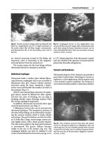
Fig.
12.14.
Right subclavian vein in its long axis (trans-
verse scan of the thorax). This vein is free and has a
large caliper, favorable to catheterization. Note that the
lung surface
(arrow)
is
not far
Above all, a nonthrombosed vein can be col-
lapsed by probe pressure (Fig.
12.5),
provided the
vein is sandwiched between the probe, under the
clavicle,
and the free hand of the operator above it.
The nearer the sternum, the more difficult this
maneuver
is.
For instance, the proximal end of the
subclavian veins cannot be compressed. Doppler
may be of help here. However, two-dimensional
ultrasound is never at a loss for solutions: when a
valvule is visualized in this noncompressible area,
one should observe its spontaneous dynamics.
Frank movements of this valvule (the free subcla-
vian valvule sign) will obviously indicate patency
of the vein and will obviate the need for Doppler.
The discussion in the previous section should the-
oretically render this vein more attractive. The
subclavian vein is localized in a longitudinal sub-
clavian approach, visible on the transverse scan of
the subclavian vessels. The vein differs from the
artery
(Fig.
12.14) in many
details:
like the internal
jugular
vein,
it does not have
a
perfectly round sec-
tion, nor perfectly parallel walls, but wide move-
ments, possible inspiratory
collapse.
Valves
can be
observed here. An echoic flow with visible echoic
particles is again sometimes seen. Differentiating
the vein from the artery using their respective
location is more hazardous since the vessels cross
each
other.
Very near the vein (too near for some)
is the lung: a hyperechoic horizontal structure
with a dynamic pattern and followed by air arti-
facts (see Chaps. 15-18).
Subclavian Venous Thrombosis
As at the internal jugular level, subclavian throm-
bosis can be easily identified using the static
approach
alone.
However, spontaneous echogenic-
ity at this level
is
inferior to that of the cervical area
(Fig. 12.15). A mild compression maneuver will
show the absence of venous compressibility. The
features of internal jugular thrombosis are found
at the subclavian area. However, the frequency of
subclavian venous thromboses appears strikingly
lower than internal jugular thromboses. Except
insufficiency of the method, which is improbable,
we
very rarely encounter patent subclavian venous
thromboses after local catheterization.
Subclavian thrombosis may generate pulmonary
embolism [6].
Id Chapter 12 Upper Extremity Central Veins
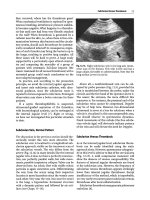
Fig.
12.15.
Occlusive thrombosis of
the
subclavian vein,
short
axis.
The vein is incompressible. The right figure,
in time-motion, depicts a very sensitive sign of occlu-
sive thrombosis: complete absence of respiratory dy-
namics of the vein
Ultrasound and Central Venous Catheterization
Ultrasound offers considerable help in central
venous catheterization. Not only does it allow
approaching the zero fault level, but it also has
many effects: a considerable gain in time, more
comfort for the patient, and substantial savings.
Two
methods are
available.
Ultrasound before nee-
dle insertion, which allows one to choose the most
adequate of six possible sites of insertion: ultra-
sound-enlightened catheterization. Ultrasound
during needle insertion is referred to as ultra-
sound-assisted catheterization.
Contribution of Ultrasound Before Internal
Jugular or Subclavian Catheterization:
Ultrasound-Enlightened Catheterization
The static approach alone of the vessel one intends
to puncture is already rich in information. It has
been proven that large veins are easier to catheter-
ize than small ones
[8].
Obviously, any catheteriza-
tion should be preceded by an ultrasound verifica-
tion of the vein, and every
ICU
should have a small
simple device only for this application.
The best site can be
chosen.
As
previously men-
tioned, observation shows that asymmetry is the
rule at the internal jugular level. It is generally
frank.
A
large venous lumen is sometimes associ-
ated with a contralateral very small, possibly
hypoplastic
one.
These data
have
been investigated
[9].
Asymmetry, defined as a cross-sectional area
greater than twice that of the contralateral vein.
was present in 62% of cases, to the benefit of the
right side in only 68% of cases. This study also
highhghted that
23%
of the internal jugular veins
had, at admission in the
ICU,
a
cross-sectional area
less than 0.4 cm^ (supine patient). Systematic use
of the right side thus means that a small vein will
be encountered in a quarter of
cases.
Such a small
area, which was only slightly increased by the
Trendelenburg maneuver, indicated foreseeable
difficulties in blind emergency catheterization.
Other disorders can explain a priori difficulties
in catheterization:
• Thrombosed vein.
• Aberrant location of the vein related to the
artery, which affects 8.5% of cases [10].
• Inspiratory collapse of the vein. Ultrasound
provides a clear image of this situation. In this
setting, no experimental studies are required
to predict that there is a major risk of gas
embolism here. Note that the increase in inspi-
ratory caliper (i.e., in the sedated patient) is
always correlated with a centrifuge flow of blood
during disconnection of the devices. Let us
specify that this route, which had the reputa-
tion of having constant dimensions even in
hypovolemic patients, can be discovered to be
completely collapsed when studied by ultra-
sound.
• Permanent complete collapse. Is it possible to
even visualize such veins? Experience will often
make it recognizable by very subtle handling of
the probe, which should not flatten the
vein.
The
vein is sometimes enlarged, from
0
to
1
or
2
mm
at inspiration (in mechanical ventilation). The
traditional Trendelenburg maneuver will not be
always effective.
At
the internal jugular level, it is
possible to compress the neck using one's free
hand, just over the clavicle: a small jugular vein
can then appear, but this small caliper may dis-
courage one from the catheterization.
Central Internal Jugular or Subclavian
Ultrasound-Assisted Catheterization
Blind insertion of an internal jugular or subclavian
catheter failed in
10%-19%
of
cases,
and compUca-
tions occurred in
5%-ll%
of
cases,
depending on
whether the operator was experienced
[11].
Other
studies have demonstrated that the failure rate
increases with the gravity of the emergency, up to
38%
in case of cardiac arrest
[12].
Loss
of time and
Central Internal Jugular or Subclavian Ultrasound-Assisted Catheterization
77
complications can severely penalize the patient.
Note that the physician, although next to the
patient, cannot help in case of instability: the
patient remains inaccessible during the entire pro-
cedure.
Permanent ultrasound guidance is mandatory
when a needle is inserted in a central vein. It was
described long ago
[13],
with many studies follow-
ing that have demonstrated the advantages of
ultrasound
[14].
This method is of
little
interest to
physicians who have never encountered technical
difficulties. In our experience, the ultrasound-
guided procedure's single drawback is its simplici-
ty.
Regardless of how clever we were before adopt-
ing this method, we have found that the ability to
find any vein in
a
few seconds
was
sufficient reason
for developing this technique.
Obviously, before learning ultrasound-guided
catheterization, the physician should have a
working knowledge of blind techniques for three
reasons. First, the ultrasound unit can break
down. Second, the ultrasound-assisted procedure,
although very efficient, does not improve one's
techniques in bUnd procedures, since the land-
marks are completely different in both approach-
es.
Third, one must have experienced stressful sit-
uations using the blind approach to fully appreci-
ate the comfort that ultrasound guidance provides.
Basic details of interventional procedures can
be found in Chap. 26. The probe is applied just
proximal to the site of needle insertion. Asepsis
must be
absolute.
A
simple sterile glove surround-
ing the probe is an unacceptable solution. A 45°
angle is made between probe and
needle.
The vein
should be visualized in its long axis, and needle
insertion is monitored using a longitudinal scan,
aiming at the probe landmark (Fig. 12.16). Using
this approach, the needle and the target are visual-
ized over the entire length (Fig.
12.17).
The artery
will not appear in the screen.
Available techniques in the literature describe a
system of servo control to the probe and use a
transversal approach. The artery is visible beside
the
vein,
but the progression of the needle is blind.
The needle can pierce a superficial structure with
impunity. Moreover, the servo control is restrictive
rather than liberating in our experience. Last, the
usual dedicated devices are limited to this use only,
and the quality
is
often suboptimal. In
practice,
we
avoid this technique.
We previously used a simple and quick method
at the internal jugular vein: make a skin landmark
at the area of the vein, switch off the ultrasound
Fig.
12.16.
The operator's hand holding the probe ex-
poses the vein in its long axis and remains strictly
motionless
over the
thorax.
The
operator's hand holding
the needle is
firmly
positioned in front of the probe's
landmark.
The
needle
is
then easily inserted toward the
vein.
For more clarity, gloves and sterile sheath are not
shown in this fictitious procedure
Fig.
12.17.
Subclavian venous catheterization. The body
of the needle is hardly visible in this scan (which does
not reflect the real-time pattern) through the superfi-
cial layers
(black arrows)
and the tip of the needle has
reached the venous lumen
(white arrow)
unit, insert the needle. However, this method was
valid only if the caliper of the vein was large
enough. In fact, if the internal jugular vein is large,
it will be easily catheterized with blind methods
[8].
To
sum
up,
if ultrasound identifies
a
large vein,
it will have the advantage of predicting easy
catheterization using the usual blind techniques.
Note that identifying ultrasound-assisted land-
marks followed by blind cannulation has been
used by other teams at the subclavian level [15].
This approach seems highly hazardous since a
small error in angulation will definitely result in
failure. In spite of this questionable methodology,
it
was
concluded that ultrasound was of no benefit
78 Chapter 12 Upper Extremity Central Veins
in this setting.
We
think ultrasound deserves
a
bet-
ter chance.
When should ultrasound-assisted catheteriza-
tion be proposed?
• After failure of
a
blind attempt
• When an adequate vein is not found using ultra-
sound
• If costs must be controlled, since ultrasound
uses 40% less material than blind techniques
[16]
• In patients with official contraindications to the
blind technique (see next section).
• In any situation where time must not be wasted
• More generally, if one wishes to avoid any risk or
discomfort to the patient
Ultrasound-Guided Subclavian Catheterization
We prefer the subclavian route, since the risk of
infection
is
lower.
Physicians rightly fear this route,
reputed to be dangerous, since immediate compli-
cations are more dramatic than in others. How-
ever, we think that the classic contraindications
(impaired hemostasis, impaired contralateral lung,
obesity, etc.) are no longer contraindications if
ultrasound is used. Ultrasound thus benefits from
all the advantages of the subclavian route with no
drawbacks. In addition, thrombotic risks seem to
be
low,
and the patient's comfort is enhanced.
In a personal study of
50
procedures carried out
in ventilated patients, the success rate was 100%
[
17].
In
72%
of
cases,
frank flux was obtained in the
syringe in less than 20 s, in 16% of cases in less
than
1
min. In 12% of cases that were considered
laborious,
success was nonetheless obtained in less
than 5 min. In other words, ultrasound has accus-
tomed us to immediate
successes,
and
5
min
is
con-
sidered a rather long and laborious procedure. It is
crucial to specify that the patients in this study
were consecutive. Among them, 13 patients were
plethoric (with a distance from the skin to the sub-
clavian vein ^ 30 mm). They were all successfully
catheterized, with an immediate procedure in
11
of
them.
When the procedure is over, absence of pneu-
mothorax (if needed) is checked using ultrasound
(see Chap. 16). Checking that the catheter is not
ectopic is similar. An ectopic position can also be
immediately recognized during catheterization,
since a metallic guidewire is perfectly visible
(Fig.
12.18).
It is thus wise to set the sterile sheet in
order to have access to both the subclavian and the
Fig.
12.18.
This figure is included
to show the
character-
istic pattern of a metallic guidewire or catheter (same
pattern) in the venous lumen. This type of material
generates a continuous hyperechoic mark with a frank
posterior shadow. In addition, note the substantial
venous thrombosis surrounding this internal jugular
catheter
jugular
areas.
If the point of insertion of the needle
is chosen rather distal to the medial
line,
the risk
of
ectopic positioning in the jugular vein decreases,
as
does,
theoretically, any risk of subclavian pinch-
off syndrome.
Ultrasound guidance at the subclavian level is
also mentioned by other teams [18], but studies
conducted in the intensive care setting are rare.
Real-time analysis is rich in information. One
can see the needle arriving in contact with the
proximal venous
wall,
pushing this
wall,
then pen-
etrating the
vein.
Sometimes the proximal and dis-
tal walls are pressed against one another, and the
needle pierces the vein. We have observed more
dramatic phenomena: repeated puncture of
a
sub-
clavian vein with a large caliper can cause a sub-
sequent decrease in lumen size, and therefore be
impossible to recognize, a chain of events that
occur as if there were complete spasm. Obviously,
this can only
create a
vicious circle that reduces the
chances of success.
The needle is not always visualized during its
penetration. This problem will be evoked in
Chap.
26.
Ultrasound-Assisted Internal Jugular
Catheterization
One can of course use the previous technique at
this
level,
the basic rules remain unchanged [19].
Vena Cava Superior
and
Left Brachiocephalic
Vein
79
Emergency Insertion of a Short Central Venous
Catheter
An additional weapon can be used in the extreme
emergency. Under sonographic guidance, we can
insert a 60-mm, 16-gauge catheter in a central vein.
In our areas, such material
is,
alas,
difficult to find,
and in
practice,
we
make have them custom-made.
Using this certainly temporary and hardly academ-
ic,
but potentially lifesaving method, the problem
of
central venous access can be resolved in a few
instants, avoiding transosseous access or others.
Can Ultrasound Replace Radiograph Monitoring
After Insertion of
a
Central Catheter?
What do we ask of the traditional bedside radi-
ograph? First and foremost, pneumothorax infor-
mation. Ultrasound will check for absence of
pneumothorax in a few seconds and with more
accuracy than
a
bedside radiograph (see
Chap.
16).
Second, is the catheter in an ectopic position?
Where has the catheter gone? In a majority of
cases,
it enters the jugular internal vein (after
a
sub-
clavian insertion); ultrasound can detect this dur-
ing the procedure. If it enters the cardiac cavities
and the right auricle is easily
visible,
the end of the
catheter
is
also
visible.
If
not,
measuring the length
of the catheter to be inserted beforehand provides
clinical landmarks; combined with common sense,
this complication is nearly impossible.
The other causes of malpositioning are very
rare.
Poor outflow is a valuable clinical sign of
insertion in a small-caliper vessel (a condition
hard to imagine if the catheter has been inserted
with ultrasound
guidance).
If the monitoring radi-
ograph is requested the next day, or during a new
situation, using ultrasound reduces cumulative
irradiation and costs.
In practice,
we
no longer request follow-up radi-
ographs and have not done so for many
years
[20].
Vena Cava Superior and Left
Brachiocephalic Vein
The vena cava superior is looked for at the supra-
clavicular fossa, with the probe applied toward the
neck. Generally, analysis is disappointing, because
the vein is surrounded by hindering structures
(lung).
However, some patients have good anato-
my. The Pirogoff confluent, the vena brachiocephal-
Fig.
12.19.
Vena cava superior
(arrows)
whose
first
3
cm
are visible in this view. Depending on the quality of
exposure, one can recognize the aorta inside the vein,
the right pulmonary artery posterior to the vein, and
sometimes the right auricle
ica, can then be recognized, and, more central, the
supra-aortic trunks, the right pulmonary artery
(passing posterior to the vein) and last the right
auricle (Fig. 12.19).
Direct signs of venous thrombosis will be hard
to detect since this area is not very accessible and
cannot be compressed. Doppler can be helpful.
However, several indirect signs are available to
indicate good patency or an obstacle: permanent
dilatation, without inspiratory collapse (in a spon-
taneously breathing patient) of the upper veins
[21,22].
Logically, inspiratory collapse of the sub-
clavian vein indicates absence of an obstacle. The
sniff test consists in sudden inspiration by the nose
[21],
which should normally yield jugular and sub-
clavian
collapse.
This
test
is
hard to carry out in the
critically ill patient, since his cooperation is need-
ed. In addition, as with any sudden maneuver, one
can ask if this test is insignificant if there is, for
instance,
floating
venous thrombosis that had been
stable until then.
When the suspicion of thrombosis is high, a
transesophageal examination can clearly analyze
the vena cava superior.
Atelectasis is not a rare situation in the ICU. It
can make the mediastinum accessible to ultra-
sound (see
Fig.
17.1
l,p
121).
A
floating
thrombosis
in the vena cava superior
was
thus diagnosed using
the right parasternal route (Fig. 12.20) in a patient
with recent complete right atelectasis. The patient
was promptly positioned in the right lateral decu-
bitus,
with the hope that a detached thrombus
would choose the right lung.
80 Chapter 12 Upper Extremity
Central
Veins
General Limitations of Ultrasound
As regards the internal jugular and subclavian
veins,
the examination is hindered by parietal
emphysema, local dressings, a tracheostomy, and
cervical collars. Massive hypovolemia makes the
veins hard to detect.
References
Fig.
12.20.
Vena cava, superior location in a patient with
right lung atelectasis. Right parasternal
route.
A
throm-
bus is visible (arrow) within the venous lumen, and is
highly mobile in real time.
PAy
right branch of the pul-
monary artery
Fig.
12.21.
Complete venous thrombosis at the humeral
level in an ICU patient with unexplained fever.
This
pat-
tern is clear when sought. Longitudinal scan at the arm
The left brachiocephalic vein is sometimes visi-
ble anterior to the aortic cross using a suprasternal
route. This segment is not easy to compress. If a
local thrombosis is suspected (in the case of
a
large
left arm, for example), only static analysis will be
contributive: direct detection of a thrombosis,
absence of spontaneous collapse, or absence of the
free valvule sign.
Upper Extremity Veins
Humeral vein thrombosis can be a source of fever
after peripheral catheterization (Fig. 12.21).
1.
Wing
V,
Scheible W (1983) Sonography of jugular
vein
thrombosis.
Am J
Roentgenol 140:333-336
2.
Dauzat M (1991) Ultrasonographie vasculaire dia-
gnostique.
Vigot,
Paris
3.
Chastre
J,
Cornud
F,
Bouchama A, Viau
F,
Benacerraf
R, Gibert
C
(1982) Thrombosis as a complication of
pulmonary-artery catheterization via the internal
jugular
vein.
New Engl
J
Med 306:278-280
4.
Yagi K, Kawakami M, Sugimoto T (1988) A clinical
study of thrombus formation associated with cen-
tral venous catheterization. Nippon Geka Gakkai
Zasshi 89:1943-1949
5.
Horattas MC, Wright DJ, Fenton AH, Evans DM,
Oddi
MA,
Kamienski
RW,
Shields EF (1988) Chang-
ing concepts of deep venous thrombosis of the
upper extremity: report of
a
series and review of the
literature. Surgery 104:561-567
6. Monreal
M,
Lafoz
E,
Ruiz
J, Vails R, Alastrue
A
(1991)
Upper-extremity deep venous thrombosis and pul-
monary emboUsm: a prospective study. Chest 99:
280-283
7.
Ricome JL, Thomas H, Bertrand D, Bouvier AM,
Kalck F (1990) Echographie avec ponction pour le
diagnostic des thromboses jugulaires sur catheter.
Rean Soins Intens Med Urg
6:532
8. Lichtenstein D (1994) Relevance of ultrasound in
predicting the ease of central venous line insertions.
Eur
J
Emerg 7:46
9. Lichtenstein D, Saifi R, Augarde R, Prin S, Schmitt
JM,
Page B, Pipien I, Jardin F (2001) The internal
jugular veins are asymmetric. Usefulness of ultra-
sound before catheterization. Intensive Care Med
27:301-305
Denys
BG,
Uretsky
BF
(1991) Anatomical variations
of internal jugular vein location: impact on central
venous
access.
Crit Care Med 19:1516-1519
Sznajder JI, Zveibil FR, Bitterman H, Weiner P,
Bursztein S (1986) Central vein catheterization,
failure and complication rates by 3 percutaneous
approaches.
Arch Intern Med 146:259-261
12.
Skolnick ML (1994) The role of sonography in the
placement and management of jugular and sub-
clavian central venous catheters. Am J Roentgenol
163:291-295
13.
Denys
BG,
Uretsky
BF,
Reddy
PS,
Ruffner
RJ,
Shandu
JS,
Breishlatt
WM
(1991) An ultrasound method for
safe and rapid central venous access. N Engl J Med
21:566
10
11
References
81
14.
Randolph AG, Cook DJ, Gonzales CA, Pribble CG
(1996) Ultrasound guidance for placement of cen-
tral venous catheters: a meta-analysis of the litera-
ture.
Grit Care Med 24:2053-2058
15.
Mansfield
PF,
Hohn
DC,
Fornage
BD,
Gregurich
MA,
Ota DM (1994) Complications and failures of sub-
clavian vein catheterization. N Engl J Med 331:
1735-1738
16.
Thompson DR, Gualtieri E, Deppe S, Sipperly ME
(1994) Greater success in subclavian vein cannula-
tion using ultrasound for inexperienced operators.
Grit Care Med 22:A189
17.
Lichtenstein D, Saifi R, Meziere G, Pipien I (2000)
Catheterisme echo-guide de la veine sous-claviere
en reanimation. Rean Urg [Suppl
9]
2:184
18.
Nolsoe
C,
Nielsen
L,
Karstrup
S,
Lauritsen K (1989)
Ultrasonically guided subclavian vein catheteriza-
tion.
Acta Radiol 30:108-109
19.
Slama
M,
Novara
A,
Safavian
A,
Ossart
M,
Safar
M &
Fagon JY (1997) Improvement of internal jugular
vein cannulation using an ultrasound-guided tech-
nique. Intensive Care Med 23:916-919
20.
Maury E, Guglielminotti J, Alzieu M, Guidet B &
Offenstadt G (2001) Ultrasonic examination: an
alternative to chest radiography after central venous
catheter insertion? Am J Respir Grit Care Med
164:403-405
21.
Gooding GAW, Hightower DR, Moore EH, Dillon
WP,
Lipton MJ (1986) Obstruction of the superior
vena cava or subclavian
veins:
sonographic diagno-
sis.
Radiology 159:663-665
22.
Grenier P (1988) Imagerie thoracique de Tadulte.
Flammarion, Paris
CHAPTER
13
Inferior Vena Cava
Draining half of the systemic blood toward the
heart and the necessary crossroads of lower
extremity thromboses, the inferior vena cava (JVC)
has a clear strategic situation. Ultrasound occupies
a major place in the search for thromboses, but
also in assessing the JVC dimensions, a possible
marker of the circulating blood volume, as well as
other more marginal applications.
The iliac veins are discussed in Chap. 14.
The Normal Inferior Vena Cava
The inferior vena cava can be separated by the
renal veins into supra- and infrarenal portions.
The infrarenal portion analysis is conditioned
by
gas,
frequent in this
area.
However, the free hand
of the operator (and not the probe itself) can drive
most gas away by applying gentle pressure. The
suprarenal portion is often visible using the liver
acoustic window. It makes its way vertically, at the
right of the aorta, receives the hepatic veins and
opens into the right auricle (see Fig. 4.2, p.
19).
A
spontaneous echoic flow can sometimes be ob-
served. This flow can hesitate, or even be inverted
at inspiration (in mechanically ventilated patients),
an obvious sign of tricuspid regurgitation. This
echoic flow is possibly explained by agglomerated
blood cells [1] and can be massive (Fig.
13.1).
Fine
analysis of the content of the inferior vena cava is
generally
possible.
Extrinsic
obstacles,
catheters or
caval filters can be observed (Fig. 13.2).
The venous caliper is modified by respiratory
and cardiac rhythms. There is usually inspiratory
collapse in the spontaneously breathing subject.
These variations in caliper are a sign of venous
patency.
A
compression maneuver
is
perfectly
pos-
sible,
but the pressure should be brought by the
operator's free hand with spread fingers, with the
probe applied between two
fingers.
A
compression
by the probe alone would possibly damage the
probe, and it can be harmful for the patient. This
Fig.
13.1.
Inferior vena cava, longitudinal scan. In this
vein, an echoic
flow
with visible particles goes toward
the right cavities. In addition, there is a bulge in the
upper portion of
the vein (arrows)y a
frequent variant
of
the normal (saber profile).
Note
that a measurement of
the vein caliper at this level would yield misleading
information in predicting central venous pressure
Fig.
13.2.
lumen
Catheter
(arrow)
within the inferior vena cava
Inferior
Vena
Cava Diameter
and
Central Venous Pressure
83
maneuver
pays
off for subjects with favorable mor-
photype: the inferior vena cava can be easily col-
lapsed. Note that such a maneuver does not affect
the instantaneous blood pressure. The infrarenal
segment can also be collapsed this way. If gentle
pressure does not succeed, it seems wise not to
insist.
Thromboembolic Disorders
The technique is the same as for the upper or low-
er extremity veins. The only difference is that the
static approach should be called a pseudo-static
approach, so to speak, as the frequent necessity to
drive digestive gas off can alter some parameters.
Thrombosis will give signs:
• Static in the static approach:
• Endoluminal echoic irregular pattern
(Fig. 13.3).
• Dynamic in the static approach:
• Absence of spontaneous inspiratory changes
(see »Normal and Pathological Patterns«
below).
• In the dynamic approach:
- Noncompressible vein. This maneuver is
redundant and should not be performed if
previous approaches have identified
a
throm-
bosis.
Fig.
13.3.
Massive thrombosis of the infrarenal inferior
vena
cava.
Transverse
scan of the umbilical area. Anteri-
or to the rachis
(R)
and at the right of the aorta
(A),
the
venous lumen of the inferior vena cava is
filled
with
echoic material, indicating here a recent thrombosis.
Note
that
this
recent
thrombus is still
soft. Hence,
a
com-
pression maneuver may
collapse
the
venous
lumen, with
doubtful consequences.
Young
patient with polytrauma
Caval Filter and Ultrasound
When local conditions are good, the correct posi-
tion of
a
caval filter and its relations with the renal
veins can be accurately assessed (Fig. 13.4).
If transportation of
a
critically
ill
patient or irra-
diation in a pregnant woman must be avoided, it
could be advantageous to insert caval filters at
the bedside, using ultrasound guidance. Once the
floating infrarenal thrombus is identified, and
once the indication is adequate (this would war-
rant an entire
chapter),
one
operator inserts the fil-
ter while another locates the main landmarks
using ultrasound. As for the pilot-bombardier
relation in a
B25,
the two operators should be per-
fectly trained since the roles are permanently
inversed.
The inferior vena cava can be round or flat-
tened; see the next section.
Fig. 13.4. Caval filter, perfectly identified within the
lumen of the suprarenal
JVC
(arrow).
Epigastric trans-
verse
scan.
One
can imagine the possibility of inserting
this device at the bedside
Inferior Vena Cava Diameter and Central Venous
Pressure
This long section gives clues for accurate measure-
ment of the caliper of the inferior vena
cava,
which
should take only a few seconds.
The accuracy of central venous pressure as a
marker of circulating blood volume will not be dis-
cussed here. It could warrant another chapter in
itself.
Recently, this data has been ignored, as it
appears old-fashioned to some. A discussion of
modern hemodynamics can be read in Chap. 28.
84 Chapter
13
Inferior
Vena Cava
Fig. 13.5.
Correlation between expiratory caliper of the
inferior
vena cava
at the
left
renal vein
(VCI)
and central
venous pressure
(PVC)
in
59
ventilated patients
Fig. 13.6.
Irregular pattern, mostly collapsed, of the in-
ferior vena cava. Hypovolemic patient. Note the bulge
(saber profile) at the left of
the
image
Our aim is to provide simple noninvasive data to
the intensivist who may find it useful [2]. Ultra-
sound measurement of the
IVC
caliper
lies
between
the invasive method of inserting a central venous
pressure system and the more invasive trans-
esophageal approach.
Circulating blood volume is mainly located
(65%) in the venous
system.
We
therefore imagine
that a variation in this volume will affect this sec-
tor, the IVC being an ultrasound-accessible por-
tion. A flattened pattern in the obviously hypov-
olemic patients having been regularly observed,
we investigated this parameter in 54 ventilated
patients (Fig.
13.5).
A
caliper less than 10 mm was
correlated with a central venous pressure under
10 cm H2O with an 84% sensitivity, a 95% speci-
ficity, an 89% positive predictive value and a 92%
negative predictive value [3]. Figure 13.5 shows
that the relation is better for the small caliper val-
ues.
Some studies have been conducted in this
field
[4-7],
but most came from cardiologic, non-
critical, spontaneously breathing, laterally posi-
tioned patients, with measurements made at the
hepatic vein level, making any comparison diffi-
cult. Only one study dealt with ventilated patients
and indicated that a caliper of
=^
12
mm always pre-
dicted a central venous pressure
=^ 10
mmHg [7].
Measurement Technique
Simple requirements are necessary for a both
accurate and reliable information.
1.
The patient remains supine. Lateral decubitus
would squash the
IVC
by the liver.
2.
The IVC should be sought in a longitudinal axis
first. A probably frequent mistake is the confu-
sion between the IVC and a hepatic vein (see
Fig. 4.3,
p
20).
Several profiles exist:
-
A
regular profile.
- A saber profile (Fig.
13.1).
This frequent find-
ing, with a bulge when the IVC receives the
hepatic veins, should be recognized and the
operator should remain far from this area,
whose measurement would give erroneous
information. In addition, the venous tissue
pro-
gressively becomes cardiac tissue in this area.
- An irregular, moniUform profile (Fig. 13.6).
3.
The probe is then applied in a transverse
axis.
A
measurement in a longitudinal axis would
expose to overestimation of the caliper, when
the vein is not perfectly located in a frontal axis.
4.
The left renal vein should be looked for
(Fig.
13.7).
This
landmark has two advantages: it
is a reliable place, and
we
are definitely far from
the hepatic bulge.
5.
Measurement should be from face to face, not
from border to border.
6. An end-expiratory measurement is needed (see
»Normal and Pathological Patterns« below).
7.
The increase in caliper with heart beats was not
taken into account in our practice.
In addition, we did not index IVC caliper with
body surface for two reasons. Risk is involved in
determining these data in a critically ill, unstable
patient, since it is necessary to weigh the patient.
Second, IVC dimensions are not correlated with
the morphotype [8]. Human eye diameter varies
little in relation to weight and height as well.
Inferior
Vena Cava
Diameter
and
Central Venous
Pressure
85
Fig.
13.7.
This transverse epigastric view shows the renal
veins'
point of
arrival.
The left renal vein is particularly
visible, passing between the aorta and the superior
mesenteric artery
(v),
the point where we chose to mea-
sure the
JVC
caliper.
Here,
an expiratory caliper of
8
mm
(arrows)
indicates low central venous pressure
Fig. 13.8.
Inspiratory collapse of the inferior vena cava.
Time-motion acquisition, showing a 12-mm diastoUc
caliper (V) that collapsed to 4 mm at inspiration in a
patient with major bleeding and spontaneous breath-
ing
Normal and Pathological Patterns
In spontaneous ventilation, inspiratory caliper
diminishes. This is seen in ambulatory abdominal
examinations, as the patient is fasting
(i.e.,
in mod-
erate hypovolemia).
In mechanical ventilation, inspiratory caliper
increases, for positive thoracic pressure creates an
obstacle to venous return. Inspiratory collapses are
found in nonsedated patients.
The expiratory caliper seems more constant. It
does not vary after intubation of a patient, where-
as inspiratory caliper is usually seriously disrupted.
Inspiratory collapse of a spontaneously breath-
ing patient (Fig. 13.8) can be explained by a dysp-
nea with use of accessory respiratory muscles,
since the inspiratory collapse of thoracic pressure
creates aspiration of the systemic blood, with the
Venturi effect. This situation is striking in acute
asthma (where fluid therapy is not at all con-
traindicated). However, not all dyspneic patients,
even with substantial use of accessory respiratory
muscles, have inspiratory collapse.
An enlarged IVC (Fig. 7.1, p 41), with enlarged
hepatic veins, is seen in right heart failure or
hypervolemia, or can again be normal. Central
venous pressure can be low but is rarely so.
A flattened IVC in a shocked patient (Fig. 13.7)
is correlated with low central venous pressures,
and indicates a hypovolemic part.
When the central venous pressure is rapidly
altered, by fluid therapy, variations of PEEP or
Fig. 13.9.
Caliper of the inferior vena cava (VCI) when
the central venous pressure
(PVC)
is altered
disconnecting the ventilator, IVC caUper follows
(Fig. 13.9).
Advantages
of
the Ultrasonic Method
One should first note that the possible errors of
this noninvasive method should be compared with
the numerous errors in the measurements and
interpretations of central venous pressure or
wedge pressure [9]. Then the advantages can be
delineated:
• These data are immediately available.
• The technique is simple (simple unit, without
Doppler).
• There is no invasive procedure.
• The measurement does not affect the treatment
(whereas measurement of the central venous
86
Chapter 13 Inferior
Vena Cava
pressure means clamping the catheter for a
short time).
• The first measurement can be used. Conversely,
the first information given by central venous
pressure is not very useful: the intensivist mod-
ifies therapeutic plans as its value evolves (a way
to implicitly recognize the imprecision of this
first value).
• A hydrostatic zero does not need to be defined,
although this point can be debated. The supposed
projection of the right auricle varies depending
on habit. Error can be substantial in patients with
widened anteroposterior thorax. Each measure-
ment of the central venous pressure requires a
number of verifications such as the height of the
bed. These points cannot be checked a posteriori.
We could list more of these points.
• The intensivist tries to estimate a volume (the
blood volume). Central venous pressure pro-
vides a pressure (a rather indirect parameter).
IVC measurement provides a distance in mil-
limeters, which is a less indirect parameter. In
discordant patients (in the right-hand column
of Fig. 13.5), one can then wonder which pa-
rameter is misleading. It is in fact tempting to
consider that the information given by a part of
a volume is nearer the truth than the one given
by a simple pressure.
In practice, this parameter will be integrated with
others (heart or lung behavior; see Chaps. 17, 20,
28).
We will conclude with this remark: even if a
patient cannot benefit from cardiac or lung ultra-
sound examination, any abdominal ultrasound
test performed in a critically ill patient should
include the degree of IVC filling.
References
1.
Dauzat M (1991) Ultrasonographie vasculaire dia-
gnostique.
Vigot,
Paris
2.
Magder
S
(1998) More respect for the CVP (Editori-
al).
Intensive Care Med 24:651-653
3.
Lichtenstein D, Jardin F (1994) Appreciation non
invasive de la pression veineuse centrale par la me-
sure echographique du calibre de la veine cave inferi-
eure en reanimation. Rean Urg
3:79-82
4.
Mintz
GS,
Kotler
MN,
Parry
WR,
Iskandrian
AS,
Kane
SA (1981) Real-time inferior vena caval ultrasono-
graphy: normal and abnormal findings and its use in
assessing right-heart function. Circulation 64:1018-
1025
5.
Moreno
F,
Hagan
G,
Holmen
J,
Pryop
A,
Strickland R,
Castle H (1984) Evaluation of size and dynamics of
inferior vena cava as an index of right-sided cardiac
function.
Am J
Cardiol 53:579-585
6. Nakao
S,
Come
P,
Mckay
R,
Ransil
B
(1987) Effects of
positional changes on inferior vena caval size and
dynamics and correlations with right-sided cardiac
pressure.
Am J
Cardiol 59:125-132
7.
Jue J, Chung
W,
Schiller N (1992) Does inferior vena
cava size predict right atrial pressures in patients
receiving mechanical ventilation?
J
Am Soc Echocar-
diogr 5:613-619
8. Sykes AM, McLoughlin RF, So B, Cooperberg PL,
Mathieson
JR,
Gray
RR,
Brandt
R
(1995) Sonographic
assessment of infrarenal inferior vena caval dimen-
sions.
J
Ultrasound Med 14:665-668
9. Teboul JL(1991) Pression capillaire pulmonaire. In:
Dhainaut JF &, Payen D Hemodynamique, con-
cepts et pratique en reanimation. Masson, Paris, pp.
107-121
CHAPTER
14
Lower Extremity Veins
The length of this chapter should in no way
obscure its simplicity.
The main problem of thromboemboUc disease
is the risk of sudden death due to undiagnosed
pulmonary embolism. The clinical data are notori-
ously insufficient
[1,2],
A
routine test that is both
accurate and applicable at the bedside is therefore
of great interest. Phlebography
is
being increasing-
ly replaced by ultrasound
[3],
which means using
the Doppler mode. However, using a rigorous but
simple approach without the Doppler mode, the
problem can be solved in most cases.
We
deliber-
ately do not discuss Doppler in this chapter.
Numerous studies have been conducted on the
advantages of Doppler in thromboembolic disease.
However, the studies dealing with the critically ill
patient admitted in the ICU (in particular, the med-
ical ICU) are rare. In this patient, it is illusory to
consider symptomatic and asymptomatic patients.
Pulmonary embolism affects 50,000 patients
yearly in France and kills
5,000
patients.
Untreated
pulmonary embolism has a
40%
risk of mortality,
angiography has a 0.04%-2% risk, and anticoagu-
lant treatment approximately
2%.
Faced with the specter of pulmonary embolism
and its various presentations, and desirous of nev-
er being surprised by this disorder reputed to be
so pernicious, we have decided to no longer ask
the question of whether an ultrasound exami-
nation of the venous system should be ordered.
This examination
is
fully part of our routine exam-
ination of every admitted patient and is repeatedly
performed. In some instances, the quasi-fortuitous
discovery of venous thrombosis immediately clar-
ifies situations that before were murky.
fore,
only the anterior approach in a supine patient
can be routinely used. We always do transverse
scans of the
veins.
The examination is always bilat-
eral and comparative.
As usual, we use the same device and the same
5-MHz
microconvex probe described in previous
chapters.
As
for any vein, the probe will be held like
a pen in order to control the pressure over the skin.
Quick and easy exploration is possible from the
groin to the knee. The iliac and calf portions will
be studied separately. The inferior vena cava was
discussed in Chap.
13.
A
vein is recognized from:
• Anatomical topography
• The constant presence of the satellite artery,
which is round, sometimes pulsatile, sometimes
calcified
• Its tubular structure
• The complete flattening of the vein when pres-
sure
is
appUed with the probe (see next section),
a major point
• Visualization of
valvules
at times (Fig. 14.1)
Echogenicity, on the other hand, is not informa-
tive,
especially for small
veins,
which can be either
Examination Technique: Recognizing the Vein
As opposed to the ambulatory patient, the critical-
ly ill patient cannot be examined sitting, with the
legs hanging down, or in ventral decubitus. There-
Fig. 14.1 . Common femoral
vein,
longitudinal
scan,
with
no
compression.
The
arrow
designates
a
valvule
88 Chapter 14 Lower Extremity
Veins
Fig.
14.2.
Transverse scan of the common femoral vessels
at the groin (without compression). The artery is at the
left of the image, the vein at the
right.
The absence of
apparent separation between the two vessels is due to a
common tangency artifact, hence this peanut pattern
Fig.
14.3.
Transverse scan of the superficial femoral ves-
sels at the low area of the femur. The femur is an imme-
diately recognized, large arc-like structure with frank
acoustic shadow
(F).
An adequate compression maneu-
ver identifies the
vein,
here under the artery (A)
hypoechoic or echoic if surrounded by echoic
structures.
Once such a structure has been identified as a
vein,
it
is
scanned step by
step
to the
extremity.
The
study can begin at the groin, since the femoral
pulse makes
a
practical landmark. From top to bot-
tom, the following portions will be identified:
• The common femoral vein outside the femoral
pulse. At this level, vein and artery seem to be
one,
with a peanut pattern, since the interface,
which separates vein from artery, is usually not
visible (Fig. 14.2).
• The superficial femoral vein follows, vertical up
to the knee, inside the femur, rarely split
(Fig. 14.3).
• The deep femoral vein leaves the main femoral
axis toward the femur, where it quickly disap-
pears.
•
The
popliteal vein follows the superficial femoral
after the Hunter canal (Fig. 14.4). The shorter
the probe, the easier the popliteal vein can be
explored in a supine patient.
Calf vein analysis raises problems that will be dis-
cussed in »The Calf Problem.« Other veins such as
the gastrocnemial veins are not discussed.
Fig.
14.4.
Posterior (transversal) approach of the popli-
teal fossa, showing the vein
(V),
generally
single,
and the
artery (A)
Examination Technique:
the Compression Maneuver
A three-step approach should again be used. The
two first steps are less important than at the upper
extremity level.
1.
Static aspect in static
analysis.
From the groin to
the feet, the static pattern is rarely informative
since the echogenicity
is
naturally increased.
2.
Dynamic patterns in static
analysis.
Here again,
spontaneous anomalies should be searched for,
but will rarely
be
encountered.
The
Signs
of Femoropopliteal Thrombosis 89
3.
Dynamic pattern in dynamic analysis: the com-
pression maneuver. This is the basic maneuver,
which can be used at almost every level (see
Fig.
12.5).
A
small surface probe is particularly
useful
here.
A
transverse scan is required, since
minor deviation of the axis will not make the
vein disappear from the screen, as would a lon-
gitudinal scan (by the out-of-plane effect).
Compression should be gently applied for three
basic reasons:
- Mild pressure is more than enough to col-
lapse a normal vein.
- Strong pressure can collapse an artery, espe-
cially if
the
blood pressure is low.
- The safety of
a
high-pressure maneuver
is
not
established. A partially detached thrombus
may be dislodged [4], and ultrasound must
remain a safe, noninvasive procedure. We
estimate that it is wise to limit pressure to
0.5-1 kg/cml
The lower femoral segment (Hunter canal) is tra-
ditionally reputed to be inaccessible to compres-
sion.
This is
untrue.
If the free hand of the operator
creates a counter-support opposite the probe, this
segment can be collapsed, and even more using a
moderate, controlled pressure. Some training will
ensure that the operator feels where to place both
hands.
The behavior of a normal vein during the com-
pression maneuver is characteristic: the upper and
lower walls get closer and eventually seem to slap
against each other, resulting in a complete collapse
of the venous lumen. The operator should be
accustomed to feeling the necessary pressure to
obtain this result. Since we did not use Doppler in
our studies, this maneuver can be called the do-it-
without-Doppler. For many
years,
using only two-
dimensional ultrasound has allowed us to confirm
or invalidate diagnoses of venous
thrombosis.
This
opinion is shared by others
[5,6].
Since the first and second steps can generally be
bypassed, only the compression technique is often
directly performed. In this way, routine explo-
ration of
a
normal femoral venous axis should not
exceed
15
s.
The
practice of working on contiguous
sections every millimeter is laudable but not very
profitable, since thrombosis usually involves sever-
al centimeters of venous segment
[7].
Our observa-
tions clearly confirm this notion. However, venous
thrombosis is visible until the critical moment
when it is no longer visible. In some cases of high
suspicion of pulmonary embolism, the discovery
of a centimeter-long area of femoral vein throm-
bosis can definitely confirm the
diagnosis.
We
call
this the miasma sign. The miasma sign is usually
found at more distal areas such as the
calf.
The Signs of Femoropopliteal Thrombosis
Static signs are rarely suggestive at this level, since
the echogenicity of femoropopliteal
veins
is usually
subject to the parasites of
the
surrounding tissues.
The venous caliper can be enlarged, a possibly
informative sign [8, 9]. In some instances, an
echoic heterogeneous pattern is recognized within
the enlarged lumen, thus making the diagnosis
obvious, and the compression maneuver useless.
Dynamic signs in static analysis are not always
clear, since a floating thrombosis rarely includes
these narrow segments - it should be searched for
at the ihac end of the thrombosis.
Conversely, the diagnosis is usually made dur-
ing the dynamic maneuver.
The
best sign of venous
thrombosis is the absence of collapse with mild
probe pressure. This sign has been constant in our
observations (Fig. 14.5).
The literature relates sensitivity and specificity
near 100% for the diagnosis of venous thrombosis
limited to this area [10]. However, studies do not
specify the necessary degree of the pressure, and
Fig.
14.5.
Left iliofemoral thrombosis. In this transverse
scan, registered slightly over the groin, the distal por-
tion of
the
iliac vein is enlarged by an echoic heteroge-
neous completely occlusive material. This sole pattern
renders the compression technique redundant. Hence,
the risk-benefit ratio of
this
maneuver is
inverted.
Note
at the
right
of
the image
an arterial catheter (two paral-
lel
hyperechoic lines)
90
Chapter 14 Lower Extremity Veins
they include Doppler findings. Doppler was of no
use in our experience to confirm or invalidate
venous thrombosis.
Other signs seem secondary.
A
recent thrombo-
sis should be hypoechoic, an old one echoic. This
distinction lacks reliability for some [9], and we
have joined this opinion.
A
recent thrombosis can
be deformed by the probe pressure
[3].
A throm-
bosed vein will not be modified by a Valsalva
maneuver.
Difficulties can arise from poorly echoic veins.
Certain maneuvers can help in difficult cases:
• Comparing a suspect area with the contralater-
al area. Some patients may need more pressure
than usual, in case of extreme plethora, for
instance. In rare cases, however, bilateral, sym-
metric thrombosis can occur, and this situation
can confuse the young operator.
• FiUing the venous lumen when the vein seems
empty:
- Using fluid therapy.
- Lowering the feet using the balance pedal of
the bed.
- Manually compressing the common femoral
vein at the groin (after checking its patency):
for the time being blood engorges the venous
sector in the lower extremity.
Fig.
14.6.
Through a peritoneal effusion, the right iliac
vessels are clearly outlined
The Iliac Level
Iliac veins are classically difficult to access with
ultrasound because of the abundance of local gas.
In addition, iliac segments can be incompressible
even in the absence of gas. This feature is, to
our knowledge, unpredictable from one patient to
another. In a majority of cases, iliac veins can be
followed and compressed over a more or less long
portion.
In a highly echoic patient, and if one carries out
the pelvic examination with both hands (one hold-
ing the probe, one gently driving away the gas),
the inferior vena cava and the two iliac veins can
be analyzed (Fig. 14.6). The vein can suddenly
become visible, once a gas has been driven away.
The pressure exerted by
the
free hand should drive
away
gas
without squashing the vein (for
a
pseudo-
static approach, see p
82).
Experience alone deter-
mines the adequate pressure.
A peritoneal effusion is a fortuitous condition
that makes iliac venous exploration easier: the vas-
cular axes are isolated from the bowel.
Fig, 14.7. Floating iliac thrombosis (M). The floating
character
is
perfectly objectified using the time-motion
mode,
at the
right
(arrow).
Compression of such
a
struc-
ture
may
not
be
sufficiently innocuous
Once the iliac veins are
exposed,
two
approaches
are available: static and dynamic. The static
approach consists in directly detecting the throm-
bosis,
if an echoic irregular tubular mass can be
described within the venous lumen (Fig. 14.5) or if
floating structures are identified (Fig. 14.7). The
dynamic approach,
i.e.,
the
compression maneuver,
is effective in some patients and completely ineffec-
tive in others. Static visualization of an obvious,
more or
less
floating
thrombosis precludes dynam-
ic
analysis,
which becomes useless and dangerous.
It is in the iliac or caval segments, almost never
in the femoropopliteal segments, that a floating
thrombosis can be observed. With experience, it
becomes clear that the diagnosis of floating throm-
bosis is best with two-dimensional ultrasound.
The Calf Problem
91
Other maneuvers are possible, Doppler except-
ed.
A
Valsalva or sniff-test maneuver, in a sponta-
neously breathing patient - if not too tired - will
either increase or collapse the iliac lumen. With
mechanical ventilation, the inspiratory caliper
normally increases slightly. An echoic flow with
dynamic particles can again be seen at the femoral
level. All these maneuvers provide information,
although approximate, but which can exclude
complete obstruction of
the
iliac or caval axes.
In some cases, the iliac veins are impossible to
analyze. When is it a problem? In a non-trauma
patient, isolated iliac thrombosis without femoral
extension is reputed to be extremely rare, not to
say nonexistent [1, 11]. In fact, if there was local
catheterization, it is not rare to find iliac thrombo-
sis with a common femoral origin. In the restrict-
ed field of thromboses occurring in pelvic disor-
ders,
for example, in the obstetrical context, and in
the traumatized patient, it appears risky to set
aside analysis of iliac
segments.
A
venography or a
Doppler complement should then be required. But,
before indicating venography, all the possibilities
of
a
simple technique should be used.
The Calf Problem
This section can be omitted if it is not of clinical
relevance to the reader.
The main problem at the calf is that its explo-
ration is dehcate, time-consuming and risky, with
no guaranteed
result.
Echogenicity
varies
from one
patient to another. The veins are small and numer-
ous (two for one artery, i.e., six veins for each leg).
Their route is
sinuous.
At
this
level,
operator expe-
rience is required. However, this apparent problem
can be considerably tempered.
The techniques available in the literature are
not applicable in the critically ill: the posterior
approach in ventral decubitus, prone position, etc.
A
posterior approach made in a supine patient, by
raising the foot, will empty the
veins.
Although not
described in the textbooks to our knowledge, we
use a method adapted to a patient immobilized in
the supine position - the usual position of the crit-
ically
ill.
We
use the anterior approach, between the
tibia and fibula. In a transverse scan, both bones
are easily recognized, as well as the interosseous
membrane. Just anterior to it passes the anterior
tibial group. Reputed to be only slightly or not at
all emboligenic, this segment is usually occulted.
Posterior to the membrane, through the posterior
Fig. 14.8. Right calf veins, transverse scan, anterior
approach. Interosseous membrane is straight between
the two
bones.
About
2
cm posterior, through a muscle
(the posterior tibial muscle), the tibial posterior and
fibular
veins are
visible.
T,
shadow of
the
tibia.
P,
shadow
of the fibula
tibial muscle, one can observe the fibular group
outside and the tibial posterior inside (Fig.
14.8).
A
regular scanning by the probe from top to bottom
localizes these vessels more easily. Arteries are
usually not detected with our
5-MHz
probe. Last,
the free hand of the operator holds the calf just in
front of
the
probe for the compression maneuvers.
Sometimes,
the static approach is not contributive,
and only the dynamic approach makes it possible
to recognize normal veins, since the collapse of
small structures may be easier to recognize than
the structures themselves. If needed, the head of
the bed can be raised, or a tourniquet can be
appUed at the knee, or venous engorgement can
be generated simply by compressing the common
femoral vein.
Here, as elsewhere, Doppler was not used. Some
authors use only the compression technique at this
level,
with an
85%
sensitivity [12].
Basically, four scenarios are possible at the end
of
this
exploration:
1.
The three venous groups are identified. They
are compressible all along the
calf.
One can
reasonably conclude that the examination is
normal.
2.
A structure is identified. It is tubular, incom-
pressible, echoic, enlarged (a very suggestive
pattern if larger than 6 mm), and unilateral
(Fig. 14.9). Calf thrombosis is quasi-certain. If
this image is prolonged by an image clearly
identified as a normal vein (the sequel
sign),
the
92 Chapter 14 Lower Extremity
Veins
Fig. 14.9. Calf venous thrombosis. In this transverse
scan,
a
tubular, enlarged structure
is
visible,
at the nor-
mal place of
a
posterior calf
vein.
Above
all,
this struc-
ture
is
not compressible
(arrows)
diagnosis of thrombosis seems certain. In a
patient with acute respiratory disorder without
femoral thrombosis, this finding can be decisive
for the diagnosis of pulmonary embolism.
3.
No tubular group is identified. No conclusion is
possible.
One could postulate that
a
thrombosed
vein is enlarged, thus visible, but this remains to
be confirmed.
4.
At
least one portion of one vein
is
identified and
compressible.
This information can be obtained
in a few seconds. It already rules out massive,
complete thrombosis of the leg
veins.
The ultra-
sound report will describe a calf venous system
free in at least
25%,
50%,
or 75% of its volume.
The practical use of this approach will be dis-
cussed in the next section.
How Can the Problem of Calf Veins Be Tempered?
Calf vein detection can have an impact on the
ICU's choice of equipment. Several arguments
should be considered. In other words, is the calf
problem a real problem?
1.
In all the cases where proximal thrombosis has
been detected, calf exploration is useless since
the treatment is not altered. In lower extremity
thrombosis, the area over the popliteal level is
affected in
95%
of cases [11].
2.
The subpopliteal level is considered not emboli-
genic [
13].
No
fatal case of pulmonary embolism
has been reported from an isolated leg venous
thrombosis
[14-18].
Lethal pulmonary embolism
should come from the iliofemoral levels [19-21].
3.
Calf thrombosis extends to the femoral veins in
20%
of cases, and this extension always occurs
before pulmonary embolism
[18].
If this notion
is taken into account, an extremely simple solu-
tion
exists:
when the calf level has not been well
analyzed, one should monitor the distal femoral
vein at the Hunter canal at regular intervals
(every
24
or
48
h).
A
few seconds are required. If
femoral thrombosis is detected by such moni-
toring, curative treatment can then be instigat-
ed. This procedure is conditioned by the pres-
ence or absence of a small ultrasound device at
the bedside.
4.
As seen
above,
patency of at last one part of the
calf
veins
can be checked in a few instants. This
may have immediate consequences since the
risk of embolism from calf thrombosis is usual-
ly considered insignificant. If a portion of the
calf veins is patent, this insignificant risk falls
again and tends toward zero. Detection at any
price of segmental calf thrombosis using inva-
sive procedures or even Doppler equipment,
or blind emergency treatment of isolated calf
venous thrombosis will expose the patient to
iatrogenic consequences. Let us then recall that
pulmonary angiography is a risky examination
[22],
that spiral
CT
has
a
low sensitivity and that
heparin therapy has an
11%
risk of major bleed-
ing and
a
lethal risk between
0.7%
and 1.8% [23,
24].
In other
words,
it may not be useful to diag-
nose or treat in extreme emergencies calf
venous thrombosis that is not massive or that is
nonexistent.
5.
If the physician in charge of the patient finds it
essential to know the exact status of the calf
veins,
the gold standard remains leg venogra-
phy.
Experience suggests that the benefit will be
small, and the drawbacks heavy (see »The Place
of Venography« below).
6. Assuming that venography and blind heparin
therapy both raise concerns, let us now ignore
leg vein status. What then happens if there is
a small thrombosis and if this thrombosis
embolizes, precisely without passing by the
inevitable step of simple extension? It creates a
small, distal pulmonary embolism. The patient
feels a low thoracic pain, but we can logically
suppose that no more severe disorder will fol-
low. This small discomfort should maybe be
considered as less deleterious than the conse-
quential drawbacks of traditional approaches.
In a dyspneic patient of course, or in a patient
with a poor margin of tolerance (chronic respi-
The Place of Venography 93
ratory insufficiency), this reasoning should pos-
sibly be nuanced. In other words, it should be
assumed that there are pulmonary emboU and
pulmonary emboli. Small pulmonary emboh
with no residual thrombosis should possibly be
considered - and managed - differently from
severe pulmonary emboli as well as small pul-
monary emboli with major, unstable venous
thromboses.
We must remain aware that with simple logistics,
the calf problem is not a true problem. The inten-
sivist can take an interest in this segment or not,
but if so, the investment will be small, since the
same small equipment provides an answer to this
question.
Usual Emergency Procedure
In a case of severe shock without a clear explana-
tion, a blind fibrinolysis is sometimes planned. In
such cases, the following method seems to be the
quickest. Femoral axes should first be analyzed,
including popliteal axes, since these segments are
the most often involved, and their analysis is
extremely rapid. In case of normality, internal
jugular veins will be analyzed. If normal, the infe-
rior caval and iliac veins will be included in the
analysis. Lastly, subclavian veins and then calf
veins will be investigated. This procedure, which
may at first sight appear rather untidy, is based
on logic and empiricism and will be well worth
the effort from the moment a thrombosis is
detected.
Limitations of Ultrasound
Iliac and calf vein analysis is uncertain.
If the compression maneuver
is
correct,
the
only
false-positive cases are the rare venous tumors.
Fresh thrombosis may theoretically be com-
pressible and yield false-negative results. The old
notion of the double femoral vein (with only one
thrombosed channel and the illusion of a normal
single vein) has not raised any problems to date.
In a minority of cases, femoral veins cannot be
recognized, in some very plethoric patients or in
deep hypovolemia. Old thrombosis isoechoic to
the surrounding tissues should be a limitation
[16],
but scanning can usually recognize a tubular
structure, even isoechoic.
In the trauma patient, access can be difficult
because there are numerous obstacles: orthopedic
material and dressings, for
example.
The compres-
sion maneuver can be harmful here.
One major limitation remains that is rarely
mentioned: detection of a patent vein means that
there is no thrombosis, but it can also mean that
there is no longer
thrombosis,
which is not exactly
the same.
The True Place of Doppler
Information provided by the Doppler device may
be largely redundant. Its high volume, high cost,
high complexity, increased risk of infections (if
buttons are prominent), and unproven innocuous-
ness must be remembered. Given that the Doppler
technique
is
highly operator-dependent,
we
beheve
that if two-dimensional and Doppler information
agree, one technique is useless. If they disagree,
which one should be trusted?
Doppler can be advantageous in the trauma
patient, since the compression maneuver may be
harmful. At the iliac level, if the clinical suspicion
is real, Doppler may supersede venography. How-
ever, in case of multiple gases, it will not solve the
problem.
For some, Doppler shortens the examination
(an opinion we do not share) and contributes
information on flow [5] or the extent of occlusion
in thromboses [
10].
The
immediate practical use of
this information seems doubtful.
The Place of Venography
Venography has a clear advantage: it provides an
objective document. Certain teams still prefer
venography to ultrasound, especially with young
traumatized patients, where anticoagulation is
never insignificant.
However,
venography:
• Means transportation of
a
critically ill patient.
• Means pelvic irradiation, iodine allergy or other
accidents, and possibly pulmonary embolism.
• Transgresses (as does bedside chest X-rays) the
first rule of radiology: any structure should be
analyzed in two perpendicular planes. There-
fore,
an anterior or posterior thrombosis is
easily missed in a single anteroposterior view.
In addition, several areas cannot be opacified:
deep femorals,
twins,
gastrocnemial
veins,
etc.
94 Chapter 14
Lower
Extremity Veins
• Is operator-dependent in its interpretation.
Troublesome divergences between observers
are reported, from 10% to 35%
[25].
Our experi-
ence confirms a high rate of
errors.
This is com-
pounded if
20%
[5] to 30% [26] of venographies
are normal in pulmonary embolism.
• Involves a difference in cost.
• Is not a pleasant examination.
To sum up, if ultrasound has limitations, veno-
graphy has other limitations, also a problem.
Interventional Ultrasound
Ultrasound can help in femoral vein catheteriza-
tion in exactly the same manner as for the upper
axes.
A
few seconds suffice to check that the vein is
not thrombosed, collapsed, at an aberrant location,
or when the arterial pulse is missing.
Conclusions
Our daily experience shows that compression
ultrasound is a rapid, easy and reliable method.
Large screening for venous thrombosis in any new
or chronic patients is therefore feasible.
We usually combine lower-extremity vein analy-
sis with examination of the internal jugular and
subclavian axes. This approach provides a nearly
complete overview of the deep venous axes in an
acceptably short time. The alternative would be
venography of the lower and upper extremities,
with front and profile acquisition. Such a test is
very unlikely to become routine.
References
1.
Haeger K (1969) Problems of acute deep vein
thrombosis: the interpretation of signs and symp-
toms.
Angiology 20:219-223
2.
Kakkar VV (1975) Deep venous thrombosis: detec-
tion and prevention. Circulation 51:8-12
3.
Dauzat M (1991) Ultrasonographic vasculaire dia-
gnostique.
Vigot,
Paris
4.
Perlin SJ (1992) Pulmonary embolism during com-
pression ultrasound of the lower extremity. Radio-
logy 184:165-166
5.
Cronan JJ (1993) Venous thromboembolic disease:
the role of ultrasound, state of the art. Radiology
186:619-630
6. Lensing
AW,
Prandoni
P,
Brand]es
D,
Huisman PM,
Vigo M, Tomasella G, Krekt J, Wouter Ten Gate J,
Huisman
MV,
BuUer HR (1989) Detection of deep-
vein thrombosis
by
real-time B-mode ultrasonogra-
phy
N
Engl
J
Med 320:342-345
7.
Markel A, Manzo RA, Bergelin RO, Strandness DE
(1992) Pattern and distribution of thrombi in acute
venous
thrombosis.
Arch Surg 127:305-309
8. Murphy TP, Gronan JJ (1990) Evaluation of deep
venous thrombosis: a prospective evaluation with
ultrasound. Radiology 177:543-548
9. Mantoni
M
(1989) Diagnosis of
deep
venous throm-
bosis by duplex sonography. Acta Radiol 30:575-579
10.
Vogel P, Laing FG, Jeffrey Jr RB, Wing VW (1987)
Deep venous thrombosis of the lower extremity:
ultrasound evaluation. Radiology 163:747-751
11.
Rose
SCy
Zwiebel
JZ,
Miller
FJ
(1994) Distribution of
acute lower extremity deep venous thrombosis in
symptomatic and asymptomatic patients: imaging
implications.
J
Ultrasound Med 13:243-250
12.
Yucel EK, Fisher
JS,
Egglin TK, Geller SQ Waltman
AG
(1991) Isolated calf venous thrombosis: diagno-
sis with compression ultrasound. Radiology 179:
443-446
13.
Alpert JS, Smith R, Garlson J, Ockene IS, Dexter L,
Dalen JE (1976) Mortality in patients treated for
pulmonary embolism.
JAMA
236:1477-1480
14.
Moser
KM,
LeMoine JR (1981) Is embolic risk con-
ditioned by location of deep venous thrombosis?
Ann Intern Med 94:439-444
15.
Appelman PT, De Jong TE, Lampmann LE (1987)
Deep venous thrombosis of the
leg:
ultrasound find-
ings.
Radiology 163:743-746
16.
Gronan J J, Dorfman
GS,
Grusmark J (1988) Lower-
extremity deep venous thrombosis: further experi-
ence with and refinements of ultrasound assess-
ment. Radiology 168:101-107
17.
Meibers DJ, Baldridge ED, Ruoff BA, Karkow WS,
Granley J J (1988) The significance of calf muscle
venous thrombosis.
J Vase
Surg 12:143-149
18.
Philbrick JT, Becker DM (1988) Galf deep venous
thrombosis: a wolf in sheep's clothing? Arch Intern
Med 148:2131-2138
19.
Browse
NL,
Thomas
ML
(1974) Source of non-lethal
pulmonary
emboli.
Lancet l(7851):258-259
20.
De Weese JA (1978) Ilio-femoral venous thrombec-
tomy. In: Bergan
JJ, Yao
ST (eds) Venous problems.
Mosby Year Book,
St.
Louis,
p 423-433
21.
Mavor
GE,
Galloway
JMD
(1969) Iliofemoral venous
thrombosis: pathological considerations and surgi-
cal management. Br
J
Surg 56:45-59
22.
Stein PD, Athanasoulis G, Alavi A, Greenspan RH,
Hales
GA,
Saltzman
HA,
Vreim
GE,
Terrin
ML,
Weg
JG (1992) Gomplications and validity of pulmonary
angiography in acute pulmonary embolism. Gircu-
lation 85:462-468
23.
Levine MN, Hirsh J, Landefeld S, Raskob G (1992)
Hemorrhagic complications of anticoagulant thera-
py Ghest 102 [Suppl]:352S-363S
24.
Mant M, O'Brien B, Thong KL, Hammond GW,
Birtwhistle
RV,
Grace
MG
(1977) Haemorragic com-
plications of heparin therapy. Lancet 1(8022):1133-
1135