Handbook of Diagnostic Endocrinology - part 9 ppsx
Bạn đang xem bản rút gọn của tài liệu. Xem và tải ngay bản đầy đủ của tài liệu tại đây (417.09 KB, 36 trang )
278 Wierman
B together regulate spermatogenesis in the presence of high intratesticular
levels of testosterone (4). Testosterone is a prohormone and is converted by 5α-
reductase to dihydrotestosterone, a more potent androgen, or by aromatase into
estradiol. Although androgens were thought to be the major sex steroid hormone
in men, recent studies in animals and humans without an estrogen receptor
(ERα) suggest that estradiol plays a critical role in normal spermatogenesis and
hormone feedback in the male (5,6). LH levels are controlled primarily by
GnRH from the hypothalamus and negative feedback from testosterone and
estradiol from the testes (7–9). FSH levels, in contrast, are controlled by GnRH,
gonadal steroid hormone feedback and the actions of the gonadal peptides,
inhibin B, activin A, and follistatin derived from both the gonad and the pituitary
(10). A critical feature of this endocrine system is the negative feedback of
steroid hormones on hypothalamic and pituitary hormone production (2). Tes-
tosterone levels are secreted in a circadian rhythm with increases at night (3).
Another feature is the requirement for an episodic pattern of hormone secretion
for normal reproductive function (1–3). Continuous production of GnRH-
induced LH secretion turns off the system and is the basis for GnRH analogues
used as reversible medical castration in hormone-dependent malignancies such
as prostate cancer. This episodic pattern of hormonal signaling is important to
remember when obtaining samples for hormone levels.
Fig. 1. Diagram of the hypothalamic-pituitary-testicular axis. GnRH from hypotha-
lamic neurons activates the gonadotropin subunit genes (α, LHβ, FSHβ) to release LH
and FSH from the pituitary. These in turn stimulate spermatogenesis and production of
sex steroids (testosterone, estradiol) and the gonadal peptide, inhibin B.
14/Wierman/277-294/F 12/3/02, 11:24 AM278
Chapter 14/Hypogonadism 279
Table 1
Classification of Erectile Dysfunction
Vascular
Neurogenic
Psychogenic
Iatrogenic
Hormonal (hypogonadism)
NORMAL PHYSIOLOGY OF ERECTION
There are two critical events during erection. Dilation of the arterial bed with
decreased resistance to allow increased blood flow is coupled with relaxation of the
trabecular smooth muscle to compress the venous outflow (11,12). The cavernosal
relaxation is mediated by adrenergic receptors activated by norepinephrine
released from sympathetic nerves. The autonomic nerves, once thought to be the
primary control system, are now thought to act as modulators of the sympathetic
activation to maintain flaccidity. Instead, it is the nonadrenergic, noncholinergic
system that mediates erection (13,14). Nitric oxide (NO) is released both from
the endothelium and the local nerve endings in the corpora cavernosa to trigger
smooth muscle relaxation via activation of guanylate cyclase and the genera-
tion of cyclic guanine monophosphate (cGMP), resulting in an erection (15).
The components of the normal physiology are relevant to the new treatment
options for erectile dysfunction and for those under active investigation.
CLASSIFICATION OF ERECTILE DYSFUNCTION
Erectile dysfunction is defined as the inability to achieve or maintain erection
sufficient to permit satisfactory intercourse (16). The prevalence of this disorder
increases with age and with associated co-morbidities, such as diabetes, athero-
sclerosis, hyperlipidemia, or hypertension (17). Impotence can be generally clas-
sified into five categories: vascular, neurogenic, psychogenic, iatrogenic (due to
a medication the physician prescribes or the patient takes), or hormonal (Table 1).
By the time a patient presents for evaluation, he usually, if not always, has
multifactorial erectile dysfunction. Table 2 outlines the strategy for evaluation.
Vascular
Associated vascular disease is the most common underlying etiology of
patients presenting with impotence and occurs in up to 40% of men (16,17).
Arterial insufficiency results in impaired blood flow to the cavernosal muscles.
A careful history and physical examination can detect the presence of macro- or
microvascular disease. A history of hypertension, hyperlipidemia, or diabetes
predicts an underlying vascular component to erectile dysfunction.
14/Wierman/277-294/F 12/3/02, 11:24 AM279
280 Wierman
Neurogenic
Both trauma, such as spinal cord injury, or systemic diseases, such as diabetes
or primary neurologic diseases, impair the normal process of erection (16–19).
Disorders that impact on the adrenergic, sympathetic, or nonadrenergic non-
cholinergic NO system all result in erectile dysfunction. Additionally, radical pros-
tatectomy, pelvic irradiation, and disorders that cause a peripheral neuropathy,
such as toxins and alcohol are associated with impotence (16–19).
Psychogenic
By the time a patient presents to a health care professional for evaluation of
erectile dysfunction, there is almost uniformly a psychogenic component to the
process (16–19). Performance anxiety can play a role with underlying normal
sexual functioning. An acute onset of impotence associated with a major life
stressor is a clue for a predominant psychogenic etiology. Patients with underly-
ing primary psychoses or neuroses often have decreased libido and erectile dys-
function when their disease is poorly controlled. In addition, medications given
to treat the psychiatric illness are associated with similar symptoms making the
underlying trigger often difficult to clarify.
Iatrogenic
Iatrogenic refers to ingestion of compounds or drugs by the patient pre-
scribed by a physician or taken on their own that impair erectile function
(11,12,18,19). These include antihypertensive medications such as diuretics,
β-blockers, and verapamil. Anti-androgens such as cimetidine, flutamide, and
spironolactone can cause gynecomastia and impotence. Most psychiatric medi-
cations affect libido, elevate prolactin, and can cause hypogonadism as well as
erectile dysfunction. Some over-the-counter medications, including herbal and
health food products, cause impotence, although the exact mechanism has not
been elucidated. It is important to review all medications, vitamins, or health
products with each patient.
Table 2
Approach to the Patient with Impotence
Complete history
Review of medications
Careful physical examination
Laboratory:
LH, FSH, testosterone +/– prolactin, TSH
Glucose
Lipid profile
Liver function tests
14/Wierman/277-294/F 12/3/02, 11:24 AM280
Chapter 14/Hypogonadism 281
Hormonal
Previously, it was argued that hypogonadism is a rare cause of erectile
dysfunction, occurring in less than 10% of men (16,17,19,20). However, these
studies were conducted in urological practices that included younger men with
predominantly psychogenic etiologies. With the renewed interest in erectile
dysfunction by primary care physicians, the educational programs available to
the patients, and new treatment options, the number of patients presenting with
a hormonal component to their erectile dysfunction is increasing. We performed
a retrospective review of hormonal measurements in patients presenting to our
Impotence Clinic at the Denver VA Medical Center and found that 48% had some
endocrine abnormality contributing to their erectile dysfunction (21). Thus, it is
our practice to exclude hypogonadism as a contributing factor in all men present-
ing with impotence. Similarly, Buvat and coworkers suggest screening with a
testosterone and prolactin level (22). A discussion of the differential diagnosis
of patients presenting with hypogonadism is given below.
CLASSIFICATION OF MALE HYPOGONADISM
There are several ways to classify hypogonadism. Many have used primary
and secondary to refer to defects at the level of the testes or central loci. How-
ever, this classification is confusing, since congenital and acquired disorders
can also be thought of as primary and secondary. A more straightforward
approach is to base classification on gonadotropin levels: those associated with
low or normal LH and/or FSH (hypogonadotropic hypogonadism) suggests a
problem at the level of the brain or pituitary; high LH and/or FSH (hyper-
gonadotropic hypogonadism) suggests a testicular problem. The disorders can
then be divided into whether they are inherited or acquired. In addition, one can
ask whether the defect is mechanical or hormonal. The approach to disorders of
male hypogonadism is presented in Tables 3 and the classifications of such
disorders in Tables 4 and 5.
Table 3
Approach to the Patient with Hypogonadism
Complete history
Review of medications
Careful physical examination
Laboratory:
LH, FSH, testosterone
Prolactin, thyroid function panel, free α-subunit,insulin-like growth
factor (IGF)-1, cortisol
MRI if hypothalamic or pituitary disorder
Karyotype if suspected Klinefelter’s, or genetic screening if familial
14/Wierman/277-294/F 12/3/02, 11:24 AM281
282 Wierman
Hypogonadotropic Hypogonadism: Low or Normal LH
and/or FSH and Low Testosterone (Table 4)
HYPOTHALAMIC DISORDERS
Congenital GnRH Deficiency. GnRH deficiency or idiopathic hypogonado-
tropic hypogonadism is a disorder of the GnRH pulse generator (3). It occurs in
1/10,000 men and 1/80,000 women (23). The disorder can occur in an X-linked,
autosomal dominant, autosomal recessive, and sporadic fashion. The patient
presents with a failure to undergo sexual maturation. Patients with associated
midline defects and anosmia are said to have Kallmann’s syndrome (24).
Although one might expect the disorder to be due to a mutation in the GnRH gene,
attempts to identify patients with mutations in the gene have been unsuccessful
(25). Developmental biologists were the first to provide a clue to the underlying
defect. They showed that the GnRH neuronal population is born in the olfactory
placode, and the cells must migrate across the cribiform plate into the forebrain
and the hypothalamus during development (26). Investigators have shown that
the X-linked form of Kallmann’s syndrome is due to a mutation in the KAL gene
whose product has structural features of a neuronal cell adhesion molecule (26–
29). An understanding of the exact physiologic role of the KAL protein has been
hampered by the fact that the gene is not expressed in rodents (29). Efforts are
underway to identify additional molecules that are important in the neuronal
migration of the GnRH population that may be miss-expressed in patients with
other more common forms of GnRH deficiency syndrome.
Other hypothalamic disorders are associated with hypogonadism. These
include Prader-Willi, in which patients have hyperphagia, morbid obesity, and
obstructive sleep apnea (30). Similarly, acquired obesity can be associated with
hypoventilation and hypogonadism (31,32).
The rare disorder X-linked adrenal hypoplasia, caused by mutations in the
DAX-1 gene is associated with hypogonadotropic hypogonadism as well as
adrenal insufficiency (33,34). Studies in a mouse model suggest the primary
Table 4
Causes of Hypogonadotropic Hypogonadism
Hypothalamic disorders:
Congenital GnRH deficiency
Acquired GnRH deficiency
Tumors of the hypothalamus: craniopharyngiomas, dysgerminomas
Pituitary disorders:
Genetic mutations in the GnRH receptor or gonadotropin subunit genes
Pituitary tumors: prolactin, adrenocorticotrophic hormone (ACTH), growth
hormone (GH)
Infiltrative diseases of the pituitary: hemachromatosis, sarcoid
14/Wierman/277-294/F 12/3/02, 11:24 AM282
Chapter 14/Hypogonadism 283
defect is in the control of secretion of GnRH, since gonadotropin synthesis and
secretion was restored with exogenous GnRH administration (35).
Acquired GnRH Deficiency. Acquired GnRH deficiency may occur after
radiation or surgery to the hypothalamus (36,37). The hypothalamus is more
sensitive to the effects of radiation than the pituitary, and thus, patients we have
previously labeled as having panhypopituitarism after radiation, often have their
defect at the level of the hypothalamus. The patients have multiple hypothalamic
defects resulting in multiple pituitary deficiencies and require lifelong hormone
replacement.
Alternatively, men can have acquired defects in the GnRH pulse generator.
Although hypothalamic amenorrhea is a well-recognized disorder in women due
to the effects of stress, excessive exercise, or eating disorders to inhibit normal
reproductive function, it was previously thought to be rare in men. This was
based on the fact that GnRH-induced LH pulse frequency of every 2 h is fairly
stable in men in contrast to the need for a changing hypothalamic input in the
female to maintain normal cyclicity (3). Recent studies however, have docu-
mented the acute reversible alteration in GnRH-induced gonadotropin secretion
in men with severe stress or with illness (38,39). Additionally, Nachtigall,
Crowley and coworkers reported a nonreversible type of acquired GnRH defi-
ciency in men (40). They studied a group of men who had undergone a normal
puberty, but then experienced loss of GnRH-induced gonadotropin secretion.
Several clinical and biochemical features identified men with this disorder. These
included: higher testicular vol (18 vs 3 mL), higher baseline serum testosterone
level (78 vs 49 ng/dL), and higher serum inhibin B (119 vs 60 pg/mL) (40).
Tumors of the Hypothalamus. Craniopharyngiomas are tumors that are
located at the level of the hypothalamus and pituitary. Patients may present at any
age with partial or complete pituitary insufficiency (41). The patients often have
associated hyperprolactinemia and, depending on the timing of the development
of the tumor, can present with delayed or incomplete puberty or acquired
hypogonadism. Patients with dysgerminomas or harmartomas of the hypothala-
mus or pineal gland may present with either precocious sexual development or
acquired hypogonadism (42).
P
ITUITARY DISORDERS
Defective GnRH Receptor or Gonadotropin Subunit Gene Expression. It
has recently been appreciated that there are genetic disorders that cause defects
in the reproductive axis, in addition to those associated with GnRH deficiency.
A family has been described with mutations in the first and third intracellular
loop of the GnRH receptor gene (43). The brother and sister presented with
hypogonadotropic hypogonadism and delayed or absent puberty. A homozygous
mutation in the LH β-subunit gene has also been reported to cause male hypogo-
nadism (44), and several women with delayed puberty and hypogonadism have
been described with mutations in the FSH β-subunit gene (45,46).
14/Wierman/277-294/F 12/3/02, 11:24 AM283
284 Wierman
Pituitary Tumors. Tumors of the pituitary cause hypogonadism by either
mass effect and destruction of gonadotropes or by hormonal production, which
inhibits the GnRH pulse generator. The most common type of pituitary tumor is
the prolactinoma, which occurs in 40% of patients (47). Although in men, the
tumors tend to be macroadenomas (greater than 1 cm in diameter), the mecha-
nism of decreased testosterone levels is not by mass effect. Instead, prolactin acts
at all levels of the hypothalamic-pituitary-testicular axis to inhibit function. Stud-
ies in hyperprolactinemic men showed that exogenous GnRH administration
with a GnRH pump induced normal reproductive function (48). These studies
confirm that the major effect of excess prolactin is at the level of the hypothala-
mus. Recent studies have shown the presence of prolactin receptors on GnRH
neuronal cell lines, consistent with the direct effects of prolactin on the GnRH
pulse generator. Prolactin has an independent effect on libido that is poorly
understood. Thus, men given testosterone replacement for hypogonadism asso-
ciated with a prolactinoma often will have persistent decreased libido until the
prolactin level is normalized.
Other pituitary tumors are often present in men with hypogonadism. Cushing’s
syndrome, with excess cortisol production from an endogenous or exoge-
nous source, results in inhibition of the reproductive axis. Again, the effects of
excess cortisol are to suppress GnRH secretion and induce hypogonadism (49,50).
Patients with acromegaly and growth hormone-producing tumors often have
decreased testosterone levels. These patients usually have large tumors, so that
the effects may be due to mass effect or may due to the fact that the tumors
co-secrete prolactin (51). Finally, patients with glycoprotein-secreting pituitary
tumors frequently have associated erectile dysfunction and hypogonadism, but
with elevated gonadotropins. This will be discussed in further detail below.
Infiltrative Diseases of the Pituitary. There are many uncommon disorders
that involve infiltration of the pituitary and gonadotropin deficiency. The most
common of these is hemachromatosis, in which excess iron is deposited selectively
in gonadotropes (52,53). The carrier frequency is 1/250, and the heterozygote can
present with the constellation of clinical features when exposed to excess alco-
hol. These include severe hypogonadism with prepubertal testosterone levels, loss
of body hair, diabetes, bronze discoloration of the skin, cardiomyopathy and arthr-
opathy, in addition to progressive liver disease and cirrhosis. Aggressive phle-
botomy is occasionally associated with reversal of the features early in the disease
process (53,54). Delayed diagnosis requires life-long androgen replacement.
Other diseases that infiltrate the pituitary and cause hypogonadism include the
granulomatous diseases, such as sarcoid (55). These patients more commonly
present with hyperprolactinemia and diabetes insipidus, due to the presence of
granulomas in the pituitary stalk and hypothalamus. An autoimmune process
termed lymphocytic hypophysitis has been associated with acquired hypo-
gonadotropic hypogonadism (56). Infectious agents, such as histoplasmosis,
14/Wierman/277-294/F 12/3/02, 11:24 AM284
Chapter 14/Hypogonadism 285
tuberculosis, and rarely, coccidiomycosis or cryptosporosis, can infiltrate the
pituitary, often affecting the production of multiple pituitary hormones (55).
Additionally, hematopoietic tumors, such as leukemias and lymphomas, have
occasionally been reported to involve the pituitary to cause hypofunction (57).
Finally, tumors may metastasize to the pituitary (57). These often invade via the
posterior pituitary and present with posterior as well as anterior pituitary dys-
function. Tumors with a predilection for the pituitary include: prostate, lung,
breast, melanoma, and renal cell cancer.
Table 5
Causes of Hypergonadotropic
Hypogonadism
Klinefelter’s syndrome
Intrauterine/testicular hypofunction
Genetic defects in gonadotropin action
Mechanical disorders of the testes
Infiltrative diseases of the testes
Glycoprotein-secreting pituitary tumors
Hypergonadotropic Hypogonadism: High LH
and/or FSH and Low Testosterone (Table 5)
KLINEFELTER’S SYNDROME
Klinefelter’s syndrome is a chromosomal disorder (XYY) of nondysjunction
that is associated with ultimate hypogonadism (58,59). There is lack of spermatic
development and tubules, resulting in small testes at any stage of pubertal devel-
opment. Initially, the Leydig cells function to produce low levels of gonadal
steroids; however, with time, there is progressive tubular fibrosis and decline in
androgen production. Men with Klinefelter’s present with delayed or halting
puberty, eunochoid body habitus, gynecomastia, and small testes. They are
infertile and, ultimately, need androgen replacement. Patients with a mosaic karyo-
type, XXY/XY, have less of the clinical hallmarks, do not have associated gyneco-
mastia, and have less severe and a later onset of their hypogonadism (58,59).
I
NTRAUTERINE/TESTICULAR HYPOFUNCTION
There are several disorders that occur across gestation that result in absent or
abnormal testicular function by birth (59,60). Testicular agenesis is associated
with absent testes. Vanishing testes syndrome is seen with testicular remnants that
disappear soon after birth. Finally, infants with cryptochidism are thought to have
had a late insult to the system. With orchiopexy, the reproductive axis in these
boys can function normally; although studies have suggested that they have sub-
tle deficiencies in spermatogenesis. With aging, Leydig cell function declines,
14/Wierman/277-294/F 12/3/02, 11:24 AM285
286 Wierman
and patients often require androgen replacement. The underlying insult can be
timed by the severity of the defect with the understanding that testicular develop-
ment occurs prior to ovarian development at 9–11 wk of gestation.
G
ENETIC DEFECTS IN GONADOTROPIN ACTION
Mutations in the gonadotropin subunit receptor genes have recently been
identified that result in abnormal pubertal development. Some mutations of the
LH receptor are constitutively active, resulting in gonadotropin-independent
precocious puberty or testitoxicosis in boys (61). Inactivating mutations in the
LH receptor result in Leydig-cell hypoplasia and under-masculinization in males
(62–64). Inactivating FSH receptor mutations are associated with primary gonadal
failure in males and hypergonadotropic hypogonadism in females (65).
M
ECHANICAL DISORDERS OF THE TESTES
Torsion of the testes can occur at any age. Normal sexual function can be
achieved with only one gonad, so that hypogonadism only occurs when the vas-
cular supply is compromised to both gonads or the remaining gonad is impaired
due to another underlying problem.
I
NFILTRATIVE DISORDERS OF THE TESTES
Similar to the pituitary, the testes is the locus for a wide variety of disorders
(reviewed in ref. 60). Iron deposition occurs in the testes of patients with
hemachromatosis, but the patients usually present with the pituitary rather than
the primary testicular defect. Mumps occurring after puberty is associated with
risk for subsequent hypogonadism. Additionally, infections, such as tuberculo-
sis, human immunodeficiency virus (HIV), histoplasmosis, and others, have
been reported to infiltrate the male gonad. Leukemia and lymphoma infiltration
is often seen, but is of unclear clinical relevance.
G
LYCOPROTEIN-SECRETING PITUITARY TUMORS
Although most patients with elevated gonadotropin and low testosterone
levels have a testicular locus of their hypogonadism, some patients with a
pituitary disorder present with similar laboratory abnormalities. These are
patients with glycoprotein-secreting pituitary tumors, which produce some
component of the glycoprotein hormones: α-subunit, LH-β-subunit or FSH-β-
subunit or rarely thyroid-stimulating hormone (TSH)-β-subunit (66–68). These
tumors, previously called nonfunctional tumors, occur in 30–35% of pituitary
tumors. They occur most commonly in older men and present with erectile
dysfunction, hypogonadism, and mass effects causing headache or visual dis-
turbance (66–68). Based on the secretory pattern of the glycoprotein tumor, the
patient may have elevated FSH with or without elevated LH or α-subunit levels
and low, normal, or high testosterone levels. Unfortunately, this is the same
14/Wierman/277-294/F 12/3/02, 11:24 AM286
Chapter 14/Hypogonadism 287
pattern of hormonal abnormalities seen in men with early or late testicular
failure. Attempts to use other markers, such as elevated prolactin levels, as a
signal of stalk compression or gonadotropin response to thyrotropin-releasing
hormone (TRH) have been disappointing in discriminating patients with pitu-
itary tumors (66–68). Magnetic resonance imaging (MRI) is the test of choice
to exclude a tumor in a patient with hypergonadotropic hypogonadism and
symptoms of a mass effect. The tumors are often large at the time of clinical
detection and are treated with transphenoidal surgical resection with occa-
sional need for postoperative radiation. After surgery, androgen and other
pituitary hormone replacement is often required depending on the status of the
residual normal pituitary.
Table 6
Disorders that Present
with Variable Patterns of Hypogonadism
Aging
Diabetes
Alcohol
Liver disease
Disorders that Present with Variable Patterns
of Hypogonadism (Table 6)
DIABETES
Patients with diabetes often present with a combination of erectile dysfunction
with or without hypogonadism (69). Early in the disease process, the lack of
metabolic control is associated with a mixed erectile disorder, which is reversed
with improved blood glucose control. Later in the disease process, the patient
presents with multifactorial erectile dysfunction and hypogonadism, which can
be hypogonadotropic or hypergonadotropic, and require androgen replacement.
A
GING
Many studies now show a gradual decline in androgen production from the
testes with age (70,71). Studies conflict on the timing of the process and the exact
number of men that are affected, in contrast to the uniform pattern of ovarian
failure seen at menopause in women (70,71). Both hypogonadotropic and
hypergonadotropic patterns have been reported. Earlier data was flawed by the
inclusion of sick hospitalized men with other disorders that underlie their hypo-
gonadism. Since co-morbid conditions increase with aging, however, the evalu-
ation of all men for androgen deficiency is warranted.
14/Wierman/277-294/F 12/3/02, 11:25 AM287
288 Wierman
ALCOHOL AND LIVER DISEASE
Excess alcohol has widespread effects on the reproductive axis. It has direct
toxic effects on the Leydig cells, decreasing testosterone production (72–74).
Additionally, the associated central effects inhibit gonadotropin production
(72). Thus, a pattern of high or low gonadotropins with low testosterone can be
observed.
TREATMENT ISSUES
Gonadotropin Replacement
In patients with a hypothalamic or pituitary defect, restoration of fertility as
well as androgen replacement, are options (75–77). Induction or reinduction of
spermatogenesis can be performed using pulsatile GnRH administration or a
combination of human menopausal gonadotropins (HMGs) and human chori-
onic gonadotropins (hCGs). Studies have shown that both are effective, but
require parenteral administration and are costly. Successful spermatogenesis
is predicted by the size of the testes upon initiation of therapy (75–77), reflect-
ing the severity of the gonadotropin deficiency. When fertility is not desired,
androgen replacement options are similar to those with a primary testicular
defect (see below).
Androgen Replacement
Androgen replacement should be considered for hypogonadism not only for
restoration of sexual function, but for effects on muscle mass, respiratory drive,
maintenance of bone mass, and cardioprotection (78). Testosterone histori-
cally was available by intramuscular (IM) injection of depotestosterone 200–
300 mg IM every (q) 2 to 3 wk. Trough levels just below the normal range,
obtained before the 4th dose, help to maintain an optimal level of replacement.
Yearly prostatic exams, lipid profiles, and complete blood counts (CBCs) are
recommended to avoid side effects of worsening of benign prostatic hypertro-
phy, increase in low density lipoprotein (LDL) cholesterol levels, or poly-
cythemia. Testosterone therapy is contraindicated in patients with underlying
prostatic cancer. The availability of testosterone patches, and, more recently,
gel formulations has revolutionized androgen replacement. These products
allow a more steady-state replacement strategy without the highs and lows of
intramuscular administration. Side effects of contact dermatitis and lack of
ability to fine tune the dosing regimen with currently available androgen
patches remain problems that should be overcome with future improvements
in the drug delivery systems.
14/Wierman/277-294/F 12/3/02, 11:25 AM288
Chapter 14/Hypogonadism 289
Erectile Dysfunction
The currently available treatment options for erectile dysfunction include me-
chanical devices and local or systemic drugs to modulate penile blood flow.
Vacuum devices use suction to induce an erection and constriction rings to main-
tain the erection. Although cumbersome, they are safe and effective (79,80).
Intracavernosal injections of alprostadil are useful in diabetic patients who are
comfortable with injections and often have a peripheral neuropathy that diminishes
the major side effect of pain after the injection (81). Other side effects include risk
of bleeding, infection, and priapism, all which occur only rarely. Compliance with
long-term use has been poor with recent studies, suggesting that only 32% of
patients continued on the therapy (82). The intraurethral formulation of alprostadil
(MUSE) has not been as successful as initial reports suggested (83,84).
The major advancements in the treatment of erectile dysfunction are the oral
therapies. Yohimbine hydrochloride is an α-adrenergic antagonist and has been
shown to be effective only in psychogenic impotence. The drug was noted to be
20% more effective than placebo when given at 5.4 mg 3×/d (85). In these
patients, combination with trazadone, a serotonin agonist, may increase effec-
tiveness (85). The recent availability of sildenafil (Viagra) has popularized the
problem of erectile dysfunction. Sildenafil works by inhibiting Type 5 phos-
phodiesterase, which breaks down cGMP, the downstream target of NO (86,87).
Studies have shown the higher effectiveness in those with psychogenic impo-
tence, spinal cord injury, and those men with partial rather than complete erectile
dysfunction (86,87). Patients with diabetes or after urological surgery have less
response to the drug. Side effects include headache, dyspepsia, blue discolora-
tion of vision, and postural hypotension. Thus, the drug is contraindicated in
patients on nitrates. Additionally, the drug half-life can be potentiated by other
medications, with aging, or with renal or hepatic disease. Longer clinical expe-
rience has suggested that for men with stable coronary artery disease, sildenafil
had no deleterious effects on clinical symptoms, exercise capacity, or exercise-
induced ischemia assessed by echocardiograpy (88). New agents for erectile
dysfunction include oral apomorphine (Ixense, Uprima), an opioid antagonist for
psychogenic impotence (89), and phentolamine, which is used to block the nore-
pinephrine-mediated smooth muscle relaxation and vasodilation (90).
SUMMARY
Thus, after a careful history, physical exam, and selected laboratory tests, one
can classify patients with erectile dysfunction and/or hypogonadism into specific
categories that allow appropriate therapeutic interventions (91). Research is
underway to use the new advances in the understanding of the physiology of
14/Wierman/277-294/F 12/3/02, 11:25 AM289
290 Wierman
erection and in disorders of the hypothalamic pituitary gonadal axis to target
more specifically the treatment choices.
REFERENCES
1. Belchetz PE, Plant TM, Nakai Y, Keogh EJ, Knobil E. (1978) Hypophysial responses to
continuous and intermittent delivery of hypothalamic gonadotropin-releasing hormone. Sci-
ence 1978;202:631–633.
2. Marshall JC, Kelch RP. Gonadotropin-releasing hormone: role of pulsatile secretion in the
regulation of reproduction. N Engl J Med 1986;315:1459–1468.
3. Crowley WF Jr, Whitcomb RW, Jameson JL, Weiss J, Finkelstein JS, O’Dea LS. Neuroendo-
crine control of human reproduction in the male. [review]. Rec Prog Horm Res 1991;47:27–62.
4. Hayes FJ, Hall JE, Boepple PA, Crowley WF Jr. Clinical review 96: differential control of
gonadotropin secretion in the human: endocrine role of inhibin. J Clin Endocrinol Metab
1998;83:1835–1841.
5. Couse JF, Korach KS. Estrogen receptor null mice: what have we learned and where will they
lead us? Endocr Rev 1999;20:358–417.
6. Smith EP, Boyd J, Frank GR, et al. Estrogen resistance caused by a mutation in the estrogen-
receptor gene in a man. N Engl J Med 1994;331:1056–1061.
7. Finkelstein JS, O’Dea LS, Whitcomb RW, Crowley WF Jr. Sex steroid control of gonadotro-
pin secretion in the human male. II. Effects of estradiol administration in normal and gonadot-
ropin-releasing hormone-deficient men. J Clin Endocrinol Metab 71991;3:621–628.
8. Finkelstein JS, Whitcomb RW, O’Dea LS, Longcope C, Schoenfeld DA, Crowley WF Jr. Sex
steroid control of gonadotropin secretion in the human male. I. Effects of testosterone admin-
istration in normal and gonadotropin-releasing hormone-deficient men. J Clin Endocrinol
Metab 1991;73:609–620.
9. Bagatell CJ, Dahl KD, Bremner WJ. The direct pituitary effect of testosterone to inhibit
gonadotropin secretion in men is partially mediated by aromatization to estradiol. J Andrology
1994;15:15–21.
10. Mather JP, Moore A, Li RH. Activins, inhibins, and follistatins: further thoughts on a growing
family of regulators. Proc Soc Exp Biol Med 1997;215:209–222.
11. Krane RJ, Goldstein I, Saenz de Tejada I. Medical progress: impotence. N Engl J Med
1989;321:1648–1659.
12. Korenman SG. New insights into erectile dysfunction: a practical approach. Am J Med
1998;105:135–144.
13. Ignarro LJ. Nitric oxide as the physiological mediator of penile erection. J NIH Res 1992;4:59–62.
14. Rajfer J, Aronson WJ, Bush PA, et al. Nitric oxide as a mediator of relaxation of the corpus
cavernosum in response to nonadrenergic, noncholinergic neurotransmission. N Engl J Med
1992;326:90–94.
15. Burnett AL, Lowenstein CJ, Bredt DS, et al. Nitric oxide: a physiologic mediator of penile
erection. Science 1992;257:401–403.
16. NIH Consensus Conference. Impotence. JAMA 1993;270:83–90.
17. Feldman HA, Goldstein I, Hatzichristal DG, et al. Impotence and its medical and psychosocial
correlates: results of the Massachusetts Male Aging Study. J Urol 1994;151:54–61.
18. Saenz de Tejada I, Goldstein I, Azadzoi K, et al. Impaired neurogenic and endothelium-
mediated relaxation of penile smooth muscle from diabetic men with impotence. N Engl J Med
1989;320:1025–1030.
19. Wierman ME. Advances in the diagnosis and management of impotence. Adv Intern Med
1999;44:1–17.
20. Korenman SG, Morley JE, Mooradian AD, et al. Secondary hypogonadism in older men: its
relation to impotence. J Clin Endocrinol Metab 1990;71:963–969.
14/Wierman/277-294/F 12/3/02, 11:25 AM290
Chapter 14/Hypogonadism 291
21. Mahoney J, Bruder JM, Balanoff A, et al. Effectiveness of laboratory assessment of the
pituitary-gonadal axis in patients with impotence [abstract 0038]. Clin Res 1994;42:250.
22. Buvat J, Lemaire A. Endocrine screening in 1,022 men with erectile dysfunction: clinical
significance and cost-effective strategy. J Urol 1997;158:1764–1767.
23. Waldstreicher J, Seminara SB, Jameson JL, et al. The genetic and clinical heterogeneity of
gonadotropin-releasing hormone deficiency in the human. J Clin Endocrinol Metab
1996;81:4388–4395.
24. Kallmann F, Schoenfeld WA, Barrera SE. The genetic aspects of primary eunuchoidism. Am
J Mental Defic 1944;48:203–236.
25. Weiss J, Crowley WF, Jameson JL. Structure of the GnRH gene in patients with idiopathic
hypogonadotropic hypogonadism. J Clin Endocrinol Metab 1989;69:299–303.
26. Schwanzel-Fukada M, Bick M, Pfaff DW. Luteinizing hormone-releasing hormone (LHRH)-
expressing cells do not migrate normally in an inherited hypogonadal (Kallmann) syndrome.
Mol Brain Res 1989;6:311–326.
27. Franco B, Guioli S, Pragliola A, et al. A gene deleted in Kallmann’s syndrome shares homol-
ogy with neural cell adhesion and axonal path-finding molecules. Nature 1991;353:529–536.
28. Legouis R, Hardelin J-P, Levilliers J, et al. The candidate gene for the X-linked Kallmann
syndrome encodes a protein related to adhesion molecules. Cell 1991;67:423–435.
29. Rugarli EI, Ballabio A. Kallmann syndrome: from genetics to neurobiology. JAMA
1993;270:2713–2716.
30. Bray GA, Dahms WT, Swerdloff RS, Fiser RH, Atkinson RL, Carrel RE. The Prader-Willi
Syndrome: a study of 40 patients and a review of the literature. Medicine 1983;62:59–80.
31. Vermeulen A, Kaufman JM, Deslypere JP, Thomas G. Attenuated luteinizing hormone (LH)
pulse amplitude but normal LH pulse frequency, and its relation to plasma androgens in
hypogonadism of obese men. J Clin Endocrinol Metab 1993;76:1140–1146.
32. Santamaria JD, Prior JC, Fleetham JA. Reversible reproductive dysfunction in men with
obstructive sleep apnoea. Clin Endocrinol 1988;28:461–470.
33. Kruse K, Sippell WG, Schnakenburg KV. Hypogonadism in congenital adrenal hypoplasia:
evidence for a hypothalamic origin. J Clin Endocrinol Metab 1984;58:12–17.
34. Muscatelli F, Strom TM, Walker AP, et al. Mutations in the DAX-1 gene give rise to both
X-linked adrenal hypoplasia congenita and hypogonadotropic hypogonadism. Nature
1994;372:672–676.
35. Ikeda Y, Luo X, Abbud R, Nilson JH, Parker KL. The nuclear receptor steroidogenic factor
1 is essential for the formation of the ventromedial hypothalamic nucleus. Mol Endocrinol
1995;9:478–486.
36. Samaan NA, Bakdash MM, Caderao JB, Cangir A, Jesse RH Jr, Ballantyne AJ. Hypopituitar-
ism after external irradiation. Ann Intern Med 1975;83:771–777.
37. Constine LS, Woolf PD, Cann D, et al. Hypothalamic-pituitary dysfunction after radiation for
brain tumors. N Engl J Med 1993;328:87–94.
38. Woolf PD, Hamill RW, McDonald JV, Lee LA, Kelly M. Transient hypogonadotropic
hypogonadism caused by critical illness. J Clin Endocrinol Metab 1985;60:444–450.
39. Spratt DI, Bigos ST, Beitins I, Cox P, Longcope C, Orav J. Both hyper- and hypogonadotropic
hypogonadism occur transiently in acute illness: bio- and immunoactive gonadotropins. J Clin
Endocrinol Metab 1992;75:1562–1570.
40. Nachtigall LB, Boepple PA, Pralong FP, Crowley WF Jr. Adult-onset idiopathic hypogonado-
tropic hypogonadism—a treatable form of male infertility. N Engl J Med 1997;336:410–415.
41. Sklar CA. Craniopharyngioma: endocrine abnormalities at presentation. Pediatr Neurosurg
1994;1:18–20.
42. Raisanen JM, Davis RL. Congenital brain tumors. Pathology 1993;2:103–116.
43. de Roux N, Young J, Misrahi M, Genet R, Chanson P, Schaison G, Milgrom E. A family with
hypogonadotropic hypogonadism and mutations in the gonadotropin-releasing hormone
receptor. N Engl J Med 1997;337:1597–1602.
14/Wierman/277-294/F 12/3/02, 11:25 AM291
292 Wierman
44. Weiss J, Axelrod L, Whitcomb RW, Harris PE, Crowley WF, Jameson JL. Hypogonadism
caused by a single amino acid substitution in the β subunit of Luteinizing hormone. N Engl
J Med 1992;326:179–183.
45. Matthews CH, Borgato S, Beck-Peccoz P, et al Primary amenorrhoea and infertility due to a
mutation in the beta-subunit of follicle-stimulating hormone. Nat Genet 1993;5:83–86.
46. Layman LC, Lee E-J, Peak DB, et al. Delayed puberty and hypogonadism caused by mutations
in the follicle-stimulating hormone beta-subunit gene. N Engl J Med 1997;337:607–611.
47. Carter JN, Tyson JE, Tolis G, et al. Prolactin-secreting tumors and hypogonadism in 22 men.
N Engl J Med 1978;299:847–852.
48. Bouchard P, Lagoguey M, Brailly S, Schaison G. Gonadotropin-releasing hormone pulsatile
administration restores luteinizing hormone pulsatility and normal testosterone levels in males
with hyperprolactinemia. J Clin Endocrinol Metab 1985;60:258–262.
49. Luton J, Thieblot P, Valcke J, Mahoudeau JA, Bricaire H. Reversible gonadotropin deficiency
in male Cushing’s disease. J Clin Endocrinol Metab 1977;45:488–495.
50. MacAdams MR, White RH, Chipps BE. Reduction of serum testosterone levels during chronic
glucocorticoid therapy. Ann Int Med 1986;104:648–651.
51. Molitch ME. Clinical manifestations of acromegaly. Endocrinol. Metab Clin. North Am.
1992;21:597–614.
52. Kelly TM, Edwards CQ, Meikle AW, Kushner JP. Hypogonadism in hemochromatosis:
reversal with iron depletion. Ann Int Med 1984;101:629–632.
53. Tavill AS. Clinical implications of the hemochromatosis gene. N Engl J Med 1999;341:755–756.
54. Cundy T, Butler J, Bomford A, Williams R. Reversibility of hypogonadotropic hypogonadism
associated with genetic haemochromatosis. Clin Endocrinol 1993;38:617–620.
55. Bell NH. Endocrine complications of sarcoidosis. Endocrinol Metab Clin North Am
1991;20:645–654.
56. Cosman F, Post KD, Holub DA, Wardlaw SL. Lymphocytic hypophysitis: report of 3 new
cases and review of the literature. Medicine 1989;68:240–256.
57. McDermott MT. Infiltrative diseases of the pituitary gland. In Wierman ME, ed. Diseases of
the Pituitary: Diagnosis and Treatment. Humana, Totowa, NJ, 1997; pp. 305–322.
58. Smyth CM, Bremner WJ. Klinefelter syndrome. Arch Intern Med 1998;158:1309–1314.
59. Diemer T, Desjardins C. Developmental and genetic disorders in spermatogenesis. Hum
Reprod 1999;Update 5:120–140.
60. Griffin JE, Wilson JD. Disorders of the testes and the male reproductive tract. In Wilson JD, Foster
DW, eds. Williams Textbook of Endocrinology W.B .Saunders, Philadelphia, 1992, pp. 799–852.
61. Kremer H, Martens JW, van Reen M, et al. A limited repertoire of mutations of the luteinizing
hormone (LH) receptor gene in familial and sporadic patients with male LH-independent
precociouis puberty. J Clin Endocrinol Metab 1999;84:1136–1140.
62. Laue L, Wu SM, Kudo M, et al A nonsense mutation of the human luteinizing hormone
receptor gene in Leydig cell hypoplasia. Hum Mol Genet 1995;4:1429–1433.
63. Kremer H, Kraaij R, Toledo SP, et al Male pseudohermaphroditism due to a homozygous
missense mutation of the luteinizing hormone receptor gene. Nat Genet 1995;9:160–164.
64. Latronico AC, Anasti J, Arnhold IJP, et al. Testicular and ovarian resistance to luteinizing
hormone caused by inactivating mutations of the luteinizing hormone-receptor gene. N Engl
J Med.1996;334:507–512.
65. Aittomaki K, Lucena JL, Pakarinen P, et al. Mutation in the follicle-stimulating hormone
receptor gene causes hereditary hypergonadotropic ovarian failure. Cell 1995;82:959–968.
66. Snyder PJ. Gonadotroph cell adenomas of the pituitary. Endocr Rev 1985;6:552–563.
67. Jameson JL, Klibanski A, Black PM, et al. Glycoprotein hormone genes are expressed in
clinically nonfunctioning pituitary adenomas. J Clin Invest 1987;80:1472–1478.
68. Snyder PJ, Bigdeli H, Gardner DF, et al. Gonadal function in fifty men with untreated pituitary
adenomas. J Clin Endocrinol Metab 1979;48:309–314.
14/Wierman/277-294/F 12/3/02, 11:25 AM292
Chapter 14/Hypogonadism 293
69. Dunsmuir WD, Holmes SA. The aetiology and management of erectile, ejaculatory, and
fertility problems in men with diabetes mellitus. Diabet Med 1996;13:700–708.
70. Neaves WB, Johnson L, Porter JC, Parker CR, Petty CS. Leydig cell numbers, daily sperm
production and serum gonadotropin levels in aging men. J Clin Endocrinol Metab
1984;59:756–763.
71. Swerdloff RS, Hever D. Effects of aging on male reproductive function. In Korenman SG, ed.
Endocrine Aspects of Aging. Elsevier Biomedical, New York, 1982, pp. 119–135.
72. Korenman SG, Morley JE, Mooradian AD, et al. Secondary hypogonadism in older men: its
relation to impotence. J Clin Endocrinol Metab 1990;71:963–969.
73. Van Thiel DH, Cobb CF, Herman GB, Perez I, Gavaler JS. An examination of various mecha-
nisms for ethanol-induced testicular injury: studies utilizing the isolated perfused rat testes.
Endocrinology 1981;109:2009–2015.
74. Iranmanesh A, Velhuis JD, Samojlik E, Rogol AD, Johnson ML, Lizarralde G. Alterations in
the pulsatile properties of gonadotropin secretion in alcoholic men. J Androl 1988;9:207–214.
75. Burris AS, Rodbard HW, Winters SJ, Sherins RJ. Gonadotropin therapy in men with isolated
hypogonadotropic hypogonadism: the response to human chorionic gonadotropin is predicted
by initial testicular size. J Clin Endocrinol Metab 1988;66:1144–1151.
76. Whitcomb RW, Crowley WF. Diagnosis and treatment of isolated gonadotropin-releasing
hormone deficiency in men. J Clin Endocrinol Metab 1990;70:3–7.
77. Schopohl J, Mehltretter G, von Zumbusch R, et al. Comparison of gonadotropin-releasing
hormone and gonadotropin therapy in male patients with idiopathic hypothalamic hypogo-
nadism. Fertil Steril 1991;56:1143–1145.
78. Bagatell CJ, Bremner WJ. Androgens in men—uses and abuses. N Engl J Med 1996;334:707–714.
79. Baltaci S, Aydos K, Kosa A, et al. Treating erectile dysfunction with a vacuum tumescence
device: a retrospective analysis of acceptance and satisfaction. Br J Urol 1995;76:757–760.
80. Vrijhof HJ, Delaere KP. Vacuum constriction devices in erectile dysfunction: acceptance and
effectiveness in patients with impotence of organic or mixed aetiology. Br J Urol 1994;74:102–105.
81. Linet OI, Ogring FG. Efficacy and safety of intracavernosal alprostadil in men with erectile
dysfunction. N Eng J Med 1996;334:873–877.
82. Sundaram CP, Thomas W, Pryor LE, et al. Long-term follow-up of patients receiving injection
therapy for erectile dysfunction. Urology 1997;49:932–935.
83. Padma-Nathan N, Hellstrom WJ, Kaiser FE, et al. Treatment of men with erectile dysfunction
with transurethral alprostadil. Medicated Urethral System for Erection (MUSE) Study Group.
N Engl J Med 1997;336:1–7.
84. Fulgham PF, Cochran JS, Denman JL, et al. Disappointing initial results with transurethral
alprostadil for erectile dysfunction in a urology practice setting. J Urol 1998;160:2041–2046.
85. Montorsi F, Strambi L, Guazzoni G, et al. Effect of yohimbine-trazodone in psychogenic impo-
tence: A randomized double-blind, placebo-controlled study. Urology 1994;44:732–736.
86. Boolell M, Gepi-Attee S, Gingell JC, et al. Sildenafil, a novel effective oral therapy for male
erectile dysfunction. Br J Urol 1996;78:257–261.
87. Goldstein I, Lue TF, Padma-Nathan H, et al. Oral sildenafil in the treatment of erectile dys-
function. N Engl J Med 1998;338:1397–1404.
88. Arruda-Olson AM, Mahoney DW, Nehra A et al. Cardiovascular effects of sildenafil during
exercise in men with known or probable coronary artery disease: a randomized crossover trial.
JAMA 2002;287:766–777.
89. Altwein JE, Keuler FU. Oral treatment of erectile dysfunction with apomorphine SL. Urology
International 2001;67:257–263.
90. Wagner, G., Lacy, S., Lewis, R., et al. (1994) Buccal phentolamine: a pilot trial for male
erectile dysfunction at three separate clinics. Int J Impot Res 6:D78–D80.
91. Wierman ME, Cassel CK. Erectile dysfunction: a multifaceted disorder. Hosp Pract
1998;33:65–90.
14/Wierman/277-294/F 12/3/02, 11:25 AM293
294 Wierman
14/Wierman/277-294/F 12/3/02, 11:25 AM294
Chapter 15/Menstrual Dysfunction 295
From: Contemporary Endocrinology: Handbook of Diagnostic Endocrinology
Edited by: J. E. Hall and L. K. Nieman © Humana Press Inc., Totowa, NJ
295
15
Menstrual Dysfunction
Drew V. Tortoriello, MD
and Janet E. Hall, MD
CONTENTS
THE PHYSIOLOGY OF NORMAL MENSTRUAL FUNCTION
MENSTRUAL DYSFUNCTION IN REPRODUCTIVE AGED WOMEN
ABNORMAL BLEEDING IN CHILDHOOD
ABNORMAL BLEEDING IN POSTMENOPAUSAL WOMEN
CONCLUSION
REFERENCES
THE PHYSIOLOGY OF NORMAL MENSTRUAL FUNCTION
A pattern of regular ovulatory menstrual cycles is achieved through the
exquisite functional and temporal integration of hormonal secretion from the
hypothalamus, the pituitary, and the ovary. This classic endocrine cascade is
initiated by pulsatile secretion of gonadotropin-releasing hormone (GnRH)
from the hypothalamus into the pituitary portal venous system. The subsequent
release of follicle-stimulating hormone (FSH) and luteinizing hormone (LH)
from the anterior pituitary stimulates ovarian follicular development, ovula-
tion, and corpus luteum formation. The uterus in turn responds to ovarian
steroids by endometrial proliferation, vascularization, and glandular develop-
ment. In the absence of implantation, ovarian hormonal support wanes, and
endometrial shedding ensues. This pattern of events is accompanied by dra-
matic changes in LH, FSH, estradiol, progesterone, inhibin A, and inhibin B
across normal menstrual cycles (1) (Fig. 1). In addition, the pulsatile stimula-
tion of pituitary hormone secretion by GnRH results in pulsatile secretion of
LH and to a lesser extent FSH, which also varies across the cycle, reflecting
changes in the frequency of the GnRH pulse generator (2) (Fig. 2).
The median menstrual cycle length of the American woman is 28 d, with a
range between 25–35 d considered normal. The 7-yr intervals immediately fol-
lowing menarche and preceding menopause are marked by the greatest amount
15/Hall/295-322/10.31F 12/3/02, 12:04 PM295
296 Tortoriello and Hall
Fig. 1. Dynamic changes in LH, FSH, inhibin B, inhibin A, estradiol (E2), and proges-
terone (P4) across the menstrual cycle reflect the integration of the hypothalamic-pitu-
itary-ovarian axis. Adapted with permission from ref. 1.
of cycle variability. The intermenstrual interval is shortest between the ages of
36 and 40 (3). Fluctuations in the length of the follicular phase are primarily
responsible for the variations in cycle length noted between women and across
the reproductive life span of individual women. The follicular phase begins on
the first day of menses and encompasses the period of multiple follicular recruit-
ment, dominant follicle emergence, and endometrial proliferation. The luteal
phase of the cycle begins with ovulation and is characterized by the emergence
of the progesterone-secreting corpus luteum. Luteal phase duration is more con-
stant, lasting between 10 and 16 d in 95% of cycles. Ovulatory cycles are often,
but not always, associated with moliminal symptoms, the term used for the
15/Hall/295-322/10.31F 12/3/02, 12:04 PM296
Chapter 15/Menstrual Dysfunction 297
Fig. 2. The pattern of LH secretion across the menstrual cycle is presented in the boxes and in relation to serum levels of LH, FSH,
estradiol (E2), and progesterone (P4) during the early, mid, and late follicular phase (EFP, MFP, LFP, respectively), at the mi
dcycle
(MCS), and during the early, mid, and late luteal phase (ELP, MLP, LLP, respectively). Pulsatile LH reflects underlying GnRH
secretion and indicates the dynamic changes that occur during normal cycles in women. Reproduced with permission from ref. 2.
297
15/Hall/295-322/10.31F 12/3/02, 12:04 PM297
298 Tortoriello and Hall
combination of bloating, breast tenderness, and food cravings that may occur
premenstrually.
This intricate system achieves its precision predominantly through multiple
negative feedback pathways. Secretion of estradiol and inhibin B and/or inhibin
A from the ovary restrain transcription of gonadotropin subunit genes in the
pituitary, thus limiting its responsiveness. Estradiol is also likely to inhibit GnRH
pulsatility at the level of the hypothalamus, either directly or through a paracrine
neuronal effect (4–6). In the presence of estrogen, progesterone secretion from
the ovary diminishes GnRH pulse frequency through interaction with the
β-endorphin system (7). In contrast to these inhibitory influences, the initiation
of the pre-ovulatory LH surge is uniquely dependent upon the positive feedback
that a prolonged elevation of serum estradiol exerts upon the pituitary (8).
Dysfunction at any level of the reproductive system is sufficient to induce
menstrual irregularities. Hormonal disorders with seemingly only indirect rela-
tionships with the reproductive system, as well as systemic disease, often have
a negative impact on reproductive function. Any such disruption may alter the
timing of menarche or the frequency and volume of menstrual flow. Menstrual
cycle disturbances occur relatively infrequently, with estimates of between 2–8%
in large studies. However, both the delayed onset of menstrual function and
subsequent menstrual cycle dysfunction serve as sensitive bioassays for general
and reproductive health in women.
A framework for the diagnosis and therapy of these disorders can best be
constructed by combining functional and anatomic approaches, as will be
described below. The first step in determining the etiology of abnormal men-
strual function is a thorough history and physical examination (Tables 1 and 2).
Based on these findings, appropriate laboratory or imaging studies can be
obtained. Although the primary focus of this chapter will be dysfunctional men-
strual patterns in women of reproductive age, it is prudent to also review the
differential diagnoses for vaginal bleeding occurring outside of this period, which
by its very nature is abnormal and frequently anatomic in nature.
MENSTRUAL DYSFUNCTION IN REPRODUCTIVE AGED WOMEN
Amenorrhea/Oligomenorrhea
Amenorrhea refers to the absence of menses. A woman is said to be experienc-
ing primary amenorrhea if she has never menstruated. The first menstrual period
or menarche occurs relatively late in the series of developmental milestones that
characterize normal pubertal development and the onset of reproductive develop-
ment. Menarche is generally preceded by pubarche (the onset of pubic hair, which
is dependent on both adrenal and gonadal maturation) and thelarche (breast de-
velopment which is sensitive to very low levels of estrogen secretion). Menarche
occurs at 12.7 yr on average in the United States and younger in African-Ameri-
15/Hall/295-322/10.31F 12/3/02, 12:04 PM298
Chapter 15/Menstrual Dysfunction 299
Table 1
History
Developmental history
• Growth, pubertal development.
Previous menstrual history
• Last menstrual period.
• Age and weight at menarche.
• Characteristics of recent cycles (duration, molimina).
• Sexual activity and use of contraception.
Health/lifestyle factors
•Medications, illnesses.
• Pregnancy, uterine instrumentation.
• Stress, diet, weight changes, exercise patterns.
Localizing symptoms
• Presence and pace of androgenic symptoms.
• Nausea, breast tenderness, weight gain.
• Galactorrhea or visual symptoms.
• Significant loss or increase in weight.
• Hot flashes or vaginal dryness.
• Anorectic behavior or perceptions.
Table 2
Physical Examination
General
• Height, weight, arm span.
• Somatic features of Turner’s Syndrome.
• Secondary sexual characteristics (Tanner staging).
Skin
• Axillary and pubic hair development.
• Acne, hirsutism (Ferriman-Galwey score), oiliness, acanthosis nigricans.
• Signs of weight loss, hypercarotenemia, lanugo hair, dental caries.
• Centripetal obesity, pigmented striae.
Visual fields
Breasts
• Development.
• Galactorrhea.
Pelvic Examination
• Normal external and internal genitalia.
• Vaginal atrophy.
• Ovarian or uterine enlargement.
15/Hall/295-322/10.31F 12/3/02, 12:04 PM299
300 Tortoriello and Hall
can girls. Primary amenorrhea is diagnosed if menarche has not occurred by age
14 in the absence of growth or development of secondary sexual characteristics,
or by age 16 regardless of the presence of normal growth and development and
the presence of secondary sexual characteristics.
A previously menstruating woman is said to have secondary amenorrhea if
menses have ceased for a length of time minimally equivalent to a total of 3 of
the usual cycle intervals, generally 3 mo. The relative frequency of presentation
of disorders at each level of consideration is influenced by whether amenorrhea
is primary or secondary (9,10), with abnormalities originating at the level of the
ovary, and outflow tract disorders being relatively more common in primary
amenorrhea (Fig. 3). It is important to note that most processes commonly thought
to be associated with secondary amenorrhea can also result in primary amenor-
rhea. In addition, many of the mechanisms underlying the complete absence of
menses may also predispose to an abnormal lengthening of the cycle (>36 d) or
oligoamenorrhea.
Strict adherence to the criteria for timing of evaluation is at certain times
unwarranted and even problematic. Occasionally, physical stigmata on exami-
nation readily identify a diagnosis that should be treated immediately. Equally
frequent is the patient who presents with such severe anxiety regarding her
condition that to delay evaluation until the strict criteria are met would be coun-
terproductive. It is also appropriate to expedite the investigation of amenorrhea
in a woman over the age of 35 who wants to conceive, due to decreased success
rates in older reproductive aged women.
It is convenient to divide the differential potential diagnoses of amenorrhea/
oligoamenorrhea into two categories, those stemming from an abnormality of the
reproductive tract, and those that lead to ovulatory dysfunction. Appropriate
tests to investigate the cause of amenorrhea are listed in Table 3. Gonadotropin
levels are used to help locate the site of the primary abnormality based on the
absolute requirement for GnRH stimulation of pituitary gonadotrope secretion
and the negative feedback of gonadal steroids and peptides on gonadotropin
secretion.
Disorders Associated with Reproductive Tract Abnormalities
Although they comprise a small percentage of cases, it is particularly impor-
tant to consider disorders involving the uterus or outflow tract in the differential
diagnosis of menstrual disorders. This is particularly true with primary amenor-
rhea, but is also relevant to the diagnosis of secondary amenorrhea. An overt
outflow tract anomaly may be immediately diagnosed on pelvic examination.
Actual obstruction may occur with an imperforate hymen, cervical stenosis, or
a transverse vaginal septum. Such conditions present with moliminal symptoms
and severe dysmenorrhea but without vaginal bleeding. A tender mass may be
palpated on examination that is consistent with a hematocolpos or hematometra.
15/Hall/295-322/10.31F 12/3/02, 12:04 PM300
Chapter 15/Menstrual Dysfunction 301
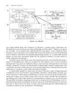
Fig. 3. The reproductive system is tightly integrated, requiring input from the hypotha-
lamic GnRH pulse generator and pituitary and feedback from the ovary, for regular
ovulatory function. The uterus acts as an end organ for the effects of ovarian steroids. The
incidence of menstrual disorders involving each level of this system differs depending on
whether amenorrhea is primary or secondary. Hypothalamic and pituitary disorders will
be hypogonadotropic while disorders resulting from ovarian failure will be associated
with high levels of FSH and LH.
Table 3
Diagnostic Testing
First-order tests
• β-hCG.
• Prolactin.
• FSH.
Second-order tests
• LH.
• Estradiol or progestin challenge (10 mg medroxyprogesterone for 5 d to induce
withdrawal bleed).
Other tests if clinically indicated
• TSH.
• Androgens: Testosterone, dehydroepiandrosterone sulfate (DHEAS), 17-
hydroxyprogesterone, urinary 17-ketosteroids.
• Pelvic ultrasound/hysterosalpingogram.
• Cranial MRI.
15/Hall/295-322/10.31F 12/3/02, 12:04 PM301
302 Tortoriello and Hall
The retrograde menstrual flow can also create a hematoperitoneum, thereby
increasing the risk of severe endometriosis. These conditions are corrected with
surgical and/or hysteroscopic resection.
Mullerian agenesis (Mayer-Rokitansky-Kuster-Hauser syndrome) is a congenital
anomaly consisting of the absence or hypoplasia of the uterus and/or vagina. Those
patients with a normal or rudimentary uterus will have cyclic abdominal pain. These
patients are genetically female and, therefore, have functioning ovaries. Radiologic
imaging is indicated, as approximately one-third of patients will have urinary tract
abnormalities, and 12% will have skeletal anomalies, usually involving the spine.
Complete androgen insensitivity or testicular feminization accounts for approx
10% of all cases of primary amenorrhea. This condition is characterized by a
female phenotype but male karyotype. There exists a congenital insensitivity to
androgens, transmitted by means of a maternal X-linked recessive gene respon-
sible for the androgen intracellular receptor (11). Wollfian duct structures do not
form; however there is no absence of antimullerian hormone, and therefore, these
patients usually do not have uteri or tubes. The testes often descend to the level of
the internal inguinal ring. Only the lower one-third of the vagina is present, for this
portion derives from the urogenital sinus. The diagnosis is likely when a pheno-
typic female presents with breast development, primary amenorrhea, scanty or
absent pubic and axillary hair, a shortened vagina, and an absent uterus and cervix.
Patients characteristically have elevated gonadotropins, especially LH, mildly
elevated testosterone, and high estradiol. Spermatogenesis does not occur, and
hence, these patients are infertile. Testicular malignant transformation is a concern.
To allow female secondary sexual characteristics to reasonably develop, gonadec-
tomy is usually not recommended until the mid to late teens, however, the optimal
timing of gonadectomy remains controversial. After gonadectomy, hormonal
replacement therapy is indicated.
Asherman’s syndrome is a complete or partial obliteration of the uterine cavity
with the formation of intrauterine adhesions or synechiae. Patients with this disor-
der present with menstrual irregularities, usually amenorrhea or scanty bleeding
and infertility. The vast majority of cases follow instrumentation or surgery of the
uterus. It generally is iatrogenically induced by an overzealous postpartum curet-
tage and may be more prevalent with severe hypoestrogenism. It has also been
linked to infectious causes, as with puerperal endometritis, genital tuberculosis,
schistosomiasis, or from an intra-uterine device. The diagnosis is suspected by
failure of withdrawal bleeding after the administration of exogenous estrogen and
progesterone, and is diagnosed by visualization of synechiae on hystero-
salpingogram (sharp angled filling defects) or hysteroscopy. The preferred treat-
ment involves hysteroscopically directed resection of intrauterine adhesions with
high-dose estrogen therapy to foster endometrial overgrowth (12). Repeated
attempts may be necessary to restore menses, but eventually about 75% of patients
achieve a successful pregnancy.
15/Hall/295-322/10.31F 12/3/02, 12:04 PM302