New Concepts in Diabetes and Its Treatment - part 6 pot
Bạn đang xem bản rút gọn của tài liệu. Xem và tải ngay bản đầy đủ của tài liệu tại đây (458.28 KB, 27 trang )
Chapter X
Belfiore F, Mogensen CE (eds): New Concepts in Diabetes and Its Treatment.
Basel, Karger, 2000, pp 135–151
Diabetic Retinopathy
Toke Bek
Department of Ophthalmology, A
˚
rhus University Hospital, A
˚
rhus, Denmark
Introduction
Diabetes mellitus is a systemic disease that affects all parts of the eye.
The majority of these changes have a mild course with no permanent influence
on visual function. Transitory changes in the refraction of the lens occur
secondary to changes in the blood sugar, and can be prevented by optimizing
the metabolic control. Diabetic cataract can nowadays be operated with few
complications and with a good visual result, and diabetic eye muscle palsy
disappears spontaneously within weeks leaving no adverse consequences for
the visual function.
Diabetic complications in the retina, diabetic retinopathy, is a somewhat
different matter. This complication is presently one of the leading causes of
blindness in the western world. From a phylogenic point of view, the retina
is an advanced part of the brain, and damage to its neuronal tissue is therefore
irreversible and leads to permanent reduction of the visual function. This
implies that preventive measures are the cornerstone in the clinical manage-
ment of diabetic retinopathy. However, the preventive efforts should be effective
at many levels ranging from elimination of risk factors, initiation of screening
programmes, optimization of treatment intervention, and by educating diabetic
patients in self-care and good life habits. Once vision-threatening changes have
developed the patient should be promptly referred to a specialist for clinical
evaluation and initiation of relevant treatment to stop or limit the visual
damage.
This chapter will present an overview of current knowledge related to the
clinical management of diabetic retinopathy. The chapter will be introduced
with a brief account of the clinical and epidemiologic characteristics of the
disease, followed by a description of the practical management of prevention,
screening, diagnostics, and treatment of diabetic retinopathy.
135
Table 1. Nomenclature used for diabetic retinopathy
Retinopathy Inside the retina In front of the retina
(background retinopathy) (neovascularizations)
Not vision-threatening Simple
7
Vision-threatening Maculopathy Proliferative retinopathy
Clinical Appearance
The clinical evaluation and classification of diabetic retinopathy is based on
inspection oftheretina throughtheoptics of theeye. Themorphological changes
thusobservedintheretinaarecomplexandheterogeneouswhichisreflectedinthe
nomenclature used to describe diabetic retinopathy (table 1). Basically, diabetic
retinopathycan bedividedintoearly changesthatarenotaccompanied byreduc-
tion in vision and late changes accompanied by visual reduction.
Early Changes Not Accompanied by Visual Reduction
The most usual name for this retinopathy stage is nonproliferative diabetic
retinopathy, but older terms are also used, such as simple retinopathy,or
background retinopathy which alludes to the fact that the changes remain inside
the ocular background.
Nonproliferative diabetic retinopathy is caused by changes in the retinal mi-
crocirculation leading to compromised barrier function of the retinal capillaries.
The changes first appear temporally from the fovea consisting of capillary mic-
roaneurysms and small intraretinal haemorrhages (fig. 1). The increased capillary
permeabilityleads tothe developmentof whitish hard exudatesconsisting oflipo-
protein from the bloodstream (fig. 2). Additionally, cotton-wool spots may de-
velop. These are localized unsharply delimited whitish areas in the superficial
parts of the retina representing intracellular material that hasaccumulated in the
nerve fibres because of disturbances in their axoplasmic flow (fig. 3). The retinal
changes characterizing nonproliferative diabetic retinopathy are reversible, and
often noticeable dynamic changes are seen at repeated examinations, so that the
same number of lesions are present, however located in different places.
Late Changes Accompanied by Visual Reduction
Nonproliferative diabetic retinopathy can develop into one or both of
two different types of retinopathy accompanied by visual reduction, namely
proliferative diabetic retinopathy and diabetic maculopathy.
136Bek
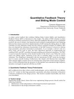
Fig. 1. Minimal nonproliferative diabetic retinopathy in a right eye. A few red dots
representing haemorrhages and/or microaneurysms are seen temporally in the macular area
which is the dark area surrounding the dark spot in the centre of the image (arrows).
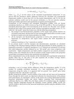
Fig. 2. Slight nonproliferative diabetic retinopathy in a left eye. Several whitish hard
exudates have developed inside the macular area.
Proliferative Diabetic Retinopathy
Proliferative diabetic retinopathy develops secondary to occlusion of the
retinal capillaries in the retinal periphery with a consequent stimulation of
vascular new growth. Clinically, both a preproliferative and a true proliferative
stage can be differentiated.
137Diabetic Retinopathy
Fig. 3. Moderate nonproliferative diabetic retinopathy in a right eye. Larger haemor-
rhages and whitish lesions with fluffy borders representing cotton-wool spots have developed.
Fig. 4. Preproliferative diabetic retinopathy in a right eye. Many large haemorrhages
are seen temporally in the macular area and there is calibre variation of the lower temporal
branch vein (arrow). Hard exudates within one disk diameter of the fovea indicate the
presence of clinically significant macular oedema.
Preproliferative diabetic retinopathy is characterized by many cotton-wool
spots, larger blot haemorrhages temporally in the macular area, and a variety
of vascularabnormalities. Theseabnormalitiesare intraretinalmicrovascularab-
normalities (IRMA vessels) often representing arteriovenous shunt vessels, and
beading and loop formation on the larger venules (fig. 4). These abnormalities
138Bek
Fig. 5. Proliferative diabetic retinopathy in a left eye. A large neovascularization has
developed from the optic disk and has given rise to a preretinal haemorrhage that extends
arcuately along the lower temporal branch vein.
develop secondary to changes in the retinal haemodynamics that result from
occlusion of the capillary bed in the retinal midperiphery and periphery. At this
stage the retinopathy will often become proliferative within a few months.
Proliferative diabetic retinopathy is characterized by outgrowth of new
vessels from the larger venules in the retina and on the optic nerve head (fig. 5).
This neovascular process is assumed to be caused by growth stimulation from
cytokines released from the ischaemic areas in the retinal periphery where the
capillary bed is occluded. However, the newly formed blood vessels do not
grow out to replace the occluded retinal vessels. Rather, they grow aberrantly
into the vitreous body. Preretinal neovascularizations can lead to visual reduc-
tion because of spontaneous haemorrhages into the vitreous body. The cause
of this vascular rupture is unknown, but may be caused by attachments of
the new vessels to the posterior hyaloid membrane that break secondary to
movements of the vitreous body. Finally, neovascularizations may contain
connective tissue that shrinks and causes tractional retinal detachment (fig. 6).
In some cases the vasostimulatory cytokines released in the retinal periph-
ery diffuse to the anterior eye chamber to cause neovascularization in the iris
(rubeosis iridis) (fig. 7) and in the anterior chamber angle. The resulting
blocking of the aqueous drainage from the eye will lead to neovascular glau-
coma. The high intraocular pressure may endanger the intraocular blood flow
and consequently the visual function, and if the rise in intraocular pressure
is rapid, severe acute pain may develop.
139Diabetic Retinopathy
Fig. 6. Severe proliferative diabetic retinopathy in a left eye. The new vessels contain
whitish fibrous tissue that covers most of the view of the fundus. Shrinkage of this fibrous
tissue may lead to tractional retinal detachment.
Fig. 7. Iris rubeosis. New vessels in the iris (arrows) have made the pupil immobile.
The small pupil together with the white cataractous lens seen behind the pupil opening
makes inspection as well as treatment of the fundus background impossible.
Diabetic Maculopathy
Diabetic maculopathy is nonproliferative diabetic retinopathy complicated
by retinal oedema. When the oedema area becomes large enough or is too
close to the fovea, it becomes vision-threatening (fig. 4) and is termed clinically
significant macular oedema (table 2). The oedema may be exudative or ischae-
140Bek
Table 2. Clinically significant macular oedema is defined as presence of one or both of
the criteria shown
1. Oedema and/or exudates within one-half disk diameter from the fovea
2. Oedema and/or exudates with a size of one disk diameter or more, part of which is located
within a zone of one disk diameter from the fovea
mic. The exudative form is most frequent, and it may be both focal or diffuse.
Exudative diabetic maculopathy is accompanied by hyperpermeability of the
macular vessels. When the oedema, the exudates, and the haemorrhages extend
towards the fovea, central vision may become threatened, partly by blocking
light access to the retinal photoreceptors, and partly because of a direct de-
structive effect on the neuronal components of the retina. Ischaemic oedema
develops secondary to occlusion of macular capillaries similarly to capillary
occlusion in the retinal periphery, with a subsequent fallout of neuronal func-
tion in the affected area. If the areas close to the fovea are included, visual
acuity may drop. Frequently, mixed types of maculopathy occur with exudative
and ischaemic retinal oedema located in different parts of the macular area.
Epidemiology
Almost all persons having diabetes mellitus will eventually develop non-
proliferative diabetic retinopathy. In countries with good diabetes care, reti-
nopathy does not develop until after 10 years of diabetes duration, whereas
retinopathy may be present at the time of diagnosis in type 2 diabetes. Nonpro-
liferative diabetic retinopathy can later be complicated by one or both of the
two late complications, proliferative diabetic retinopathy and diabetic macu-
lopathy. In type 1 diabetes the most frequent vision-threatening complication is
proliferative diabetic retinopathy, whereas in type 2 diabetes the most frequent
vision threat is diabetic maculopathy.
Prevention
Preventing the development of diabetic retinopathy is one of the basic
pillars in the management of diabetic eye complications, since the damage
that occurs to the retina is irreversible.
The two most significant factors now known to limit the risk of developing
diabetic retinopathy are tight regulation of the blood glucose and of the blood
pressure. For many years it was suspected that exposition to hyperglycaemia
accelerated the development of diabetic retinopathy, but it was not established
141Diabetic Retinopathy
Table 3. Characteristics of diabetic
retinopathy
Is frequent
Can be prevented
Can be detected
Can be treated
Screening is cost-efficient
until a few years ago in the Diabetes Control and Complications Trial (DCCT)
study that the risk of developing retinopathy is considerably lowered by tight
glycaemic control. Recently, several studies have been published unanimously
showing that the risk of developing retinopathy in type 1 as well as in type 2
diabetes can be considerably lowered by antihypertensive treatment. Further-
more, it has been shown that treatment with especially ACE inhibitors can
reduce the risk for developing retinopathy with an effect that adds to the
antihypertensive effect. However, these studies have not been conducted far
enough to show that this intervention also has an effect on the visual prognosis.
Pregnancy is a definite risk factor for the development of diabetic retinopa-
thy. A tight regulation of the blood glucose during pregnancy alone can slow
and often halt the development of retinopathy completely, suggesting that the
risk of developing retinopathy is to a large extent caused by disturbances in the
diabetic metabolism in pregnancy. Since the risk for developing diabetic retino-
pathy during pregnancy increases with increasing duration of diabetes, diabetic
women should be counselled to have children as early as possible in life.
A multitude of studies have been conducted to identify new preventive
measures for diabetic retinopathy. These studies have for example shown that
aspirin and aldose reductase inhibitors have no beneficial effect on diabetic
retinopathy. More recent studies have shown that pharmaceutical intervention
on second messengers such as protein kinase C might be a future treatment
modality for diabetic retinopathy, and these hypotheses are presently under
investigation in clinical trials.
Screening
Background
Even when optimal preventive measures are undertaken, some patients
will unavoidably develop vision-threatening retinopathy. Since these changes
may not be recognized by the patient before they have advanced to a stage
where vision damage is irreversible, early detection is important. Diabetic
retinopathy fulfills a number of criteria that makes it appropriate to screen
for this complication among the diabetic population (table 3).
142Bek
Methods
Screening for diabetic retinopathy is performed by inspecting the ocular
background through the optics of the eye, supplemented by measurement of
the visual acuity.
Examination of the Ocular Background
Inspection of the ocular background can be done by ophthalmoscopy that
enables a qualitative assessment of retinopathy. Alternatively, photography of
the ocular background allows a semiquantitative analysis by comparison with
standard photographs, or a quantitative computerized analysis of the retinal
changes.
Examination of the ocular background by ophthalmoscopy has been
known for almost 150 years, and this technique is therefore one of the oldest
known examination methods in ophthalmology. During ophthalmoscopy the
retina is illuminated continuously, and inspection is either done directly or
indirectly through a lens positioned with its focal point in the pupil plane of
the examined eye. The relevance of doing ophthalmoscopy in diabetic patients
was realized during the fifties where it became usual for diabetic patients
to survive long enough to develop retinal complications. The advantage of
ophthalmoscopy is that only simple equipment is needed for the examination.
The disadvantages of this method is that the severity of the retinal lesions
cannot be documented in detail, that the retinal changes are difficult to quan-
tify, and that the quality and conclusion of the examination depend on the
experience and attitude of the examiner. In spite of these weaknesses, ophthal-
moscopy has until now been the most important examination technique for
early detection of diabetic retinopathy, and globally it is still the most widely
used method.
During the last decades, increasing focus has been directed at a different
technique to screen for diabetic retinopathy by examination of the ocular back-
ground with fundus photography. This method has a number of advantages.
Firstly,theretinalchanges are documentedsothatit ispossibleto re-evaluatethe
retinopathy,and thegrader canconsult otherspecialists ata latertime.Secondly,
retinal photography enables a standardized and centralized semiquantitative
evaluation of the severity of the changes, and thirdly, photography enables an
evaluation of even minimal changes in retinopathy. Fourthly, ophthalmologists
need nothave primarypatientcontact. Thus,with technicians doingthe photog-
raphy and opthalmologists performing the evaluation of the photographs, more
examinations can be carried out with the same specialist resources. Finally, it
has been shown that for other than retinal specialists the sensitivity in detecting
vision-threatening retinal changes is higher when the retinopathy is evaluated
from retinal photographs than by ophthalmoscopy.
143Diabetic Retinopathy
With the current development within computerized image analysis it can
be expected that withina few years it willbe possible to replace semiquantitative
grading of fundus photographs with a fully computerized quantification of
the fundus photographic changes. Many initiatives have been taken to start
this process, and the results achieved hitherto appear promising.
Stereoscopic examination of the ocular background is done by examining
the same part of the retina from different angles with the examiner’s two eyes,
thus giving an impression of the depth relation of the retinal structures. This
can be done directly by binocular inspection of the ocular background, or
indirectly by studying stereo photographs of the ocular background. The
validity of this technique depends on the examiner’s stereo vision which shows
great interindividual variation. The significance of this type of examination
lies in its potential for detecting retinal oedema. Until now there has been
no documentation of the value of stereoscopic examination of the ocular
background for screening for diabetic retinopathy.
Measurement of Visual Acuity
In most countries there is general agreement that measurement of the
visual acuity should be part of the routine screening examination for diabetic
retinopathy. The visual acuity may be valuable as a supplement to the inspec-
tion of the ocular background, especially if it has to be decided whether the
patient should be referred for further evaluation by an ophthalmologist. Thus,
in exudative diabetic maculopathy, hard exudates and retinal oedema may be
located in the border zone of being clinically significant. A reduced or declining
visual acuity in these cases will speak infavour of referral for further evaluation.
Similarly, in ischaemic maculopathy with no hard exudates and questionable
macular oedema, the visual acuity may be a valuable help in determining
whether incipient retinal damage needs referral for further evaluation.
Organization
In order to ensure that screening efforts are efficient it is necessary that:
(1)thehealthsystemisorganizedtopermittheestablishmentofefficientscreening
programmes; (2) sufficient resources are made available in the short term (they
will always pay back in thelongterm); (3)qualified personnel is available to carry
out thescreeningexaminations andevaluations,and (4)patientsaretaughtabout
the advantages of screening, and are given motivation to participate.
Iceland is a positive example of a country where all these factors have
been optimal. This country has succeeded in setting up a screening programme
where, in principle, all the country’s diabetic patients are known and followed.
In most countries, however, screening efforts do not live up to expectations,
for the most part due to social or geographic differences. Generally, screening
144Bek
is of a high quality near centres with high expertise and impact in the commu-
nity, and similarly of a poorer quality in peripheral areas.
An optimization of screening efforts meets with various barriers in differ-
ent countries. In some countries it is the structure of the health system that
hinders free access to retinopathy screening for the whole diabetic population,
whereas in other countries problems inherent in health system structure or
geography can be solved within the existing framework. The exploitation of
new technology may play a central role for removal of these barriers. For
example, it is conceivable that the ongoing developments within teleophthal-
mology will enable the setting up of decentral screening clinics for diabetic
retinopathy from which fundus photographs taken by technicians can be trans-
mitted electronically to a central place for evaluation. Such an organization
would be a huge step towards more efficient care in areas where the bottleneck
is shortage of qualified specialists, and would solve problems with transporta-
tion of patients over long distances.
Screening Interval
Patientswithtype1diabetesmellitusshouldbeexaminedatleastoncea year
whendiabetesdurationislongenoughforthedevelopmentofvision-threatening
changes to be conceivable. This critical diabetes duration is only a few years in
some societies with poor diabetes care, e.g. in some of the former east block
countries, but up to 10 years in societies where diabetes care is optimal.
Patients with type 2 diabetes mellitus should be screened at the time of
diagnosis, and then every other year if no or minimal retinopathy is found at
the initial examination. In both diabetes types the screening interval should
be shorter when there is progression of retinopathy or changes appear that
possibly in the near future will require treatment.
Economy
Several health economic analyses have shown that screening for diabetic
retinopathy is very cost-efficient. Thus, from a socio-economic point of view,
the ability to rescue a few cases from blindness each year is sufficent to balance
the total cost of screening efforts in an area with several thousand diabetic
patients, not to mention the personal and socialconsequencesfor the individual
diabetic patient who can preserve visual health.
Diagnostics
If a screening examination leads to suspicion of proliferative diabetic
retinopathy or maculopathy that potentially threatens central vision, the pa-
145Diabetic Retinopathy
tient should be referred for further evaluation with an ophthalmologist having
facilities for fluorescein angiography and photocoagulation treatment.
In diabetic maculopathy it is necessary to do fluorescein angiography to
distinguish between exudative maculopathy, that can be treated by photocoagu-
lation, and ischaemic maculopathy that is not treatable. Fluorescein angiogra-
phy is performed by intravenous injection of the tracer compound fluorescein
which is transported and distributed in the bloodstream to reach the eye in
a few seconds. Under normal circumstances, fluorescein cannot pass the blood-
retina barrier and therefore remains inside the bloodstream. However, in exu-
dative diabetic maculopathy the blood-retina barrier is broken down and areas
where fluorescein leaks out of the bloodstream into the retinal tissue and the
vitreous can be recognized. In ischaemic maculopathy, however, areas with
focal occlusion of retinal capillaries are seen in the macular area. When the
ocular background is inspected by ophthalmoscopy or by evaluation of fundus
photographs, the type of maculopathy can often be diagnosed. Thus, exudative
maculopathy is almost always associated with hard exudates, while the ocular
background in ischaemic maculopathy almost always appears slightly yellowish
combined with many intraretinal haemorrhages and no exudates. However,
since mixed types with both exudative and ischaemic maculopathy may occur,
angiography is an important tool to differentiate and locate leakage and capil-
lary occlusion.
Exudative diabetic maculopathy should be treated with retinal photoco-
agulation when there is clinically significant macular oedema (table 2), or
when exudates or oedema are otherwise suspected to threaten central vision.
Clinically significant macular oedema has previously been difficult to describe
quantitatively since no technique was available to quantify retinal thickness.
However, in recent years this has changed, and several new methods making
this possible have now been developed. One of the most promising of these
methods is optical coherence tomography that detects the phase shift of light
reflected from different surfaces in the retina and transforms this signal to a
colour code that expresses the reflectivity and depth of different retinal levels.
The method has a depth resolution of approximately 10 m and gives a precise
indication of whether there is retinal oedema, and the method can be used to
quantify the effect of therapeutic intervention.
When the diagnosis of proliferative diabetic retinopathy is certain, no
more diagnostic evaluation is required and retinal photocoagulation can be
initiated immediately. In less clear cases, differential diagnostic alternatives
should be carefully considered, the most frequent being shunt vessels or other
intraretinal microvascular abnormalities. On the basis of the criteria shown in
table 4, the presence or not of neovascularizations requiring photocoagulation
treatment can almost always be established clinically.
146Bek
Table 4. Characteristicsofnewvesselsinproliferative diabeticretinopathyrequiringphoto-
coagulation treatment and intraretinal microvascular abnormalities not requiring treatment
New vessels requiring photocoagulation Intraretinal microvascular abnormalities not
treatment requiring photocoagulation
Preretinal Intraretinal
May cross their feeder vessel Do not cross their feeder vessel
Always emerge from larger venules Connect venules and arterioles
Are recursive back to venule of origin Are not recursive
Displays extensive branching Branching pattern normal
In proliferative diabetic retinopathy complicated with vitreous haemor-
rhage it may be difficult to get a view of the ocular background. In these cases
ultrasound B-scan examination is useful to establish whether the vitreous
opacities are associated with retinal detachment, in which case operation will
give no benefit for vision.
Treatment
Retinal Photocoagulation
Retinalphotocoagulationis theonlyknown treatment modalitywitha docu-
mented effect on diabetic retinopathy. The treatment itself, however, may incur
impairment of vision and should therefore only be performed by ophthalmolo-
gists withspecial interestand training withinthis field.The mechanism ofaction
of retinal photocoagulation is unknown, but the effect can be achieved with any
light source that destroys the outer retinal layers after absorption in the retinal
pigment epithelium. Retinal photocoagulation is usually performed using the
blue line of an argon laser which is mounted on a slit lamp so that treatment can
be applied through a contact glass. The contact glass enables the viewing of the
ocular background by eliminating the corneal refraction, enables treatment of
the retinal periphery through built-in angled mirrors, and dampens voluntary
or reflectory eye movements. The treatment is done by applying burns with a
distance of one burn in between but avoiding retinal vessels, and the energy of
the burns is adjusted to produce a distinct retinal whitening.
Proliferative Diabetic Retinopathy
In proliferative diabetic retinopathy, treatment should be panretinal, mean-
ing that the whole retina peripherally from the temporal arcades should be
147Diabetic Retinopathy
treated. In most cases, this treatment will arrest the neovascular growth, and
often lead to a regression of the new vessels. This effect is assumed to be a
result of a destruction of the peripheral parts of the retina. The elimination
of the ischaemic retinal areas that release the vasostimulatory factors eliminates
the stimulus for neovascular growth. For panretinal photocoagulation a spot
size of 300–500 m is usually employed with which approximately 2,500–3,500
applications are needed to fill out the retinal periphery. The treatment is
applied in at least two sittings, partly because the redistribution of the choroidal
blood flow induced by the treatment may impair central retinal function, and
partly because sittings lasting more than 15 min are tiring for both the patient
and the treating ophthalmologist. The risk to consider with this treatment is
accidental photocoagulation in the foveal area, which is less likely to occur
when treatment is done through angled mirrors. During treatment the patient’s
eye is subjected to a strong blaze, and there may occasionally be a distinct
stinging pain when the laser treatment is applied to the retinal areas along
the vertical and horizontal meridians. After treatment the patients often experi-
ence a shrinkage of the peripheral visual field and impaired night vision which
can be directly attributed to the destructive effect of the laser applications in
the retinal periphery. In more rare cases, retinal photocoagulation applied to
the retinal periphery may, for unknown reasons, lead to a lowering of central
vision.
Exudative Diabetic Maculopathy
In diabetic maculopathy, laser treatment is applied corresponding to the
lesions in the macular area. The treatment is performed differently dependent
on the individual appearance and location of the lesions, but also dependent
on varying ideas of how diabetic maculopathy should be interpreted. The
principle of the treatment strategy is to apply a laser grid pattern corresponding
to the area with retinal oedema, however sparing a central zone out to approxi-
mately 500 m from the fovea. In some centres, treatment is only applied in
a horseshoe temporally around the centre, thus sparing the papillomacular
bundle. If the papillomacular bundle is treated, one should be careful not to
apply burns with so high an energy that they extend transretinally to cause
destruction to the nerve fibres coursing to the fovea. If the oedema area is
small, as for example inside circinate conglomerates of hard exudates, the
treatment grid becomes small, perhaps consisting of single points, and treat-
ment becomes focal.
The mechanism of action of macular laser photocoagulation is unknown,
but the treatment leads to disappearance of hard exudates and oedema. The
total resolution of these lesions is slow, however, and it may take from weeks
to years. The treatment causes blazing, but is not otherwise associated with
148Bek
any appreciable discomfort. The risk involved with macular photocoagulation
is that the applications can accidentally be given in the foveal area with a
consequent reduction in central vision. Even when treatment is applied safely
outside the foveal area, there may be a risk of visual reduction. Thus, if the
patient has excentric fixation due to lowered visual acuity, treatment may
unwittingly be applied corresponding to the new fixation area resulting in a
further reduction of vision.
When there is coexistence of proliferative diabetic retinopathy and diabetic
maculopathy, both panretinal photocoagulation and macular photocoagula-
tion should be given. There is no conclusive documentation for whether panret-
inal or macular photocoagulation should be given first. A suggestion that
diabetic maculopathy should be worsened when panretinal photocoagulation
is done first has received some focus, but the issue has not been finally clarified
since there is evidence in support of both this and the opposite view.
Vitrectomy
When proliferative diabetic retinopathy has resulted in vitreous haemor-
rhage or retinal traction from connective tissue in the new vessels, there is
indication for vitrectomy. During vitrectomy, thin instruments are introduced
through the sclera in order to cut and remove fibrous strings and opaque
vitreous, and to apply laser treatment. With modern techniques this operation
can be done in local anaesthesia in an outpatient setting with no appreciable
discomfort. When the purpose of the operation is only to remove a vitreous
haemorrhage, full restitution of the visual function to the level before the
haemorrhage developed will often result, while permanent damage to the
visual function will most often have developed when there is tractional retinal
detachment.
Neovascular Glaucoma
Proliferative diabetic retinopathy that has progressed to neovascular glau-
coma should be treated immediately with panretinal photocoagulation. Most
often, neovascular glaucoma is associated with severe visual reduction and if
vascular new growth has immobilized the iris, perhaps combined with cataract,
the diseasehascome beyond therapeutic reach with laserand vitrectomy (fig. 7).
The therapeutic goal in this situation is to keep the intraocular pressure normal
primarily in order to preserve residual vision, but also to keep the patient free
of pain and thereby avoid a cosmetically disfiguring enucleation. The primary
treatment is administration of local or systemic drugs to lower the intraocular
pressure. Destruction of the ciliary body by transscleral heating or freezing re-
duces aqueous production, or alternatively an artificial outflow channel can be
made to replace the trabecular meshwork channel closed by new vessels.
149Diabetic Retinopathy
Psychosocial Aspects
Diabetes mellitus is a burdening chronic disease with profound influence
on daily life. The disease is associated with many contacts to the health-care
system and the necessary routine eye controls combined with the threat of
losing vision may be an additional burden. Consequently, some patients may
need to repress or forget their disease for periods, during which they drop out
of the diabetes care system. For these patients there may be a risk of developing
vision-threatening retinal changes, in spite of the fact that the organization
of retinopathy screening to detect early changes has been set up optimally.
This risk is so much higher because the patients who drop out of the eye
controls often also neglect the metabolic regulation, which further increases
the risk of developing vision-threatening complications. The problem is not
easy to solve, but resources should be used to give appropriate information
about adverse consequences of disease neglect, and ideally psychological assis-
tance should be offered.
Impairment of vision secondary to diabetes mellitus often affects younger
persons of family supportive age. Therefore, in addition to the personal con-
sequences, diabetic retinopathy also has great social and economic implications
for the patients’ relatives and for the society as a whole. Preserved vision may
make the difference that enables self-monitoring of blood glucose or home
dialysis, enables daily doings, and enables the filling out of a job position.
Therefore, it is of paramount importance that visual loss is prevented, but
also, that diabetic patients who have already experienced visual reduction are
offered help to come to terms with their situation, to manage their daily life,
and perhaps be rehabilitated to fill out a job with demands that match the
visual ability.
Conclusion
It appears from the foregoing account that in most countries there are
significant unexploited potentials for reducing the risk of visual loss secondary
to diabetic retinopathy. Presently, the most remarkable shortcoming is the
lack of detection of vision-threatening retinopathy in the diabetic population,
largely caused by organisational limitations and lack of long-term health
economic thinking. An optimization of this field requires that patients and
medical personnel bring this problem to the attention of health politicians.
Another significant limitation for initiating a rational fight aginst visual loss
secondary to diabetic retinopathy is the limited knowledge of the pathophysio-
logy of the disease. Huge and significant research efforts have been initiated
150Bek
to solve this puzzle, and major advances have been made. A detailed account
of this field, however, is beyond the scope of this chapter.
In most recent years much attention has been directed at the importance
of systemic factors for the development of diabetic complications, inclusively
diabetic retinopathy. It is now well established that a tight glycaemic regulation
can delay the development of diabetic retinopathy, and lately focus has been
given to the effect of reducing the blood pressure. The diabetologic expertise
has thus become one of the cornerstones in the preventive efforts against
diabetic eye disease, signalling that optimal diabetes care of the future will
probably depend on a close cooperation between diabetologists and ophthal-
mologists. With such a collaboration, the ophthalmological evaluation of reti-
nopathy might be an effective measure for the diabetological regulation of
systemic factors such as blood glucose, blood pressure, or other metabolic
parameters. Together with an optimized education and motivation of the
diabetic patient, and appropriate treatment of already developed vision-
threatening retinopathy, it can be hoped that once in a not too far future
diabetic retinopathy will be demoted to a rare cause of visual impairment and
blindness.
Suggested Reading
Aiello LP, Gardner TW, King GL, Blankenship G, Cavallerano JD, Ferris FL, Klein R: Diabetic retinopa-
thy. Diabetes Care 1998;21:143–159.
Diabetes Control and Complications Trial Research Group: The effect of intensive diabetes treatment on
the progression of diabetic retinopathy in insulin-dependent diabetes mellitus. Arch Ophthalmol
1995;113:36–51.
Early Treatment Diabetic Retinopathy Study Research Group: Report No 1: Photocoagulation for diabetic
macular oedema. Arch Ophthalmol 1985;103:1796–1806.
Kohner EM, Bek T, Aldington S: Diabetic Retinopathy. Diagnosis, Management and Reference Images
(CD-ROM). Amsterdam, Elsevier, 1999.
Kohner EM, Porta M: Screening for Diabetic Retinopathy in Europe: A Field Guide Book, Geneva,
WHO, 1992, pp 1–51.
Dr. Toke Bek, Department of Ophthalmology, A
˚
rhus University Hospital,
DK–8000 A
˚
rhus C (Denmark)
Tel. +45 89493223, Fax +45 86121653, E-Mail
151Diabetic Retinopathy
Chapter XI
Belfiore F, Mogensen CE (eds): New Concepts in Diabetes and Its Treatment.
Basel, Karger, 2000, pp 152–173
Nephropathy and Hypertension in
Diabetic Patients
Carl Erik Mogensen
Medical Department M (Diabetes and Endocrinology), Kommunehospitalet,
A
˚
rhus University Hospital, A
˚
rhus, Denmark
Introduction
Strict and steady near normoglycemia over many years is of paramount
importance for the prevention and postponement of renal disease, as well as
other complications in most patients with type 1 and type 2 diabetes. Later,
several other factors appear to affect progression in renal disease of which
blood pressure (BP) elevation seems most important. This seems also to be
the case for macrovascular complications along with dyslipidemia, smoking
and, as mentioned, hyperglycemia. Incipient renal disease in diabetes, as judged
by the occurrence of microalbuminuria, is frequently characterized by hyper-
tension starting with increase in BP from a normal level. The increase, however,
is often subtle and may only be detectable by careful and continuous mon-
itoring, e.g. by 24-hour ambulatory recordings. Elevation of BP is found in
both types of diabetes, but there appear to be several distinctions between
type 1 and type 2 diabetes; some of these variations are clearly explained by
the different etiology and nature of the diabetic state. In type 2 diabetic patients,
higher age, increased body weight, as well as syndrome X abnormalities are
important factors. Though hypertension secondary to renal dysfunction is
also frequently seen in type 2 diabetic patients, the renal genesis of hypertension
is much clearer and more common in the relatively younger type 1 diabetic
patients. Indeed a vicious circle seems to be operating in both types of diabetes
and differences between type 1 and type 2 diabetes regarding nephropathy are
far fewer than reported earlier. It should be noted that dietary protein intake
may also be a modulating factor, but further studies on intervention are
needed. These factors – glycemic control, BP elevation and to some extent
152
dietary proteins, and the modification by treatment – will be the main issues
for discussion here.
BP, Glomerular Pressure and Potential Genetic Factors
In the past decades there has been a growing interest in the nature of
diabetic renal disease, mainly focusing on BP, glomerular pressure and protein
leakage as related to structural and biochemical abnormalities. A recently
published volume intends to cover almost every aspect of renal disease and
hypertension in diabetes. One key point is interesting here; in general, two or
more risk factors must coincide to provoke fast and serious organ damage.
In terms of diabetic nephropathy this means that some degree of poor glycemic
control may not always be clinically noxious enough per se, unless some other
risk factors, especially elevated BP or possibly poorly defined genetic elements
coexists. However, increased glomerular pressure seems to be a decisive factor,
whether caused directly by poor glycemic control, dietary proteins or systemic
hypertension, in particular with loss of renal vascular autoregulation that may
be seen in diabetes. Other risk factors may contribute to renal and especially
vascular damage in diabetes, e.g. smoking, lipid abnormalities or obesity, again
highlighting the importance of the metabolic syndrome, or syndrome X mainly
in type 2 diabetes.
Diabetic renal disease may tend to cluster in families, possibly partly
reflecting that poor metabolic control also predominates in certain families.
This could also relate to ACE gene or other gene polymorphism, but genetic
association to diabetic renal disease and its progression may not be strong and
has recently been challenged. From a clinical point of view, ACE genotyping is
hardly relevant. Based on a meta-analysis, Tarnow et al. concluded that the
ACE/ID polymorphism may contribute to the genetic susceptibility to diabetic
nephropathy in Japanese type 2 diabetic patients, whereas it does not play a
major role in the initiation ofdiabetic nephropathy inCaucasiantype 2 patients.
In Caucasian type 1 diabetic patients, comparison of data is complicated by
differences between study populations, but a trend towards a protective effect
of the II genotype on the development of increased urinary albumin excretion
rate was observed, but there is considerable overlap between genotypes. How-
ever, a progression also during antihypertensive treatment is somewhat faster
with the DD genotype. Whether this is related to actual BP during treatment
is unclear.
Comparing the different risk factors – apart from poor metabolic control
– BP elevation seems to be not only the most important index of actual or
subsequent organ damage, but also the most readily measurable (sometimes
153Nephropathy and Hypertension in Diabetic Patients
Fig. 1. Interplay of genetics and risk factors.
with 24 h ambulatory BP) as well as modifiable risk factor (fig. 1). Virtually
all studies agree that standard medical antihypertensive treatment is able to
reduce BP in diabetes, and many studies have confirmed the original observa-
tions of a beneficial impact of antihypertensive treatment on the course of
renal disease, both in incipient and overt type 1 diabetic patients. Interestingly,
ACE inhibitors may be particularly beneficial, although this has been ques-
tioned by some. Certainly, the side effect profiles usually favor the use of these
agents often combined with diuretics both in incipient and overt nephropathy.
These considerations also apply to cardiovascular events in hypertensive type
2 diabetic patients. Combination therapy including -blockers often has to be
used to reduce BP as well as albuminuria efficiently.
Changing Cumulative Incidence of Renal Disease in Diabetes
The cumulative incidence of diabetic nephropathy used to be high ( 35%)
but seems to have declined over recent years, especially in certain areas where
only very few patients in a given cohort developed nephropathy. However, this
observation could not be confirmed by other groups. The explanation is not
clear, but certainly the so-called natural history may be considerably modified
by more intensive intervention throughout the course of diabetes. To a large
extent, this relates to metabolic control and BP elevation as major factors,
but other issues are of importance, e.g. smoking, that may vary considerably.
Also race is of importance and diabetic nephropathy is more commonly seen
in African-Americans. Indeed new studies among the Pima Indians suggest
that with long follow-up periods practically all patients will develop renal
154Mogensen
disease. This information is important because it has been suggested that
there may be important susceptibility factors that could relate to genetics.
Comparison has been made to eye diseases where practically all patients sooner
or later develop lesions. However, there are important modifications since
usually renal disease is judged by albuminuria and not by morphology, e.g.
on biopsies, and in fact the cumulative incidence of diabetic retinopathy and
nephropathy could be almost the same, if histological as well as ophthalmologic
examinations are used. It has also been discussed why some people seem
to escape diabetic nephropathy even if they are in poor control. A feasible
explanation is that in order to produce important clinical disease two factors
must be present, namely high BP as well as high glucose. If the combination
of high BP and high blood glucose is present the clinical experience is that
almost all patients will develop clinically relevant nephropathy and also reti-
nopathy.
Recent studies underscore the role of good metabolic control also in more
advanced nephropathy. This has been documented in several studies, and also
recently by Mulec et al. These results are in concert with information from
Denmark, Gothenburg and London. Clearly with advanced nephropathy
elevated BP is of importance, and combining the two risk factors in overt
nephropathy, huge differences in progression may be observed. With poor
control of glycemia and especially poor BP control, the fall rate is high
( 10–12 ml/min/year or even more), but with efficient control of blood glucose
and BP fall may be close to 1–2 ml/year which is close to the age-related
reduction. Obviously, it is not possible to obtain perfect metabolic control in
all patients, especially in those at risk or with nephropathy because the very
background for developing complications is the poor control which may not
be easy to modify even after development of complications. This is exemplified
in a study from the UK, The Microalbuminuria Collaborative Study. The
combined deleterious effect of poor glycemic control and BP control is indeed
also clear from the important UKPDS intervention study in type 2 diabetes.
In summary, one could argue that the concept of ‘natural history’ may
be wrong unless it is used specifically in patients who are in specifically defined
glycemic control. However, if risk factors such as hyperglycemia and BP eleva-
tion can be controlled, few patients may actually develop proteinuria and
eventually end-stage renal disease both in type 1 and type 2 diabetes. Also
with advanced nephropathy, glycemic control seems very important.
However, an intensified strategy requires considerable resources not only
from the health-care providers but also from the patients. The recent Steno
Study used the new concept of multifactorial intervention with a good result
on renal and retinal diseases. It may be easier toimplementlong-term treatment
with ACE inhibitors or otherantihypertensive agents, alsointhe normoalbumi-
155Nephropathy and Hypertension in Diabetic Patients
nuric state as recently proposed by Ravid et al. However, both strategies should
be exercised in the clinical setting as discussed below.
Notes on Key Risk Factors: Blood Glucose
Perfect metabolic control, that is blood glucose as well as concentrations
of other metabolites and hormones within normal range, is presently almost
impossible to obtain in the majority of diabetic patients. Even in the DCCT
(The Diabetes Control and Complications Trial Research Group) optimized
management in type 1 diabetic patients only rarely resulted in perfect glycemic
control. The same may be the case in type 2 diabetes, where a somewhat better
control may be possible. Under standard care conditions, HbA
1c
values may
be 50% or most often even higher than normal reference values in most
patients. However, good metabolic control remains a key factor in preventing
retinopathy and nephropathy, and progression of nephropathy, also when
severe damage is present after fall in GFR. Further long-term studies are
needed in type 2 diabetic patients but the same relation seems to exist here,
especially early in the course of the renal disease. Long-term renal data from
the UKPDS would be highly interesting.
Notes on Key Risk Factors: BP Level in Treated Diabetes
Nowadays very high BP levels are rarely observed in the clinic in treated
diabeticpatients.High pressures aremostoftenencountered inpopulations with-
outanystructuredcareforcomplications.Withappropriateantihypertensivepro-
grams the degree of elevation of BP is usually not very pronounced at least when
compared to the past. This is for instance corroborated by new studies where 24-
hour BP recordings in diabetics are carefully compared to nondiabetics. When
diabetics who do not receive antihypertensive treatmentare selected, it isobvious
thatBP elevationisnotpronounced, around5mmHgonaverageinmicroalbumi-
nuric patients. Clearly such data are biased, because patients who are already in
treatment are excluded. On the other hand, even minor BP elevation may lead to
vascular and glomerular damage, especially when accompanied by other risk
factors,e.g. hyperglycemia.A correlation existsbetween albuminuria andBP and
the association is amplified when 24 ambulatory BP values are used rather than
conventional BP measurements. Diabetic patients may be exquisitely susceptible
to systemic BP elevation because the normal protection exerted by the afferent
renal arteriolarvasculatureislikely tobe compromised inmanydiabeticpatients,
and a vicious circle will develop in such conditions.
A few decades ago, BP elevation was usually much higher. A very pro-
nounced fall in recorded BP has been observed in diabetes clinics during recent
years, as evidenced by a Danish study, where BP levels in cohorts of patients
in the 1960s were compared to patients in the 1980s.
156Mogensen
Interesting differences exist between the two types of diabetes. In type 1
diabetes the prevalence of hypertension is strongly correlated with the degree
of albuminuria. With normal albumin excretion rate, BP is close to normal
which has been confirmed in recent studies using 24-hour BP recordings. With
the occurrence of microalbuminuria there is a considerable increase in the
prevalence of elevated BP, and even more marked changes are seen with overt
diabetic nephropathy.
In type 2 diabetes the situation is more variable, although there is usually
some association between albuminuria and BP level. However, the correlation
is weaker, and it is also important to recognize that the prevalence of BP
elevation is much higher in the elderly type 2 diabetic patients; even at the
time of clinical diagnosis about 40% of patients had elevated BP or were
receiving antihypertensive treatment. In a control population without diabetes
this figure may be 20%. Interestingly BP elevation in type 2 diabetic patients
is usually of systolic nature, at least in some studies. Effective treatment may
be difficult with high initial values and therapeutic goals should be modified,
with a stepwise reduction in BP.
Without treatment the rate of increase in BP with time is recorded to be
high in type 1 diabetic patients with microalbuminuria or overt proteinuria,
supporting the idea that a self-perpetuating process exists. This increase is
most pronounced in type 1 diabetic patients; clear data are more difficult to
obtain in type 2 diabetes, because so many patients are treated with antihyper-
tensive drugs and discontinuation of treatment is notjustifiable. Still an increase
is seen, especially with 24-hour monitoring. In type 1 diabetes, BP may increase
by 3–4 mm Hg/year with microalbuminuria, and around 6 mm Hg/year with
overt renal disease. Such data may be difficult to reproduce today, simply
because so many patients are early and effectively treated.
Notes on Key Risk Factors: Dietary Proteins
With some variations from country to country, traditional diabetic dietary
management often results in a high protein intake (sometimes 50% higher
than the average background population). This may not be an appropriate
strategy because a dietary pattern like that may aggravate the course of renal
disease.
Microalbuminuria as an Important Intermediary Endpoint
A major question in all types of clinical management is to define parame-
ters that can be considered important markers in terms of disease activity.
This is of special importance in intervention trials, but also in the treatment
157Nephropathy and Hypertension in Diabetic Patients
of patients, where results from already conducted trials are rapidly reflected
in practical management. An outline of the natural history of renal disease
in type 1 diabetic patients is given in table 1.
Hypertensive and proteinuric diabetic patients usually carry a very poor
prognosis. It has also become clear that abnormal albuminuria in the microal-
buminuric range (20–200 g/min) is an important long-term predictor for poor
outcome. A decisive parameter is the fall rate of GFR as measured by exact
and reproducible techniques. Doubling of S-creatinine has also been used.
Obviously, an even more solid endpoint is end-stage renal failure (ESRF)
and/or death, but in patients with early clinical proteinuria or microalbuminu-
ria, this is (fortunately) a distant endpoint since the development of ESRF
may last at least one or two decades, especially after it has been shown that
antihypertensive treatment postpones end-stage renal disease.
Strong evidence suggests that abnormal albuminuria (even slight eleva-
tion) is a key parameter and an important intermediary endpoint in the
monitoring of all diabetic patients, not only because it relates so closely to
the more advanced endpoints, but also because this parameter can be used
both in the treatment strategies in controlled clinical trials, and in the day-to-
day management of patients. Importantly, new studies show that glomerular
structural damage can be arrested by early antihypertensive treatment
(-blockers or ACE inhibitors) in microalbuminuric patients. This is an ex-
tremely important finding again supporting the use of early antihypertensive
medication.
GFR Fall in Type 1 Diabetes Related to Abnormal Albuminuria and/or
BP Elevation
Patients with completely normal albumin excretion rate usually preserve
normal renal function (GFR) over many years, at least one or two decades.
It should be noted here that there may, however, be a small probably age-
related reduction in GFR. Also patients with persistent microalbuminuria
usually maintain intact GFR, though a subsequent fall in GFR can be
predicted, with progression to macroalbuminuria and possibly partly related
to previous hyperfiltration. Only with the development of proteinuria (ma-
croalbuminuria) is there a significant decline in GFR. Antihypertensive treat-
ment may reduce or even normalize albumin excretion, and thus lead to
misclassification of patients. Albumin excretion may again increase if treat-
ment is stopped for some reason. Feldt-Rasmussen et al. observed a significant
drop in GFR with the development of clinical nephropathy but most of their
patients received antihypertensive treatment which modifies the level of UAE
158Mogensen
Table 1. Stages in the development of renal changes and lesions in diabetes mellitus (mainly type 1 or younger type 2)
Stage Chronology Main structural Glomenular Dextran clearance Albumin exception
Blood pressure Reversible by strict Arrestable or
changes or lesions filtration rate (% of GFR)
insulin treatment reversible by
AHT
b
baseline UAE
a
exercise-
induced UAE
1 Acute renal Present at Increased Increased by Normal May be Increased, but Normal Yes No hypertension
hypertrophy- diagnosis of kideney size 20–50% increased, but reversible
present
hyperfunction diabetes Increased
reversible
Microcirculatory
(reversible with glomerular size
changes
good control)
modifiable
2 Normo- Almost all On renal
Increased by Normal Normal by definition May be Normal (BP as Hyperfiltration Filtration fraction
albuminuria patients normo- biopsy,
20–50% (15–20 g/min may abnormal after in background reduced and UAE may be
(UAE=20 g/ albuminuric in increased BM
be abnormal) a few years population) reduced
min) first 5 years thickness
Increase by 1
mm Hg/year
3 Incipient diabetic Typically after Further BM Still supra- Normal Increase 20%/year Abnormal Incipient Microalbuminuria Microalbuminuria
nephropathy, 6–15 years thickening and normal values, (of glomerular aggravation of increase, 3 stabilized, GFR reduced
UAE 20–200 g/ (in 35% of mesangial predicted to
origin) baseline UAE, mm Hg/year also stable (if HbA
1c
Prevention of fall
min patients) expansion, decline with
related to BP (if untreated) is reduced).
in GFR
arrestable with development of
increase Structural damage
AHT proteinuria
slower
4 Proteinuria, After 15–25 Clear and
Decline 10 Abnormal to Progressive
Pronounced High BP, Higher fall in GFR Progression
clinical overt years (in 35% pronounced ml/min/year high molecular clinical proteinuria
c
increase in BP increase by 5 with poor control reduced (aiming
diabetic of patients) abnormalities
with clear dextrans (non- of glomerular origin mm Hg/year at 135/85 mm Hg)
nephropathy
proteinuria
c
specific and only
(if untreated)
with low GFR)
5 End-stage renal Final outcome, Glomerular
=10 ml/min Not studied Often some decline Not studied High No No
failure after 25–30 closure and
due to nephron (if untreated)
years or more advanced
closure
glomerulopathy
BM>Basement membrane; UAE>urinary albumin excretion rate; AHT>antihypertensive treatment.
a
The best clinical marker of early renal involvement.
b
Mostly ACE inhibition + diuretics.
c
Without antihypertensive treatment.
159
Nephropathy and Hypertension in Diabetic Patients