PATHOLOGY OF VASCULAR SKIN LESIONS - PART 3 pps
Bạn đang xem bản rút gọn của tài liệu. Xem và tải ngay bản đầy đủ của tài liệu tại đây (1.26 MB, 34 trang )
54 Sangüeza and Requena / Pathology of Vascular Skin Lesions
The bone lesions are radiologically translucent and histopathologically consist of enchon-
dromas. Of prime importance is the risk of malignant transformation of the enchondromas
into chondrosarcomas, which occur in approximately 15% of the patients (21).
Other cutaneous lesions described in patients with Maffucci’s syndrome include café-
au-lait macules (22) and cystic lymphatic malformations (23). In addition to chondro-
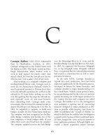
Fig. 16. (A) Dramatic upper limb deformity with large venous malformations involving the entire
right upper extremity in a patient with Maffuci’s syndrome. (B) Close-up view of the lesions of
the forearm.
05/Sangüeza/27-72/F 01/14/2003, 11:21 AM54
Chapter 5 / Cutaneous Vascular Malformations 55
sa
rcoma, other malignant neoplasms have been reported in patients with Maffucci’s
syndrome including fibrosarcoma (24), angiosarcoma (21), lymphangiosarcoma (25),
osteosarcoma (22), malignant ovarian neoplasms (21), gliomas (21), adenocarcinoma of
pancreas (24), and other multiple primary malignancies (21,22,24).The differential
diagnosis of Maffucci’s syndrome has to be established with Ollier disease’s, in which
there is dyschondroplasia without cutaneous vascular lesions (25).
Venous malformations are also prominent in and are the main cutaneous manifesta-
tion of Klippel-Trenaunay syndrome. The dominant features of this syndrome include
cutaneous capillary and venous malformations, congenital varicose veins, and hypertro-
phy of the involved limb (26) (Fig. 17). When, in addition to the aforementioned features,
there is an arteriovenous fistula, the disorder is termed Parkes Weber syndrome (27).
Klippel-Trenaunay syndrome affects males and females equally. Most commonly the
malformation is unilateral, and the lower limb is the most commonly involved area.
However, in rare cases the upper and lower limbs or the upper limb alone are affected;
bilateral involvement has also been reported, and occasionally the disease affects the
entire trunk. Regardless of the location, the malformation is present at birth, although it
may not be clinically apparent at that time. Almost all reported cases are sporadic, although
a few cases with a familial tendency have been described (28).
Cutaneous lesions of Klippel-Trenaunay syndrome may consist of one or several port
wine stains over the affected limb (26,27,29–35), but in addition, it is common to find
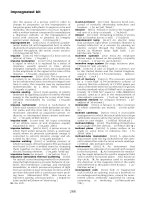
Fig. 17. (A) Venous malformation involving the entire lower right extremity in a patient with
Klippel-Trenaunay syndrome. (B) Close-up view of the lesions involving the leg.
05/Sangüeza/27-72/F 01/14/2003, 11:21 AM55
56 Sangüeza and Requena / Pathology of Vascular Skin Lesions
large venous ectatic vessels and vesicular lymphatic lesions (36). These vascular malfor-
mations do not blanch significantly under pressure. In many cases varicosities are asso-
ciated with the venous malformations. The varicosities start from a plexus of veins of the
dorsum and lateral side of the foot and extend up a variable distance on the leg. Incom-
petent perforating veins and deep vein abnormalities, which consist of occlusion by a
fibrous band, agenesis, or atresia, are also seen in these patients. The involved limb is
usually hypertrophic, and this enlargement is mostly caused by muscle hypertrophy,
thickened skin, excessive subcutaneous fat, the bulkiness of the abnormal vascular tissue,
and sometimes concomitant lymphedema. Usually there is little increase in bone diam-
eter in the hypertrophic limb. Patients with this syndrome occasionally complain of
profuse sweating of the skin involved in the vascular malformation, and the affected areas
may also feel warmer than normal.
Other systems and organs may show abnormalities in patients with Klippel-Trenaunay
syndrome (37); these anomalies usually occur within or adjacent to the area involved by
the vascular malformation. They can affect any mesodermal and ectodermal structure,
suggesting a more generalized dysplasia of the structures subject to a common teratoge-
nic influence. Venous thrombosis is common in patients with Klippel-Trenaunay syn-
drome, and therefore these patients have frequent episodes of pulmonary embolism. The
simultaneous occurrence of Klippel-Trenaunay syndrome and Fabry’s disease has been
described in the same patient (38).
Gorham’s syndrome (39) is a rare, nonfamilial disorder, that affects both sexes equally.
It is characterized by the development of venous and lymphatic malformations in the
skin, mediastinum, and bones (40,41). The osseous lesions cause osteolysis with fibrosis
and may lead to the disappearance of entire bones. Roentgenograms demonstrate lytic
lesions on the involved bones with little or no sclerosis. Cutaneous lesions usually develop
in the areas adjacent to the involved bones and may be accompanied by local muscular
atrophy. Usually, Gorham’s syndrome is self-limited (42), although an aggressive vari-
ant with a poor prognosis has been described (43).
Bannayan-Zonana syndrome is a rare autosomal dominant disorder characterized by
benign macrocephaly, lipomas, and cutaneous and visceral vascular malformations (44).
The cutaneous lesions are usually deeply situated, bluish nodules (45–47), but lesions
resembling superficial lymphatic malformations and angiokeratomas have also been de-
scribed (48). Visceral involvement may be massive, resulting in life-threatening obstruction
of vital organs, including the gastrointestinal tract and the central nervous system (48). The
macrocephaly is not associated with hydrocephalus, and most patients remain intellectually
normal, although mental retardation has been described in some cases (49).
Riley-Smith syndrome is an autosomal dominant condition described in five members
of the same family. It consists of macrocephaly without hydrocephalus, pseudo-
papilledema, and cutaneous capillary, venous, and lymphatic malformations (50).
Cutaneous vascular lesions may be present either at birth or appear shortly thereafter. The
abdominal wall, hands, feet, and thighs are the most commonly involved sites. The patients
remain intellectually and neurologically normal. This syndrome is similar to the
Bannayan-Zonana syndrome, except that patients with the Riley-Smith syndrome have
pseudopapilledema and do not have systemic lipomatous lesions.
In 1980, Ruvalcaba et al. (51) described two male patients thought to be affected with
hamartomatous intestinal polyps and spotted pigmentation of the penis. Based on the
description of these two patients and other cases from the literature (52,53); Cohen (54)
05/Sangüeza/27-72/F 01/14/2003, 11:21 AM56
Chapter 5 / Cutaneous Vascular Malformations 57
suggested that the condition described by Ruvalcaba et al. (51) was a distinctive entity
and coined the name Ruvalcaba-Myhre syndrome. Since then, additional cases have been
reported under the name of Ruvalcaba-Myhre-Smith syndrome (55–57). In 1988, Dvir
et al. (58) described a boy with macrocephaly, pseudopapilledema, lipoangiomatosis,
and spotted pigmentation of the penis. Because the patient had clinical features of three
syndromes (Bannayan-Zonana, Riley-Smith, and Ruvalcaba-Myhre-Smith), the authors
proposed that the three conditions were simply different expressions of a single
heredofamilial disorder. Cohen (59) supported this unifying theory and suggested that the
“new” syndrome be named after the first authors of the three original reports, i.e.,
Bannayan-Riley-Ruvalcaba syndrome. Subsequently, additional reports of Bannayan-
Riley-Ruvalcaba syndrome have appeared in the literature, lending further support to the
unifying concept (60–62). Recently, patients with Bannayan-Riley-Ruvalcaba syndrome
and facial tricholemmomas have been described, raising the possibility that Bannayan-
Riley-Ruvalcaba syndrome and Cowden disease may represent different alleles at the
same genetic locus or mutations of two genes in a common pathway (62).
Other rare miscellaneous syndromes that may show cutaneous venous malformations
include zosteriform venous malformations grouped in a unilateral dermatomal distribu-
tion (63,64); hereditary neurocutaneous vascular malformations syndrome (65), which
is transmitted as an autosomal dominant trait and it is characterized by the presence of
multiple cutaneous vascular malformations associated with intracranial arteriovenous
malformations; venous malformations on the face and anterior trunk associated with
sternal cleft and atrophic scar on the median abdominal raphe (66); retroauricular
hemangiomatous branchial clefts associated with several facial and neurosensorial
anomalies (67); sacral vascular malformations associated with renal, genital, osseous,
and neurologic malformations (68); cutaneous vascular malformations associated with
vascular anomalies of the retina and optic nerve (69); and several members of a family
affected by venous malformations involving the mouth, skin, and soft tissues, inherited
as an autosomal dominant trait and with no other associated anomalies (70).
H
ISTOPATHOLOGIC FEATURES
Histopathologically, venous malformations generally consist of ectatic blood vessels
of irregular size and shape involving the deep dermis and subcutaneous fat (Fig. 18).
Some of the involved blood vessels show thin walls, whereas others exhibit a thick layer
of smooth muscle in their walls. Thrombosis and phleboliths are common, and areas of
extravasated erythrocytes, deposits of hemosiderin, and extravascular calcifications are
also frequent findings.
Some of the cutaneous lesions of the aforementioned complex syndromes associated
with venous malformations may show specific histopathologic features. Large blue fa-
cial lesions with the clinical appearance of venous malformations showing glomus cells
surrounding the vascular structures are better interpreted as glomangiomas (2). In some
patients with blue rubber bleb nevus, the cutaneous lesions may also show multiple
glomangiomas (18,71–73). The gastrointestinal lesions of patients with blue rubber bleb
nevus show similar histopathologic features to those of the cutaneous lesions. The case
described as “blue rubber bleb nevus with vascular lesions suggesting a link to the Osler-
Rendu-Weber syndrome” (74) is better interpreted as an example of blue rubber bleb
nevus with telangiectatic cutaneous lesions but not related to the Osler-Rendu-Weber
syndrome.
05/Sangüeza/27-72/F 01/14/2003, 11:21 AM57
58 Sangüeza and Requena / Pathology of Vascular Skin Lesions
Histopathologically, the cutaneous lesions of Maffucci’s syndrome consist of large,
blood-filled vascular channels lined with flat endothelial cells. The walls of the vascular
spaces vary from thin, delicate, irregularly outlined walls to thick, fibrous, and smooth
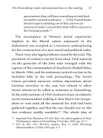
Fig. 18. Histopathologic features of a venous malformation. (A) Scanning power view showing
dilated vascular spaces in both superficial and deep dermis. The deeper component shows conges-
tive blood vessels. (B) Higher magnification of the deeper component shows congestive dilated
blood vessels. (C) Still higher magnification shows thin-walled blood vessels and hemosiderin
deposition on adjacent dermis.
05/Sangüeza/27-72/F 01/14/2003, 11:21 AM58
Chapter 5 / Cutaneous Vascular Malformations 59
muscle-containing walls. Several cases of spindle cell hemangiomas in patients with
Maffuci’s syndrome have been described (75–83). Spindle cell hemangioma consists of
well-circumscribed but not encapsulated nodules that combine features of hemangioma
and Kaposi’s sarcoma. Dilated blood vessels appear as thin veins that sometimes contain
organized thrombi and phleboliths within their lumina. Interspersed among the dilated
blood vessels there are fascicles of spindle cells mimicking Kaposi’s sarcoma, but within
the fascicles there are also round cells with prominent vacuolated cytoplasm. Sometimes
vacuolization of the cytoplasm of round cells is so marked that they may be mistaken for
entrapped fatty tissue.
Histopathologically, the cutaneous vascular lesions of patients with Bannayan-Riley-
Ruvalcaba syndrome show different combinations of capillary, venous, and lymphatic
malformations (51).
T
REATMENT
Small venous cutaneous malformations may be treated by simple surgical excision,
but in those cases in which the vascular malformation is associated with other internal
abnormalities, a careful follow-up of the patient is required. It is usually impossible to
remove large extensive venous malformations surgically without causing severe scarring
and other complications. In those cases involving the limbs, elastic stocking use is man-
datory and should be started early in infancy.
Management of patients with blue rubber bleb nevus depends on the individual case.
Resection of the involved bowel segment may be required in patients with recurrent
melena and anemia. Painful cutaneous lesions of glomangiomas may be treated by
excision, cryosurgery, or laser therapy (13).
Patients with Maffucci’s syndrome require careful follow-up, with radiologic and
histopathologic examination of any rapidly enlarging bone lesion for early diagnosis of
chondrosarcoma. Surgical excision of the cutaneous vascular malformations may be
indicated to improve the appearance of the patient. Spindle cell hemangioma is a benign
lesion and excision is curative.
Superficial venous varicosities of patients with the Klippel-Trenaunay syndrome may
be treated by ligation and stripping to relieve the local pain, but recurrences are common
(33). Before excision of the superficial veins, a radiographic exploration should be performed
to demonstrate that there is neither absence nor hypoplasia of the deep venous system.
Patients with Klippel-Trenaunay syndrome should receive antithrombotic prophylasis
prior to any surgery owing to the high risk of thromboembolic complications (33).
No effective treatment has been found for patients with Gorham’s syndrome, although
radiotherapy may be helpful for bone pain. Patients with Bannayan-Riley-Ruvalcaba
syndrome should be explored for detection of neurologic or any other associated internal
malformation, and genetic counseling should be given to the family.
References
1. Boon LM, Mulliken JB, Vikkula M, et al. Assignment of a locus for dominantly inherited venous
malformations to chromosome 9p. Hum Mol Genet 1994;3:1583–7.
2. Mounayer C, Wassef M, Enjolras O, Boukobza M, Mulliken JB. Facial “glomangiomas”: large venous
malformations with glomus cells. J Am Acad Dermatol 2001;45:239–45.
3. Boon LM, Brouillard P, Irrthum A, et al. A gene for inherited cutaneous venous anomalies (“gloman-
giomas”) localizes to chromosome 1p21-22. Am J Hum Genet 1999;65:125–33.
4. Kern S, Niemeyer C, Darge K, Merz C, Laubenberger J, Uhl M. Differential of vascular birthmarks by MR imaging.
An investigation of hemangiomas, venous and lymphatic malformations. Acta Radiol 2000;41:453–7.
05/Sangüeza/27-72/F 01/14/2003, 11:21 AM59
60 Sangüeza and Requena / Pathology of Vascular Skin Lesions
5. Boyd JB, Mulliken JB, Kaban LB, Upton J III, Murray JE. Skeletal changes associated with vascular
malformations. Plast Reconstr Surg 1984;74:789–97.
6. Enjolras O, Ciabrini D, Mazoyer E, Laurian C, Herbreteau D. Extensive pure venous malformations in
the upper or lower limb: a review of 27 cases. J Am Acad Dermatol 1997;36:219–25.
7. Bean WB. Blue rubber bleb nevi of the skin and gastrointestinal tract. In: Vascular Spiders and Related
Lesions of the Skin. Springfield, MO, CC Thomas, 1958:178–85.
8. Berlyne GM, Berlyne N. Anaemia due to “blue rubber bleb” naevus disease. Lancet 1960;2:1275–7.
9. Munkvad M. Blue rubber bleb nevus syndrome. Dermatologica 1983;167;307–9.
10. Walshe MM, Evans CD, Warin RP. Blue rubber bleb naevus. BMJ 1966; 2:931–2.
11. Baker AL, Kahn PC, Binder SC, et al. Gastrointestinal bleeding due to blue rubber bleb nevus syndrome.
Gastroenterology 1971;61:530–4.
12. McCauley RGK, Leonidas JC, Bartoshesky LE. Blue rubber bleb nevus syndrome. Radiology
1979;133:375–7.
13. Olsen TG, Milroy SK, Goldman L, et al. Laser surgery for blue rubber bleb nevus. Arch Dermatol
1979;115:81–2.
14. Baiocco FA, Gamoletti R, Negri A, et al. Blue rubber bleb nevus syndrome: a case with predominant
ENT localization. J Laryngol Otol 1984;98:317–9.
15. McCarthy JC, Goldberg MJ, Zimbler S. Orthopedic dysfunction in the blue rubber bleb nevus syndrome.
J Bone Joint Surg 1982;64A:280–3.
16. Rennie JG, Shortland JR, Mahood JM, et al. Periodic exophthalmos associated with blue rubber bleb
nevus syndrome. Br J Ophthalmol 1982;66:594–8.
17. Satya-Murti S, Navada S, Eames F. Central nervous system involvement in blue rubber bleb nevus
syndrome. Arch Neurol 1986;43:1184–6.
18. Sakurane HF, Sugai T, Saito T. The association of blue rubber bleb nevus and Maffucci’s syndrome.
Arch Dermatol 1967;95:28–36.
19. Maffucci A. Di un caso di encondroma ed angioma multiple: contribuzione alla genesi embrionale dei
tumori. Mov Med Chir Nap 1891;13:399–412.
20. Tilsley DA, Burden PW. A case of Maffucci’s syndrome. Br J Dermatol 1981;105:331–6.
21. Lewis RJ, Ketcham AS. Maffucci’s syndrome: functional and neoplastic significance: case report and
review of the literature. J Bone Joint Surg 1973; 55A:1465–79.
22. Bean WB. Dyschondroplasia and hemangiomata (Maffucci’s syndrome). II. Arch Intern Med
1958;102:544–50.
23. Suringa DWR, Ackerman AB. Cutaneous lymphangiomas with dyschondroplasia (Maffucci’s syn-
drome): a unique variant of an unusual syndrome. Arch Dermatol 1970;191:472–4.
24. Johnson JL, Webster Jr, Sippy HI. Maffucci’s syndrome (dyschondroplasia with hemangiomas). Am J
Med 1960;28:864–6.
25. Nardell SG. Ollier’s disease: dyschondroplasia. BMJ 1950;2:555–7.
26. Klippel M, Trenaunay P. Du noevus variqueux osteohypertophique. Arch Gen Med 1900;3:641–72.
27. Parkes Weber F. Angioma formation in connection with hypertrophy of limbs and hemi-hypertrophy.
Br J Dermatol 1907;19:231–5.
28. Lindenauer SM. Klippel-Trenaunay syndrome. Ann Surg 1965;162:303–10.
29. Cosman B. Clinical experience in the laser therapy of port-wine stains. Lasers Surg Med 1980;1:133–52.
30. Harper PS. Sturge-Weber syndrome with Klippel-Tranaunay syndrome. Birth Defects 1971;7:314.
31. Schofield D, Zaatari GS, Gay BB. Klippel-Trenaunay and Sturge-Weber syndromes with renal heman-
gioma and double inferior vena cava. J Urol 1986;136:442–5.
32. Adam JS, Cunliffe WJ. The Klippel-Trenaunay-Weber syndrome presenting with cutaneous bleeding.
Acta Derm Venereol 1981;62:176–7.
33. Baskerville PA, Ackroyd JS, Thomas ML, et al. The Klippel-Trenaunay syndrome: clinical, radiological
and haemodynamic features and management. Br J Surg 1985;72:232–6.
34. Phillips GN, Gordon DH, Mortin EC, et al. The Klippel-Trenaunay syndrome: clinical and radiological
aspects. Radiology 1978;128:429–34.
35. Viljoen D, Saxe N, Peran J, et al. The cutaneous manifestations of the Klippel-Trenaunay-Weber syn-
drome. Clin Exp Dermatol 1987;12:12–7.
36. Servelle M. Klippel-Trenaunay syndrome. Ann Surg 1985;201:365–76.
37. Young AE, Ackroyd J, Baskerville P. Combined vascular malformations. In: Mulliken JB, Young AE, eds.
Vascular Birthmarks. Hemangiomas and malformations. Philadelphia, WB Saunders, 1988:247–74.
38. Germain DP. Co-occurrence and contribution of Fabry disease and Klippel-Trenaunay-Weber syn-
drome to a patient with atypical skin lesions. Clin Genet 2001;60:63–7.
05/Sangüeza/27-72/F 01/14/2003, 11:21 AM60
Chapter 5 / Cutaneous Vascular Malformations 61
39. Gorham LW, Wright AW, Shultz HH, et al. Disappearing bones: a rare form of massive osteolysis. Am
J Med 1954;17:674–82.
40. Frost JF, Caplan RM. Cutaneous haemangiomas and disappearing bones with a review of cutaneo-
visceral haemangiomatosis. Arch Dermatol 1965;92:501–8.
41. Gellis SS, Feingold M. Hemangiomas with osteolysis (Gorham’s disease: vanishing bone disease). Am
J Dis Child 1978;132:715–6.
42. Mendez AA, Keret D, Robertson W, et al. Massive osteolysis of the femur (Gorham’s disease): a case
report and review of the literature. J Pediatr Orthop 1989;9:604–8.
43. Haferkamp O. Uber das Syndrome generalisierte maligne Haemangiomatosis mit Osteolysis. Krebforsch
1962;64:418–26.
44. Bannayan GA. Lipomatosis, angiomatosis, and macrocephalia: a previously undescribed congenital
syndrome. Arch Pathol 1971;92:1–5.
45. Higginbottom MC, Schultz P. The Bannayan syndrome: an autosomal dominant disorder consisting of
macrocephaly, lipomas, hemangiomas, and risk for intracranial tumors. Pediatrics 1982;69:632–4.
46. Miles HR, Zonana J, MacFarland J. Macrocephaly with hamartomas: Bannayan-Zonana syndrome. Am
J Med Genet 1984;19:225–34.
47. Zonana J, Rimoin DL, Davis DC. Macrocephaly with multiple lipomas and hemangiomas. J Pediatr
1976;89:600–3.
48. Klein JA, Barr RJ. Bannayan-Zonana syndrome associated with lymphangiomatous lesions. Pediatr
Dermatol 1990;7:48–53.
49. Saul RA, Stevenson RE, Bley R. Mental retardation in the Bannayan syndrome. Pediatrics 1982;69:642–4.
50. Riley HD, Smith WR. Macrocephaly, pseudopapilloedema and multiple hemangiomata: a previously
undescribed heredofamilial syndrome. Pediatrics 1960;26:293–300.
51. Ruvalcaba RHA, Myhre S, Smith DW. Sotos syndrome with intestinal polyposis and pigmentary changes
of the genitalia. Clin Genet 1980;18:413–6.
52. Halal F. Male to male transmission of cerebral gigantism. Am J Med Genet 1982;12:411–9.
53. Halal F. Cerebral gigantism, intestinal polyposis, and pigmentary spotting of the genitalia. Am J Med
Genet 1983;15:161.
54. Cohen MM Jr. The large-for-gestational-age (LGA) infant in dysmorphic perspective. In: Willey AM,
Carter TP, Kelly S, Porter IH, eds. Clinical Genetics: Problems in Diagnosis and Counseling. New York,
Academic,1982:153–69.
55. DiLiberti JH, Weleber RG, Budden S. Ruvalcaba-Myhre-Smith syndrome: a case with probable auto-
somal-dominant inheritance and additional manifestations. Am J Med Genet 1983;15:491–5.
56. DiLiberti JH, D’Agostino AN, Ruvalcaba RHA, Schimschock KR. A new lipid storage myopathy
observed in individuals with the Ruvalcaba-Myhre-Smith syndrome. Am J Med Genet 1984;18:163–7.
57. Gretzula JC, Hevia O, Schachner LS, et al. Ruvalcaba-Myhre-Smith syndrome. Pediatr Dermatol
1988;5:28–32.
58. Dvir M, Beer S, Aladjem M. Heredofamilial syndrome of mesodermal hamartomas, macrocephaly, and
pseudopapilledema. Pediatrics 1988;81:287–90.
59. Cohen MM Jr. Bannayan-Riley-Ruvalcaba syndrome: renaming three formerly recognized syndromes
as one etiologic entity. Am J Med Genet 1990;35:291.
60. Gorlin RS, Cohen MM, Leven LS. Bannayan-Riley-Ruvalcaba syndrome (Bannayan-Zonana syndrome,
Ruvalcaba-Myhre, Riley-Smith syndrome). In: Gorlin RS, Cohen MM, Leven LS, eds. Syndromes of
the Head and Neck. New York, Oxford University Press, 1990:336–8.
61. Gorlin RJ, Cohen MM Jr, Condon LM, Burke BA. Bannayan-Riley-Ruvalcaba syndrome. Am J Med
Genet 1992;44:307–14.
62. Fargnoli MC, Orlow SJ, Semel-Concepcion J, Bolognia JL. Clinicopathologic findings in the Bannayan-
Riley-Ruvalcaba syndrome. Arch Dermatol 1996;132:1214–8.
63. Steinway DM, Fretzin DF. Acquired zosteriform cavernous hemangiomas: brief clinical observations.
Arch Dermatol 1977;113:848–9.
64. Wilkin JK. Unilateral dermatomal cavernous hemangiomatosis. Dermatologica 1980;161:347–54.
65. Hurst J, Baraitser M. Hereditary neurocutaneous angiomatous malformations: autosomal dominant
transmission in two families. Clin Genet 1988;33:44–8.
66. Hersh JH, Waterfill D, Rutledge J, et al. Sternal malformation/vascular dysplasia association. Am J Hum
Genet 1985;21:177–84.
67. Hall BD, deLorimier A, Foster LH. Brief clinical report: a new syndrome of hemangiomatous branchial
clefts, lip pseudoclefts and unusual facial appearance. Am J Med Genet 1983;14:135–8.
05/Sangüeza/27-72/F 01/14/2003, 11:21 AM61
62 Sangüeza and Requena / Pathology of Vascular Skin Lesions
68. Goldberg NS, Hebert AA, Esterly NB. Sacral hemangiomas and multiple congenital anomalies. Arch
Dermatol 1986;122:684–7.
69. Goldberg RE, Pheasant TR, Shields JA. Cavernous hemangioma of the retina: a four generation pedigree
with neurocutaneous manifestations and an example of bilateral retinal involvement. Arch Ophthalmol
1979;97:2321–4.
70. Pasyk KA, Argenta LC, Erikson RP. Familial vascular malformations: report of 25 members of one
family. Clin Genet 1984;24:221–7.
71. Rice J, Fisher D. Blue rubber-bleb nevus syndrome. Generalized cavernous hemangiomas or venous
hamartoma with medulloblastoma of the cerebellum. Case report and review of the literature. Arch
Dermatol 1962;86:503–11.
72. Fretzin DF, Potter B. Blue rubber bleb nevus. Arch Intern Med 1965;116:924–9.
73. Hagood MF, Gathright JB Jr. Hemangiomatosis of the skin and gastrointestinal tract: report of a case.
Dis Colon Rectum 1975;18:141–6.
74. Rosenblum WI, Nakoneczna I, Konerding HS, Nochlin D, Ghatak NR. Multiple vascular malformation
in the “blue rubber bleb naevus” syndrome. A case with aneurysm of vein of Galen and vascular lesions
suggesting a link to the Weber-Osler-Rendu syndrome. Histopathology 1978;2:301–11.
75. Weiss SW, Enzinger FM. Spindle cell hemangioendothelioma: a low-grade angiosarcoma resembling
a cavernous hemangioma and Kaposi’s sarcoma. Am J Surg Pathol 1986;10:521–30.
76. Scott GA, Rosai J. Spindle cell hemangioendothelioma. Report of seven additional cases of a recently
described vascular neoplasm. Am J Dermatopathol 1988;10:281–8.
77. Lawson JP, Scott G. Case report 602. Skeletal Radiol 1990;19:158–62.
78. Murakami J, Sarker AB, Teramoto N, Horie Y, Tagucmi K, Akagi T. Spindle cell hemangioendothe-
lioma: a report of two cases. Acta Pathol Jpn 1993;43:529–34.
79. Pellegrini AE, Drake RD, Qualman SJ. Spindle cell hemangioendothelioma: a neoplasm associated with
Maffucci’s syndrome. J Cutan Pathol 1995;22:176–6.
80. Fukunaga M, Ushigome S, Nikaido T, Ishikawa E, Nakamori K. Spindle cell hemangioendothelioma:
an immunohistochemical and flow cytometric study of six cases. Pathol Int 1995;45:589–95.
81. Hisaoka M, Koumo H, Aoki T, Hashimoto H. DNA flow cytometric and immunohistochemical analysis
of proliferative activity in spindle cell haemangioendothelioma. Histopathology 1995;27:451–6.
82. Fanburg JC, Meis-Kindblom JM, Rosenberg AE. Multiple enchondromas associated with spindle cell
hemangioendotheliomas: an overlooked variant of Maffucci’s syndrome. Am J Surg Pathol
1995;19:1029–38.
83. Niechajev IA, Hansson LI. Maffucci’s syndrome. Scand J Plast Reconst Surg 1982;16:215–9.
05/Sangüeza/27-72/F 01/14/2003, 11:21 AM62
Chapter 5 / Cutaneous Vascular Malformations 63
Fig. 19. Superficial lymphatic malformation involving the posterior aspect of the thigh.
Fig. 20. Superficial lymphatic malformation involving the anterior aspect of the right forearm.
Numerous small vesicle-like lesions grouped in a plaque.
6. SUPERFICIAL CUTANEOUS LYMPHATIC MALFORMATIONS
CLINICAL FEATURES
Superficial cutaneous lymphatic malformations are localized lesions of the cutaneous,
subcutaneous, or submucosal lymphatic vessels. These lesions have been referred to in
the past as “lymphangiomas,” which is an inaccurate term.
The lesions are usually present at birth or appear shortly thereafter; they can be located
in any anatomic site but have a predilection for the axillary folds, shoulders, neck, proxi-
mal parts of the extremities (Figs. 19 and 20), and tongue (1–3). Superficial lymphatic
malformations (inaccurately termed “lymphangioma circunscriptum”) are the common-
est variant of cutaneous lymphatic malformation. Clinically, the lesion consists of
numerous small vesicle-like lesions, often with a verrucous surface, grouped in a plaque.
Sometimes, owing to the presence of blood vessels, purplish areas can be seen within the
lesion. The stereotypical superficial lymphatic malformation is accompanied by dilated
lymphatic cisterns located in the subcutaneous fat, which results in swelling of the tissue
05/Sangüeza/27-72/F 01/14/2003, 11:22 AM63
64 Sangüeza and Requena / Pathology of Vascular Skin Lesions
beneath the superficial vesicles (4,5). It is believed that the superficial vesicles are sac-
cular dilations of the superficial lymphatics secondary to the increased pressure from the
pulsating cisterns localized underneath (1). MRI is useful in demonstrating the size and
invasiveness of these lesions (6). In rare instances, superficial lymphatic malformations
extend deeply and can be associated with visceral lymphatic malformations involving the
mediastinum (7) or the bladder wall (8). Superficial lymphatic malformations may be
associated with Becker’s nevus (9), and have been described in patients with Maffucci’s
(10) and Cobb’s syndrome (11).
H
ISTOPATHOLOGIC FEATURES
Histopathologically, superficial lymphatic malformations are situated immediately
beneath the epidermis, but they may also involve areas of the reticular dermis (Fig. 21).
They consist of dilated lymph vessels, lined by a discontinuous layer of flat endothelial
cells (2,12). Sometimes the lymphatic vessels form clusters in the papillary dermis,
which gives a papillated or verrucous appearance to the skin surface.
Superficial lymphatic malformations are sometimes difficult to distinguish histopatho-
logically from angiokeratomas, especially when the lesions have been traumatized, which
results in the presence of erythrocytes within the ectatic lumina. The usual immunohis-
tochemical markers for endothelial cells, such as factor VIII-related antigen, Ulex
europaeus, and CD31 do not differentiate between blood and lymphatic vessels (13). In
these cases, the use of the new endothelial marker vascular endothelial growth factor
receptor-3 can be helpful. This marker is expressed by the lymphatic endothelium but not
by the endothelial cells lining blood vessels or neoplasms with blood endothelial differ-
entiation (14,15).
Ultrastructural studies of superficial lymphatic malformations demonstrate the pres-
ence of a fragmented basal lamina and anchoring filaments (13).
T
REATMENT
Most of the time, superficial cutaneous lymphatic malformations do not require treat-
ment, and it is probably best to leave them untreated. Surgical removal of the superficial
vesicles tends to be disappointing, especially if there is a deep component, since removal
is followed by local recurrence. Some lesions have been effectively treated with radio-
therapy (16), but there is a report of a lymphangiosarcoma arising in a preexisting superfi-
cial lymphatic malformation following X-ray therapy (17), and radiotherapy is not
currently recommended. The best cosmetic results have been achieved with argon laser (18)
or carbon dioxide laser (19–21). Combined therapy, which consists of carbon dioxide
laser vaporization for the superficial component of the lesion and transcutaneous sclero-
therapy with doxycycline for the deeper cisterns, has also been applied with good results (22).
References
1. Whimster J. The pathology of lymphangioma circumscriptum. Br J Dermatol 1976;94:473–86.
2. Flanagan BP, Helwig EB. Cutaneous lymphangioma. Arch Dermatol 1977;113:24–30.
3. Peachey RDG, Lim CC, Whimster JW. Lymphangioma of the skin: a review of 65 cases. Br J Dermatol
1970;83:519–27.
4. Russell B, Pridie RB. Lymphangioma circumscriptum with involvement of deep lymphatics. Br J
Dermatol 1967;79:300.
5. Palmer LC, Strauch WG, Welton WA. Lymphangioma circumscriptum: a case with deep lymphatic
involvement. Arch Dermatol 1978;114:394–6.
6. McAlvany JP, Jorizzo JL, Zanolli D, et al. Magnetic resonance imaging in the evaluation of lymphan-
gioma circumscriptum. Arch Dermatol 1993;129:194–7.
05/Sangüeza/27-72/F 01/14/2003, 11:22 AM64
Chapter 5 / Cutaneous Vascular Malformations 65
Fig. 21. Histopathologic features of a superficial cutaneous lymphatic malformation. (A) Low-
power view shows dilated vascular structures at all levels of the dermis. (B) Higher magnification
shows dilated thin-walled vessels with a lymphatic appearance lined by a single discontinuous
layer of endothelial cells.
7. Mordehai J, Kurzbart E, Shinhar D, Sagi A, Finaly R, Nares AJ. Lymphangioma circumscriptum. Pediatr
Surg Int 1998;13:208–10.
8. Irvine AD, Sweeney L, Corbett JR. Lymphangioma circumscriptum associated with paravesical cystic
retroperitoneal lymphangioma. Br J Dermatol 1996;134:1135–7.
9. Oyler RM, Davis DA, Woosley JT. Lymphangioma associated with Becker’s nevus: a report of coin-
cident hamartomas in a child. Pediatr Dermatol 1997;14:376–9.
10. Suringa DW, Ackerman AB. Cutaneous lymphangiomas with dyschondroplasia (Maffucci’s syndrome).
A unique variant of an unusual syndrome. Arch Dermatol 1970;101:472–4.
11. Shim JH, Lee DW, Cho BK. A case of Cobb syndrome associated with lymphangioma circumscriptum.
Dermatology 1996;193:45–7.
12. Bauer BS, Kernahan DA, Hugo NE. Lymphangioma circumscriptum: a clinicopathologic review. Ann
Plast Surg 1981;7:318–26.
05/Sangüeza/27-72/F 01/14/2003, 11:22 AM65
66 Sangüeza and Requena / Pathology of Vascular Skin Lesions
13. Pearson JM, McWilliam LJ. A light microscopical, immunohistochemical, and ultrastructural compari-
son of hemangioma and lymphangioma. Ultrastruct Pathol 1990;14:497–504.
14. Lymboussaki A, Partanen TA, Olofsson B, et al. Expression of the vascular endothelial growth factor
C receptor VEGFR-3 in lymphatic endothelium of the skin and in vascular tumors. Am J Pathol
1998;153:395–403.
15. Folpe AL, Veikkola T, Valtola R, Weiss SW. Vascular endothelial growth factor receptor-3 (VEGFR-3):
a marker of vascular tumors with presumed lymphatic differentiation, including Kaposi’s sarcoma,
kaposiform and Dabska-type hemangioendotheliomas, and a subset of angiosarcomas. Mod Pathol
2000;13:180–5.
16. O’Cathail S, Rostom AY, Johnson ML. Successful control of lymphangioma circumscriptum by super-
ficial X-rays. Br J Dermatol 1985;113:611–5.
17. King DT, Duffy DM, Hirose FM, Gurevitch AW. Lymphangiosarcoma arising from lymphangioma
circumscriptum. Arch Dermatol 1979;115:969–72.
18. Landthaler M, Haina D, Waidelich W, Braun-Falco O. Behandlung zirkumskripter lymphangiome mit
dem Argonlaser. Hautarzt 1982;33:266–70.
19. Bailin PL, Kantor GR, Wheeland RG. Carbon dioxide laser vaporization of lymphangioma
circumscriptum. J Am Acad Dermatol 1986;14:257–62.
20. Eliezri YD, Sklar JA. Lymphangioma circumscriptum: review and evaluation of carbon dioxide laser
vaporization. J Dermatol Surg Oncol 1988;14:357–64.
21. Haas AF, Narurkar VA. Recalcitrant breast lymphangioma circumscriptum treated by ultrapulse carbon
dioxide laser. Dermatol Surg 1998;24:893–5.
22. Wimmershoff MB, Schreyer AG, Glaessl A, et al. Mixed capillary/lymphatic malformation with coex-
isting port-wine stain: treatment utilizing 3D MRI and CT-guided sclerotherapy. Dermatol Surg
2000;26:584–7.
05/Sangüeza/27-72/F 01/14/2003, 11:22 AM66
Chapter 5 / Cutaneous Vascular Malformations 67
7. CYSTIC LYMPHATIC MALFORMATIONS (CYSTIC HYGROMAS)
Cystic lymphatic malformations are contrasted with superficial lymphatic malforma-
tions, in being deeply located and consisting of subcutaneous painless nodules covered
by normal skin (1–3). The lesion may be painful, especially when the tumor is subject to
pressure or is bumped. Cases of cystic lymphatic malformations have been described in
patients with Maffucci’s syndrome (4).
C
LINICAL FEATURES
Cystic hygroma is a variant of subcutaneous lymphatic malformation whose shape and
character are determined by its anatomic location. These lesions most commonly occur
in the neck, axilla (Fig. 22), and groin, areas where the presence of loose connective tissue
allows for the expansion of the lymphatic channels (5). Cystic hygroma is usually present
at birth or appears in early infancy. They present as a large fluid-filled cystic mass that
may be diagnosed by transillumination. Cystic hygromas of the posterior triangle of the
neck have been associated with hydrops fetalis, Turner’s syndrome (45X0 karyotype),
congenital malformations, several varieties of chromosomal aneuploidy, and fetal death
(6). Since aneuploidic conditions may recur in subsequent pregnancies, cytogenetic
analysis of fetuses born with cystic hygroma is mandatory.
H
ISTOPATHOLOGIC FEATURES
Histopathologically, cystic lymphatic malformations are made up of irregular, dilated,
and interconnected lymphatic vessels localized in the subcutaneous fat (Fig. 23). Some
of these vessels contain bundles of smooth muscle in their walls (7). Nodular collections
of lymphocytes, sometimes with germinal centers, may be present within the surrounding
connective tissue.
Cystic hygroma lesions consist of large uni- or multilocular cystic cavities surrounded
by a loose connective tissue stroma. In some areas the stroma may be denser and even
sclerotic as a result of compression by the lymphatic cysts.
Fig. 22. Clinical appearance of a lymphatic malformation on the axilla, with a deep component of
subcutaneous nodules and a superficial component of an angiomatous appearance.
05/Sangüeza/27-72/F 01/14/2003, 11:22 AM67
68 Sangüeza and Requena / Pathology of Vascular Skin Lesions
TREATMENT
The small superficial lymphatic malformations may be adequately managed by sur-
gery (8,9), cryotherapy (10), radiotherapy (11), and laser phototherapy (12). Larger and
subcutaneous lesions of cystic hygroma show a high rate of recurrence after surgery, and
unless the lesion causes severe signs or symptoms, such lesions are best left untreated.
Complete regression of cystic hygromas has also been reported with intracystic injec-
tions of sclerosing substances with no sequels (13).
References
1. Flanagan BP, Helwig EB. Cutaneous lymphangioma. Arch Dermatol 1977;113:24–30.
2. Peachey RDG, Lim CC, Whimster JW. Lymphangioma of the skin: a review of 65 cases. Br J Dermatol
1970;83:519–27.
3. Harkins GA, Sabiston DC. Lymphangioma in infancy and childhood. Surgery 1960;47:811–22.
4. Suringa DWR, Ackerman AB. Cutaneous lymphangiomas with dyschondroplasia (Maffucci’s syn-
drome): a unique variant of an unusual syndrome. Arch Dermatol 1970;191:472–4.
5. Bill AH, Sumner DS. A unified concept of lymphangioma and cystic hygroma. Surg Gynecol Obstet
1965;120:79–86.
6. Chervenak FA, Isaacson G, Blakemore KJ, et al. Fetal cystic hygroma cause and natural history. N Engl
J Med 1983;309:822–5.
7. Russell B, Pridie RB. Lymphangioma circumscriptum with involvement of deep lymphatics. Br J
Dermatol 1967;79:300.
Fig. 23. Histopathologic features of a deep lymphatic malformation. (A) Scanning power view
shows dilated vascular structures involving the deeper dermis and subcutaneous tissue. (B) Higher
magnification demonstrates that the vascular structures have thin walls and eosinophilic homoge-
neous material in the lumina, which is lymph.
05/Sangüeza/27-72/F 01/14/2003, 11:22 AM68
Chapter 5 / Cutaneous Vascular Malformations 69
8. Edwards JM, Peachey RDG, Kinmonth JB. Lymphangiography and surgery in lymphangioma of the
skin. Br J Surg 1972;59:36–41.
9. Jordan PR, Sanderson KV, Wilson JSP. Surgical treatment of lymphangioma circumscriptum: a case
report. Br J Plast Surg 1977;30:306–7.
10. Nanda Kumar H, Bhaskar Roa C, Kukreja R. Management of lymphangioma by cryoprobe: a case report.
J Indian Dent Assoc 1982;54:25–7.
11. O’Cathial S, Rostom AY, Johnson ML. Successful control of lymphangioma circumscriptum by super-
ficial x-rays. Br J Dermatol 1985;113:611–5.
12. Bailin PL, Kantor GR, Wheeland RG. Carbon dioxide laser vaporization of lymphangioma
circumscriptum. J Am Acad Dermatol 1986;14:257–62.
13. Ogita S, Tsuto T, Tokiwa K, Takahashi T. Intracystic injection of OK-432. A new sclerosing therapy for
cystic hygroma in children. Br J Surg 1987;74:690–1.
05/Sangüeza/27-72/F 01/14/2003, 11:22 AM69
70 Sangüeza and Requena / Pathology of Vascular Skin Lesions
Fig. 24. Lymphangiomatosis resulting in deformity of the right leg of a young woman.
8. LYMPHANGIOMATOSIS
This is a rare disorder characterized by the presence of abnormal lymphatic channels
in a diffuse or multifocal arrangement. These lesions usually involve both soft tissue and
parenchymal organs. Most cases of lymphangiomatosis with both bone and visceral
involvement are associated with a poor prognosis and high mortality rate (1). In contrast,
patients with involvement solely of soft tissues and bones of the extremities, show slow
clinical progression and have a good prognosis (2), although there is a report of a patient
with disseminated lymphangiomatosis of the skin and bone who developed disseminated
intravascular coagulation (3). The lesions appear either at birth or in early infancy.
C
LINICAL FEATURES
Clinically, the lesions are fluctuant and sponge-like, with progressive swelling of the
affected limb (Fig. 24). The overlying skin is usually normal, but it may become second-
arily involved and develop a verrucous surface, areas of pigmentation, or vesicle forma-
tion. Examples of lymphangiomatosis have been described in patients with kaposiform
hemangioendothelioma (4,5).
H
ISTOPATHOLOGIC FEATURES
Histopathologically, the lesions of lymphangiomatosis consist of lobules of intercon-
necting widely dilated lymphatic channels lined by a single, attenuated layer of endothe-
05/Sangüeza/27-72/F 01/14/2003, 11:22 AM70
Chapter 5 / Cutaneous Vascular Malformations 71
Fig. 25. Histopathologic features of lymphangiomatosis involving the skin. (A) Scanning magni-
fication shows irregular dilated vascular structures replacing the subcutaneous fat. (B) Higher
magnification demonstrates that these vascular structures have walls of variable thickness con-
taining an eosinophilic homgeneous material within the vascular lumina.
lial cells. The lesions can involve dermis, subcutaneous fat (Fig. 25), and sometimes
underlying soft tissue and bone. When the lesions are present in the dermis, the lymphatic
spaces dissect between collagen bundles and around preexisting dermal and subcutane-
ous structures, resembling a well-differentiated angiosarcoma (1,2). However, the endot-
helial cells lack atypia. The lumina of the lymphatic channels either appear empty or
05/Sangüeza/27-72/F 01/14/2003, 11:22 AM71
72 Sangüeza and Requena / Pathology of Vascular Skin Lesions
contain a proteinaceous eosinophilic material. In one case, there was prominent intra- and
extravascular extramedullary hematopoiesis with large amounts of hemosiderin in the
stroma despite the absence of any apparent intra- or extravascular erythrocytes (2).
Immunohistochemical studies have demonstrated that the endothelial cells of the
abnormal lymphatic channels stain positive for factor VIII-related antigen and Ulex
europaeus I lectin, whereas positivity for CD31 and CD34 is variable from case to case
(1,2). These immunohistochemical results confirm the fact that there are no reliable
endothelial markers to distinguish between blood vessel and lymphatic endothelium.
T
REATMENT
Treatment of patients with lymphagiomatosis of the limbs consists of surgical reduc-
tion with extensive excision of the skin, subcutaneous tissue, and fascia. Although sig-
nificant clinical improvement is achieved, in some cases the swelling recurrs slowly over
succeeding years (2).
References
1. Romani P, Shah A. Lymphangiomatosis and immunohistochemical analysis of four cases. Am J Surg
Pathol 1993;17:329–35
2. Singh Gomez C, Calonje E, Ferrar DW, Browse NL, Fletcher CDM. Lymphangiomatosis of the limbs.
Clinicopathologic analysis of a series with a good prognosis. Am J Surg Pathol 1995;19:125–33.
3. Lauret P, Monconduit M, Sonlica J. Lymphangiomatose cutanée et osseuse disseminée avec coagulation
intra-vasculaire disseminée. Ann Dermatol Venereol 1978;105:759–63.
4. Zukerberg LR, Nickoloff BJ, Weiss SW. Kaposiform hemangioendothelioma of infancy and childhood.
An aggressive neoplasm associated with Kasabach-Merritt syndrome and lymphangiomatosis. Am J
Surg Pathol 1993;17:321–8.
5. Mentzel T, Mazzoleni G, Dei Tos AP, Fletcher CD. Kaposiform hemangioendothelioma in adults. Clini-
copathologic and immunohistochemical analysis of three cases. Am J Clin Pathol 1997;108:450–5.
05/Sangüeza/27-72/F 01/14/2003, 11:22 AM72
Chapter 6 / Cutaneous Lesions with Dilations 73
73
Cutaneous Lesions Characterized
by Dilation of Preexisting Vessels
CONTENTS
SPIDER ANGIOMA (NEVUS ARANEUS)
C
APILLARY ANEURYSM-VENOUS LAKE
TELANGIECTASES
ANGIOKERATOMAS
LYMPHANGIECTASES
1. SPIDER ANGIOMA (NEVUS ARANEUS)
CLINICAL FEATURES
Spider angioma, also known as nevus araneus, is present in approximately 10–15% of
adults and young children. The face, neck, upper trunk, and arms are the regions most
frequently involved; however, in children, the hands and fingers are the preferred sites
(1). A higher incidence of spider angiomas is seen in pregnant women and in patients with
chronic liver disease (2–4). In pregnant women, the lesions usually disappear at the end
of the pregnancy without therapy. In patients with liver disease, the development of these
lesions has been attributed to alcohol, increased plasma levels of estrogen, vascular
dilation, and neovascularization. To date only an increased plasma level of substance P
has been demonstrated in patients in whom cirrhosis and spider angiomas coexist (5).
Clinically, spider angiomas are characterized by a central, slightly elevated, red punctum
or “body” of the spider, from which the blood vessels or “legs” of the spider radiate
(Fig. 1). Occasionally, pulsation can be observed in the central punctum.
H
ISTOPATHOLOGIC FEATURES
Histopathologically, spider angiomas consist of a central ascending arteriole, which
ends in a thin-walled ampulla just beneath the epidermis. From this ampulla, thin, delicate
arterial branches radiate peripherally into the papillary dermis. Occasionally glomus
cells can be identified in the wall of the central arteriole (6,7). In one case a giant solitary
spider angioma had an overlying pyogenic granuloma (8).
T
REATMENT
Electrodesiccation of the central punctum is usually followed by extinction of the
spider angioma, but recurrences are fairly common. Laser therapy has also been used for
treatment of spider angiomas in children (9).
6
06/Sangüeza/73-98/F 01/14/2003, 11:47 AM73
74 Sangüeza and Requena / Pathology of Vascular Skin Lesions
References
1. Wenzl JE, Burgert EO Jr. The spider nevus in infancy and childhood. Pediatrics 1964;33:227–32.
2. Whiting DA, Kallmeyer JC, Simson IW. Widespread arterial spiders in a case of latent hepatitis with
resolution after therapy. Br J Dermatol 1970;82:32–6.
3. Witte CL, Hicks T, Renert W, Witte MH, Butler C. Vascular spider: a cutaneous manifestation of
hyperdynamic blood flow in hepatic cirrhosis. South Med J 1975;68:246–8.
Fig. 1. (A) Clinical appearance of a spider angioma on the dorsum of the nose. (B) Close-up view
of the lesion.
06/Sangüeza/73-98/F 01/14/2003, 11:47 AM74
Chapter 6 / Cutaneous Lesions with Dilations 75
4. Li CP, Lee FY, Hwang SJ, et al. Spider angiomas in patients with liver cirrhosis: role of alcoholism and
impaired liver function. Scand J Gastroenterol 1999;34:520–3.
5. Li CP, Lee FY, Hwang SJ, et al. Role of substance P in the pathogenesis of spider angiomas in patients
with nonalcoholic liver cirrhosis. Am J Gastroenterol 1999;94:502–7.
6. Bean WB. The arterial spider and similar lesions of the skin and mucous membranes. Circulation
1953;8:117–29.
7. Schuhmachers-Brendler R. Beitrag zur morphologishen Pathologie und therapie des Naevus-araneus
Rezidivs. Dermatol Wochenschro 1959;139:167–74.
8. Okada N. Solitary giant spider angioma with an overlying pyogenic granuloma. J Am Acad Dermatol
1987;16:1053–4.
9. Tan OT, Gilchrest BA. Laser therapy for selected cutaneous vascular lesions in the pediatric population:
a review. Pediatrics 1988;82:652–62.
06/Sangüeza/73-98/F 01/14/2003, 11:47 AM75
76 Sangüeza and Requena / Pathology of Vascular Skin Lesions
Fig. 2. Multiple capillary aneurysms on the face of an adult man.
2. CAPILLARY ANEURYSM-VENOUS LAKE
Capillary aneurysm and venous lake probably represent two different stages in the
development of the same lesion. Capillary aneurysm was first described by Epstein et al.
(1) in 1956. These authors emphasized the clinical similarity of this lesion with malignant
melanomas. In the same year, Bean and Walsh (2) described venous lakes.
C
LINICAL FEATURES
Capillary aneurysm is classically referred to as a suddenly growing dark papule on
the face of elderly patients (3) (Fig. 2), although similar lesions may also occur in the
oral mucosa (4). Most of the time these are solitary lesions, but multiple lesions have
been reported (5). The majority of capillary aneurysms are asymptomatic, but occa-
sionally the affected patients may complain of tenderness or pruritus. Venous lakes are
small, dark blue, dome-shaped, soft papules occurring in elderly patients on skin
exposed to sun. The lesions are easily compressed. The face, ears, and lips (Fig. 3) are
the most common sites (2).
H
ISTOPATHOLOGIC FEATURES
Capillary aneurysm presents with a widely dilated thin-walled venule just beneath the
epidermis, lined by a single layer of endothelial cells. No smooth muscle or elastic tissue
is discernible in the vessel wall. The presence of a thrombus within the lumen is charac-
06/Sangüeza/73-98/F 01/14/2003, 11:47 AM76
Chapter 6 / Cutaneous Lesions with Dilations 77
Fig. 4. Histopathologic features of a capillary aneurysm. (A) A dilated and congestive blood vessel
involving the entire dermis. (B) Higher magnification shows the thin vessel wall. Probably venous
lake and capillary aneurysm are just two different stages of evolution of the same lesion.
Fig. 3. Venous lake on the lower lip of an elderly woman.
teristic of this lesion (3,6) (Fig. 4). In many cases the thrombus undergoes recanalization
and shows areas of papillary endothelial hyperplasia. Most of the vessels involved in
capillary aneurysms are venules, so the term capillary is a misnomer. Venous lakes are
histopathologically identical to “capillary” aneurysms, except for the absence of a lumi-
nal thrombus (2) (Fig. 5). In most cases there is severe sun damage in the adjacent dermis.
Venous lakes result from faulty elastic tissue in elderly patients (7). As mentioned earlier,
capillary aneurysms and venous lakes are the same lesion. Capillary aneurysms represent
06/Sangüeza/73-98/F 01/14/2003, 11:47 AM77
78 Sangüeza and Requena / Pathology of Vascular Skin Lesions
the early lesion in which there is superficial vein thrombosis with subsequent dilation.
When the thrombus lyses, usually before excision, the lesion acquires the appearance of
a venous lake. In short, “capillary” aneurysm and venous lake are two different stages in
the development of superficial venous varicosities.
T
REATMENT
Sclerosing injections and compression therapy have been ineffective (5). Simple
excision is curative of lesions of capillary aneurysm-venous lake. Careful cryotherapy,
electrocautery, or therapy with argon laser also give good results (8).
References
1. Epstein E, Novy FG, Skahen RA, Krause ME. Melanoma-simulating nodules due to capillary aneu-
rysms. Cali Med 1956;85:22–5.
2. Bean WB, Walsh JR. Venous lakes. Arch Dermatol 1956;74:459–63.
3. Epstein E, Novy FJ Jr, Allington HV. Capillary aneurysms of the skin. Arch Dermatol 1965;91:335–41.
4. Weathers DR, Fine RH. Thrombosed varyx of oral cavity. Arch Dermatol 1971; 104:427–30.
5. Pokorny M, Vanek J, Pavcova S. Multiple kapillare aneurysmen. Hautartz 1987;38:541–3.
6. Weiner HA. Capillary aneurysms of the skin. Arch Dermatol 1966;96:670–3.
7. Alcalay J, Sandbank M. The ultrastructure of cutaneous venous lakes. Int J Dermatol 1987;26:645–6.
8. Neumann RA, Knobler RM. Venous lakes (Bean-Walsh) of the lips—treatment experience with the
argon laser and 18 months follow-up. Clin Exp Dermatol 1990;15:115–8.
Fig. 5. Histopathologic features of a venous lake. (A) Low-power shows a dilated vascular channel
in the upper dermis. (B) Higher magnification shows a thin-walled vascular structure.
06/Sangüeza/73-98/F 01/14/2003, 11:47 AM78