Chapter 061. Disorders of Granulocytes and Monocytes (Part 3) pps
Bạn đang xem bản rút gọn của tài liệu. Xem và tải ngay bản đầy đủ của tài liệu tại đây (95.99 KB, 5 trang )
Chapter 061. Disorders of Granulocytes
and Monocytes
(Part 3)
Pelger-Hüet anomaly. In this benign disorder, the majority of granulocytes
are bilobed. The nucleus frequently has a spectacle-like, or "pince-nez,"
configuration.
In severe acute bacterial infection, prominent neutrophil cytoplasmic
granules, called toxic granulations, are occasionally seen. Toxic granulations are
immature or abnormally staining azurophil granules. Cytoplasmic inclusions, also
called Döhle bodies (Fig. 61-3), can be seen during infection and are fragments of
ribosome-rich endoplasmic reticulum. Large neutrophil vacuoles are often present
in acute bacterial infection and probably represent pinocytosed (internalized)
membrane.
Neutrophils are heterogeneous in function. Monoclonal antibodies have
been developed that recognize only a subset of mature neutrophils. The meaning
of neutrophilheterogeneity is not known.
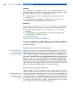
The morphology of eosinophils and basophils is shown in Fig. 61-6.
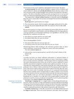
Figure 61-6
Normal eosinophil and basophil. The eosinophil contains large, bright
orange granules and usually a bilobed nucleus. The basophil contains large purple-
black granules that fill the cell and obscure the nucleus
Marrow Release and Circulating Compartments
Specific signals, including IL-1, tumor necrosis factor α (TNF-α), the CSFs,
complement fragments, and chemokines, mobilize leukocytes from the bone
marrow and deliver them to the blood in an unstimulated state. Under normal
conditions, ~90% of the neutrophil pool is in the bone marrow, 2–3% in the
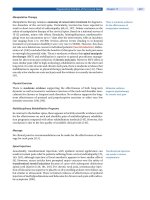
circulation, and the remainder in the tissues (Fig. 61-7).
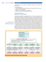
Figure 61-7
Schematic neutrophil distribution and kinetics between the different
anatomic and functional pools
The circulating pool exists in two dynamic compartments: one freely
flowing and one marginated. The freely flowing pool is about one-half the
neutrophils in the basal state and is composed of those cells that are in the blood
and not in contact with the endothelium. Marginated leukocytes are those that are
in close physical contact with the endothelium (Fig. 61-8). In the pulmonary
circulation, where an extensive capillary bed (~1000 capillaries per alveolus)
exists, margination occurs because the capillaries are about the same size as a
mature neutrophil. Therefore, neutrophil fluidity and deformability are necessary
to make the transit through the pulmonary bed. Increased neutrophil rigidity and
decreased deformability lead to augmented neutrophil trapping and margination in
the lung. In contrast, in the systemic postcapillary venules, margination is
mediated by the interaction of specific cell-surface molecules called selectins.
Selectins are glycoproteins expressed on neutrophils and endothelial cells, among
others, that cause a low-affinity interaction, resulting in "rolling" of the neutrophil
along the endothelial surface. On neutrophils, the molecule L-selectin [cluster
determinant (CD) 62L] binds to glycosylated proteins on endothelial cells [e.g.,
glycosylation-dependent cell adhesion molecule (GlyCAM1) and CD34].
Glycoproteins on neutrophils, most importantly sialyl-Lewis
x
(SLe
x
, CD15s), are
targets for binding of selectins expressed on endothelial cells [E-selectin (CD62E)
and P-selectin (CD62P)] and other leukocytes. In response to chemotactic stimuli
from injured tissues (e.g., complement product C5a, leukotriene B
4
, IL-8) or
bacterial products [e.g., N-formylmethionylleucylphenylalanine (f-metleuphe)],
neutrophil adhesiveness increases, and the cells "stick" to the endothelium through
integrins. The integrins are leukocyte glycoproteins that exist as complexes of a
common CD18 βchain with CD11a (LFA-1), CD11b (called Mac-1, CR3, or the
C3bi receptor), and CD11c (called p150, 95 or CR4). CD11a/CD18 and
CD11b/CD18 bind to specific endothelial receptors [intercellular adhesion
molecules (ICAM) 1 and 2].