The principles of toxicology environmental and industrial applications 2nd edition phần 2 ppt
Bạn đang xem bản rút gọn của tài liệu. Xem và tải ngay bản đầy đủ của tài liệu tại đây (834.71 KB, 61 trang )
Calculated from the terminal slope of a plot of the natural logarithm of the concentration in the
central compartment as a function of time, this half-life is designated the biological half-life. It
is the parameter most frequently used to characterize the in vivo kinetic behavior of an exogenous
compound.
Other features of chemical kinetic behavior or of mode of administration may be incorporated into
the model as appropriate. For example, there may be more than one peripheral tissue compartment, as
in Figure 2.1; or absorption, which is never truly instantaneous even for intravenous injection, may be
first-order instead. An oral exposure, in which the rate of absorption is usually considered to be directly
proportional to the amount remaining available in the GI tract, is an example of first-order uptake.
The important group of models that incorporate non-first-order kinetics should also be mentioned.
Absorption and distribution are conventionally considered to be passive, first-order processes unless
observation dictates otherwise. However, elimination often is not first-order. Frequently this is because
excretion or metabolism is saturable, or capacity-limited, due to a limitation on the maximum number
of active transport sites in organs of excretion or the maximum number of active sites on metabolizing
enzymes. When all active elimination sites are occupied, the elimination process is said to be saturated.
Kinetically it is a zero-order process, operating at a constant maximum rate independent of the amount
or concentration of the chemical in the body. At very low concentrations at which relatively few
elimination sites are occupied, capacity-limited kinetics reduces to pseudo-first-order kinetics. Capac-
ity-limited kinetics is often referred to as
Michaelis–Menten kinetics
, after the authors of an early paper
analyzing and interpreting this type of kinetic behavior. Classical kinetic models incorporating
Michaelis–Menten elimination have been developed.
Figure 2.7 Plot of the logarithm of the concentration versus time for the linear one-compartment open model. C
0
is the concentration at time t = 0, assuming instantaneous distribution. (Reproduced with permission from
O’Flaherty, 1981, Figure 2.15a.)
2.4 DISPOSITION: DISTRIBUTION AND ELIMINATION
47
Most industrial or environmental exposures are not acute. Acute exposures do occur, but chronic
exposures are much more frequent in both industrial and environmental settings. When exposure is
approximately constant and continuous over a long period of time (e.g., if a contaminant is widely
dispersed in ambient air), a steady state or “ plateau” level will eventually be reached in all tissues. As
long as elimination processes remain first-order (typical, e.g., of excretion by glomerular filtration in
Figure 2.8 The linear two-compartment open model, where C
1
and C
2
are the concentrations in the central and
peripheral compartments, respectively, and k
12
and k
21
are the rate constants for transfer between the two
compartments. (Reproduced with permission from O’Flaherty, 1981, Figure 2.22.)
Figure 2.9 Plot of the logarithm of the concentration versus time for the linear two-compartment open model,
showing ln C as a function of time for the central (C
1
) and peripheral (C
2
) compartments. (Reproduced with
permission from O’Flaherty, 1981, Figure 3-24b.)
48
ABSORPTION, DISTRIBUTION, AND ELIMINATION OF TOXIC AGENTS
the kidney, or of loss of a volatile chemical in expired air), this steady state should be directly
proportional to both the magnitude of exposure and the biological half-life.
If exposure were truly constant, the plateau level would be constant also. More commonly, exposure
is intermittent, in which case blood concentrations at steady state will cycle in a way that reflects the
absorption and elimination characteristics of the compound as well as the exposure pattern (Figure
2.10). However, on a larger timescale this cycling will take place about a constant mean that is
predictable from the equivalent constant exposure rate and the biological half-life. This is one of the
reasons why biological half-life is such an important attribute. Together with exposure rate, it
determines mean steady-state blood level irrespective of whether exposure is continuous or intermit-
tent. However, the individual exposed to large amounts of a substance at wide intervals will experience
greater peak concentrations in blood and tissues following each new exposure than will an individual
exposed to the same total amount as frequent small exposures. If the large peak concentrations are
associated with toxicity or with saturation of elimination processes, then it becomes important to
consider the pattern of administration as well as the equivalent mean exposure rate.
Physiologically Based Kinetic Models
Physiologically based kinetic (PBK) models are simplified
but anatomically and physiologically reasonable models of the body. Tissues are selected or grouped
according to their perfusion (blood flow) characteristics and whether they are sites of absorption or
elimination (by excretion or metabolism). The model design process is facilitated by reference to
compilations of anatomic and physiologic data, including tissue and organ perfusion rates, that are
now widely available.
Within this general structural framework, the kinetic behavior of the selected chemical is modeled.
A key question is how the chemical is taken up into tissues. When flow-limited kinetics are assumed,
the chemical is presumed to be in equilibrium between each tissue group and the venous blood leaving
Figure 2.10 The relationship between average concentration C
__
(
n
)
, calculated for repetitive administation, and the
time course of concentration change during continuous administration of a hypothetical compound. C
max
and C
min
are the maximum and minimum concentrations in each time interval between doses, assuming instantaneous
distribution of each successive dose. (Reproduced with permission from O’Flaherty, 1981. Figure 5-4.)
2.4 DISPOSITION: DISTRIBUTION AND ELIMINATION
49
the tissue. This equilibrium will vary from tissue to tissue and may also vary from species to species.
Simple partitioning phenomena, such as into body lipid stores, can be described by defining partition
coefficients, whose values can be determined experimentally at steady state in vivo or in vial
equilibration experiments in vitro. More complex partitioning, such as capacity-limited binding of a
metal to specific binding sites in tissues, must be defined appropriately. Estimates of dissociation
constants may be required.
Diffusion-limited kinetics can also be accommodated within the framework of PBK models. In
diffusion-limited kinetics, the process of transfer across the membrane separating tissue from blood is
the rate-limiting step in tissue uptake. The distinction between flow-limited and diffusion-limited
tissue-uptake kinetics is roughly analogous to the distinction between ventilation-limited and flow-
limited absorption in the lung.
The metabolism of the compound must also be known. Metabolic parameters are more likely than
anatomic or physiologic parameters to be species-specific or even tissue-specific. The differences may
be quantitative or qualitative. Capacity-limited metabolism, absorption, and/or excretion can be
incorporated into PBK models as needed.
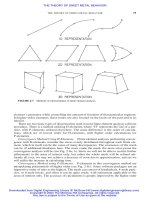
Figure 2.11 is a schematic diagram of a PBK model that might be designed for a volatile lipophilic
chemical. Arrows designate the direction of blood flow, with arterial blood entering the organs and
tissue groups and mixed venous blood returning to the lung to be reoxygenated. Organs of entry (lung,
liver), excretion (kidney, intestine, lung), and metabolism (liver), and tissue of accumulation (fat) for
this chemical class are explicitly included in the model. Other tissues are lumped into well-perfused
and poorly perfused groups. Note that uptake into the liver is considered to take place both by way of
the portal vein coming from the intestine and by way of the hepatic artery. An enterohepatic recycling
Absorption Excretion
Lung
Fat
Well-perfused Tissues
Poorly-perfused Tissues
Metabolism
Liver
Intestine
Excretion
Kidney
Excretion
Figure 2.11 Schematic diagram of a physiologically-based model of the kinetic behavior of a volatile chemical
compound.
50
ABSORPTION, DISTRIBUTION, AND ELIMINATION OF TOXIC AGENTS
between liver and intestine is also included in the model. These features of the model are choices made
by the model developer, and reflect the known physicochemical behavior of the agent whose kinetics
are being modeled. Models for other chemicals will be quite different. A model for a nonvolatile
chemical would not include an explicit lung compartment, while models for bone-seeking elements
like lead and uranium include bone as a distinct tissue.
In a sense, classical and PBK models work in opposite directions. In classical descriptive kinetics,
model compartments having no necessary relationship to actual tissue volumes and clearances having
no necessary relationship to tissue blood flow are inferred from a set of concentration data. In contrast,
the PBK model is constructed from basic anatomic, physiologic, physicochemical, and metabolic
building blocks. It is then used to simulate concentrations under a defined set of conditions, and its
predictions are compared with observations. If the predictions are not accurate, some premise of the
model is at fault. The need for model revision can afford insight into the processes that control the
kinetic behavior of the chemical.
A PBK model for dichloromethane (DCM) forms the basis of a current human health risk
assessment. DCM is metabolized by two pathways, a capacity-limited oxidative pathway and first-
order conjugation with glutathione (for descriptions of these biotransformation processes, see Chapter
3). Either pathway was thought potentially capable of generating reactive intermediates involved in
the tumorigenicity of DCM in mice. Andersen et al. (1987) demonstrated that tumorigenicity correlated
well with the activity of the glutathione pathway, but not with the activity of the oxidative pathway.
These investigators scaled a PBK model developed for DCM from mouse to human and from high
dose to low dose in order to predict, based on studies carried out at high doses in mice, the risk associated
with human environmental exposure to DCM. The mouse-to-human scaling of metabolism relied on
experimentally-determined human metabolic parameter values.
Their physiologic foundation and the inclusion of species-specific physiologic and metabolic
mechanisms, when these are known, confer on PBK models a flexibility that allows their use for
route-to-route, dose-to-dose, and species-to-species extrapolations such as this one, for which classical
models would be wholly inappropriate.
Biotransformation
Biotransformation is one of the two general elimination mechanisms. Biotransformation reactions in
general can be divided into two classes: phase I and phase II reactions. Phase I reactions are catabolic
or breakdown reactions (oxidation, reduction, and hydrolysis) that generate or free up a polar functional
group. They produce metabolites that may be excreted directly or may become substrates for phase II
reactions. Phase II reactions, which are often coordinated with phase I activity, are synthetic reactions
in which an additional molecule is covalently bound to the parent or the metabolite, which usually
results in a more water-soluble conjugate. Biotransformation reactions, and the factors that influence
them, are discussed in detail in Chapter 3.
Excretion
Excretion takes place simultaneously with biotransformation and, of course, with distribution. The
kidney is probably the single most important excretory organ in terms of the number of compounds
excreted, but the liver and lung are of greater importance for certain classes of compounds. The lung
is active in excretion of volatile compounds and gases. The liver, because it is a key biotransforming
organ as well as an organ of excretion, is in a unique position with regard to the elimination of foreign
chemicals.
Excretion in the Kidney
About 20 percent of all dissolved compounds of less than protein size are
filtered by the kidney in the glomerular filtration process. Glomerular filtration is a passive process; it
does not require energy input. Filtered compounds may be either excreted or reabsorbed. Passive
reabsorption in the kidney, as elsewhere, is a diffusion process. It is governed by the usual principles.
2.4 DISPOSITION: DISTRIBUTION AND ELIMINATION
51
Thus, lipid-soluble compounds are subject to reabsorption after having been filtered by the kidney.
The degree of reabsorption of electrolytes will be strongly influenced by the pH of the urine, which
determines the amount of the chemical present in a nonionized form.
It is to be expected that some control could be exerted over the rate of excretion of weak acids and
bases by adjusting urine pH. This type of treatment can be used very effectively in some cases.
Alkalinization of the urine by administration of bicarbonate has been used to treat salicylic acid
poisoning in humans. Alkalinization causes the weak acid to become more fully ionized; the ionized
molecule is excreted in the urine rather than reabsorbed.
There are also active secretory and reabsorptive processes in the renal tubules of the kidney. These
processes are specialized to handle endogenous compounds; active reabsorption helps to conserve the
essential nutrients, glucose and amino acids. These pathways can also be used by exogenous
compounds, provided the compounds have the structural and electronic configurations required by the
carrier molecules.
The renal clearance represents a hypothetical plasma volume cleared of solute by the net action of
all renal mechanisms during the specified period of time. A compound such as creatinine that is filtered
but not secreted or reabsorbed is cleared in adult humans at a rate of about 125 mL/min. Compounds
that are reabsorbed as well as filtered have clearances less than the creatinine clearance. Compounds
that are actively secreted can have clearances as large as the renal plasma flow, about 600 mL/min.
The presence of disease in the kidney can affect the half-life of a compound eliminated via the
kidney, just as the presence of disease in the liver can affect the half-life of a compound that is largely
biotransformed.
Excretion in the Liver
The liver is both the major metabolizing organ and a major excretory organ.
Large fractions of many toxicants absorbed from the gastrointestinal tract are eliminated in the liver
by metabolism or excretion before they can reach the systemic circulation, the hepatic first-pass effect.
In addition, metabolites formed in the liver may be excreted into the bile before they themselves have
had a chance to circulate. Although it does not excrete as many different compounds as the kidney
does, the liver is in an advantageous position with regard to excretion, particularly of metabolites.
There are at least three active systems for transport of organic compounds from liver into bile: one
for acids, one for bases, and one for neutral compounds. Certain metals are also excreted into bile
against a concentration gradient. These transport processes are efficient and can extract protein-bound
as well as free chemicals. The characteristics that determine whether a compound will be excreted in
the bile or in the urine include its molecular weight, charge, and charge distribution. In general, highly
polar and larger compounds are more frequently found in the bile. The threshold molecular weight for
biliary excretion is species-dependent. In the rat, compounds with molecular weights greater than about
350 can be excreted in the bile. Those having molecular weights greater than about 450 are excreted
predominantly in the bile, while compounds with molecular weights between 350 and 450 are
frequently found in both urine and bile.
Once a compound has been excreted by the liver into the bile, and thereby into the intestinal tract,
it can either be excreted in the feces or reabsorbed. Most frequently the excreted compound itself,
being water-soluble, is not likely to be reabsorbed directly. However, glucuronidase enzymes of the
intestinal microflora are capable of hydrolyzing glucuronides, releasing less polar compounds that
may then be reabsorbed. The process is termed
enterohepatic circulation
. It can result in extended
retention of compounds recycled in this manner. Techniques have been developed to interrupt the
enterohepatic cycle by introducing an adsorbent that will bind the excreted chemical and carry it
through the gastrointestinal tract.
Certain factors influence the efficiency of liver excretion. Liver disease can reduce the excretory
as well as the metabolic capacity of the liver. On the other hand, a number of drugs increase the rate
of hepatic excretion by increasing bile flow rate. For example, phenobarbital produces an increase in
bile flow that is not related to its ability to induce metabolizing enzymes. Whether the increased rate
of bile flow will increase the rate of elimination of a compound that is both metabolized and excreted
by the liver depends on whether the rate-limiting step is the enzyme-catalyzed biotransformation or
52
ABSORPTION, DISTRIBUTION, AND ELIMINATION OF TOXIC AGENTS
the transfer from liver to bile. If transfer from liver to bile is the rate-limiting step, enhancement of the
rate of bile flow will enhance the rate of excretion.
Excretion in the Lung
The third major organ of elimination is the lung, the key organ for the
excretion of volatile chemical compounds. Pulmonary excretion, like pulmonary absorption, is by
passive diffusion. For example, the rate of transfer of chloroform out of pulmonary blood is directly
proportional to its concentration in the blood. Essentially, pulmonary excretion is the reverse of the
uptake process, in that compounds with low solubility in the blood are perfusion-limited in their rate
of excretion, whereas those with high solubility are ventilation-limited. Highly lipophilic chemicals
that have accumulated in lipid depots may be present in expired air for a very long time after exposure.
Other Routes of Excretion
Skin, hair, sweat, nails, and milk are other, usually minor routes of
excretion. Hair can be a significant route of excretion for furred animals, and indeed the amount of a
metal in hair, like the amount of a volatile compound in exhaled air, can be used as an index of exposure
in both laboratory animals and humans. Hair is not quantitatively an important route of excretion in
humans, however. Sweat and nails are only rarely of interest as routes of excretion, simply because
loss by these routes is quantitatively so slight.
Milk may be a major route of excretion for some compounds. Milk has a relatively high fat content,
3–5 percent or even higher, and therefore compounds that are lipophilic may be excreted in milk to a
significant extent. Some of the toxicants known to be present in milk are the highly lipid-soluble
chlorinated hydrocarbons: for example, the polychlorinated biphenyls (PCBs) and DDT. Certain heavy
metals may also be excreted in milk. Lead is thought to be secreted into milk by the calcium transport
process.
2.5 SUMMARY
This chapter has conveyed some of the general biochemical and physiological principles that govern
absorption, distribution, and elimination of toxic agents, in particular
•
The importance of lipid solubility, molecular size, and degree of ionization to the rate at
which a molecule moves through a membrane by passive transfer or diffusion.
•
The characteristics of other transfer processes such as facilitated diffusion, active transport,
phagocytosis, and pinocytosis.
•
Absorption from the gastrointestinal tract with particular emphasis on the importance of pH
as a determinant of absorption of ionizable organic acids and bases as well as on compound-
specific and host-related factors such as lipid solubility and molecular size, the presence of
villi and microvilli in the intestine, the possibility that the compound can be absorbed by
facilitated or active transport mechanisms, and the action of gastrointestinal enzymes or
intestinal microflora.
•
Factors determining the rate of diffusion across the skin.
•
Absorption of solid and liquid particulates and of gases and vapors in the lung.
•
Simple classical and physiologically based kinetic models describing disposition (distribu-
tion, metabolism, and excretion).
•
Excretion from kidney, liver (including enterohepatic circulation), and lung, and by less
general routes such as skin, hair, sweat, nails, or milk.
2.5 SUMMARY
53
REFERENCES AND SUGGESTED READING
Abernethy, D. R., and D. J. Greenblatt, “Drug disposition in obese humans: An update,” Clin. Pharmacokinet.
11:
199–212 (1986).
Andersen, M. E., H. J. Clewell, M. L. Gargas III, F. A. Smith, and R. H. Reitz, “Physiologically-based
pharmaco-kinetics and the risk assessment process for methylene chloride,” Toxicol. Appl. Pharmacol.
87:
185–205 (1987).
Bragt, P. C., and E. A. van Dura, “Toxicokinetics of hexavalent chromium in the rat after intratracheal administration
of chromates of different solubilities,” Ann. Occup. Hyg.
27:
315–322 (1983).
Brewster, D., M. J. Humphrey, and M. A. McLeavy, “ The systemic bioavailability of buprenorphine by various
routes of administration,” J. Pharm. Pharmacol.
33:
500–506 (1981).
Brodie, B. B., H. Kurz, and L. S. Shanker, “The importance of dissociation constant and lipid-solubility in
influencing the passage of drugs into the cerebrospinal fluid,” J. Pharmacol. Exp. Therap.
130:
20–25 (1960).
Chamberlain, A. C., M. J. Heard, P. Little, D. Newton, A. C. Wells, and R. D. Wiffen. Investigations into Lead from
Motor Vehicles, AERE. Publication N2R9198, Harwell, England, 1978.
Crouthamel, W. G., J. T. Doluisio, R. E. Johnson, and L. Diamond, “ Effect of mesenteric blood flow on intestinal
drug absorption,” J. Pharm. Sci.
59:
878–879 (1970).
English, J. C., R. D. R. Parker, R. P. Sharma, and S. G. Oberg, “ Toxicokinetics of nickel in rats after intratracheal
administration of a soluble and insoluble form,” Am. Ind. Hyg. Assoc. J.
42:
486–492 (1981).
Gariépy, L., D. Fenyves, and J P. Villeneuve, “Propranolol disposition in the rat: Variation in hepatic extraction
with unbound drug fraction,” J. Pharm. Sci.
81:
255–258 (1992).
Gregus, Z., and C. D. Klaassen, “Disposition of metals in rats: A comparative study of fecal, urinary, and biliary
excretion and tissue distribution of eighteen metals,” Toxicol. Appl. Pharmacol.
85:
24–38 (1986).
Guidotti, G., “ The structure of membrane transport systems,” Trends Biochem. Sci.
1:
11–12 (1976).
Hamilton, D. L., and M. W. Smith, “ Inhibition of intestinal calcium uptake by cadmium and the effect of a low
calcium diet on cadmium retention,” Environ. Res.
15:
175–184 (1978).
Herrmann, D. R., K. M. Olsen, and F. C. Hiller, “ Nicotine absorption after pulmonary instillation,” J. Pharm. Sci.
81:
1055–1058 (1992).
Hirom, P. C., P. Millburn, and R. L. Smith, “Bile and urine as complementary pathways for the excretion of foreign
organic compounds,” Xenobiotica
6:
55–64 (1976).
Hogben, C. A. M., D. J. Tocco, B. B. Brodie, and L. S. Shanker, “ On the mechanism of intestinal absorption of
drugs,” J. Pharmacol. Exp. Therap.
125:
275–282 (1959).
Hussain, A. A., K. Iseki, M. Kagoshima, L. W. Dittert, “Absorption of acetylsalicylic acid from the rat nasal cavity,”
J. Pharm. Sci.
81:
348–349 (1992).
King, F. G., R. L. Dedrick, J. M. Collins, H. B. Matthews, and L. S. Birnbaum, “ Physiological model for the
pharmacokinetics of 2,3,7,8-tetrachlorodibenzofuran in several species,” Toxicol. Appl. Pharmacol.
67:
390–
400 (1983).
Lien, E. J., and G. L. Tong, “Physicochemical properties and percutaneous absorption of drugs,” J. Soc. Cosmet.
Chem.
24:
371–384 (1973).
Nebert, D. W., A. Puga, and V. Vasiliou, “ Role of the Ah receptor and the dioxin-inducible [Ah] gene battery in
toxicity, cancer, and signal transduction,” Ann. NY Acad. Sci.
685:
624–640 (1993).
Nelson, D. R., T. Kamataki, D. J. Waxman, F. P. Guengerich, R. W. Estabrook, R. Feyereisen, F. J. Gonzalez, M.
J. Coon, I. C. Gunsalus, O. Gotoh, K. Okuda, and D. W. Nebert, “The P450 superfamily: Update on new
sequences, gene mapping, accession numbers, early trivial names of enzymes, and nomenclature,” DNA Cell
Biol.
12:
1–51 (1993).
O’Flaherty, E. J., Toxicants and Drugs: Kinetics and Dynamics, Wiley, New York, 1981.
O’Flaherty, E. J., “ Physiologically based models for bone-seeking elements. IV. Kinetics of lead disposition in
humans,” Toxicol. Appl. Pharmacol.
118:
16–29 (1993).
Rollins, D. E., and C. D. Klaassen, “Biliary excretion of drugs in man,” Clin. Pharmacokinet.
4:
368–379 (1979).
Schanker, L. S., and J. J. Jeffrey, “Active transport of foreign pyrimidines across the intestinal epithelium,” Nature
190:
727–728 (1961).
54
ABSORPTION, DISTRIBUTION, AND ELIMINATION OF TOXIC AGENTS
Sha’afi, R. I., C. M. Gary-Bobo, and A. K. Solomon, “ Permeability of red cell membranes to small hydrophilic
and lipophilic solutes,” J. Gen. Physiol. 58: 238–258 (1971).
U.S. Environmental Protection Agency, Update to the Health Risk Assessment Document and Addendum for
Dichloromethane: Pharmacokinetics, Mechanism of Action and Epidemiology, EPA 600/8-87/030A (1987).
Wagner, J. G., “ Properties of the Michaelis-Menten equation and its integrated form which are useful in
pharmacokinetics,” J. Pharmacokinet. Biopharmaceut. 1: 103–121 (1973).
Williams, R. T., “Interspecies scaling,” in T. Teorell, R. L. Dedrick, and P. G. Condliffe, eds., Pharmacology and
Pharmacokinetics, Plenum, New York, 1974, Table IV, p. 108.
REFERENCES AND SUGGESTED READING
55
3
Biotransformation: A Balance between
Bioactivation and Detoxification
BIOTRANSFORMATION: A BALANCE BETWEEN BIOACTIVATION AND DETOXIFICATION
MICHAEL R. FRANKLIN and GAROLD S. YOST
This chapter identifies the fundamental principles of foreign compound (xenobiotic) modification by
the body and discusses
•
How xenobiotics enter, circulate, and leave the body
•
The sites of metabolism of the xenobiotic within the body
•
The chemistry and enzymology of xenobiotic metabolism
•
The bioactivation as well as inactivation of xenobiotics during metabolism
•
The variations in xenobiotic metabolism resulting from prior or concomitant exposure to
xenobiotics and from physiological factors
The body is continuously exposed to chemicals, both naturally occurring and synthetic, which have
little or no value in sustaining normal biochemistry and cell function. These chemical substances
(xenobiotics) can be absorbed from the environment following inhalation, ingestion in food or water,
or simple exposure to the skin (Figure 3.1). Biotransformation or metabolism of the chemicals allows
the elimination of the absorbed chemicals to occur. Without this process, chemicals that were readily
absorbed through lipid membranes because of a high octanol/water partition coefficients would fail to
leave the body. They would be passively reabsorbed through the lipid membrane of the kidney tubule
instead of remaining in, and passing out with, the urine (Figure 3.2). In addition, they would not be
subject to active transport mechanisms capable of actively secreting many xenobiotic metabolites.
Thus, an important objective of biotransformation is to promote the excretion of chemicals by the
formation of water-soluble metabolites or products. Biotransformation can also alter the biological
activity of chemicals, including endogenous chemicals released in the body, such as steroids and
catecholamines, both by structural alteration and by enhancing their partition away from cellular
compartments, membranes, and receptors. Thus biotransformation helps to both terminate the biologi-
cal activity of chemicals and increase their ease of elimination.
Biotransformation
is defined as the chemical alteration of substances by reactions in the living
organism. For convenience, the conversion of xenobiotics is divided into two phases: metabolic
transformations (phase I reactions) and conjugation with natural body constituents (phase II reactions)
(Figure 3.3). The reactions of both of these phases are predominantly enzyme-catalyzed. A xenobiotic
does not necessarily undergo metabolism by a sequential combination of phase I followed by phase II
reactions for successful elimination. It may undergo phase I metabolism alone, phase II alone, and
occasionally, phase I reactions subsequent to phase II conjugations are encountered.
An important objective of biotransformation is to promote the excretion of absorbed chemicals by
the formation of water-soluble drug metabolites or products (
p
in Figure 3.1). Increased water solubility
is derived primarily from the phase II reactions since most conjugates exist in the ionized state at
physiological pH levels. This promotes excretion (
e
in Figure 3.1) by decreasing xenobiotic reabsorp-
Principles of Toxicology: Environmental and Industrial Applications, Second Edition
, Edited by Phillip L. Williams,
Robert C. James, and Stephen M. Roberts.
ISBN 0-471-29321-0 © 2000 John Wiley & Sons, Inc.
57
tion from the renal tubule following glomerular filtration or active secretion (
f
and
s
, respectively, in
Figures 3.1 and 3.2) and from the gastrointestinal tract following biliary secretion. Biotransformation
also decreases the entry of xenobiotics into cells of all organs and makes them more suitable for
secretion by active transport mechanisms into the bile and urine. Active secretion requires both energy
and a carrier protein and is capable of forcing molecules up a chemical gradient. Of the carrier
molecules, those that recognize and transport organic acids have particular importance for drug
conjugates since they can carry glucuronides, sulfate esters, and amino acid conjugates. While
Figure 3.1 Diagram of major sites of xenobiotic absorption, metabolism, and excretion.
Abbreviations
:
a
= major absorption sites;
e
= excretion sites;
f
= filtration sites;
m
= major metabolism site;
p
= metabolic product;
s
= secretion sites;
x
= xenobiotic.
58
BIOTRANSFORMATION: A BALANCE BETWEEN BIOACTIVATION AND DETOXIFICATION
excretion of xenobiotics into the urine largely terminates the exposure of the body to the chemical,
excretion in the bile may not always result in efficient drug elimination because enterohepatic
recirculation may occur. This can result in the prolonged effects and persistence seen with some drugs
and chemicals. Enterohepatic recirculation most often involves the secretion of xenobiotic conjugates
in the bile and their hydrolysis by enzymes from the host or microorganisms in the gastrointestinal
tract. This deconjugation releases the free xenobiotic, which is often sufficiently lipid soluble (high
octanol/water partition coefficient), to be reabsorbed. The reabsorbed xenobiotic returns in the portal
circulation to the liver where it is reconjugated, resecreted, and so on. The same reabsorption can also
occur if an unmetabolized lipid soluble xenobiotic is secreted in the bile.
As stated above, the conversion of xenobiotics is divided into the two phases of metabolic transformation
and conjugation (Figure 3.3). The main chemical reactions involved in phase I or metabolic transformation,
in approximate order of capacity or importance, are oxidation, hydrolysis, and reduction. Of the phase II or
conjugation reactions, glucuronidations are generally the most prevalent in mammals, with the other
conjugations having lesser overall capacity. All conjugation reactions, except with glutathione, involve the
participation of energy-rich or activated cosubstrates. Conjugation with the cellular nucleophile, glutathione,
is an especially important mechanism for the sequestering of electrophilic intermediates generated during
phase I metabolism, and it can occur, albeit less efficiently, in the absence of enzyme.
As mentioned above, with reference to the generation of electophilic metabolites, biotransformation can
have a variety of effects on the biological reactivity of the xenobiotic. The chemical can be inactivated or
detoxified, can be changed into a more toxic substance (bioactivated), or can be changed into other chemical
entities having effects that differ both quantitatively and qualitatively from the parent compound (Table 3.1).
Generally, phase II metabolites are inactive, but important exceptions exist. Phase I metabolites
may or may not be inactive, and many are more reactive than the original xenobiotic. The greater
reactivity can be viewed as an unfortunate necessary prerequisite to conjugation, which is the step
contributing most to the facilitation of excretion (Figure 3.4).
Figure 3.2 The role of metabolism in increasing urinary excretion.
BIOTRANSFORMATION: A BALANCE BETWEEN BIOACTIVATION AND DETOXIFICATION 59
Figure 3.3
Xenobiotic metabolism summary; reaction characteristics and
flowchart.
60
TABLE 3.1 Pharmacologic Effects with Xenobiotic Metabolism
Phase I Phase II
Active to Inactive
Amphetamine —P450
→
phenylacetone Acetaminophen —UGT/ST
→
Cocaine —esterase
→
benzoylecgonine Aflatoxin 2,3-epoxide —GST
→
8 glutathionyl-9
hydroxyaflatoxin
Hexobarbital —P450
→
Morphine —UGT
→
morphine-3-glucuronide
Phenytoin —P450
→
Testosterone —ST
→
Active to Active
Acetylsalicylic acid —esterase
→
salicylic acid
Codeine —P450
→
morphine Morphine —UGT
→
morphine-6-glucuronide
Heroin —esterase
→
morphine Procainamide —AT
→
N-acetylprocainamide
Primidone-P450
→
phenobarbital Thiobarbital —P450
→
barbital
Inactive to Active
Chloral hydrate —reductase
→
trichloroethanol
Prontosil —reductase
→
sulfanilamide
Sulindac —reductase
→
sulfide
Inactive to Toxic
Acetaminophen —P450
→
N-acetyl-p-benzoquinine imine
N-Hydroxyacetylaminofluorene —ST
→
Acetylhydrazine —P450
→
acetylcarbonium ion N-Hydroxymethylaminoazobenzene —ST
→
Aflatoxin —P450
→
aflatoxin-8,9 epoxide Tetrachloroethylene —GST
→
Malathion —P450
→
malaoxon Tolmetin —UGT
→
Nitrofurantoin —reductase
→
hydroxylamine
Benzo(a)pyrene 7,8-diol —P450
→
benzo(a)pyrene 7,8-diol 9,10-epoxide
Dimethylnitrosamine —P450
→
methyldiazohydroxide
Figure 3.4 The balance of reactivity and excretability in xenobiotic metabolism.
BIOTRANSFORMATION: A BALANCE BETWEEN BIOACTIVATION AND DETOXIFICATION 61
3.1 SITES OF BIOTRANSFORMATION
Xenobiotic metabolism occurs in all organs and tissues in the body. Because many of the chemicals
metabolized can have deleterious effects on the body, xenobiotic metabolism can be considered a
defense mechanism that hastens the elimination of a toxic chemical and thus terminates the exposure.
When viewed as a defense mechanism, it is not surprising that the exposure is best terminated at the
point of exposure. These are the so-called portals of entry (shown as sites of absorption [
a
] for
xenobiotics [
X
] in Figure 3.1), and constitute mainly the skin, lung, and intestinal mucosa. While drug
metabolizing enzymes are present in all these tissues (Table 3.2), and at relatively high activity in some,
particularly intestine and lung, the liver is by far the most important tissue for xenobiotic metabolism
(site [
m
] in Figure 3.1).
Although it is not the first tissue of the body to be exposed to chemicals, the liver receives the entire
chemical load absorbed from the gastrointestinal tract, which is the predominant portal of entry for
most xenobiotics (Figure 3.1). The xenobiotic metabolizing enzymes are present in high concentrations
and the organ itself has large bulk, approximately 5 percent of the total body weight. Xenobiotics
absorbed from the lungs and skin can also quickly move to the liver for metabolism. Once in the liver,
the highly vascular nature of the tissue and the intimate contact between blood and hepatocytes, which
contain the xenobiotic metabolizing enzymes, allows for the rapid diffusion of chemicals in and
metabolites out (Figure 3.5).
Although not a portal of entry, the kidney is an organ where xenobiotics are likely to be
concentrated during the excretion process, and this may be the reason for the relatively high level
of xenobiotic metabolizing enzymes in this tissue. Although the data presented in Table 3.2 are
from laboratory animals, there is little evidence to contraindicate the existence of a similar
distribution pattern in humans.
Within the liver, hepatocytes or parenchymal cells are the major site of drug biotransformation,
and within these cells it is the endoplasmic reticulum, which occupies about 15 percent of the
hepatocyte volume and contains 20 percent of the hepatocyte protein, which houses the bulk of
the critical drug metabolizing enzyme activity. (The nonparenchymal cells, including endothelial
and Kupffer cells, constitute 35 percent of liver cell number but only contribute 5–10 percent of
liver mass. Their drug metabolizing enzyme activities are typically less than 20 percent of that in
hepatocytes).
When liver is carefully homogenized, fragments of the endoplasmic reticulum are converted to
microsomes (an artifact of cell disruption). The drug-metabolizing enzymes located in the endoplasmic
reticulum are often referred to as
microsomal enzymes
, and it is often stated that chemicals are
metabolized by liver microsomes. Enriched microsomal fractions are usually obtained by differential
sedimentation, either as a suspension with cytoplasm (10,000
g
supernatant) or as a sediment free of
cytosol (105,000
g
precipitate) (Table 3.3).
Many important xenobiotic metabolizing enzymes reside in the cytoplasm and microsomal frac-
tions (Figures 3.3 and 3.6).
Oxidations and glucuronidations are the most common reactions occurring in microsomes. The
terminal oxidase responsible for many of the oxidations, cytochrome P450, represents about 5 percent
of the microsomal protein under normal conditions; more if induction has occurred (see text below).
Other flavoproteins necessary for cytochrome P450 function and epoxide hydrolase, an enzyme
important in the further metabolism of epoxides formed by cytochrome P450–dependent oxidation,
are also conveniently located in the endoplasmic reticulum (Figure 3.6). Microsomal metabolism in
tissues other than liver is seldom quantitatively important in overall drug elimination, but local
generation of active metabolites may be important in drug-induced tissue damage, carcinogenesis, and
other effects. Enzymes located in the cytoplasm of the hepatocyte catalyze a wide variety of both phase
I and phase II reactions. Dehydrogenases and esterases are examples of phase I enzymes found
predominantly in the cytosol. The sulfotransferase and glutathione transferase enzymes also depicted
in Figure 3.6 serve as examples of phase II enzymes that are similarly located.
62
BIOTRANSFORMATION: A BALANCE BETWEEN BIOACTIVATION AND DETOXIFICATION
Figure 3.5 Diagrammatic rendition of hepatic lobule blood flow.
TABLE 3.2 Drug-Metabolizing Enzyme Activities
a
in Various Organs
Lung
Intestine
Mucosa Liver
Kidney
Cortex Brain
Rabbit
Cytochrome P450 0.4 0.34 1.45 0.33 0.02
UDP-glucuronosyltransferase (
p
-nitrophenol)
b
0.4 — 6.6 2.9
Glutathione
S
-transferase (DCNB)
b
5.3 — 21.9 7.4
Rat
Cytochrome P450 0.09 0.05 0.84 0.12 0.01
Ethoxyresorufin demethylase (P4501A) 0.003 0.001 0.034 0.001
Erythromycin demethylase (P4503A) — 0.12 0.47 0.06
UDP-glucuronosyltransferase (
p
-nitrophenol)
b
0.8 — 4.4 3.3
Glutathione
S
-transferase (DCNB)
b
2.1 — 76.4 3.8
a
All activities are expressed on a per milligram of protein basis (DCNB = 1,2-dichloro 4-nitrobenzene).
b
Litterst CL, Mimnaugh EG, Reagan RL, Gram TE,
Drug Metab. Disp.
3:
259 (1975).
3.1 SITES OF BIOTRANSFORMATION
63
Without exception, the xenobiotic metabolizing enzymes occur in multiple forms (isozymes), often
with differing substrate selectivities. The presence of specialized isozymes, which can more efficiently
metabolize a specific range of chemicals, may enable those specific chemical challenges to be met
more effectively. With differing substrate selectivities, often comes different sensitivity to inhibitors.
The presence of multiple forms thus carries the advantage of not having all the metabolism of all
compounds metabolized by that route or chemical reaction being subject to inhibitory influences at
the same time. It has also been found that the synthesis of individual isozymes can be under different
regulatory influences. The body can thus meet a chemical challenge with a finely tuned response to
increase the production of only that enzyme best equipped to counter or neutralize the challenge.
TABLE 3.3 Preparation of Subcellular Fractions for Xenobiotic Metabolism Studies
Step Procedure Result
1 Liver pieces homogenized in 4 volumes of 0.25 M
sucrose in Potter Elvehjem glass–Teflon
homogenizer
Tissue structure disrupted and hepatocytes sheared.
2 Homogenate centrifuged at 2000
g
for 10 min Unbroken cells, connective tissue, and nucleii
sedimented
32000
g
supernatant centrifuged at 10,000
g
for 15 min Heavy mitochondria sedimented as pellet
4 10,000
g
supernatant centrifuged at 18,000
g
for 15
min
Light mitochondria sedimented as pellet
5 18,000
g
supernatant centrifuged at 105,000
g
for 60
min
Microsomes sedimented as pellet leaving nonturbid
cytosol in 0.2 M sucrose supernatant
Figure 3.6 Diagram of the subcellular localization and organization of major xenobiotic metabolizing enzymes
and necessary cofactors.
Abbreviations (clockwise) are ST = sulfotransferase; PAPS = adenosine 3
′
-phosphate 5
′
-phosphosulfate; GST = glutathione
S
-transferase; GSH = glutathione; AlcDH = alcohol dehydrogenase; ES = esterase; FP
1
= NADH cytochrome
b
5
reductase; b
5
=
cytochrome
b
5
; P450 = cytochrome P450; mEH = microsomal epoxide hydrolase; FP
2
= NADPH-cytochrome P450/
c
reductase;
UGT = UDP-glucuronosyltransferase; UDPGA = uridine 5
′
-diphosphoglucuronic acid; FP
3
= flavin-dependent monooxygenase.
64
BIOTRANSFORMATION: A BALANCE BETWEEN BIOACTIVATION AND DETOXIFICATION
3.2 BIOTRANSFORMATION REACTIONS
There is multiple redundancy in metabolism. There may be more than one site of attack on a xenobiotic
(e.g., amine and ester group of cocaine), there may be more than one metabolic reaction at a single
site (e.g., sulfation and glucuronidation of the phenolic group of acetaminophen), and more than one
enzyme/isozyme capable of catalyzing a single reaction at a single site. An example of the complexity
of possible metabolism of a relatively simple hypothetical chemical is shown in Figure 3.7. From
considerations in this chapter so far, it can be seen that the subcellular location of a metabolic reaction does
not dictate the nature of the reaction. Both oxidations and hydolyses, albeit by different enzymes, occur in the
cytoplasm and endoplasmic reticulum. Likewise, so do conjugations when considered collectively, but a
specific form of conjugation may occur only in a single fraction (e.g., sulfation in the cytoplasm). The enzymes
are therefore considered in the following paragraphs by the nature of the chemical reaction that they catalyze,
and only for phase I oxidations is the subcellular location used as a convenient subdivision.
Phase I; Oxidations
Microsomal
Microsomal oxidations are predominantly catalyzed by a group of enzymes called
mixed-function oxidases
or
monooxygenases
. The terminal oxidase is generally a hemoprotein called
cytochrome P450
but can be a flavoprotein.
Figure 3.7 Possible metabolic conversions of a simple hypothetical xenobiotic.
3.2 BIOTRANSFORMATION REACTIONS
65
Cytochrome P450 is a collective term for a group of related hemoproteins, all with a molecular
weight (MW) around 50,000 daltons, which as will be seen later, differ in their substrate selectivity
and in their ability to be induced and inhibited by drugs and chemicals (Table 3.4).
Cytochrome P450–catalyzed oxidations are categorized by the nature of the atom that is oxidized
(see Figure 3.8). Subsequent to the oxidation, the oxygen atom from molecular oxygen may be retained
within the major fragment of the chemical or it may be eliminated by molecular rearrangement (e.g.,
O
and
N
dealkylations).
Whatever the atom oxidized, or the name given to the reaction, the cytochrome P450–mediated
oxidation involves the same cyclic three-step series (Figure 3.9).
Step 1.
The xenobiotic [
X
] first binds to the cytochrome at a substrate binding site on the protein and
alters the conformation sufficiently to enable the efficient transfer of electrons to the heme from
NADPH via a nearby (see Figure 3.6) flavoprotein, NADPH cytochrome P450 reductase. (The
activity of this FAD- and FMN-containing flavoprotein is often determined experimentally using
exogenously added mitochondrial cytochrome
c
rather than microsomal cytochrome P450 as the
electron acceptor and so is often identified as NADPH cytochrome
c
reductase). The conformational
change can sometimes be seen in vitro (in the absence of electron transfer) as an alteration of the
heme from a low-spin to a high-spin state, which results in a blue shift in the absorbance maximum
of the hemoprotein. The gain at 390 nm and loss at 420 nm, when seen by difference spectroscopy,
is termed a
type I binding spectrum
(not to be confused with phase I metabolism).
Step 2.
The reduction of the heme iron from its normal ferric state to the ferrous state allows a
molecule of oxygen (O–O) to bind (the binding of CO rather than oxygen to ferrous cytochrome
P450 in the in vitro situation provides a characteristic absorbance maximum around 450 nm, which
gives this cytochrome its name).
Step 3.
The ternary complex of xenobiotic, cytochrome, and oxygen receives another electron, either
through the same flavoprotein as before or through an alternative path involving a different
flavoprotein in which the electron is first passed through cytochrome
b
5
, another cytochrome
present in the endoplasmic reticulum (see Figure 3.6). This alternate pathway for the second electron
can also use NADH as the pyridine nucleotide electron donor. The addition of the second electron
to the ternary complex results in a eventual splitting of the molecular oxygen, one atom of which
oxidizes the chemical, the other atom picking up protons to form water, returning ferric cytochrome
P450 to repeat the cycle.
Flavoprotein-catalyzed oxidations differ from cytochrome P450–catalyzed oxidations in mechanism
and in substrate selectivity. For the flavoproteins (a 65,000-dalton protein containing only FAD), the
enzyme forms an activated oxygen complex (“cocked gun” ) and the addition of a metabolizable
chemical discharges this, in the process of becoming oxidized. The electrophilic oxygenated species
attacks nucleophilic centers. A wide range of chemicals can thus be metabolized by this flavoprotein;
the important feature for metabolism being a heteroatom (nitrogen, sulfur) presenting a lone pair of
electrons (Table 3.5).
Some compounds are metabolized both by flavin-containing monooxygenases and cytochrome
P450 but to different products. An example is dimethylaniline, which is metabolized to the
N
-oxide
by the flavoprotein and is
N
-demethylated by cytochrome P450.
Nonmicrosomal
Oxidations in other subcellular organelles can be catalyzed by flavoproteins (e.g.,
monoamine oxidase in mitochondria) or pyridine nucleotide linked dehydrogenases (e.g., alcohol and
aldehyde dehydrogenases in cytoplasm).
Dehydrogenase-catalyzed oxidations do not involve molecular oxygen. The oxidation of the
chemicals or drugs occurs through electron transfer to a pyridine nucleotide, usually NAD
+
. Most of
the dehydrogenases are cytoplasmic in location. The most noteworthy of this class of enzymes in
humans is the dehydrogenase responsible for the metabolism of ethanol. In contrast to the major
66
BIOTRANSFORMATION: A BALANCE BETWEEN BIOACTIVATION AND DETOXIFICATION
TABLE 3.4 Important Cytochrome P450s
a
Subfamily
Forms Present in
Tissue
Substrates/
Reactions Inducers InhibitorsRat Rabbit Human Mouse
1A 1,2 1,2 1,2 1,2 liver, EH ethoxyresorufin,
phenacetin deE, caffeine 3
deM, benzopyrene,
aflatoxin, cooked-food
heterocyclic amines, NBI
PCB, PAH, TCDD, 3MC,
isosafrole, omeprazole
7,8-benzoflavone,
ellipticine, furafylline
2A 1,2 4,5 PAH (2A1)
2B 1,2 4 9,10,13 pentoxyresorufin,
benzphetamine
PB
2C 6,7 liver, GI PB
8,9,10 tolbutamide PB, RIF sulfaphenazole
11,12,13 sex, maturation,
17,18,19
S
-mephenytoin 4-OH,
debrisoquine 4-OH, RIF
tranylcypromine
quinidine
2D 6 9,10,11,
12,13
liver, EH sparteine,
dextromethorphan,
bufuralol 1
′
-OH
2E 1 1,2 1 1 liver, EH chlozoxazone, ethanol
dimethylnitrosamine,
acetaminophen, CCl
4
,
4-nitrophenol-OH
ethanol, disulfiram
ketones, pyridine,
isoniazid
2F 1 2 lung 3-methylindole
naphthalene
67
TABLE 3.4 Important Cytochrome P450s
a
Subfamily
Forms Present in
Tissue
Substrates/
Reactions Inducers InhibitorsRat Rabbit Human Mouse
3A 1,2 6 3,4,5,7 11,13 erythromycin
N
deM,
TAO MI complex,
cyclosporine, quinidine,
testosterone, and cortisol
6
β
-OH, mephenytoin
(rat), benzphetamine,
nifedipine, and other
dihydropyridines
glucocorticoids DEX,
PCN, macrolides, TAO
clotrimazole,
phenobarbital
cimetidine,
naringenin
4A 1,2,3,8 4,5,6,7 9,11 10,12 liver, kidney Lauric acid and
prostaglandin
ω
-OH
peroxisome proliferators,
DEHP, clofibrate
pregnancy (4A4)
4B 1 1 1 lung valproic acid
a
Abbreviations
: deE = deethylation
deM = demethylation
DEHP = di(2-ethylhexyl)phthalate
DEX = dexamethasone
EH = extrahepatic
GI = gastrointestinal tract
Kid = kidney
MI = metabolic-intermediate
3MC = 3-methylcholanthrene
NBI = N-benzylimidazole
OH = hydroxylation
PAH = polycyclic aromatic hydrocarbons
PCB = polychlorinated biphenyls
PCN = pregeneolone 16
α
carbonitrile
PB = phenobarbital
RIF = rifampicin
TAO = troleandomycin
TCDD = 2,3,7,8-tetrachlorodibenzo-p-dioxin
68
Figure 3.8 Cytochrome P450–catalyzed oxidations.
3.2 BIOTRANSFORMATION REACTIONS
69
microsomal oxidizing enzyme, these enzymes are not subject to extensive induction (see discussion
later).
Monoamine oxidases, which are usually mitochondrial in location, oxidize by electron transfer to
a flavin group. Monoamine oxidases are responsible for the normal metabolism of neurotransmitters,
and exposure to agents, which are also metabolized by this enzyme, (e.g., tyramine) can result in
toxicities or pharmacological effects arising from accumulation of the unmetabolized neurotransmitter.
A neurotoxin of much recent interest, 1-methyl-4-phenyl-1,2,3,6-tetrahydropyridine (MPTP), which
leads to Parkinson’s syndrome, is bioactivated by monoamine oxidase B (a form selectively inhibited
by deprenyl and located in serotonergic neurons in the brain). Environmental compounds or drugs that
are also tetrahydropyridines have been speculated to be causative agents in Parkinson’s disease in the
elderly.
Phase I; Hydrolyses
Hydrolysis reactions are catalyzed by esterases and amidases. While both can be microsomal, esterases
are predominantly cytosolic in location. Hydrolysis of amides and esters produces two reactive centers,
Figure 3.9 The cytochrome P450 oxidation cycle.
TABLE 3.5 Compounds Metabolized by the Flavin-Containing Monooxygenases
Heteroatom Class Examples
Nitrogen Tertiary amine N-Dimethylaniline, imipramine, amitryptyline
Secondary amine N-Methylaniline, desipramine, nortryptyline
Sulfur Thiocarbamides Thiourea, propylthiouracil, methimazole
Thioamides Thioacetamide
Thiols Dithiothreitol,
β
-mercaptoethanol
Sulfides Dimethylsulfide
70
BIOTRANSFORMATION: A BALANCE BETWEEN BIOACTIVATION AND DETOXIFICATION
both of which are suitable for conjugation, if the metabolites are not first excreted as the phase I products
(Figure 3.10).
Epoxide hydrolase activity is predominantly microsomal, but an enzyme is also present in the
cytosol.
Most hydrolyses occur to a significant extent in tissues other than liver. Their quantitative
importance is variable, depending on the chemical challenge. One significant extrahepatic location of
esterases is in the blood (plasma and erythrocytes), and of great concern is the enzyme normally
responsible for the hydrolysis of acetylcholine. Blockade of this enzyme is the mode of action of many
insecticides and “ nerve gases.”
Figure 3.10 Hydrolytic and reductive phase I reactions.
3.2 BIOTRANSFORMATION REACTIONS
71
Phase I; Reductions
Reductive metabolism in the liver endoplasmic reticulum can occur through the mediation of both
hemoprotein (cytochrome P450) and flavoproteins. Reductions of azo and nitro groups are the most
commonly encountered (Figure 3.10), but reduction of disulfides, sulfoxides, epoxides, and
N
-oxides
can also occur. In many instances, the products of reductive metabolism can be reoxidized under
aerobic conditions.
Phase II; Glucuronidation
Glucuronidations are catalyzed by a group of closely related 55,000-dalton isozymes, termed
UDP-
glucuronosyltransferases
, located within the endoplasmic reticulum. They catalyze the transfer of
glucuronic acid from a uridinediphosphoglucuronic acid (UDPGA) cofactor to a carboxyl or hydroxyl
(phenol), or less often an amine group on the xenobiotic (or phase I metabolite) (Figure 3.3). The
UDPGA is generated from the abundant carbohydrate supply in the liver as glucose-1-phosphate, and
following the reaction with UTP, the resultant UDP-glucose is oxidized. The formation of the
glucuronide does not involve the acid group of glucuronic acid, so the conjugate retains acid and ionized
character at physiological pH, providing dramatic enhancement of water solubility and excretability
to the xenobiotic. Glucuronides are actively secreted into bile and in the proximal tubule of the kidney.
Xenobiotics conjugated as glucuronides can be released as either a phase I metabolite or the original
molecule by the action of glucuronidases of both mammalian and microbial origin.
UDP-glucuronosyltransferases occur in multiple forms. The most common classification utilized
for the enzymes responsible for the metabolism of xenobiotics are those (GT1) that conjugate planar
phenols (e.g., 1-naphthol, 4-nitrophenol) and are induced by polycyclic hydrocarbon-like molecules
(see Table 3.6) and those (GT2) that conjugate nonplanar phenols (e.g. morphine, chloramphenicol)
and are induced by phenobarbital and similar compounds. There are other forms which appear to be
more selective for endogenous substrates, notably those for the 17 hydroxysteroids (testosterone), the
3 hydroxysteroids (androsterone) and bilirubin. More recent studies using the powerful techniques of
molecular biology have provided a more rational classification system, but to aid the reader in
understanding the bulk of existing literature, the old system has been used in this chapter. Like
cytochrome P450s, UDP-glucuronosyltransferases are often substrate selective rather than substrate
specific, being able to metabolize a wide range of compounds poorly (e.g., 4-nitrophenol is conjugated
by almost all isozymes) while metabolizing substrates with particular characteristics very efficiently.
Also like cytochrome P450s, more than one form may be induced by a xenobiotic inducing agent (both
bilirubin and testosterone as well as morphine conjugations are induced by phenobarbital).
Phase II; Sulfation
Sulfate conjugation is an important alternative to glucuronidation for phenolic compounds and
occasionally arylamines. Sulfate availability within the cell may be limited, so this conjugation pathway
decreases in importance with higher xenobiotic or phenolic metabolite concentrations. The 3′-phos-
phoadenosine-5′-phosphosulfate (PAPS) cofactor from which the sulfate group is transferred is
generated from ATP and inorganic sulfate. The sulfate can be derived from the sulfur containing amino
acids, cysteine and methionine. The enzymes catalyzing the sulfate conjugations are a family of
cytosolic 64,000-dalton enzymes, termed
sulfotransferases
, and are one of the exceptions to the major
groups of drug metabolizing enzymes in that they appear to not be induced by xenobiotic compounds
(see Table 3.6). The sulfates are completely ionized at physiological pH and easily eliminated. Much
like glucuronides, enzymes exist (termed
sulfatases
) that can break the conjugate and return the
xenobiotic, if it is phenolic, or the phase I metabolite of a xenobiotic, if it was oxidized or hydrolyzed
to that functional group.
72
BIOTRANSFORMATION: A BALANCE BETWEEN BIOACTIVATION AND DETOXIFICATION