Case Files Neurology - part 3 potx
Bạn đang xem bản rút gọn của tài liệu. Xem và tải ngay bản đầy đủ của tài liệu tại đây (326.61 KB, 50 trang )
The diagnosis of delirium is clinical, with an emphasis on evaluating level
of attention. Attention can be evaluated by serial reversal test (such as asking
the patient to spell a word backwards). The history should include a review of
medications patients take and information obtained from friends or family. The
neurological examination may not show focal signs or may show myoclonus,
dysarthria, tremor, motor abnormalities, or asterixis. Laboratory evaluation
should include a comprehensive metabolic panel, glucose, blood urea nitrogen
(BUN), liver function studies, electrolyte levels, a complete blood count (CBC)
to evaluate for infection, thyroid function studies to evaluate for endocrinopa-
thy, and ammonia to evaluate for hepatic encephalopathy. Arterial blood gas
(ABG) or pulse oximetry should be obtained if the patient has a history of lung
disease or smoking. Urine toxicology studies in those individuals with a history
of drug abuse or at risk for drug abuse should be requested as well. A CT scan
of the head or MRI brain scan needs to be performed with the choice of study
depending on ease of obtaining and clinical scenario. Other studies to consider
depending on the clinical picture include chest radiograph (evaluates for pneu-
monia), electrocardiograph (ECG) (exclude myocardial infarction or arrhyth-
mia), electroencephalograph (EEG), and lumbar puncture if there is concern for
central nervous system (CNS) infection.
The differential diagnosis for delirium is extensive (see Table 9–3) and
includes metabolic causes, infections, drug-related causes, primary neurologic
abnormalities, trauma, and perioperative causes. Importantly delirium must
be differentiated from dementia. Typically demented patients have a history
of chronic (>6 months) progression with normal attention (except advanced
cases) and level of consciousness. Perceptual disturbances and fluctuating
course are less common with dementia.
CLINICAL CASES 83
Table 9–3
SELECTED LISTING OF ETIOLOGIES OF DELIRIUM
Etiologies
Metabolic disorders: hypoglycemia, hyponatremia, uremia, hypoxia, hypo/
hypercalcemia, endocrinopathies (thyroid, pituitary), vitamin deficiencies, hepatic
encephalopathy, toxic exposures (lead, carbon monoxide, mercury, organic solvents)
Neurological: head trauma, cerebrovascular accidents, brain tumors, epilepsy,
hypertensive encephalopathy
Infections: encephalitis, meningitis, neurosyphilis, HIV, brain abscesses
Drug related: narcotics, sedatives, hypnotics, anticholinergics, antihistamine agents,
beta-blockers, antiparkinson medications, illicit drug (cocaine, amphetamines,
hallucinogens)
Perioperative: anesthetics, hypoxia, hypotension, fluid and electrolyte abnormalities,
sepsis, embolism, cardiac or orthopedic surgery
Other: cardiovascular, CNS vasculitis, dehydration, sensory deprivation
Treatment is dependent on the etiology of delirium with the use of drug-
related treatments being directed toward symptoms such as agitation, hallucina-
tions, paranoia, and so on. The most common medications used include
lorazepam, haloperidol (Haldol), or risperidone. Elderly patients who are hos-
pitalized, particularly in the ICU setting, often become disoriented and are prone
to delirium; introducing familiar faces and objects and a routine is important in
this setting.
Comprehension Questions
[9.1] An 82-year-old man presents to the emergency room with acute dis-
orientation, hallucinations, and agitation. He had been healthy until
last year when he developed diabetes mellitus and suffered a myocar-
dial infarction. His examination is normal except for the symptoms
mentioned above. Which of the following is the best next step?
A. Obtain a stat CT scan of the head followed by a lumbar puncture
B. Review his medication list and talk to family or caregivers about
his cognitive state earlier that week
C. Obtain a CBC with dialysis/plasma urea ratio (D-P), comprehen-
sive metabolic panel, and urinalysis
D. Begin treatment with risperidone
[9.2] A 21-year-old man is brought in by emergency medical services (EMS)
to the emergency room with agitation, disorientation, hyperalertness,
and recent personality changes. He is not known to have any medical
problems and had been doing well until yesterday after attending a fra-
ternity party. No one else is known to be ill, and he has not had fever or
complained of headache or other symptoms. His examination is unre-
markable except for mildly elevated blood pressure of 146/90 mmHg.
What is the diagnosis?
A. Bacterial meningitis
B. Brain tumor
C. Cerebrovascular accident
D. Hallucinogen use
[9.3] Which of the following statements is true regarding delirium?
A. Up to 60% of delirium cases result in death
B. Less than 10% of all cases presenting to the hospital involve
delirium
C. Delirium is distinguished from dementia based on a fluctuating
level of attention
D. Neuroimaging is indicated only with a history of trauma
84
CASE FILES: NEUROLOGY
Answers
[9.1] B. History is key in trying to determine etiology of delirium so obtain-
ing further information from caregivers or family including reviewing
his medication list is critical. It is possible that his symptoms are
caused by medications he is taking or that he has suffered another
myocardial infarction and complained of chest pain before having an
alteration in mental status. Obtaining a CBC with D-P, comprehensive
metabolic panel, and urinalysis are important and will need to be per-
formed but are not the next step in this patient’s evaluation.
[9.2] D. The most likely culprit of his delirium is hallucinogen use as he is
in an age group at risk for this. He does not have fever or meningismus
to suggest bacterial meningitis, and the lack of focal findings on exam-
ination argues against a brain tumor or stroke.
[9.3] C. Typically demented patients have a history of chronic (>6 months)
progression with normal attention (except advanced cases) and level of
consciousness. Perceptual disturbances and fluctuating course are less
common with dementia.
CLINICAL CASES 85
CLINICAL PEARLS
❖ Delirium is differentiated from dementia by having acute changes in
mentation with fluctuating altered levels of consciousness and
attention.
❖ Delirium has a myriad of etiologies including toxins, fluid/
electrolyte or acid/base disturbances, infections such as urinary
tract infections or pneumonia.
❖ Delirium often lasts only approximately 1 week, although it can
take several weeks for cognitive function to return to normal lev-
els. Full recovery is common.
REFERENCES
Chan D, Brennan NJ. Delirium: making the diagnosis, improving the prognosis.
Geriatrics 1999 Mar;54(3):28–30, 36, 39–42.
Mendez Ashala, M. Delirium. In: Bradley WG, Daroff, RB, Fenichel G, Jankovic J.
Neurology in clinical practice, 4th ed. Philadelphia, PA: Butterworth-
Heinemann; 2003.
Sipahimalani A, Masand PS. Use of risperidone in delirium: case reports. Ann Clin
Psychiatry 1997 Jun;9(2):105–107.
This page intentionally left blank
❖
CASE 10
A 15-year-old right-hand dominant male became briefly unconscious after being
tackled in a high school football game. He was unresponsive for approximately
30 seconds then slowly regained awareness over the following 2 minutes. He
reported no neck pain but did complain of a moderate generalized headache as
well as nausea and tinnitus. When tested on the sideline 5 minutes after his
injury he was oriented only to place and the name of his coach, did not know
the month, day, or year, could not recall who was President, and had no mem-
ory of the series of plays immediately prior to becoming unconscious. His
speech was quite slow and deliberate. His pupils were equal, round, and reac-
tive to light, and he had no facial asymmetry. Finger-to-nose testing was
somewhat slow and deliberate with mild past-pointing. His gait was mildly
wide-based and unsteady. When tested again 15 minutes after his injury he was
oriented to person, place, and time, but still had no memory of the events pre-
ceding his injury, and his gait remained unsteady. He was taken to a local
emergency room for further evaluation. Regarding the remainder of his his-
tory, he was a neurodevelopmentally normal young man who had never previ-
ously experienced loss of consciousness. He had no other medical problems
and was not taking any medications. He had not recently been ill. There was
no history of neurologic problems in the family.
◆
What is the most likely diagnosis?
◆
What is the next diagnostic step?
◆
What is the next step in therapy?
ANSWERS TO CASE 10: Cerebral Contusion
Summary: This previously healthy and neurodevelopmentally normal 15-year-
old male experienced brief loss of consciousness during a football game with
mild but persistent neurologic symptoms more than 15 minutes after the initial
injury. He is now in the emergency room for evaluation.
◆
Most likely diagnosis: Grade 3 concussion
◆
Next diagnostic step: CT scan without contrast
◆
Next step in therapy: Observation, reassurance, and education
Analysis
Objectives
1. Be aware of the basic epidemiology of concussion.
2. Understand clinical criteria for obtaining head imaging following a
concussion.
3. Know current “return-to-play” guidelines for sports-related concussions.
4. Be aware of the clinical features and usual course of the post-
concussion syndrome.
Considerations
The neurologic status of this 15-year-old male is now steadily improving fol-
lowing his sports-related concussion. There are no focal or lateralizing find-
ings on his neurologic examination to suggest a significant central nervous
system injury. Nevertheless, given his persistent retrograde amnesia (his
inability to remember the events preceding his injury), it would be prudent to
obtain a noncontrast head CT looking for hemorrhage or other significant
abnormality. He can then be observed in the emergency room until he returns
entirely to his neurologic baseline, or he could be admitted to the hospital for
overnight observation. It will be important to discuss with the family what
postconcussive symptoms they should expect as well as any symptoms that
should prompt seeking medical attention.
APPROACH TO CEREBRAL CONTUSION
Epidemiology
Although there is no universally accepted definition of concussion, the term is
generally taken to refer to a traumatic alteration in cognitive function with or
without loss of consciousness. As such, concussion is best thought of as a mild
traumatic brain injury (TBI). It is a very common occurrence, with an incidence
88 CASE FILES: NEUROLOGY
of approximately 50 people per 100,000 in the United States. More than
300,000 sports-related traumatic brain injuries occur every year, and football is
the most common venue in which they take place. It has been estimated that at
least one player experiences a concussion in every game of football. Rates of
concussion are also high in soccer, ice hockey, and basketball. While sports and
bicycle accidents are the most common causes of concussion in patients 5 to 14
years of age, falls and motor vehicle accidents are the more common precipi-
tants in adults.
Pathophysiology
Because the ascending reticular-activating system (ARAS) is a key structure
mediating wakefulness, transient interruption of its function can be partly
responsible for temporary loss of consciousness following head injury. The
junction between the thalamus and the midbrain, which contains reticular neu-
rons of the ARAS, seems to be particularly susceptible to the forces produced
by rapid deceleration of the head as it strikes a fixed object. The pathophysi-
ology of other symptoms, such as anterograde and retrograde memory diffi-
culties, is less clear. Certainly more severe traumatic brain injuries can be
associated with diffuse axonal injury as well as cortical contusions leading to
dysfunction.
Classification of Concussion
There are several different schemes available to classify concussions, but the
one most commonly used is that developed by the American Academy of
Neurology. According to this system:
• A grade 1 concussion involves no loss of consciousness and all symp-
toms resolve within 15 minutes.
• A grade 2 concussion involves no loss of consciousness but symptoms
last longer than 15 minutes.
• A grade 3 concussion involves loss of consciousness for any period
of time.
Such a grading system is useful in thinking about management as well as
in considering possible return to play for sports-related concussions. It should
be noted that this scheme is currently undergoing revision.
Initial Management of Concussion
In any patient with a head injury immediate thought must be given to whether
or not there is a concomitant cervical spine injury. If any suspicion exists then
the spine must be immobilized, and the patient transported to an emergency
room for evaluation. If a spinal injury is suspected, taking off the football hel-
met should only be performed by a health care provider experienced in its
CLINICAL CASES 89
removal. Apart from the spine, the possibility of intracranial hemorrhage is the
principal concern in the setting of a concussive injury. This is relatively uncom-
mon, complicating only 10% of such injuries, but must be considered as its
presence will change subsequent management. A noncontrast head CT is more
than sufficiently sensitive to detect clinically significant bleeding. An MRI scan
is not necessary.
An important clinical question is to determine which patients require imag-
ing and which do not. Clearly any patient with focal neurologic findings, per-
sistent mental status changes, or worsening neurologic status requires
imaging. Conversely, patients who experience only very brief transient confu-
sion without any subsequent symptoms (a grade 1 concussion) are very
unlikely to have any significant intracranial pathology. The New Orleans
Criteria recommends a head CT if any of the following are true: (1) persistent
headache, (2) emesis, (3) age: older than 60 years, (4) drug or alcohol intoxi-
cation, (5) persistent anterograde amnesia, (6) evidence of soft-tissue or bony
injury above the clavicles, or (7) a seizure. Imaging is often recommended for
children younger than 16 years of age because clear validated clinical criteria
do not yet exist.
The next issue will be for how long and in what context to observe the
patient. Clearly individuals with hemorrhage or other acute abnormalities on
imaging will require hospitalization and careful monitoring. Relatively small
surface contusions are not uncommon and are very unlikely to portend any sig-
nificant neurologic problem other than headache. Such patients should be
observed overnight in the hospital but can be discharged the next day if their
neurologic examination is normal. Patients with normal head CTs and normal
neurologic examinations who sustained a grade 1 or grade 2 concussion can
safely be discharged home from the emergency room after 2 hours of obser-
vation. The practice of discharging patients with the instruction to wake them
up at intervals to make sure that they can be aroused is not recommended. If
such monitoring is necessary, it would be better performed in a hospital
setting.
Prior to discharge it is important to clarify with the patient and the family
what symptoms are to be expected and what symptoms should prompt a phone
call or return visit. The postconcussive syndrome, discussed below, is quite com-
mon and symptoms such as headache, dizziness, irritability, and difficulty con-
centrating are to be expected. However, worsening cognitive function, new
sensory or motor symptoms, increasing drowsiness, or significant emesis should
prompt a return for further evaluation.
Postconcussion Syndrome
Following a concussion, up to 90% of patients will continue to experience
headaches and dizziness for at least 1 month. Between 30% and 80% of
patients develop a more extensive constellation of symptoms within 4 weeks
of their head injury referred to as the postconcussion syndrome (PCS). These
90 CASE FILES: NEUROLOGY
individuals report other symptoms such as irritability, depression, insomnia,
and subjective intellectual dysfunction. Fatigue, anxiety, and excessive noise
sensitivity can also be seen. Some patients report becoming unusually sensi-
tive to the effects of alcohol. Many patients who develop PCS also become
preoccupied with fears of brain damage. PCS appears to be more likely to
develop in non–sports-related concussions such as those following motor vehi-
cle accidents or falls. The peak of symptom intensity is generally 1 week after
injury, and most patients are symptom free by 3 months. However, approxi-
mately 25% of patients will still be symptomatic after 6 months, and 10%
report symptoms 1 year following injury. Particularly in patients with such
unrelenting symptoms, it remains unclear and somewhat controversial how
much is caused by psychogenic factors and how much is caused by residual
pathophysiologic effects of the initial TBI. Psychiatric consultation would
most certainly be warranted in patients with persistent PCS. More detailed
neuroimaging using an MRI should also be considered in these patients to
fully exclude significant parenchymal injury. Educating patients at the time of
their initial injury regarding common symptoms and the benign self-limited
nature of PCS is likely to be helpful.
Return to Play Guidelines
For sports-related concussions, an important consideration is when the athlete
will be able to return to playing. Guidelines to assist in this decision have been
developed by the American Academy of Neurology (AAN), although they are
currently being revised.
Grade 1 concussion should be removed from the game for at least 15 minutes
and assessed at 5 minute intervals. If there was no loss of consciousness and
the symptoms have resolved completely by 15 minutes (the definition of a
grade 1 concussion) then the athlete can return to play.
Grade 2 concussion (symptoms persisting longer than 15 minutes without
initial loss of consciousness) merits removal from the game for the remainder
of the day. If the athlete’s neurologic examination is normal, he or she may
return to play in 1 week.
Grade 3 concussion (any concussion associated with loss of conscious-
ness) merits transport to an emergency room for evaluation and possible neu-
roimaging. Following this evaluation the patient’s neurologic examination
should be repeated both at rest and after exertion. If the examination is normal
and the initial loss of consciousness was brief then the player can return after
1 week. If the loss of consciousness was more prolonged then 2 weeks are
recommended.
These recommendations apply to athletes experiencing their first concus-
sion of the season. For a second concussion, the guidelines would be to return
to play (if asymptomatic): after 1 week for a grade 1 concussion, after 2 weeks
for a grade 2 concussion, and after 1 month of being symptom free for a grade
3 concussion. Neurologic testing on the sideline should include orientation,
CLINICAL CASES 91
digit string repetition, 5-minute word recall, recall of recent events and game
events, pupillary symmetry, finger-to-nose testing, tandem gait, and Romberg
testing. These tests should be performed at rest and, if normal, also after exer-
tion (40 yard sprint, 5 push-ups, 5 sit-ups, and 5 knee bends).
Comprehension Questions
[10.1] Which of the following patients should have a head CT performed?
A. A 27-year-old who was momentarily dazed after striking his head
on a tree branch but is back to baseline within 5 minutes
B. An 18-year-old ice hockey player who did not lose consciousness
after being hit by a flying puck but did have significant dizziness
and ataxia that resolved after 30 minutes
C. A 68-year-old who slipped and hit his head on the pavement, was
unconscious for less than 30 seconds, and was back to baseline
within 5 minutes
D. A 22-year-old who suffered a grade 2 concussion 1 week ago and
who continues to have a mild to moderate headache
[10.2] Which of the following is true regarding return to play guidelines for
sports-related concussions?
A. The number of concussions experienced during a season do not
matter as long as they do not involve loss of consciousness
B. As long as an athlete is symptom-free at rest they can return to play
after a grade 2 concussion
C. Only players with a grade 1 concussion should be allowed to return
to the game that same day
D. Any loss of consciousness necessitates removing the athlete from
play for the remainder of the season
[10.3] Which of the following is true regarding postconcussion syndrome?
A. It is an uncommon sequelae of traumatic brain injury
B. A characteristic symptom would be progressively increasing
lethargy
C. It is only found in patients who are involved in litigation
D. It is usually self-limited and resolves over weeks to months
Answers
[10.1] C. Any patient who has experienced loss of consciousness should have
a head CT obtained. Also, patients older than 60 years of age should be
imaged given the higher incidence of hemorrhage with increasing age.
[10.2] C. Only players with a grade 1 concussion can be allowed to return to
the game that same day. Athletes should be tested both at rest as well
as after exertion.
92 CASE FILES: NEUROLOGY
[10.3] D. The postconcussion syndrome is a common sequelae of head injury
and usually resolves over weeks to months. It is not a form of malin-
gering. Progressively increasing lethargy would be concerning for an
evolving hemorrhage or other serious process.
CLINICAL CASES 93
CLINICAL PEARLS
❖ Concussion is a brief, transient loss of consciousness associated
with a short period of amnesia caused by blunt head trauma or
sudden deceleration.
❖ The subjective memory impairments that patients with postconcus-
sion syndrome (PCS) report are not associated with significant
memory problems on formal neuropsychologic testing. Much of
the problem with memory in PCS can actually represent diffi-
culty with concentration.
❖ Patients with a prolonged course of postconcussion syndrome have
a high rate of premorbid depression and anxiety. This is another
reason why these patients are likely to benefit from psychiatric
consultation.
❖ Whether or not repeated minor concussions can produce chronic
cognitive problems remains controversial. It is clear that recur-
rent grade 3 concussions, such as what occurs in boxing, can
result in long-term consequences.
❖ A brief convulsion occurring at the time of the initial head injury
does not require treatment with anticonvulsant medication and is
not associated with a significantly increased risk of epilepsy.
❖ The period of postconcussive amnesia is usually roughly propor-
tional to the duration of unconsciousness.
❖ A concussion is a traumatic injury to the brain as a result of a vio-
lent blow, shaking, deceleration, or spinning.
REFERENCES
Buzzini SR, Guskiewicz KM. Sport-related concussion in the young athlete. Curr
Opin Pediatr 2006;18:376.
Chachad S, Khan A. Concussion in the athlete: a review. Clin Pediatr (Phila)
2006;45:285.
Kelly JP, Rosenberg JH. The diagnosis and management of concussion in sports.
Neurology 1997;48:575.
Ropper A, Gorson K. Concussion. N Engl J Med 2007;356:166.
This page intentionally left blank
❖
CASE 11
A 68-year-old woman was brought to the emergency room after suddenly
developing speech difficulty and weakness of the right arm and leg. She was in
her usual state of health when she was observed by family members to become
mute and slump in her chair. Her past medical history is significant for hyper-
tension and angina for which she takes a beta-blocker, atenolol, and a calcium
channel blocker, amlodipine. The patient’s temperature is 36.6°C (98°F); heart
rate, 84 beats/min; and blood pressure, 172/86 mmHg. Her physical examina-
tion reveals no carotid bruit and an irregularly irregular cardiac rhythm.
Neurologic examination shows an alert, attentive patient who is able to follow
some simple commands but has severe impairment of word fluency, naming,
and repetition. There is a left gaze deviation and right lower facial droop. There
is severe weakness of the right upper extremity and, to a lesser degree, weak-
ness of the right lower extremity. The left limbs display full antigravity power
without drift for 5 seconds. An electrocardiogram reveals atrial fibrillation.
◆
Most likely diagnosis and what part of the brain is affected?
◆
What is the best next diagnostic step?
◆
What is the best next step in therapy?
ANSWERS TO CASE 11: Acute Cerebral Infarct
Summary: A 68-year-old woman presents with the sudden onset of right hemi-
paresis and aphasia, risk factors of hypertension and coronary artery disease,
and physical findings of atrial fibrillation.
◆
Most likely diagnosis: Acute left hemispheric stroke in the anterior
circulation
◆
Next diagnostic step: Head CT scan
◆
Next step in therapy: Thrombolytic therapy if ischemic stroke and
eligible by criteria
Analysis
Objectives
1. Understand the clinical presentation of stroke.
2. Be familiar with the evaluation and treatment of stroke.
3. Describe the risk factors and pathophysiology of stroke.
Considerations
The most likely diagnosis in a patient with abrupt onset of focal neurologic
deficits is an acute cerebrovascular event. This patient’s neurologic deficits,
right hemiparesis, aphasia, and gaze paresis, point to a perfusion defect in the
left middle cerebral artery territory. Focal neurologic deficits can include
hemiparesis, hemisensory loss, speech disturbance, homonymous hemianopia,
or hemiataxia. Other diagnostic considerations include a seizure with post-
ictal Todd paralysis or complicated migraine. If the acuity of onset is less cer-
tain, a brain tumor, subdural hematoma, multiple sclerosis, herpes
encephalitis, or a brain abscess can mimic a stroke albeit with a subacute
tempo. The distinction between a stroke and a transient ischemic attack rests
on the duration of symptoms. The symptoms of a transient ischemic attack
resolve within 24 hours, usually lasting from several minutes to 1 to 2 hours.
Distinguishing between an ischemic stroke and an intracerebral hemor-
rhage requires a brain imaging study, either CT or MRI. The etiologies and
treatment of ischemic stroke and intracerebral hemorrhage are quite different.
Because intervention can improve outcome, the patient should be rapidly
assessed for possible thrombolytic therapy (hemorrhagic stroke is a con-
traindication). The treatment of hemorrhagic stroke is primarily supportive
and involves the control of hypertension. Careful monitoring of intracranial
pressure, hyperventilation, and osmotic therapy, and occasionally surgical
decompression are employed.
96 CASE FILES: NEUROLOGY
APPROACH TO ACUTE CEREBRAL INFARCT
Definitions
Ischemic stroke: Cerebral infarction associated with neurologic symptoms
of greater than 24-hour duration.
Transient ischemic attack: A cerebral ischemic event associated with
focal neurologic deficits lasting less than 24 hours and generally no evi-
dence of cerebral infarction.
Intracerebral hemorrhage: A cerebrovascular event characterized by
arterial rupture and parenchymal hemorrhage.
Homonymous hemianopia: The loss of one-half of the field of view on the
same side in both eyes.
Todd’s paralysis: A brief period of transient (temporary) paralysis follow-
ing a seizure.
Clinical Approach
Stroke, or cerebrovascular accident, is a neurologic deficit of sudden onset
attributable to the loss of perfusion of a portion of the brain from vascular
occlusion or hemorrhage. Ischemic stroke is caused by vascular insuffi-
ciency, whereas hemorrhagic stroke is associated with a mass effect from
the blood clot impinging on brain tissue. Understanding the vascular supply
to the brain can help in correlating the neurologic finding to the likely artery
occluded. The carotid arteries are the vascular supply for the frontal and pari-
etal lobes and most of the temporal lobes and basal ganglia. The main
branches of the carotid artery are the middle cerebral and the anterior cerebral
arteries. The vertebrobasilar territory encompasses the brainstem, cerebellum,
occipital lobes, and thalami. The posterior inferior cerebellar artery derives
from the vertebral artery. The posterior cerebral, superior cerebellar, and ante-
rior inferior cerebellar arteries are branches of the basilar artery.
When a patient presents with weakness, numbness, or speech difficulties, a
brain imaging study such as a CT or MRI is extremely valuable to distinguish
between an ischemic stroke and an intracerebral hemorrhage and to help rule
out a stroke mimic. An electrocardiogram and laboratory studies including a
complete blood count, glucose, prothrombin time (PT), and partial thrombo-
plastin time (PTT) are also essential.
The patient should be admitted to a unit that provides neurologic and car-
diac monitoring. Intravenous fluids to maintain euvolemia (normal volume
status) should be provided, and measures implemented to avoid aspiration
pneumonia, deep venous thrombosis, and fever. Acute blood pressure eleva-
tion is often encountered in the stroke patient; in general, the blood pressure
should not be lowered in the first few days of an ischemic stroke unless
extremely elevated. Iatrogenic hypotension can exacerbate focal cerebral
ischemia and worsen neurologic outcome.
CLINICAL CASES 97
The diagnostic evaluation for an ischemic stroke can include a carotid
ultrasound, echocardiogram, magnetic resonance angiogram of the head and
neck, and/or a cerebral arteriogram. A fasting lipid panel is usually warranted,
and other laboratory studies such as serum B
12
, folate, homocysteine levels,
hemoglobin A1C, erythrocyte sedimentation rate (ESR), rapid plasma rea-
gin (RPR), HIV, and toxicology screens can be useful to identify stroke risk
factors.
Etiologies
The most common etiologies of ischemic stroke include cardiac embolism,
large vessel atherothrombosis, and small vessel intracranial occlusive disease,
although the comprehensive list of potential stroke etiologies is quite extensive
(see also Case 13). As many as 30% of ischemic strokes are cryptogenic (with-
out discernible etiology) after a thorough diagnostic evaluation.
Acknowledged sources of cardiac embolism to the brain include atrial fib-
rillation, mechanical prosthetic heart valves, acute myocardial infarction, low
left ventricular ejection fraction <30%, patent foramen ovale, and endocarditis.
Large vessel atherosclerosis can affect the carotid bifurcation, the major
intracranial vessels, or the extracranial vertebral artery. Small vessel strokes,
also known as lacunar strokes, are characterized by classic clinical syndromes
such as pure motor stroke or pure sensory stroke and related to occlusive dis-
ease of penetrating arteries in the brain usually associated with hypertension
and/or diabetes. Risk factors for stroke are similar to those of coronary heart
disease and include elderly age, hypertension, smoking, diabetes, hyperlipi-
demia, heart disease, hyperhomocysteinemia, and family history.
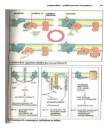
Clinical Presentation
Hemiparesis, aphasia, and gaze paresis, point to an anatomic localization in the
left middle cerebral artery territory (Fig. 11–1). Cortical symptoms such as
aphasia (impairment of the ability to use or comprehend words), neglect,
agnosia (loss of ability to recognize objects, persons, sounds, shapes or smells),
and apraxia (loss of the ability to execute or carry out learned purposeful
movements) indicate a lesion in the anterior (or carotid) circulation. Symptoms
such as diplopia, vertigo, crossed neurologic findings, and homonymous hemi-
anopia, however, suggest a posterior (or vertebrobasilar) circulation lesion.
The symptoms of an intracerebral hemorrhage cannot be reliably distinguished
from those of an ischemic stroke on clinical grounds alone. The presence of
headache, depressed level of consciousness, or extreme elevations of blood
pressure, however, can raise the suspicion of a hemorrhagic stroke.
98 CASE FILES: NEUROLOGY
Treatment
Treatment of ischemic stroke starts with assessment of eligibility for throm-
bolysis. Treatment must be initiated urgently. Intravenous tissue plasminogen
activator (t-PA) can significantly improve the odds of neurologic recovery, but
must be administered within 3 hours of onset of stroke symptoms. t-PA is asso-
ciated with a risk of intracranial hemorrhage. Thus, urgent imaging of the brain
such as CT scan is imperative to assess for hemorrhagic stroke. Contraindications
to t-PA include active bleeding, recent stroke, or history of intracerebral hemor-
rhage. Other acute stroke treatments are currently under investigation and can in
the near future include endovascular/intra-arterial and/or neuroprotective thera-
pies. Patients who are not candidates for thrombolytic therapy should be treated
with aspirin unless contraindicated.
Secondary stroke prevention should be implemented right away.
Antiplatelet drugs such as aspirin, clopidogrel, or the combination of aspirin
and extended release dipyridamole are the mainstays of stroke prevention treat-
ment for most patients with ischemic stroke and transient ischemic attack (TIA).
Patients with high-risk cardioembolic conditions such as atrial fibrillation,
CLINICAL CASES 99
Figure 11–1. Noncontrast axial CT image of a subacute left major coronary
artery infarction. (With permission from Chen MY, Pope TL, Ott DJ. Basic
radiology. McGraw-Hill Medical/Lange Clinical Science; 2004: Fig. 12–20.)
however, warrant long-term anticoagulation with warfarin, which has been
demonstrated to be superior to antiplatelet treatment for this indication.
Risk factor management is crucial to preventing recurrent stroke. Long-
term control of hypertension is especially important. Treatment should be ini-
tiated approximately 1 week after ischemic stroke. Statins for hyperlipidemia
lower the odds of stroke recurrence, and current guidelines recommend a tar-
get low density lipoprotein (LDL) of under 100 mg/dL. Carotid stenosis in a
patient with ischemic stroke or TIA is an indication for carotid endarterectomy
or, in a patient who is a poor surgical candidate, carotid stenting.
Rehabilitation is especially beneficial for patients who have gait difficulty or
aphasia, or who require assistance in activities of daily living or help resum-
ing gainful employment after a stroke.
The treatment of hemorrhagic stroke is primarily supportive and involves
control of hypertension. Intracranial pressure should be monitored and
addressed with hyperventilation and osmotic therapy, or surgical decompres-
sion when appropriate. Thrombolytic therapy is contraindicated.
Comprehension Questions
[11.1] An 81-year-old patient arrives in the emergency department with acute
left hemiparesis and neglect. What finding is most important in deter-
mining noneligibility for thrombolytic treatment?
A. Time of symptom onset of 2 hours
B. History of any previous myocardial infarction
C. Patient taking any antihypertensive medication
D. Recent gastrointestinal (GI) bleeding
[11.2] For this patient in question 11.1, what study is most useful to rule out
an intracerebral hemorrhage?
A. Electrocardiogram
B. Brain CT
C. Complete blood count
D. Cerebral arteriogram
[11.3] After receiving stroke therapy, the patient is being discharged home on
physical therapy. The usual treatment would include long-term
antiplatelet treatment; however, it is not used in this patient’s case.
Which of the following is most likely to be present such that antiplatelet
therapy is not prescribed?
A. The patient has diabetes
B. The patient has ischemic heart disease
C. The patient has carotid stenosis
D. The patient has atrial fibrillation
100
CASE FILES: NEUROLOGY
Answers
[11.1] D. A history of bleeding issues can contraindicate the use of an
anticoagulant.
[11.2] B. The head CT scan is reliable and rapid in assessing for cerebral
hemorrhagic stroke.
[11.3] D. When atrial fibrillation is present, then warfarin therapy is used
rather than antiplatelet therapy.
CLINICAL CASES 101
CLINICAL PEARLS
❖ Sudden onset of focal neurologic deficits equals stroke until proven
otherwise.
❖ Time is brain viability; treat ischemic stroke with thrombolytics within
3 hours to preserve brain tissue.
❖ Stroke risk factors are similar to those of ischemic heart disease.
❖ Cortical symptoms suggest a carotid territory stroke; brainstem or
cerebellar findings suggest a vertebrobasilar territory stroke.
REFERENCES
Mohr JP, Choi D, Grotta J, et al. pathophysiology, diagnosis, and management,
4th ed. Churchill Livingstone; 2004.
Ropper AH, Brown RH. Adams and Victor’s principles of neurology, 8th ed. New
York: McGraw-Hill; 2005.
This page intentionally left blank
❖
CASE 12
A 50-year-old female is brought by her husband to the Emergency Center after
experiencing sudden onset of severe headache associated with vomiting, neck
stiffness, and left-sided weakness. She was noted to complain of the worst
headache of her life shortly before she became progressively confused. Two
weeks ago she returned from jogging noting a moderate headache with nausea
and photophobia. She has a history of hypertension and tobacco use. On exam-
ination, her temperature is 37.6°C (99.8°F); heart rate, 120 beats/min; respira-
tion rate, 32 breaths/min; and blood pressure, 180/90 mmHg. She is stuporous
and moaning incoherently. Her right pupil is dilated with papilledema and ipsi-
lateral ptosis, and she vomits when a light is shone in her eyes. She has a left
lower face drooped and does not withdraw her left arm and leg to pain as
briskly compared to the right. Her neck is rigid. Her chest examination reveals
tachycardia and bibasilar crackles. During the examination, her head suddenly
turns to the left, and she exhibits generalized tonic-clonic activity. STAT labo-
ratory tests show a sodium level of 125 mEq/L. The electrocardiograph (ECG)
shows prolonged QT interval and T-wave inversion.
◆
What is the most likely diagnosis?
◆
What is the next diagnostic step?
◆
What is the next step in therapy?
ANSWERS TO CASE 12: Subarachnoid Hemorrhage
Summary: A 50-year-old female with a history of hypertension and tobacco
use presents with sudden onset of the worse headache of her life associated
with confusion, vomiting, neck stiffness, and left-sided weakness. She was
noted to complain of a headache 1 week ago. She is now hypertensive. Her
neurologic examination is significant for stupor, right cranial nerve III paraly-
sis, left-sided weakness, neck stiffness, and a seizure. Her workup is signifi-
cant for hyponatremia and ECG changes.
◆
Most likely diagnosis: Subarachnoid hemorrhage
◆
Next diagnostic step: Noncontrast CT of the head
◆
Next step in therapy: Cerebral angiography
Analysis
Objectives
1. Identify the epidemiology and risk factors for subarachnoid hemorrhage.
2. Understand the prognosis and complications of subarachnoid
hemorrhage.
3. Know a diagnostic and therapeutic approach to subarachnoid
hemorrhage.
Considerations
This 50-year-old woman has multiple risk factors for subarachnoid hemor-
rhage caused by an underlying aneurysm, including her age (mean age for
subarachnoid hemorrhage is 50 years of age), sex (slightly higher risk for
females), hypertension, and tobacco use. The complaint of “the worst
headache of my life” to describe its sudden severe onset is classic, and may or
may not be associated with altered mentation and focal neurologic deficits.
There is usually a history of a recent moderate headache as a result of sentinel
bleed, as in her case after running, and 60% of subarachnoid hemorrhages
occur during physical or emotional strain, head trauma, defecation, or coitus.
The clinical severity of the subarachnoid hemorrhage is graded based on the
degree of stupor, nuchal rigidity, focal neurologic deficits, and elevation of
intracranial pressure. Our patient exhibits neurogenic pulmonary edema, a rare
complication of subarachnoid hemorrhage. Her neurologic signs localize to a
ruptured right posterior communicating artery aneurysm, with the bleed caus-
ing compression of the nearby ipsilateral cranial nerve III with mydriasis, pto-
sis, and impaired extraocular movements. Her contralateral hemiparesis and
complex partial seizure with secondary generalization can result from either
parenchymal extension of the hemorrhage with edema or middle cerebral
104
CASE FILES: NEUROLOGY
artery vasospasm, all three of which are complications of subarachnoid hem-
orrhage. Hyponatremia is frequently seen on chemistries, correlating with an
elevation of atrial natriuretic factor, cerebral salt wasting, and syndrome of
inappropriate antidiuretic hormone. ECG changes, especially QT prolonga-
tion, T-wave inversion, and arrhythmias, are also systemic complications com-
mon to subarachnoid hemorrhage.
APPROACH TO SUBARACHNOID HEMORRHAGE
Definitions
Subarachnoid space: The spongy interval between the arachnoid mater and the
pia mater. The headache and nuchal rigidity is caused by chemical inflam-
mation of the pia arachnoid from blood degradation products in this space.
Sentinel bleed: Intermittent aneurysmal subarachnoid hemorrhage causing
lesser headaches that precede the “worst headache” that occurs with rup-
ture of the aneurysm.
Vasospasm: Most alarming complication of aneurysmal subarachnoid hem-
orrhage in which irritation causes constriction of major cerebral arteries,
vasospasm lethargy, and cerebral infarction. Vasospasm occurs mostly
with aneurysms rather than other causes of subarachnoid hemorrhage, and
peaks after 4 to 14 days. Transcranial Doppler can be used to detect a
change in flow velocity in an affected middle cerebral artery.
Acute communicating hydrocephalus: Complication that occurs because
of obstruction of the subarachnoid granulations in the venous sinuses by
the subarachnoid blood. CT shows enlarged lateral, third, and fourth
ventricles, with clinical signs of headache, vomiting, blurry and double
vision, somnolence, and syncope.
Clinical Approach
Etiologies
Subarachnoid hemorrhage is the underlying cause of approximately 10% of
stroke presentations and results from a number of etiologies. Ruptured saccu-
lar or berry aneurysms account for up to 80% of nontraumatic subarach-
noid hemorrhage, and portend the worst prognosis. More than three-fourths of
intracerebral aneurysms arise in the anterior circulation. The most frequent sites
of aneurysms are in the anterior communicating artery (up to one-third of
aneurysmal subarachnoid hemorrhages), followed by the bifurcation of the
internal carotid artery with the posterior communicating artery, and the bifur-
cation of the internal carotid artery with the middle cerebral artery. One-fourth
of patients will have more than one aneurysm, with risk for rupture increasing
with size of the aneurysm. Fibromuscular dysplasia is an associated etiology in
one-fourth of aneurysm patients, whereas polycystic kidney disease is related
CLINICAL CASES 105
to 3% of cases. Other risk factors for aneurysms include chronic severe hyper-
tension with diastolic blood pressure greater than 110 mmHg, liver disease,
tobacco and alcohol use, vasculitides, collagen vascular disorders such as
Marfan syndrome, infections (mycotic aneurysms), and oral contraception.
Nonaneurysmal causes of subarachnoid hemorrhage include trauma, arteriove-
nous malformations, and cocaine or amphetamine abuse.
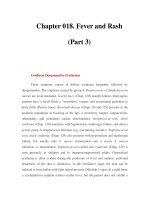
Diagnosis and Prognosis
Head CT without contrast is the most sensitive neuroimaging study for
detecting subarachnoid bleeding, appearing as hyperdensity within the cere-
bral convexities, cisterns, and parenchyma (Fig. 12–1). Intraventricular hem-
orrhage portends a worse prognosis and increased risk for hydrocephalus.
Sensitivity of CT is greatest 24 hours after the event with 50% still detectable
after 1 week. Negative head CT occurs in 10–15% of cases, and should
be further evaluated with lumbar puncture looking for xanthochromia
(yellowish discoloration of CSF) and increased red blood cells. Cerebrospinal
fluid (CSF) studies are most sensitive 12 hours after onset, but can be negative
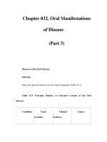
in 10–15% of patients as well, in which case the prognosis is better. CT, MRI,
or conventional angiography can be used to screen for an underlying aneurysm
(Fig. 12–2).
106
CASE FILES: NEUROLOGY
Figure 12–1. Noncontrast CT scan subarachnoid blood in the left sylvian fis-
sure (bright) and within the left lateral ventricle. (With permission from Kasper
DL, Braunwal E, Fauci A, et al. Harrison’s principles of internal medicine,
16th ed. New York: McGraw-Hill; 2004: Fig. 349–14b.)
Up to 60% of patients die in the first 30 days after a subarachnoid hem-
orrhage, 10% instantly without warning. First month mortality is 40% for
hospitalized patients, with worsening of mortality to 50–80% with rebleeding.
Severity of cases and their prognoses can be graded based upon alertness and
presence of focal signs.
Grade I subarachnoid hemorrhage (SAH) patients are alert with mild headache
and nuchal rigidity and have a 5% chance of deteriorating with a 3–5%
mortality risk.
Grade II patients have moderate-to-severe headache and nuchal rigidity,
and a 6–10% mortality.
Grade III patients have added confusion.
Grade IV patients have stupor and moderate hemiparesis.
Grade V patients are comatose with signs of severe increased intracranial
pressure, and they have the worst prognosis with 80% chance of deteri-
orating, 25–30% rebleeding rate, and 50–70% mortality. Delayed
vasospasm is a calamitous complication that occurs in up to 20% of
cases.
CLINICAL CASES 107
Figure 12–2. Conventional angiogram of the right vertebral and basilar artery
showing the large aneurysm. (With permission from Kasper DL, Braunwal E,
Fauci A, et al. Harrison’s principles of internal medicine, 16th ed. New York:
McGraw-Hill; 2004: Fig. 349–14c.)