Case Files Neurology - part 7 pot
Bạn đang xem bản rút gọn của tài liệu. Xem và tải ngay bản đầy đủ của tài liệu tại đây (248.38 KB, 50 trang )
❖
CASE 34
A 65-year-old man with a history of hypertension, coronary artery disease, and
early Alzheimer disease presents with a complaint of double vision since
yesterday. He has not experienced chest pain, chest palpitations, nausea, light-
headedness, vertigo, headache, facial weakness, hemisensory loss, hemiparesis,
loss of balance, hearing loss, tinnitus, visual loss, ptosis, or proptosis. He has
noticed that covering up either eye corrects his double vision. He has resorted to
wearing an eye patch since yesterday so that he can see and walk without falling.
In fact he was able to drive on his own on the freeway to your office much to his
family’s dismay. On further questioning you elicit the history that his double
vision occurs only on horizontal gaze and not vertical gaze. He has been com-
pliant with his medications for hypertension and coronary artery disease. On
examination, his blood pressure (BP) is 124/72 mmHg with a heart rate (HR) of
88 beats/min. He is afebrile and has a regular rate and rhythm without murmurs
on cardiac examination. There are no carotid bruits, and his peripheral pulses are
normal. His neurologic examination is notable for intact orientation and intact
motor strength. His cranial nerve examination is remarkable only for a right lat-
eral rectus palsy. Sensory examination is normal, and his deep tendon reflexes
are 2+ throughout. Plantar responses are flexor. His gait is normal. Review of his
daily blood pressure log shows stable pressures of 130/70 mmHg.
◆
What is the most likely diagnosis?
◆
What is the neurologic deficit?
ANSWERS TO CASE 34: Sixth Nerve Palsy (Ischemic
Mononeuropathy)
Summary: A 65-year-old man with hypertension, coronary artery disease,
and early Alzheimer disease presents with a 24-hour history of binocular
horizontal diplopia (double vision). He has not experienced associated
symptoms such as chest pain or headache. His examination is significant for
a normal blood pressure and heart rate and the findings of the isolated right
sixth nerve palsy.
◆
Most likely diagnosis: Sixth nerve palsy secondary to ischemic
mononeuropathy
◆
Likely neurological deficit: Sixth nerve palsy
Analysis
Objectives
1. Understand the diagnostic approach in evaluating diplopia.
2. Describe the difference between monocular and binocular diplopia.
3. Know the differential diagnosis of a sixth nerve palsy.
Considerations
This 65-year-old man with known risk factors for cerebral vascular disease
(hypertension and coronary artery disease) presents with an acute episode of
binocular diplopia. The history suggests binocular diplopia as he tells you that
covering up an eye resolves the diplopia. You are given the history that he has
diplopia only on horizontal gaze. In this particular case you are told that the
patient’s blood work and MRI brain is normal. Given the history of hyperten-
sion and coronary artery disease he is at risk for cerebrovascular disease and
ischemia. In this setting, the most likely cause of this man’s diplopia is an
ischemic mononeuropathy to the abducens nerve. In this particular case the
patient has a completely normal examination except for a sixth nerve palsy.
This makes it easy to pinpoint the location of the abnormality as the only loca-
tion for an isolated abducens nerve palsy is in the nucleus. Table 34–1 shows
locations where the sixth nerve can be affected and its associated clinical
findings.
284 CASE FILES: NEUROLOGY
APPROACH TO BINOCULAR DIPLOPIA
Definitions
Ptosis: Drooping of the eye lids
Proptosis: Abnormal protrusion of the eyeball
Diplopia: Double vision
Ischemic mononeuropathy: Isolated nerve injury from inadequate blood
flow to the nerve
CLINICAL CASES 285
Table 34–1
CLINICAL FINDINGS
ASSOCIATED CLINICAL
LOCATION FINDINGS ETIOLOGIES
Nuclear Horizontal gaze palsy, sixth Ischemia, demyelinating,
nerve dysfunction or other inflammatory, trauma,
brainstem signs vascular (aneurysm or other
vascular malformations),
neoplastic, congenital,
metabolic
Fascicle Contralateral hemisensory Ischemia, inflammatory,
loss, contralateral hemiparesis, vascular, neoplastic, trauma,
central Horner syndrome demyelinating
Subarachnoid Signs of increased intracranial Inflammatory, infectious,
space pressure (e.g., headache, toxic, vascular, neoplastic,
papilledema) or other cranial cervical traction,
neuropathies myelogram, infiltrative
Petrous apex Facial pain or fifth, seventh, Traumatic, infectious,
or eighth cranial nerves inflammatory (sarcoid),
dysfunction neoplastic (meningioma)
Cavernous sinus Sixth nerve palsy with any Ischemic, neoplastic, inflam-
combination of third, fourth matory, infectious, vascular,
or ophthalmic division of the fistula, or thrombosis
fifth cranial nerve dysfunction;
Horner syndrome
Orbit/superior Can have proptosis or optic Traumatic, infectious,
orbital fissure nerve atrophy/edema inflammatory, neoplastic
Clinical Approach
Sixth nerve palsy has a variety of causes, and clinical examination usually leads
to an accurate diagnosis. The abducens nucleus is located in the lower dorsal
pons. The motor neurons of this nucleus send axons that course anteriorly in the
pons and travel near the corticospinal tract and emerge in the sulcus between
the pons and medulla. The abducens nerve exits the pons ventrally and ascends
in the prepontine cistern via the subarachnoid space. It then rises over the
petrous apex of the temporal bone and enters the cavernous sinus laying
between the carotid artery and the ophthalmic branch of the trigeminal nerve
laterally. It finally passes into the orbit through the superior orbital fissure.
Etiology of Sixth Nerve Palsy
After the localization of the sixth nerve lesion, the next step is to determine the
etiology of the abnormality. Table 34–1 shows there are various causes for a
nuclear abducens abnormality. The evaluation includes serologic studies
including an erythrocyte sedimentation rate, antinuclear antibody (ANA),
complete blood count (CBC), glycosylated hemoglobin, and if appropriate a
2-hour glucose tolerance test. An MRI of the brain without contrast should be
ordered concomitantly. An erythrocyte sedimentation rate (ESR) and ANA can
help exclude inflammatory causes such as vasculitis; glycosylated hemoglobin
can exclude diabetes mellitus, and a CBC can exclude infectious processes. An
MRI brain and orbits can exclude vascular abnormalities such as an aneurysm
and can exclude mass lesions that are inflammatory (sarcoid), demyelinating,
neoplastic, or traumatic. An ischemic process may not be readily visualized on
imaging studies and is often a diagnosis of exclusion.
Evaluation of Diplopia
Diplopia results from lack of visual fusion. The first step in evaluating a
patient with diplopia is to determine whether it is binocular or monocular.
Binocular diplopia is usually caused by an underlying primary neurologic
problem. Monocular diplopia, conversely, is primarily caused by an oph-
thalmologic disorder such as abnormalities of the lens, cornea, vitreous
humor, or iris. Rarely, monocular diplopia can be caused by occipital lobe dis-
ease or seizures. Binocular diplopia denotes double vision arising from mis-
alignment of both eyes. Covering up one eye resolves the double vision.
Monocular diplopia, however, arises from a primary problem within one eye.
This type of diplopia does not resolve when an eye is covered.
The next step in evaluating someone with binocular diplopia is to determine
if it is horizontal or vertical. Different eye muscles are involved in moving the
eyes horizontally or vertically. There are only two muscles in each eye respon-
sible for horizontal gaze and those are the medial rectus, which is innervated
by the third nerve, and the lateral rectus, which is innervated by the sixth
286 CASE FILES: NEUROLOGY
nerve. Worsening diplopia on near vision suggests a problem with the medial
rectus, whereas diplopia that worsens when viewing distant and lateral objects
suggest a problem with the lateral rectus.
The other four eye muscles (superior rectus, inferior rectus, inferior
oblique, and superior oblique) move the eyes vertically. Individuals that pres-
ent with vertical binocular diplopia are experiencing weakness in one or sev-
eral of these muscles. Vertical diplopia that worsens on near vision suggests
a problem with either the inferior oblique or superior oblique. At this point in
the evaluation, it must be differentiated whether or not the patient’s binocular
diplopia is secondary to a medial rectus or a lateral rectus problem.
Examining extra-ocular muscles in the nine cardinal fields of gaze can read-
ily point out which of the two muscles is affected. For example, if the right
eye cannot cross the midline and look out laterally, the lateral rectus is
affected. Conversely, if the right eye cannot cross the midline and turn
inward, the medial rectus is affected.
One of these tests is called the alternate cover test and is performed by ask-
ing the patient to fixate on an object in each position of gaze. As the patient
moves the eyes in each position, deviations in the eye as each one is alternately
covered may be seen. The second test often used for evaluating binocular
diplopia is the red lens test. In this test, a red lens is placed over an eye, most
commonly the right eye, and the patient is asked to look at the nine positions
of a cardinal gaze. The key to performing this test is to understand the follow-
ing: (1) image separation will be greatest in the direction of the weak muscle
and (2) the image that is the furthest away from the midline is a false image
and corresponds to the eye with impaired motility.
Evaluating other aspects of the cranial nerve examination will help deter-
mine where the diplopia is arising from. Special attention should be given to
the eyelid, pupillary responses, symmetry of the pupillary size, abnormalities
of cranial nerves V, VII, and VIII. For example, ptosis or droopiness of the eye-
lid can suggest a third nerve problem. Likewise, pupillary asymmetry suggests
a third nerves problem. Fatigue of the eyelid can suggest myasthenia gravis.
Patients who have a head tilt can also provide you with clues as to where the
problem may lie. For example, someone with a right superior oblique palsy
may have a leftward tilt of the head.
Treatment
Treatment of the underlying disorder of sixth nerve palsy is indicated when
significant and persistent. An isolated and presumed ischemic-related sixth
nerve palsy can be observed for improvement for 1 to 3 months. Patching of
the involved eye can help alleviate diplopia symptoms temporarily. Prism ther-
apy can also be used. Some suggest using botulinum toxin as a temporizing
measure. However if these measures fail, surgery may be the only way to cor-
rect this problem.
CLINICAL CASES 287
288 CASE FILES: NEUROLOGY
Comprehension Questions
[34.1] Which of the following is most accurate regarding diplopia?
A. Binocular diplopia refers to double vision occurring from intrinsic
problems in both eyes
B. Monocular diplopia most commonly occurs because of extrinsic
eye problems
C. The green lens test is a way of evaluating binocular diplopia
D. The key in evaluating diplopia is to start by determining if it is
monocular or binocular
[34.2] A 33-year-old woman has a 3-minute seizure episode caused by her
epilepsy. There are no underlying medical disorders or brain structural
lesions. Which of the following indicates a more complicated underly-
ing neurologic problem?
A. Urinary incontinence with seizure
B. Confusion and lethargy after seizure
C. Headache after the seizure
D. Sixth nerve palsy after seizure
[34.3] A 58-year-old woman suffers from an ischemic-related sixth nerve
palsy which occurred 6 months ago. Various methods have been tried
with limited success, and the patient still has diplopia. Which of the
following is most likely to be helpful at this stage?
A. Surgery
B. Eye patch
C. Prisms
D. Prednisone at a dose of 10 mg per day
E. Botulinum toxin
Answers
[34.1] D. The key to evaluating diplopia is to assess unilateral versus bilateral.
Binocular diplopia arises from misalignment of the eye muscles on a
target.
[34.2] D. Seizures have not been reported to cause sixth nerve dysfunction
and thus, its presence indicates a more complex situation.
[34.3] A. Surgery is the best option for persistent symptoms that have not
resolved. Prednisone has not been used for sixth nerve palsies from
ischemia. It can be used for inflammatory causes of sixth nerve
abnormalities.
REFERENCES
Dorland’s Illustrated Medical Dictionary, 27th ed. Philadelphia, PA: WB Saunders;
1988.
Patel SV, Mutyala S, Leske DA, et al. Incidence, associations, and evaluation of
sixth nerve palsy using a population-based method. Ophthalmology 2004;
111:369–375.
Quah BL, Ling YL, Cheong PY, et al. A review of 5-years experience in the use of
botulinum toxin A in the treatment of sixth cranial nerve palsy at the Singapore
National Eye Centre. Singapore Med J 1999;40:405–409.
Savino PJ. Diplopia and sixth nerve palsies. Semin Neurol 1986;6:142–146.
CLINICAL CASES
289
CLINICAL PEARLS
❖ Binocular diplopia occurs from misalignment of the eye muscles on
a target and commonly denotes an underlying primary neurologic
problem within the brain parenchyma.
❖ Younger patients with sixth nerve palsies more often have malig-
nant etiologies, whereas older patients usually have more benign
etiologies.
❖ Monocular diplopia results from intrinsic eye problems, including
ocular muscles and neuromuscular junction.
❖ MRI of the brain is critical in evaluating patients with binocular
diplopia as it allows for the detection of vascular or demyelinating
processes.
This page intentionally left blank
❖
CASE 35
A 68-year-old woman presents with right facial paralysis. She states she was
well until approximately 3 days ago when she began to have right ear pain. She
has not taken any pain medication and has not had any fever. Today, she awoke
with right facial paralysis. She feels slightly dizzy and notices that she has
right-sided hearing loss. She denies any past history of ear infections. Her med-
ical history is unremarkable. She does have a past history of chicken pox as a
child. Her physical examination shows a 68-year-old woman with obvious right
facial paralysis involving her forehead and mouth. She is afebrile but is anxious
because of the loss of facial function. There is no motion in any of the branches
of the right facial nerve. Her head and neck examination finds small blisters on
an erythematous base in the right conchal bowl of the external ear. The exam-
ination of the ear canal is painful to her, but the tympanic membrane is intact.
No pus is seen in the ear canal. The left ear canal is normal. The Weber tuning
fork test lateralizes to the left ear. The Rinne test is normal in both ears. The
examination of the nose, oral cavity, throat, and neck are normal. The cranial
nerve (CN) examination is normal except for the right VII and VIII nerve prob-
lems listed above. The remaining physical examination is normal.
◆
What is the most likely diagnosis?
◆
What is the next diagnostic step?
◆
What is the next step in therapy?
ANSWERS TO CASE 35: Facial Paralysis
Summary: A 68-year-old woman presents with right facial paralysis, a 3-day
history of right ear pain, and right-sided hearing loss. There is no motion in
any of the branches of the right facial nerve. There are small blisters on an ery-
thematous base in the right conchal bowl of the external ear. The examination
of the ear canal is painful to her, but the tympanic membrane is intact. The
Weber tuning fork test lateralizes to the left ear. The Rinne test is normal in
both ears. The cranial nerve examination is normal except for the right VII and
VIII nerve problems listed above.
◆
Most likely diagnosis: Herpes zoster oticus (Ramsay Hunt syndrome)
◆
Next diagnostic test step: Tzanck smear, audiogram, consider facial
nerve electrodiagnostic studies and diagnostic imaging, if indicated
◆
Next therapeutic step: anti-herpes virus medication
Analysis
Objectives
1. Describe the clinical presentation and diagnostic approach to facial
weakness.
2. Be familiar with the differential diagnosis of facial weakness.
3. Know the treatment for Ramsey Hunt syndrome.
Considerations
This elderly woman has a history of chicken pox, blisters on her ear, hearing
abnormalities, and unilateral facial paralysis. Her entire right facial muscles
are affected, suggestive of a peripheral facial nerve palsy; a central defect usu-
ally spares the forehead. The Weber and Rinne tests are consistent with a sen-
sorineural hearing loss rather than a conductive disorder. This constellation of
findings is most consistent with Ramsey Hunt syndrome, which is reaction of
the herpes zoster affecting both CNs VII and VIII. A diligent history and phys-
ical examination should be performed to exclude other possibilities such as
central nervous system disorders, cholesteatomas, facial neuromas, and tumors
of the parotid. Corticosteroid and antiviral therapy are recommended, with the
probability of good recovery.
APPROACH TO FACIAL NERVE PARALYSIS
Definitions
Audiogram: A test that measures the level of hearing in each ear.
Bell palsy: An idiopathic form of facial paralysis, thought to be caused by
herpes simplex virus reactivation.
292
CASE FILES: NEUROLOGY
Cholesteatoma: A benign tumor composed of epithelial debris from the
tympanic membrane that becomes trapped in the middle ear.
Facial nerve electromyograph (EMG): Like EMG performed for other
nerves, a needle electrode is inserted into the facial muscles, and the
patient is asked to perform maximal facial motion effort. The elec-
tromyographer looks for compound muscle action potentials, abnormal
waves, or fibrillation potentials. Evoked potentials, such as the blink
reflex, can also be performed with EMG. An absence of motor unit
potentials signifies severe damage or loss or nerve continuity.
Fibrillation potentials are signs of a lack of facial nerve input, and are a
particularly bad prognostic sign.
Facial nerve electroneurogram (ENoG): An electrical test that evokes a
compound muscle action potential (cMAP) by stimulating the facial
nerve. The ENoG uses surface electrodes rather than needles to measure
the cMAP. Each side is stimulated at the stylomastoid foramen, and the
responses from muscle groups are measured and compared. Significant
nerve damage is indicated by a 90% or greater reduction in the cMAP.
Otorrhea: Drainage from the ear.
Postherpetic neuralgia: Neuropathic pain resulting from resolved herpes
infection.
Tzanck smear: A test that looks for intracytoplasmic particles due to viral
infection.
Vesicles: Small fluid-filled blisters on an erythematous base.
Clinical Approach
Approach to Facial Paralysis
Facial function can be characterized in many different ways. A distinction is
made between paresis, which indicates weakness, but function is still present;
and paralysis, which indicates total lack of function despite maximal effort.
The American Academy of Otolaryngology has adopted a system for grading
facial nerve function called the House-Brackmann score. Evaluation of
patients with facial paralysis is performed systematically by considering the
anatomy of the facial nerve’s pathway. The facial nerve emerges from the
brainstem at the pons to traverse the cerebellopontine angle and then through
the temporal bone. The bony course through the temporal bone is the longest
course of any nerve through bone. It emerges at the stylomastoid foramen to
pass through the substance of the parotid gland and divide into branches that
innervate the various parts of the face. Additionally, the facial nerve contains
general sensation to the ear canal and pinna, special sensation of taste from the
anterior two-thirds of the tongue, and secretomotor function of parasympa-
thetics to the submandibular gland, the lacrimal gland, and the nasal mucosa.
As a point of departure, isolated unilateral facial paralysis will be discussed.
Facial paralysis of central origin, that is, caused by stroke, is marked by fore-
head sparing. The paralysis affects the lower half of the face, but forehead
CLINICAL CASES 293
movement remains normal. This is caused by the bilateral cortical connections
to the facial nucleus in the brainstem. In such a circumstance, the examining
physician should inquire about risk factors for stroke and look for other signs
that might indicate a stroke. Facial paralysis associated with hearing loss
and/or dizziness, vertigo, or imbalance suggests cerebellopontine angle and
internal auditory canal disorders. In this circumstance, an audiogram might
show a sensorineural type of hearing loss. Further evaluation will include con-
trast enhanced MRI and possibly CT.
Because the facial nerve passes through the middle ear and temporal bone,
examination of the ear canal and tympanic membrane is of paramount impor-
tance. Otitis media and cholesteatoma can be associated with facial paralysis.
The ear examination will clearly disclose these abnormalities when present.
Acute bacterial otitis media produces a purulent middle ear effusion, which
can often produce a spontaneous tympanic membrane perforation. In these
cases, a preexisting history of otitis media is not always present, although the
history and physical examination might indicate an upper respiratory tract
infection or inflammation (as from allergic rhinitis). The physical examination
will clearly show the abnormal findings in the middle ear. Acute otitis media
is probably the most common cause of isolated facial paralysis in children.
Cholesteatoma is a benign tumor of epithelial debris that is produced when
the squamous layer of the eardrum is trapped and cannot exfoliate properly.
Cholesteatoma usually occurs in patients that have preexisting ear problems.
The physical examination in cholesteatoma will show either cheesy epithelial
debris in the ear canal or a pearly white tumor behind the ear drum. Generally,
patients with cholesteatoma will have a pre-existing history of hearing loss and
often a long history of intermittent foul-smelling, purulent otorrhea.
Cholesteatomas grow slowly, and sometimes can be present for years without
causing many symptoms. Neglected cholesteatomas can produce destruction
of the ossicles, the inner ear or the facial nerve. Complications of
cholesteatomas can include sigmoid sinus thrombosis, brain abscess, and
meningitis. CT scanning of the temporal area is helpful prior to surgical exci-
sion. Referral to an otologist-neurootologist is recommended.
Facial neuromas (schwannomas of the facial nerve) are rare, and their
occurrence is roughly 1:1,000,000 persons per year. These are benign tumors
of the facial nerve that grow slowly and produce a slowly progressive (over
several months, not days) form of facial paralysis. When these tumors occur in
the middle ear portion of the facial nerve, they produce a conductive hearing
loss. When they occur in the internal auditory canal, they can produce a sen-
sorineural form of hearing loss. Again, an audiogram and MRI with contrast
will be necessary to diagnose and discover these tumors. Referral to a neu-
rootologist is recommended.
Tumors of the parotid and skull base can produce facial paralysis.
Paralysis of an isolated branch of the facial nerve is caused by malignancy
until proven otherwise. Malignant tumors of the skin or parotid gland can pro-
duce facial paralysis either by compression or perineural invasion. Skull base
294
CASE FILES: NEUROLOGY
tumors (meningiomas, carcinomas, sarcomas, etc.) can produce facial paraly-
sis, however, this facial paralysis is usually found along with other CN find-
ings consistent with a skull base location (e.g., loss of CN IX,X,XI, or XII).
A careful history and physical examination of the involved area will usually
uncover this pathology when present. Imaging studies, such as enhanced MRI
or CT, are helpful in identifying neoplasms that affect the facial nerve. Other
special considerations in facial paralysis involve its bilateral occurrence.
Bilateral facial paralysis has a limited number of causes, principally Lyme
disease or Guillain-Barré syndrome. Herpes zoster oticus (or Ramsay Hunt
syndrome) is a frequently encountered form of facial paralysis.
Ramsey Hunt Syndrome
The sine qua non of Ramsay Hunt syndrome are vesicles in the ear associated
with facial paralysis. It is caused by reactivation of varicella-zoster virus
(VZV), the virus that causes chicken pox and shingles. This virus lingers in
sensory ganglia until reactivated. The sensory ganglion of the facial nerve is
the geniculate ganglion. Reactivation of the virus produces vesicles in its area
of sensory innervation. For the facial nerve, this can include the posterior ear
canal, conchal bowl, or even postauricular skin. (In segmental nerves, the dor-
sal ganglia contains the dormant virus, a dermatomal distribution of vesicles is
often found when it is reactivated). Reactivation can result from being
immunocompromised or in some other way “stressed.”The pain from herpes
zoster might be described as burning and can be intensely painful. This pain
can linger for up to 1 year, despite resolution of the active infection, and is
called postherpetic neuralgia.
Treatment of Ramsay Hunt syndrome involves use of anti-herpes virus
medication for 7 to 10 days. Traditionally acyclovir was used; its IV form
might still be indicated for severe infections in severely immunocompromised
patients. Because of its poor oral absorption, its oral form requires five doses
daily and is difficult for patients to maintain. Newer antiviral medications,
such as ganciclovir and valacyclovir, have better oral absorption and less fre-
quent dosing schedules. These medications are most often used for limited
episodes of Ramsay Hunt syndrome. Topical acyclovir cream might help to
speed healing of vesicles. Patients are contagious and can spread the virus to
susceptible individuals as long as vesicles are present.
Steroids are frequently prescribed for patients with facial paralysis. Often
doses of prednisone, 1 mg/kg/day for 10 to 14 days are given. Use of steroids
during an active infection such as Ramsay Hunt syndrome must be weighed
carefully. Although steroids might reduce the pain and might improve the
chance for facial recovery, the possible risks of worsening an immunocom-
promised state or of dissemination of the herpes infection to the brain (herpes
encephalitis) or eye (ocular herpes) must be considered.
Hearing loss and vestibular symptoms can occur in patients with Ramsay
Hunt syndrome. This will produce ipsilateral sensorineural hearing loss and
CLINICAL CASES 295
vestibular weakness. It is unclear if the virus spreads from one ganglion to
another (i.e., from the geniculate to the spiral or Scarpa ganglion), or if edema
and inflammation produce the associated cochleovestibular symptoms.
Nevertheless, patients with facial paralysis who complain of hearing loss
should have an audiogram.
Bell Palsy
Bell palsy is likely caused by viral infection. Herpes simplex virus has been
implicated and has been isolated from cases of Bell palsy when the facial nerve
was decompressed. For this reason, the recommendations for treating Bell
palsy include antiviral medications (ganciclovir or valacyclovir) and oral
steroids (prednisone 1 mg/kg/day for 10–14 days). The use of both forms of
medications (antiviral and steroids) has been shown to improve return of facial
function compared to either medication alone or to placebo. Although sponta-
neous rates of recovery are high, especially in patients with mild weakness,
treatment should not be withheld on the expectation of speedy and normal
recovery. Surgical treatment for Bell palsy has a checkered past. Facial nerve
decompression has been advocated for Bell paralysis for several reasons:
(1) the facial nerve has the longest bony course of any nerve, peripheral or cra-
nial; (2) this bony confinement does not allow the nerve to swell; (3) this swelling
in a confined space produces ischemia of the nerve; (4) poor regeneration occurs
once ischemia takes place; and (5) very limited and unsatisfactory methods are
available to rehabilitate the paralyzed face. Surgery is only indicated for cases of
facial paralysis where ENoG and EMG both show absence of facial function.
Regardless of cause, patients with facial paralysis need special care of the
eye on the affected side to avoid permanent vision loss. Because of the loss of
the blink reflex and decreased lacrimation, the affected eye can dry out caus-
ing exposure keratitis, which can lead to loss of vision in the affected eye.
Simple eye care consisting of artificial tears every hour while awake and
ocular lubricant (Lacri-Lube) ointment at night with eye taping can avoid per-
manent loss of vision. Ophthalmologic consultation should be sought for any
patient with facial paralysis and is mandatory in patients who complain of eye
pain, irritation, or loss of vision. Most cases of facial nerve weakness can be
fully evaluated and managed by primary care physicians. These patients
demand close attention and should be seen once or twice a week until resolu-
tion is seen. Bell palsy and Ramsay Hunt syndrome should respond relatively
rapidly (over 2 to 3 weeks) to the treatment outlined, but the greater the
weakness, the longer the recovery.
Consultation with a neurologist should be sought when the diagnosis is in
doubt. Also, consider referring patients that have (1) rapid progression (over
3 days) to complete paralysis; (2) evidence of middle ear, inner ear, or skull
base disease; (3) an initial improvement in facial weakness to have recurrence
a few weeks or months later, or (4) no return of function despite appropriate
therapy.
296
CASE FILES: NEUROLOGY
Comprehension Questions
[35.1] What is the most common cause of unilateral facial weakness in an adult?
A. Lyme disease
B. Varicella zoster reactivation
C. Acoustic neuroma
D. Herpes simplex virus reactivation
E. Noncaseating granulomas
[35.2] What is the key indicator of herpes zoster oticus (Ramsay Hunt
syndrome)?
A. Vesicles on an erythematous base found in the external ear
B. Noncaseating granulomas on lower lip biopsy
C. Circulating antibodies to Borrelia burgdorferi
D. Uveitis and parotid gland swelling
E. Loss of taste on the ipsilateral tongue
[35.3] A 69-year-old man complains of right facial weakness. A close exam-
ination of his facial movements indicates loss of the nasolabial fold and
inability to raise the upper lip on that side. His blink, forehead, and
lower lip movement are normal. What is the most likely cause of his
facial paralysis?
A. Bell palsy
B. Herpes zoster oticus
C. Malignant parotid gland tumor
D. Acoustic neuroma
E. Lyme disease
Answers
[35.1] D. By far the most common cause of acute facial weakness in an adult
is Bell palsy. This disorder is caused by reactivation of herpes simplex
virus. However, this is a diagnosis of exclusion as no accurate sero-
logic tests have been discovered that confirm the diagnosis.
[35.2] A. The pathognomonic feature of herpes zoster oticus (Ramsay Hunt
syndrome) is a vesicular eruption on an erythematous base in an area of
facial nerve sensory distribution (external ear). This disorder is caused
by reactivation of varicella-zoster virus and is treated with antiviral
medications and steroids. Inadequately treated zoster infections can
lead to poor recovery of facial function and postherpetic neuralgia.
[35.3] C. An isolated facial nerve branch paralysis is caused by malignancy
until proven otherwise. Bell palsy, herpes zoster oticus, and Lyme dis-
ease affect the entire nerve. Acoustic neuromas can cause facial paral-
ysis when they are very large, but this is very rarely seen in the modern
area. Their location in the cerebellopontine angle would produce whole
face weakness, and not an isolated branch weakness as described.
CLINICAL CASES 297
REFERENCES
Ahmed A. When is facial paralysis Bell palsy? Current diagnosis and treatment.
Cleve Clin J Med 2005;72(5):398–401, 5.
Alberton DL, Zed PJ. Bell’s palsy: a review of treatment using antiviral agents. Ann
Pharmacother 2006;40(10):1838–1842.
Austin JR, Peskind SP, Austin SG, et al. Idiopathic facial nerve paralysis: a ran-
domized double blind controlled study of placebo versus prednisone.
Laryngoscope 1993;103(12):1326–1333.
Gilden DH, Cohrs RJ, Hayward AR, et al. Chronic varicella-zoster virus ganglioni-
tis—a possible cause of postherpetic neuralgia. J Neurovirol 2003;9(3):404–407.
House JW, Brackmann DE. Facial nerve grading system. Otolaryngol Head Neck
Surg 1985;93(2):146–147.
Kuhweide R, Van de Steene V, Vlaminck S, et al. Ramsay Hunt syndrome: patho-
physiology of cochleovestibular symptoms. J Laryngol Otol 2002;116(10):
844–848.
Ohtani F, Furuta Y, Aizawa H, et al. Varicella-zoster virus load and cochleovestibu-
lar symptoms in Ramsay Hunt syndrome. Ann Otol Rhinol Laryngol
2006;115(3):233–238.
Overell JR, Willison HJ. Recent developments in Miller Fisher syndrome and
related disorders. Curr Opin Neurol 2005;18(5):562–566.
Redaelli de Zinis LO, Gamba P, Balzanelli C. Acute otitis media and facial nerve
paralysis in adults. Otol Neurotol 2003;24(1):113–117.
Sweeney CJ, Gilden DH. Ramsay Hunt syndrome. J Neurol Neurosurg Psychiatry
2001;71(2):149–154.
298 CASE FILES: NEUROLOGY
CLINICAL PEARLS
❖ Bell palsy is the most common cause of acute, unilateral facial
weakness in adults.
❖ The diagnosis of Bell palsy is a diagnosis of exclusion.
❖ Facial paralysis with vesicles on an area of facial nerve sensation is
pathognomonic for herpes zoster oticus (Ramsay Hunt syndrome).
❖ An isolated facial nerve branch weakness is a sign of malignant
tumor involving the facial nerve until proven otherwise.
❖ Patients with facial paralysis or paresis should be given instructions
regarding eye care and moisturization to avoid exposure
keratopathy.
❖ Steroid and antiviral medications should be given to patients with
either Bell palsy or Ramsay Hunt syndrome.
❖
CASE 36
A 30-year-old female plastic surgery resident presents with a 1-month history
of intermittent ptosis (droopiness of the eyelids) and fatigue. She has been on
call every third night over the past 2 months and has been attributing her
fatigue to her hectic call schedule. However she became concerned when she
acutely developed ptosis last month after being on call. She went home and
went to sleep, and by morning her ptosis had resolved. Her 6-year-old triplets
have pointed out to her that she can’t keep up with them when they’re riding
their bicycles. She has experienced three more episodes of ptosis over the past
month. They all have occurred while she has been post call and have improved
by the morning. Today for the first-time she developed ptosis while perform-
ing a complicated facial lift. Her attending asked her to stop assisting in sur-
gery and to immediately seek medical evaluation. She has not experienced
diplopia, dysarthria, dysphagia, difficulty walking up stairs, difficulty blow
drying her hair, or shortness of breath. She had always been healthy until now.
Her neurologic examination is notable for normal mental status and speech.
Her cranial nerve examination reveals bilateral ptosis on primary gaze, which
worsens with sustained upward gaze for 90 seconds. Extraocular muscles are
intact as is her facial strength. Her motor strength is normal with the exception
of 4+/5 in the deltoid muscles bilaterally. On repetitive testing of the right
iliopsoas muscle fatigability is elicited, which improves after 2 minutes of rest.
Her sensory examination and deep tendon reflexes are normal.
◆
What is the most likely diagnosis?
◆
What is the best test to confirm the diagnosis?
◆
What is the next step in therapy?
ANSWERS TO CASE 36: Ptosis (Myasthenia Gravis)
Summary: A 30-year-old healthy female presents with a 2-month history of
fatigue and a 1-month history of intermittent ptosis. She has not experienced
proximal muscle weakness, dysarthria, shortness of breath, or dysphagia. Her
examination is notable for ptosis on primary gaze, which worsens with sus-
tained upward gaze, weakness of the deltoid muscles, and fatigability of the
iliopsoas muscle, which improves with rest.
◆
Most likely diagnosis: Myasthenia gravis
◆
Best confirmatory test: Antiacetylcholine receptor antibodies
◆
Next step in therapy: Acetylcholinesterase inhibitors (pyridostigmine)
and immunosuppression
Analysis
Objectives
1. Know a diagnostic approach to ptosis and understand how associated
symptoms are helpful in determining the etiology.
2. Be familiar with the differential diagnosis of ptosis.
3. Understand the basic pathophysiology of myasthenia gravis and the
rationale for treatment.
Considerations
This 30-year-old woman developed fatigue and ptosis over a short period of
time. The most concerning symptom is ptosis as it has already interfered with
her ability to perform her duties as a resident. In this particular case, the patient
complained only of fatigue in addition to the ptosis and findings on examina-
tion are notable for fatigability and proximal muscle weakness. Based on this
the cause of ptosis can be pinpointed to either a neuromuscular junction trans-
mission disorder or myopathy. Electromyograph (EMG)/nerve conduction
study (NCS) will help differentiate between the two, and if indicative of a
neuromuscular junction issue, then the diagnosis of myasthenia gravis is most
likely. Forced vital capacity is very important in evaluating patients with sus-
pected neuromuscular disease associated with diaphragmatic weakness. In
this particular case the patient does not complain of shortness of breath; how-
ever, the history of fatigue and having difficulty keeping up with her children
while bike riding should raise the concern. Forced vital capacity is a simple
bedside test that can provide further information on the respiratory status of an
individual.
300 CASE FILES: NEUROLOGY
APPROACH TO PTOSIS
Definitions
Anti-MuSK antibodies: Muscle-specific receptor tyrosine kinase anti-
bodies. MuSK is a surface membrane enzyme critical for aggregating
acetylcholine receptors during neuromuscular junction development. It
is often seen in individuals who are seronegative for acetylcholine receptor
antibodies.
Dysarthria: Speech disorder arising from weakness, paralysis, or incoor-
dination of speech musculature.
Dysphagia: Difficulty swallowing.
Forced vital capacity: Total amount of air exhaled during a forced breath
with maximal speed and effort.
Myogenic: A disorder of muscle or muscle tissue.
Neurogenic: A disorder affecting either anterior horn cell, nerve root,
plexus, or peripheral nerve.
Mitochondrial cytopathies: A diverse group of diseases affecting the
mitochondria.
Clinical Approach
Ptosis is a symptom associated with multiple conditions. As noted in Table 36–1,
the differential diagnosis will be based on the patient’s symptoms and the clinical
findings. Ptosis is also known as blepharoptosis and results from the levator palpe-
brae superioris muscle weakness. Ptosis can occur unilaterally or bilaterally, with
the upper eyelid barely covering the upper cornea. If the upper eyelid falls below
this position it is considered to be ptosis. In some instances the upper eyelid may
only cover up part of the pupil, and in others it may cover up the entire pupil result-
ing in impaired vision. Acquired ptosis is a sign of an underlying neurologic prob-
lem that requires urgent medical evaluation.
The etiologies of ptosis include local mechanical lid abnormalities, myopa-
thy, diseases of the neuromuscular junction such as myasthenia gravis, ocu-
losympathetic lesions, third nerve palsy, third nuclear pathology, and
supranuclear lesions in the contralateral hemisphere along the territory of the
middle cerebral artery (see Table 36–1).
Associated clinical findings such as miosis, hemiparesis, or other cranial
nerve abnormalities will indicate if this is a supranuclear problem, nuclear
problem, oculosympathetic problem, third nerve dysfunction, neuromuscular
junction transmission disorder, myopathic disorder, or local infiltrative process.
The associated symptoms and findings on neurologic examination are crit-
ical in trying to establish the cause of ptosis. Isolated ptosis without other
symptoms suggests local mechanical factors as a cause. Conversely, symptoms
of proximal muscle weakness (difficulty climbing up stairs, difficulty arising
from a chair, difficulty blow drying hair, difficulty reaching over the head)
CLINICAL CASES 301
with ptosis suggest an underlying myopathy. Fatigability of muscle (repetitive
use of the same muscle leads to loss of strength) with improvement after a
short period of rest associated with ptosis suggests an underlying neuromus-
cular junction transmission disorder. Contralateral hemiparesis or hemitremor
accompanying ptosis suggests ischemic lesions in the midbrain affecting the
third nerve. Ptosis from a third nerve palsy associated with other cranial nerve
dysfunction such as IV, V, and VI are seen with cavernous sinus syndrome. The
history and clinical examination is key to evaluate patients with ptosis. In this
particular case, the patient gives a history of fatigue and ptosis; and her exam-
ination is notable for ptosis, proximal muscle weakness, and fatigability. These
features are suggestive of an underlying neuromuscular junction transmission
disorder or less likely a myopathy.
The evaluation of someone who presents with ptosis can be guided by asso-
ciated symptoms and findings on clinical examination. Serologic studies con-
sisting of a comprehensive metabolic panel and complete blood count (CBC)
with differential are helpful in ascertaining metabolic processes such as dia-
betes mellitus, hypokalemia, infections, or even malignancies. Vasculitis screen
with antinuclear antibody (ANA) and erythrocyte sedimentation rate (ESR) can
be helpful in evaluating for inflammatory processes such as systemic lupus ery-
thematosus. Thyroid function studies evaluate for thyroid disease whereas
302
CASE FILES: NEUROLOGY
Table 36–1
ETIOLOGIES OF PTOSIS
LOCATION OF LESION ETIOLOGIES
Local mechanical lid Thyroid disease, ocular surgery, infiltrative processes
abnormalities (sarcoid, amyloid), orbital cellulitis, primary or
metastatic tumors
Myopathy Mitochondrial cytopathies (Kearns-Sayre), congenital
myopathies (centronuclear myopathy), oculopharyn-
geal muscular dystrophy
Neuromuscular junction Myasthenia gravis, botulism
Oculosympathetic Horner syndrome; associated miosis
Third nerve palsy Ischemic, metabolic (diabetes mellitus), uncal hernia-
tion syndrome, posterior communicating artery
aneurysm, cavernous sinus; associated with other
cranial nerve abnormalities
Third nucleus Ischemic
Supranuclear Midbrain neoplasms (bilateral ptosis), contralateral
middle cerebral artery ischemia
serum creatine phosphokinase (CPK) is helpful in evaluating for myopathies.
Serum lactate can be helpful in screening for mitochondrial cytopathies.
Acetylcholine receptor antibodies are used to evaluate for myasthenia gravis.
An MRI of the brain is requested if there are multiple cranial nerves
involved or if there is evidence of contralateral hemiparesis. These findings are
suggestive of abnormalities in the cavernous sinus or brainstem. Ptosis associ-
ated with a third nerve palsy should always raise the concern of the posterior
communicating artery aneurysm for which an MRI of the brain and magnetic
resonance (MR) angiogram of the brain are indicated.
An electromyograph/nerve conduction study (EMG/NCS) is one of the most
important studies in evaluating patients with suspected neuromuscular diseases.
It is helpful in differentiating between a neurogenic process, myogenic process,
and a disorder of the neuromuscular junction. Additionally it provides infor-
mation as to the severity and chronicity of the process. It is a two-part study
consisting of nerve conduction studies and electromyography. Nerve conduc-
tion studies evaluate conduction velocity of a nerve between two different
points. It evaluates both motor and sensory nerves. Electromyography evaluates
the electrical properties of the muscle at rest and on contraction. This test
should only be performed in patients who have ptosis and are suspected of
having either a myopathy, peripheral neuropathy, or underlying neuromuscu-
lar junction transmission disorder. EMG/NCS is not helpful in evaluating dis-
eases of the central nervous system.
Myasthenia Gravis
Myasthenia gravis is an uncommon autoimmune disorder affecting the neuro-
muscular junction postsynaptically. It is characterized by skeletal muscle
weakness and fatigability. The prevalence of myasthenia gravis in the United
States is approximately 14.2 cases per 100,000. It is estimated that the annual
incidence of myasthenia gravis in United States is 2:1,000,000. Women are
affected more than men at a ratio of 3:2. Although myasthenia gravis can occur
at any age it tends to peak in females during the second and third decade of
life and in males during the sixth and seventh decade of life. Women have also
been noted to have a second peak during their eighth decade of life.
The classic symptoms are those of skeletal muscle weakness affecting the
ocular, facial, bulbar, respiratory, and limb muscles. The weakness quickly
fluctuates and worsens throughout the day. Importantly there is fatigability of
the muscles with recovery to the baseline strength after a short period of rest.
Approximately 75% of patients will present with ocular disturbances includ-
ing ptosis and diplopia. Up to 90% of patients with myasthenia gravis will
eventually experience ocular symptoms. Ptosis can be bilateral or unilateral
and can shift quickly from one eye to the other. Weakness of the extraocular
muscles causing diplopia can be asymmetrical.
Other common complaints include dysphagia, dysarthria, shortness of
breath, fatigue with chewing, difficulty holding head up, limb weakness, and
CLINICAL CASES 303
torso weakness. Limb weakness is most commonly proximal and presents as
having difficulty raising arms above the head, having difficulty climbing up
stairs, and having difficulty arising from a chair. Commonly affected muscles
include the neck flexors, deltoids, triceps, finger extensors, wrist extensors, hip
flexors, and foot dorsiflexors. Fatigability is frequently observed on physical
examination. Fatigability is defined as incremental weakness with repetitive
testing of a muscle’s strength.
Weakness of the pharyngeal and tongue muscles results in impaired speech
and swallowing. Speech can have a nasal quality to it or be slurred. This is
most noticeable when the patient continues to talk for prolonged periods of
time. A snarling expression on attempted smile can be present denoting facial
weakness. Additionally, weakness of the orbicularis oculi muscles can be pres-
ent on examination when the eyelids are separated against forced eye closure.
Patients do not often complain of facial weakness.
Shortness of breath results from weakness of the intercostal and diaphragm
muscles. This can become a medical emergency requiring emergent intuba-
tion. A good way of evaluating the status of respiratory muscle weakness is to
perform a forced vital capacity. Significant precautions should be undertaken
when patients are evaluated in the emergency room as they can decompensate
very quickly requiring immediate intubation.
Physiology of Myasthenia Gravis
Normally, an excitatory postsynaptic end-plate potential is generated at the
neuromuscular junction when acetylcholine (ACh) is released into the synap-
tic cleft and diffuses to the postsynaptic membrane to bind to nicotinic ACh
receptors. Once the threshold for depolarization is reached, an action potential
will be generated and spread across muscle leading to contraction.
Acetylcholinesterase clears ACh from the synaptic cleft. However, it is not the
only mechanism that clears ACh because the presynaptic membrane might
also remove ACh by reuptake.
In myasthenia gravis, an action potential is not generated at the postsynap-
tic membrane, and there is neuromuscular transmission failure that results in
weakness. Failure to generate an action potential is caused by the inability of
excitatory postsynaptic endplate potentials to reach threshold for depolariza-
tion. This is caused by a diminished amount and availability of postsynaptic
receptors. If ACh fails to bind to a sufficient number of postsynaptic ACh
receptors, the endplate potentials generated are not enough to reach threshold
for depolarization. This in essence fails to generate an action potential and thus
prevents muscle contraction causing weakness. Circulating antibodies (ACh
receptor antibodies) bind to the ACh receptor and prevent ACh from binding.
This in turn allows for cross-linking of receptors, which leads to degradation
and eventually receptor internalization. Postsynaptic membrane damage can
also occur via complement activation. The number of ACh receptors dimin-
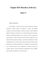
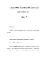
ishes over time because of these changes (Fig. 36–1).
304
CASE FILES: NEUROLOGY
Diagnostic Testing for Myasthenia Gravis
Laboratory studies for ACh receptor antibodies are the most specific and sen-
sitive test for myasthenia gravis. There are three antibodies described against
the ACh receptor: binding, blocking, and modulating. Up to 90% of patients
with generalized myasthenia gravis (affecting more than the ocular muscles)
will have a positive test for one of these antibodies. The antibody test most
commonly used to screen for myasthenia gravis is the binding ACh receptor
antibody. Recently, anti-MuSK antibodies have been associated with myasthe-
nia gravis in individuals who do not have ACh receptor antibodies. The
Tensilon test has historically been described as the classic diagnostic test.
Importantly thyroid function studies should always be performed as concomi-
tant thyroid disease is often seen in myasthenia gravis.
A simple bedside test that can be used in patients with ptosis is the ice test.
Ice is placed over the ptotic eyelid for 2 minutes. If the ptosis improves after
removing the ice a diagnosis of an underlying neuromuscular junction trans-
mission disorder can be made. Cooling improves neuromuscular junction
transmission whereas heat worsens it. This is the reason that many patients
with myasthenia gravis worsen during the summer months.
Electrodiagnostic studies with EMG/NCS can be performed to evaluate
patients with myasthenia gravis. Classically nerve conduction studies are nor-
mal. EMG can be normal or can show myopathic features. Repetitive nerve
stimulation, a part of EMG/NCS, consists of repeatedly stimulating a nerve and
recording the compound muscle action potential obtained. This test is usually
CLINICAL CASES 305
Figure 36–1. End plate from a patient with myasthenia gravis, electron
microscopy. (With permission from Ropper AH, Brown RH. Adams and Victor’s
principles of neurology, 8th ed. New York: McGraw-Hill; 2005: Figs. 36–2,
53–1, Diagrams of (A) normal and (B) myasthenic neuromuscular junctions.
With permission from Kasper DL, Braunwal E, Fauci A, et al. Harrison’s prin-
ciples of internal medicine, 16th ed. New York: McGraw-Hill; 2004: Fig. 366–1.)
Muscle
Nerve
terminal
V
M
Release
site
Axon
Normal MG
A
B
AChR
AChE
AChR
performed at 2 or 3 Hz. The ulnar, spinal accessory and facial nerves most
commonly evaluated. Greater than 10% decrement in the amplitude of the
compound muscle action potential is considered an abnormal response and
suggestive of a neuromuscular junction transmission disorder. A more special-
ized study, single fiber EMG, is the most sensitive test available for myasthe-
nia gravis; however, it is not very specific, nor commonly available.
A CT scan or MRI of the mediastinum should be performed to exclude
thymic enlargement or more importantly a thymoma. Thymectomy should
always be performed in those individuals that have thymoma. It is cur-
rently controversial as to whether or not thymectomy is of any benefit in those
individuals with myasthenia gravis that merely have thymic hyperplasia or a
normal thymic size.
Treatment of Myasthenia Gravis
The mainstay of treatment for myasthenia gravis is immunosuppressive
agents. These include corticosteroids, cyclosporine azathioprine, mycopheno-
late mofetil, intravenous immunoglobulin, and plasmapheresis. Although most
experts believe that corticosteroids are the first line of treatment there is no
general consensus as to how to administer it and at what dose. There is no gen-
eral agreement among experts regarding the timing or use of the other
immunosuppressive treatments. Anti-cholinesterase inhibitors such as pyri-
dostigmine treat only the symptom but not the disease. However, this is rou-
tinely used in patients with myasthenia gravis especially if the only symptoms
are ocular. The typical dose of this is 60 mg orally four times a day.
Comprehension Questions
[36.1] A 60-year-old man in the ICU is noted to have a brain pathology and
ptosis. Which of the following conditions is the most likely cause for
ptosis?
A. Pituitary necrosis
B. Uncal herniation
C. Central herniation
D. Arterio-venous (AV) malformation
[36.2] A critical difference between myogenic processes and disorders of the
neuromuscular junction is:
A. The finding of fatigability with improvement after rest in neuro-
muscular junction transmission disorders
B. Weakness of the ocular muscles only in neuromuscular junction
transmission disorders
C. Low CPK levels in myogenic processes
D. Elevated CPK in neuromuscular junction transmission disorders
E. Myogenic findings on EMG
306
CASE FILES: NEUROLOGY
[36.3] Individuals presenting with ptosis and multiple cranial nerve abnor-
malities should have which study performed first?
A. MRI of the brain with magnetic resonance angiography (MRA)
B. EMG/NCS
C. Serologic studies for CPK
D. ACh receptor antibodies
E. Thyroid function studies
Answers
[36.1] B. Central herniation causes compression of the diencephalon flatten-
ing the mid brain and pons whereas uncal herniation compresses the
third cranial nerve causing ptosis.
[36.2] A. Fatigability of muscles with improvement after rest is a hallmark of
neuromuscular junction transmission disorders.
[36.3] A. The presence of multiple cranial abnormalities including ptosis
speaks for a process in the central nervous system particularly the
brainstem or cavernous sinus.
CLINICAL CASES 307
CLINICAL PEARLS
❖ The etiology of ptosis is best determined by recognizing associated
symptoms that patients present with and discerning clinical find-
ings on examination.
❖ Ptosis associated with central nervous system signs and symptoms
mandates an MRI of the brain.
❖ Fatigability of muscle with improvement after a brief period of rest
is seen only with neuromuscular junction transmission disorders.
❖ Up to 90% of patients with myasthenia gravis will eventually have
ocular symptoms.
❖ Local cooling of the eye can improve function in a ptotic eyelid, sim-
ilar to a Tensilon test, and is a rapid, simple, and inexpensive test
for myasthenia gravis.
REFERENCES
Dorland’s Illustrated Medical Dictionary, 27th ed. Philadelphia, PA: WB Saunders;
1988.
Keesey JC. Clinical evaluation and management of myasthenia gravis. Muscle
Nerve 2004 Apr;29(4):484–505.
Saperstein DS, Barohn RJ. Management of myasthenia gravis. Semin Neurol 2004
Mar;24(1):41–48.