CLINICIAN’S POCKET REFERENCE - PART 7 pot
Bạn đang xem bản rút gọn của tài liệu. Xem và tải ngay bản đầy đủ của tài liệu tại đây (420.45 KB, 77 trang )
20
TABLE 20–9
(Continued)
Determination
Derivation
Normal
O
2
consumption
(Ca
O
2
− Cv
O
2
) × CO x 10
180–280 mL/min
Qs/Qt (shunt fraction)
(Cc
O
2
− Cv
O
2
) × CO x 10
0.05
(Cc
O
2
− Cv
O
2
)
ICP
Measured 0–20 mmHg
CPP
MAP −
ICP
keep >70 mmHg
Abbreviations: RAP = right atrial pressures; CVP = central venous pressure; R
VP = right ventricular pressure; PAS = pulmonar
y artery systolic; PAD = pulmonary
artery diastolic; PCWP = pulmonary capillar
y wedge pressure; CO = cardiac output;
CI = cadiac input; MAP = mean arterial pressu
re; MPAP = mean pul-
monary artery pressure; SVR = systemic
vascular resistance; PVR = pulmonary vascular
resistance; ICP = intracranial pressure; CPP
= cerebral perfusion pres-
sure; BSA = body surface area; DBP = diastolic
blood pressure; SBP = systolic blood pressure;
Fi
O
2
= inhaled O
2
; Hgb = hemoglobin; Sa
O
2
= arterial oxygen,
Sv
O
2
= mixed venous oxygen saturation; Qs =
volume of shunted blood (ie, blood shunted past
nonventilated alveoli, not participating in
gas exchange); Qt =
total cardiac output; C
CO
2
=
O
2
content of alveolar-capillary blood; C
VO
2
= mixed venous
O
2
content of pulmonary artery blood.
438
TABLE 20–10
Guidelines for Adult Critical Care Drug Infusions*
(Final Concentration)
Drug
Dilution
Flow Rate = mL/h
Usual Dose Range
Amrinone
500 mg
(2 mg/mL)
(Inocor)
250 mL
1500 µg/min = 45
LD = 0.75 µg/kg
1000 µg/min = 30
MD = 5–20 µg/kg/min
(150 mL PSS+
750 µg/min = 22.5
100 mL drug)
500 µg/min = 15
PSS only
350
µg/min = 10.5
Diltiazem
(Cardizem)
125 mg
(1 mg/mL)
Bolus = 0.25 mg/kg
125 mL
5 mg/h = 5
over 2 min; may give
10 mg/h = 10
second bolus 0.35
mg/kg 15 min after
initial bolus
(100 mL diluent 15 mg/h = 15
+25 mL drug)
MD = 5–15
mg/h
D
5
W or PSS
Dobutamine
500 mg
(2000 µg/mL)
2.5–20 µg/kg/min
(Dobutrex)
250 mL
1500 µg/min = 45
20
(continued
)
439
20
TABLE 20–10
Continued
(Final Concentration)
Drug
Dilution
Flow Rate = mL/h
Usual Dose Range
Dobutamine
1250 µg/min = 37.5
(continued)
D
5
W or PSS
1000
µg/min = 30
750 µg/min = 22.5
500 µg/min = 15
250 µg/min = 7.5
Dopamine
400 mg
(1600 µg/mL)
0.5–2.0 µg/kg/min (renal)
250 mL
1400 µg/min = 52.5
2.0–10 µg/kg/min (inotropic)
1200 µg/min = 45
10–20 µg/kg/min (vasopressor)
D
5
W or PSS
1000
µg/min = 37.5
800 µg/min = 30
600 µg/min = 22.5
400 µg/min = 15
200 µg/min = 7.5
Epinephrine
3 mg
(12 µg/mL)
Initially 1 µg/min
250 mL
4 µg/min = 20
3 µg/min = 15
Titrate to response
D
5
W or PSS
2 µg/min = 10
1 µg/min = 5
Esmolol
5000 mg
(10 mg/mL)
LD = 500 µ/kg/min over 1 minute
(Brevibloc)
500 mL
5000 µg/min = 30
MD = 50 µ/kg/min, titrate to
4000 µg/min = 24
response.
D
5
W or PSS
3000
µg/min = 18
Increase by 50
µ/kg/min increments
every 5 minutes
(continued
)
440
20
TABLE 20–10
(Continued)
(Final Concentration)
Drug
Dilution
Flow Rate = mL/h
Usual Dose Range
Isoproterenol
2 mg
(8 µg/mL)
Initially: 1–4 µg/min
(Isuprel)
500 mL
10 µg/min = 75
6 µg/min = 45
Titrate up to 20 µg/min
D
5
W or PSS
4 µg/min = 30
2 µg/min = 15
1 µg/min = 7.5
Labetalol
200 mg
(1 mg/mL)
Bolus = 20 mg over 2 min
(Trandate)
200 mL
Additional 20–80 mg may be given
(160 mL diluent
every 10 min until response or
+40 mL drug) 2 mg/min = 120
maximum of 300 mg
or
Initially 2 mg/min
D
5
W or PSS
Titrate to response
Lidocaine
2 g
(8 mg/mL)
LD = 1–1.5 mg/kg over 2 min
(Xylocaine)
250 mL
4 mg/min = 30
MD = 1–4 mg/min
3 mg/min = 22.5
Maximum 4 mg/min
D
5
W or PSS
2 mg/min = 15
1 mg/min = 7.5
Nicardipine
25 mg
(0.1 mg/mL)
Initially: 5 mg/h
(Cardene)
250 mL
5 mg/h = 50
Titrate to BP: increase rate by 2.5 mg/h
7.5 mg/h = 75
every 5–15 min
(continued
)
441
20
TABLE 20–10
(Continued)
(Final Concentration)
Drug
Dilution
Flow Rate = mL/h
Usual Dose Range
Nicardipine
D
5
W or PSS
10 mg/h = 100
Maximum: 15 mg/h
(continued)
12.5 mg/h = 125
MD 3 mg/h
15 mg/h = 150
Nitroglycerin
100 mg
(400 µg/mL)
Initially 5–10 µg/min
(Tridil)
250 mL
80 µg/min = 12
Titrate up by 10–20 µg/min every 5 min
D
5
W or PSS
60
µg/min = 9
based on current dose and patient
(glass bottle)
40 µg/min = 6
condition
20 µg/min = 3
10 µg/min = 1.5
Nitroprusside
100 mg
(400 µg/mL)
Initially: 0.3–0.5 µg/kg/min
(Nipride)
250 mL
300 µg/min = 45
200 µg/min = 30
Titrate to response every few minutes
D
5
W
150 µg/min = 22.5
Maximum: 10 µg/kg/min
100 µg/min = 15
70 µg/min = 10.5
50 µg/min = 7.5
Norepinephrine 4 mg
(16 µg/mL)
Initially: 8–12 µg/min
(Levophed)
250 mL
12 µg/min = 45
8 µg/min = 30
Titrate to response
D
5
W or PSS
6 µg/min = 22.5
4 µg/min = 15
2 µg/min = 7.5
(continued
)
442
20
TABLE 20–10
(Continued)
(Final Concentration)
Drug
Dilution
Flow Rate = mL/h
Usual Dose Range
Phenylephrine
50 mg
(200 µg/mL)
Initially: 10–50 µg/min
(Neo-Synephrine) 250 mL
100 µg/min = 30
80 µg/min = 24
D
5
W or PSS
60
µg/min = 18
Titrate to response
50 µg/min = 15
Procainamide
2 g
(8 mg/mL)
LD = 17 mg/kg over 1 h, or 100 mg
(Procan)
250 mL
4 mg/min = 30
every 5 min up to 1 g
3 mg/min = 22.5
MD = 1–4 mg/min
D
5
W or PSS
2 mg/min = 15
1 mg/min = 7.5
Vasopressin
100 units
(0.4 units/mL)
0.1–0.4 units/min
(Pitressin)
250 mL
0.4 units/min = 60
0.3 units/min = 45
Maximum 0.9 units/min
D
5
W or PSS
0.2 units/min = 30
0.1 units/min = 15
Abbreviation:
LD = loading dose; MD = maintenance dose; BP = blood pressure; PSS = physiologic saline solution; D
5
W = dextrose 5% in water
*These agents must be administered in the appropriately monitored clinical setting.
Source: Reprinted, with permission, from Thomas Jefferson University Phar
macy and Therapeutic Committee, Philadelphia, P
A.
443
This page intentionally left blank.
CARDIOPULMONARY RESUSCITATION
Emergency cardiac care guidelines from the American Heart Association now recommend
that health care providers have the following items readily available: gloves, a barrier device
or bag mask, and an automated defibrillator to handle cardiac emergencies. In cardiopul-
monary resuscitation, remember there are now two sets of ABCDs:
Primary Survey
• Airway: Assess and manage noninvasively.
• Breathing: Use positive pressure ventilations.
• Circulation: Perform chest compressions as needed.
• Defibrillation: Assess for VT/VF and defibrillate using an AED. These are also
called PADs and are becoming widely available in public areas such as airports, sta-
diums, health clubs, and shopping malls.
Secondary Survey: Uses advanced medical techniques
• Airway: Assess and manage with airway device (eg, endotracheal intubation, etc).
• Breathing: Verify tube function and placement, use positive pressure ventilation sys-
tem through tube.
• Circulation: Start IV, attach ECG, use rhythm-based ACLS medications.
• Differential Diagnosis: Search for, find, and treat problems according to AHA algo-
rithms presented in this chapter.
Adult CPR
(Victim’s age ≥8 y)
One Rescuer
1. Determine unresponsiveness (shake and shout). If the patient is unresponsive, call for
help (activate EMS system, eg, call “code,” dial 911). In trauma situation do not move
21
445
21
EMERGENCIES
Cardiopulmonary Resuscitation
Advanced Cardiac Life Support
and Emergency Cardiac Care*
Advanced Cardiac Life Support
Drugs
Electrical Defibrillation
and Cardioversion
Other Common Emergencies
* The section on basis CPR and ACLS are based on guidelines from the American Heart Association and
the International Liaison Committee on Resuscitation [Circulation 2000;102 (Sup 1)] and the Guidelines
2000 for Cardiopulmonary Resuscitation and Emergency Cardiovascular Care by the American Heart
Assocation in Collaboration with the International Liaison Committee on Resuscitation (ILCOR).
Copyright 2002 The McGraw-Hill Companies, Inc. Click Here for Terms of Use
the victim unless in immediate danger. Roll victim on to back as a unit if lying face
down. Protect the neck.
2. Kneel at the level of the victim’s shoulder. Open the airway (head-tilt, chin-lift,), deter-
mine breathlessness (“look [chest movement], listen [for air escaping], feel [for air
movement]”) for no more than 10 s. In the unresponsive victim with spontaneous respi-
ration, place the victim in the recovery position. Jaw thrust maneuver recommended as
alternative for health care providers especially if neck injury is suspected. If the victim
is breathing, place in the RECOVERY POSITION (see page 449).
3. If not breathing, give patient two slow ventilations (2 s/inspiration) while maintaining
airway. Use pocket mask or bag mask. Volume should be between 0.8–1.2 L. A barrier
device (face shield or mask with one-way valve) is recommended if mouth-to-mouth or
mouth-to-nose contact is necessary. Ventilate 10–12 breaths/min. If unable to ventilate,
reposition head and try again. If unsuccessful, perform the FOREIGN BODY OB-
STRUCTION AIRWAY SEQUENCE (see page 448).
4. Check for circulation (breathing, coughing, movement). Palpate the carotid artery no
more than 10 s to determine lack of a pulse. If pulse is present, perform rescue breath-
ing: 1 ventilation every 5 s (10–12 ventilation/min).
5. If no pulse, use four cycles of 15 compressions and two ventilations (compression rate
100/min, two ventilations 1.5–2 s each). Depth of compression 1.5–2 in. or slightly
greater to generate carotid pulse. Apply compressions to lower half of sternum using
the heels of both hands placed on top of each other.
6. After the four cycles (approximately 1 min of CPR), pause and check for return pulse
and spontaneous respirations.
7. If no pulse or respiration, resume cycles with two ventilations, then compressions, as
noted earlier.
8. Incorporate appropriate ACLS management guidelines.
Two-Rescuer Adult CPR
For laypersons
1. Second rescuer identifies him or herself. Verify that EMS has been notified. If so, sec-
ond rescuer gets into position opposite first rescuer. If EMS not notified, the second
rescuer does so before assisting first rescuer.
2. First rescuer continues CPR.
3. If and when first rescuer tires, second rescuer takes over one-person CPR as described
in the preceding section.
For health care professionals
1. Sequence to continue from one-rescuer CPR as mentioned in previous section. Second
rescuer identifies him or herself and gets into position for compressions.
2. First rescuer completes compression and ventilation cycle (15 compression and two
ventilations).
3. First rescuer then checks for spontaneous pulse and breathing, states: “No pulse con-
tinue CPR,” then ventilate once (1.5–2 s).
4. Second rescuer resumes compressions at same rate of 80–100/min.(“1 & 2 & 3 & 4 & 5
& pause,” ventilate) Ratio of five compressions to one breath. If airway is protected, do
not pause for ventilations.
5. When ready to switch, rescuer doing compressions says “switch & 2 & 3 & 4 & 5 &.”
6. Both rescuers change position simultaneously immediately after ventilation.
7. Rescuer who will perform ventilations opens airway and performs a 5-s pulse check.
8. If no pulse, give ventilation. Rescuer states “No pulse continue CPR.”
446 Clinician’s Pocket Reference, 9th Edition
21
9. In patient with unprotected airway, cricoid pressure may be applied (Sellick’s maneu-
ver) by a third rescuer (if health care professional) to help limit gastric distention.
Child CPR
(Victim’s age 1–8 y)
1. Determine unresponsiveness, and shout for help. Activate EMS system (call code or
911).
2. Open airway (head-tilt, chin-lift; jaw thrust if neck trauma is suspected), determine
breathlessness (follow “look, listen, feel” rubric as for adult). If victim is breathing,
place in RECOVERY POSITION (see page 449).
3. If victim not breathing, give two ventilations (1–1.5 s). If unable to ventilate, perform
the FOREIGN BODY OBSTRUCTED AIRWAY SEQUENCE (see page 448).
4. Check for circulation (breathing, coughing, movement). Palpate the carotid artery for
no more than 10 s to determine presence of a pulse. If pulse is present, perform rescue
breathing using pocket mask or bag-mask device (20 breaths/min).
5. If no pulse, or if pulse is <60 bpm and perfusion is poor, begin cardiac compressions at
five compressions to one ventilation at rate of 100/min. Depth of compressions less
than for an adult (1–1.5 in. or one third to one half the depth of chest).Use the heel of
one hand at the lower half of the sternum. Pause compressions for ventilations until pa-
tient is intubated.
6. Check for return of pulse and spontaneous breathing after 20 cycles (approximately
1 min).
7. Resume cycles with one ventilation (1–1.5 s each), then resume compressions.
Infant CPR
(Victim’s age, ≤1 y)
1. Determine unresponsiveness, and shout for help. Activate EMS system (call code or 911).
2. Open airway (head-tilt, chin-lift). Do not hyperextend head; however, create adequate
head-tilt to accomplish chest rise with breath. If neck trauma suspected, use jaw thrust.
If victim is breathing, place in the RECOVERY POSITION (see page 449).
3. If patient is not breathing, give two ventilations (1–1.5 s) using pocket mask or bag-
mask device. If unable to ventilate, perform the FOREIGN BODY OBSTRUCTED
AIRWAY SEQUENCE using back blows and chest thrusts as noted on page 448.
4. Check for circulation (breathing, coughing, movement). Palpate the femoral or brachial
artery for no more than 10 s to determine presence of a pulse. If pulse is present, con-
tinue rescue breathing (20 breaths/min).
5. If no pulse or if pulse is <60 bpm and perfusion is poor, begin cardiac compressions.
Draw an imaginary line between the nipples and identify where this line crosses the
sternum (intermammary line). The site of compression is one finger breadth below this
intersection. Use a compression depth of ¹₂ –1 in., using the middle and ring fingers.
Use five compressions to one ventilation (rate of compression is 100/min or 120 min
for newborns).
6. Use the mnemonic: (“1 & 2 & 3 & 4 & 5 & pause, head-tilt, chin-lift, ventilate−
continue compressions”). When patient is intubated, no need to pause.
7. Check for return of pulse and spontaneous breathing after 20 cycles (1 min).
Neonatal CPR
1. The newborn should be dried, placed head down, gently suctioned and stimulated.
2. Supplemental oxygen is useful. If baby is not breathing, ventilate 40–60 breaths/min
with gentle puff of air or with bag mask.
21 Emergencies
447
21
3. Check apical pulse. If absent or if <60 bpm and perfusion is poor, compress at a rate of
120/min. Wrap your hands around infant’s chest and compress ¹₂ –³₄ in. with thumbs
side by side at the midsternum.
4. The compression/ventilation ratio is 3:1 for intubated newborn with two rescuers. Dis-
continue compressions when rate reaches 80 bpm or greater.
Foreign Body Obstructed Airway Sequence
Adult (≥8) and Child (1–8 y)
A. Conscious victim can cough, speak, breath. Do not interfere and reassure patient.
Stand by and allow patient to clear partial obstruction.
B. Conscious victim cannot cough, speak, breath.
1. Ask “Are you choking” or “Can you speak?” Observe for “universal distress sig-
nal” for choking (hands clutched at neck).
2. Give abdominal thrusts/Heimlich maneuver. Stand behind victim. Using arms
wrapped around victim, place thumb side of fist above umbilicus but below
xiphoid. Give up to five subdiaphragmatic thrusts (Heimlich maneuver).
3. Reassess victim’s status, repeat Heimlich maneuvers as needed. If not improved
by 1 min, activate EMS.
C. Victim becomes unconscious.
1. Place in supine (face up) position. Activate EMS or if second rescuer becomes
available have that person activate EMS.
2. Open airway with tongue-jaw lift; finger sweep to clear airway, open airway
(head-tilt, chin-lift).
3. Give five abdominal thrusts/Heimlich maneuver astride victim.
D. Victim found unconscious: Cause unknown
1. Determine unresponsiveness, call for help (activate EMS).
2. Open airway (head-tilt, chin-lift), determine breathlessness (look, listen, feel).
3. Attempt to ventilate. If unsuccessful, reposition head and reattempt.
4. If unsuccessful:
a. Perform up to five Heimlich maneuvers astride victim.
b. Open mouth (tongue-jaw lift); finger sweep; open airway (head-tilt, chin-lift)
5. Attempt to ventilate, if unsuccessful, repeat sequence until ventilations are effec-
tive.
Infant
(Victim’s age, <1 y)
Victim conscious
1. Verify airway obstruction (ineffective cough, no strong cry).
2. Hold child with head lower than body, give five back blows or five gentle abdominal
thrusts. Repeat until victim becomes responsive.
Victim becomes unconscious
1. If second rescuer is available, have that person activate EMS.
2. Open airway with tongue-jaw lift, remove foreign body if visualized. Attempt to venti-
late.
3. If still obstructed, reposition head and attempt to ventilate. Give five back blows and
five abdominal thrusts. Repeat step 2 until ventilation is effective.
4. If obstruction still not relieved after 1 min, activate EMS system.
448 Clinician’s Pocket Reference, 9th Edition
21
Recovery Position
Place an unconscious person who is still breathing and who has not suffered a traumatic
neck injury in this position.
1. Kneel alongside the victim and straighten the legs.
2. Place victim’s arm that is closest to you in the “waving goodbye” position and place the
other arm across the victim’s chest.
3. Grasp the far side leg above the knee and pull the thigh up toward the body. With the
other hand, grasp the shoulder on the same side as the thigh.
4. Gently roll the patient toward you. Adjust the leg you are holding until both the thigh
and knee are at right angles to the body. Tilt the patient’s head back and use the pa-
tient’s uppermost hand to support the head and maintain a head-tilt position.
5. Continue to monitor for breathing, and call for EMS.
6. If patient stops breathing, roll on back and follow basic CPR guidelines.
ADVANCED CARDIAC LIFE SUPPORT AND EMERGENCY
CARDIAC CARE
ACLS includes the use of advanced airway management (See Endotracheal Intubation,
Chapter 13, page 268), defibrillation, and drugs along with basic CPR. Most cardiac arrests
are due to VF and are unwitnessed outside the hospital setting. ACLS protocols incorporat-
ing all these emergency cardiac care techniques are reviewed in the following algorithms for
adults:
• Universal/International ACLS algorithm (Figure 21–1)
• Comprehensive emergency cardiac care algorithm (Figure 21–2)
• Ventricular fibrillation and pulseless VT algorithm (Figure 21–3)
• Pulseless electrical activity algorithm (Figure 21–4)
• Asystole: The silent heart algorithm (Figure 21–5)
• Bradycardia algorithm (Figure 21–6)
• Tachycardia overview algorithm (Figure 21–7)
• Narrow complex SVT algorithm (Figure 21–8)
• Stable VT algorithm (Figure 21–9)
• Acute coronary syndromes algorithm (Figure 21–10)
• Acute pulmonary edema, hypotension, and shock (Figure 21–11)
Advanced Cardiac Life Support Drugs
The most commonly used agents are listed on the inside covers for quick reference.
ACE Inhibitors
INDICATIONS: These agents improve the outcome in post-MI patients.
• Enalapril (Enalaprilat IV)
SUPPLIED: Tabs 2.5, 5, 10, 20 mg; IV 1.25 mg/mL (1- and 2-mL vial)
DOSAGE: .2.5 mg PO single dose, increase to 20 mg PO bid; 1.25 mg IV over 5 min, then
1.25–5.0 mg IV q6h
• Captopril
SUPPLIED: Caps 12.5, 25, 50, 100 mg
DOSAGE: 6.25 mg PO, increase to 25 mg tid and the 50 mg PO tid as tolerated
21 Emergencies 449
21
(continued on page 461)
450 Clinician’s Pocket Reference, 9th Edition
21
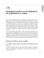
Adult Cardiac Arrest
BLS algorithm
if appropriate
Precordial thump if appropriate
Attach defibrillator/monitor
Assess rhythm
Check pulse +/
VF/VT
Attempt
defibrillation 3
as necessary
CPR
1 minute
Non-VF/VT
CPR
up to 3 minutes
Consider causes that are potentially reversible
• Hypovolemia
• Hypoxia
• Hydrogen ion — acidosis
• Hyper-/hypokalemia, other metabolic
• Hypothermia
• “Tablets” (drug OD, accidents)
• Tamponade, cardiac
• Tension pneumothorax
• Thrombosis, coronary (ACS)
• Thrombosis, pulmonary (embolism)
During CPR
• Check electrode/paddle positions and contact
• Attempt to place, confirm, secure airway
• Attempt and verify IV access
• Patients with VF/VT refractory to initial shocks:
— Epinephrine 1 mg IV, every 3 to 5 minutes
or
— Vasopressin 40 U IV, single dose, 1 time only
• Patients with non-VF/VT rhythms:
— Epinephrine 1 mg IV, every 3 to 5 minutes
• Consider: buffers, antiarrhythmics, pacing
• Search for and correct reversible causes
1
2
3
4
5,6
7
FIGURE 21–1 Universal/international advanced cardiac life support algorithm.
Abbreviations: VF = ventricular fibrillation; VT = ventricular tachycardia; BLS =
basic life support. (Reproduced, with permission, from: Circulation 2000;102
supplement 1, part 6.)
21 Emergencies 451
21
1
1
23
4,5
• Person collapses
• Possible cardiac arrest
• Assess responsiveness
Unresponsive
Begin Primary ABCD Survey
(Begin BLS Algorithm)
• Activate emergency response
system
• Call for defibrillator
• A Assess breathing (open
airway, look, listen, and feel
)
Not Breathing
• B Give 2 slow breaths
• C Assess pulse, if no pulse
• C Start chest compressions
• D Attach monitor/defibrillator
when available
No Pulse
• CPR continues
• Assess rhythm
VF/VT
Non-VF/VT
Attempt defibrillation
(up to 3 shocks if VF persists)
Non-VF/VT
(asystole or PEA)
CPR up to
3 minutes
CPR for
1 minute
Secondary ABCD Survey
• Airway: attempt to place airway device
• Breathing: confirm and secure airway device,
ventilation, oxygenation
• Circulation: gain intravenous access; give adrenergic
agent; consider antiarrhythmics, buffer agents,
pacing
Non-VF/VT patients:
— Epinephrine 1 mg IV, repeat every 3 to 5 minutes
VF/VT patients:
— Vasopressin 40 U IV, single dose, 1 time only
or
— Epinephrine 1 mg IV, repeat every 3 to 5 minutes
(if no response after single dose of vasopressin,
may resume epinephrine 1 mg IV push; repeat
every 3 to 5 minutes)
• Differential Diagnosis: search for and treat reversible
causes
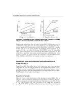
FIGURE 21–2 Comprehensive emergency cardiac care (ECC) algorithm. Abbrevi-
ations: VF = ventricular fibrillation; VT = ventricular tachycardia; BLS = basic life sup-
port; PEA = pulseless electrical activity. (Reproduced, with permission, from:
Circulation 2000;102 supplement 1, part 6.)
452 Clinician’s Pocket Reference, 9th Edition
21
Primary ABCD Survey
Focus: basic CPR and defibrillation
• Check responsiveness
• Activate emergency response system
• Call for defibrillator
A Airway: open the airway
B Breathing: provide positive-pressure ventilations
C Circulation: give chest compressions
D Defibrillation: assess for and shock VF/pulseless VT, up to 3 times
(200 J, 200 to 300 J, 360 J, or equivalent biphasic) if necessary
Rhythm after first 3 shocks?
Persistent or recurrent VF/VT
Secondary ABCD Survey
Focus: more advanced assessments and treatments
A Airway: place airway device as soon as possible
B Breathing: confirm airway device placement by exam plus confirmation device
B Breathing: secure airway device; purpose-made tube holders preferred
B Breathing: confirm effective oxygenation and ventilation
C Circulation: establish IV access
C Circulation: identify rhythm monitor
C Circulation: administer drug appropriate for rhythm and condition
D Differential Diagnosis: search for and treat identified reversible causes
• Epinephrine 1 mg IV push, repeat every 3 to 5 minutes
or
• Vasopressin 40 U IV, single dose, 1 time only
Resume attempts to defibrillate
1 360 J (or equivalent biphasic) within 30 to 60 seconds
Consider antiarrhythmics:
amiodarone (llb), lidocaine (Indeterminate),
magnesium (llb if hypomagnesemic state),
procainamide (llb for intermittent/recurrent VF/VT).
Consider buffers.
Resume attempts to defibrillate
1
2
3
4
5
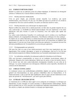
FIGURE 21–3 Ventricular fibrillation and pulseless ventricular tachycardia algo-
rithm. Abbreviations: VF = ventricular fibrillation; VT = ventricular tachycardia.
(Reproduced, with permission, from: Circulation 2000;102 supplement 1, part 6.)
21 Emergencies 453
21
Primary ABCD Survey
Focus: basic CPR and defibrillation
• Check responsiveness
• Activate emergency response system
• Call for defibrillator
A Airway: open the airway
B Breathing: provide positive-pressure ventilations
C Circulation: give chest compressions
D Defibrillation: assess for and shock VF/pulseless VT
Secondary ABCD Survey
Focus: more advanced assessments and treatments
A Airway: place airway device as soon as possible
B Breathing: confirm airway device placement by exam plus confirmation device
B Breathing: secure airway device; purpose-made tube holders preferred
B Breathing: confirm effective oxygenation and ventilation
C Circulation: establish IV access
C Circulation: identify rhythm monitor
C Circulation: administer drugs appropriate for rhythm and condition
C Circulation: assess for occult blood flow (“pseudo-EMT”)
D Differential Diagnosis: search for and treat identified reversible causes
Pulseless Electrical Activity
(PEA = rhythm on monitor, without detectable pulse)
Review for most frequent causes
• Hypovolemia
• Hypoxia
• Hydrogen ion — acidosis
• Hyper-/hypokalemia
• Hypothermia
• “Tablets” (drug OD, accidents)
• Tamponade, cardiac
• Tension pneumothorax
• Thrombosis, coronary (ACS)
• Thrombosis, pulmonary (embolism)
Epinephrine 1 mg IV push,
repeat every 3 to 5 minutes
Atropine 1 mg IV (if PEA rate is slow),
repeat every 3 to 5 minutes as needed, to a total
dose of 0.04 mg/kg
1
2
3
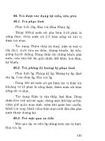
FIGURE 21–4 Pulseless electrical activity algorithm. Abbreviations: VF = ventricu-
lar fibrillation; VT = ventricular tachycardia; EMT = emergency medical treatment;
ACS = acute coronary syndrome; PEA = pulseless electrical activity. (Reproduced,
with permission, from: Circulation 2000;102 supplement 1, part 6.)
454 Clinician’s Pocket Reference, 9th Edition
21
Primary ABCD Survey
Focus: basic CPR and defibrillation
• Check responsiveness
• Activate emergency response system
• Call for defibrillator
A Airway: open the airway
B Breathing: provide positive-pressure ventilations
C Circulation: give chest compressions
C Confirm true asystole
D Defibrillation: assess for VF/pulseless VT; shock if indicated
Rapid scene survey: any evidence personnel should not attempt resuscitation?
Asystole
Secondary ABCD Survey
Focus: more advanced assessments and treatments
A Airway: place airway device as soon as possible
B Breathing: confirm airway device placement by exam plus confirmation device
B Breathing: secure airway device; purpose-made tube holders preferred
B Breathing: confirm effective oxygenation and ventilation
C Circulation: confirm true asystole
C Circulation: establish IV access
C Circulation: identify rhythm monitor
C Circulation: give medications appropriate for rhythm and condition
D Differential Diagnosis: search for and treat identified reversible causes
Epinephrine 1 mg IV push,
repeat every 3 to 5 minutes
Atropine 1 mg IV,
repeat every 3 to 5 minutes
up to a total of 0.04 mg/kg
Transcutaneous pacing
If considered, perform immediately
Asystole persists
Withhold or cease resuscitation efforts?
• Consider quality of resuscitation?
• Atypical clinical features present?
• Support for cease-efforts protocols in place?
2,3
4
5
6
7,8,9
1
FIGURE 21–5 Asystole: the silent heart algorithm. Abbreviations: VF = ventricular
fibrillation; VT = ventricular tachycardia. (Reproduced with permission from Circula-
tion 2000;102 supplement 1, part 6)
21 Emergencies 455
21
Bradycardia
• Slow (absolute bradycardia = rate <60 bpm)
or
• Relatively slow (rate less than expected
relative to underlying condition or cause)
Primary ABCD Survey
• Assess ABCs
• Secure airway noninvasively
• Ensure monitor/defibrillator is available
Secondary ABCD Survey
• Assess secondary ABCs (invasive airway
management needed?)
• Oxygen–IV access–monitor–fluids
• Vital signs, pulse oximeter, monitor BP
• Obtain and review 12-lead ECG
• Obtain and review portable chest x-ray
• Problem-focused history
• Problem-focused physical examination
• Consider causes (differential diagnoses)
Serious signs or symptoms?
Due to the bradycardia?
1,2
No Yes
No Yes
Type II second-degree AV block
or
Third-degree AV block?
Observe
6
3,4,5
Intervention sequence
• Atropine 0.5 to 1.0 mg
• Transcutaneous pacing if available
• Dopamine 5 to 20 g/kg per minute
• Epinephrine 2 to 10 g/min
7
• Prepare for transvenous pacer
• If symptoms develop, use
transcutaneous pacemaker until
transvenous pacer placed
FIGURE 21–6 Bradycardia algorithm. Abbreviations: BP = blood pressure; ECG =
electrocardiogram; AV = atrioventricular. (Reproduced, with permission, from: Circu-
lation 2000;102 supplement 1, part 6.)
456 Clinician’s Pocket Reference, 9th Edition
21
DC cardioversion
or
Amiodarone
Evaluate patient
• Is patient stable or unstable?
• Art there serious signs or symptoms?
• Are signs and symptoms due to tachycardia?
Stable Unstable
Stable patient: no serious signs or symptoms
• Initial assessment identifies 1 of 4 types of
tachycardias
Unstable patient: serious signs or symptoms
• Establish rapid heart rate as cause of signs and
symptoms
• Rate related signs and symptoms occur at many
rates, seldom <150 bpm
• Prepare for immediate cardioversion
(see page 468)
1. Atrial fibrillation
Atrial flutter
2. Narrow-complex
tachycardias
3. Stable wide-complex
tachycardia: unknown type
4. Stable monomorphic VT
and/or polymorphic VT
Evaluation focus, 4 clinical
features:
1. Patient clinically unstable?
2. Cardiac function impaired?
3. WPW present?
4. Duration <48 or >48 hours?
Attempt to establish a
specific diagnosis
• 12-lead ECG
• Clinical information
• Vagal maneuvers
• Adenosine
Attempt to establish a
specific diagnosis
• 12-lead ECG
• Esophageal lead
• Clinical information
Treatment focus: clinical
evaluation
1. Treat unstable patients
urgently
2. Control the rate
3. Convert the rhythm
4. Provide anticoagulation
Diagnostic efforts yield
• Ectopic atrial tachycardia
• Multifocal atrial tachycardia
• Paroxysmal supraventricular
tachycardia (PSVT)
Treatment of
atrial
fibrillation/
atrial flutter
(See following
table)
Treatment of SVT
(See narrow-complex
tachycardia algorithm)
Confirmed
SVT
Wide-complex
tachycardia of
unknown type
Confirmed
stable VT
Treatment of stable
monomorphic
and
polymorphic VT
(See stable VT:
monomorphic
and polymorphic
algorithm)
Preserved
cardiac function
Ejection fraction
<40% Clinical CHF
DC cardioversion
or
Procainamide
or
Amiodarone
FIGURE 21–7 Tachycardia overview algorithm. Abbreviations: VF = ventricular
fibrillation; ECG = electrocardiogram; PSVT = paroxysmal supraventricular tachycar-
dia; SVT = supraventricular tachycardia. (Reproduced, with permission, from: Circu-
lation 2000;102 supplement 1, part 6.)
21 Emergencies 457
21
Narrow-Complex Supraventricular
Tachycardia, Stable
Attempt therapeutic diagnostic maneuver
• Vagal stimulation
• Adenosine
Junctional tachycardia
Preserved
• No DC cardioversion!
• Amiodarone
• b-Blocker
• Ca
2+
channel blocker
EF <40%, CHF
• No DC cardioversion!
• Amiodarone
Paroxysmal supraventricular
tachycardia
Preserved
EF <40%, CHF
Priority order:
• Ca
2+
channel blocker
• b-Blocker
• Digoxin
• DC cardioversion
• Consider procainamide,
amiodarone, sotalol
Priority order:
• No DC cardioversion!
• Digoxin
• Amiodarone
• Diltiazem
Ectopic or multifocal
atrial tachycardia
• No DC cardioversion!
• Ca
2+
channel blocker
• b-Blocker
• Amiodarone
• No DC cardioversion!
• Amiodarone
• Diltiazem
Preserved
EF <40%, CHF
FIGURE 21–8 Narrow complex SVT algorithm. Abbreviations: EF = ejection frac-
tion; CHF = congestive heart failure. (Reproduced, with permission, from: Circulation
2000;102 supplement 1, part 6.)
458 Clinician’s Pocket Reference, 9th Edition
21
Stable Ventricular Tachycardia
Monomorphic or Polymorphic?
Monomorphic VT
• Is cardiac function impaired?
Note!
May go directly to
cardioversion
Polymorphic VT
• Is QT baseline interval prolonged?
Normal function Poor ejection fraction
Normal baseline
QT interval
Prolonged baseline
QT interval
(suggests torsades)
Medications: any one
• Procainamide
• Sotalol
Others acceptable
• Amiodarone
• Lidocaine
Normal baseline QT interval
• Treat ischemia
• Correct electrolytes
Medications: any one
• b-Blockers or
• Lidocaine or
• Amiodarone or
• Procainamide or
• Sotalol
Long baseline QT interval
• Correct abnormal electrolytes
Medications: any one
• Magnesium
• Overdrive pacing
• Isoproterenol
• Phenytoin
• Lidocaine
Cardiac function
Impaired
2
Amiodarone
• 150 mg IV bolus over 10 minutes
or
Lidocaine
• 0.5 to 0.75 mg/kg IV push
Then use
• Synchronized cardioversion
1
5
3,4
FIGURE 21–9 Stable supraventricular tachycardia algorithm. Abbreviations: VT =
ventricular tachycardia. (Reproduced, with permission, from: Circulation 2000;102
supplement 1, part 6.)
21 Emergencies 459
21
• Aspirin
• Other therapy as appropriate
• Patients with positive serum
markers, ECG changes, or
functional study: manage as
high risk
Assess the initial ECG
The 12-lead ECG is central to triage of ACS in the Emergency Department.
Classify patients as being in 1 of 3 syndromes within 10 minutes of arrival.
ST-segment elevation
or new LBBB
ST-segment depression/
dynamic T-wave inversion:
strongly suspicious for ischemia
Nondiagnostic
or normal ECG
• ST elevation ≥1 mm in 2 or
more contiguous leads
• New or presumably new
LBBB (BBB obscuring
ST-segment analysis)
• ST depression >1 mm
• Marked symmetrical T-wave
inversion in multiple precordial
leads
• Dynamic ST-T changes
with pain
• ST depression 0.5 to 1.0 mm
• T-wave inversion or flattening in
leads with dominant R waves
• Normal ECG
• >90% of patients with ischemic-
type chest pain and ST-segment
elevation will develop new
Q waves or positive serum
markers for AMI.
• Patients with hyperacute
T waves benefit when AMI
diagnosis is certain. Repeat
ECG may be helpful.
• Patients with ST depression in
early precordial leads who have
posterior MI benefit when AMI
diagnosis is certain.
High-risk subgroup with
increased mortality:
• Persistent symptoms,
recurrent ischemia
• Diffuse or widespread ECG
abnormalities
• Depressed LV function
• Congestive heart failure
• Serum marker release:
positive troponin or CK-MB+
Heterogeneous group: rapid
assessment needed by
• Serial ECGs
• ST-segment monitoring
• Serum cardiac markers
Future risk assessment
helpful
• Perfusion radionuclide imaging
• Stress echocardiography
• Reperfusion therapy
• Aspirin
• Heparin
(if using fibrin-specific lytics)
• -Blockers
• Nitrates as indicated
• Antithrombin therapy with
heparin
• Antiplatelet therapy with
aspirin
• Glycoprotein llb/llla inhibitors
• -Blockers
• Nitrates
12 3
FIGURE 21–10 Acute coronary syndromes algorithm. Abbreviations: ECG = elec-
trocardiogram; LBBB = left bundle branch block; BBB = bundle branch block; AMI =
acute myocardial infarction; MI = myocardial infarction; LV = left ventricle; CK-MB+
= positive for myocardial muscle creatine kinase isoenzyme. (Reproduced, with per-
mission, from: Circulation 2000;102 supplement 1, part 6.)
460 Clinician’s Pocket Reference, 9th Edition
21
Clinical signs: Shock, hypoperfusion,
congestive heart failure, acute pulmonary edema
Most likely problem?
Acute pulmonary edema Volume problem Pump problem Rate problem
1st — Acute pulmonary edema
• Furosemide IV 0.5 to 1.0 mg/kg
• Morphine IV 2 to 4 mg
• Nitroglycerin SL
• Oxygen/intubation as needed
Administer
• Fluids
• Blood transfusions
• Cause-specific interventions
Consider vasopressors
Bradycardia
See algorithm
Tachycardia
See algorithm
Blood
pressure?
Systolic BP
BP defines 2nd
line of action
(See below)
Systolic BP
<70 mm Hg
Signs/symptoms
of shock
Systolic BP
70 to 100 mm Hg
Signs/symptoms
of shock
Systolic BP
70 to 100 mm Hg
No signs/symptoms
of shock
Systolic BP
>100 mm Hg
•
Norepinephrine
0.5 to 30 µg/min IV
•
Dopamine
5 to 15 µg/kg per
minute IV
•
Dobutamine
2 to 20 µg/kg per
minute IV
•
Nitroglycerin
10 to 20 µg/min IV
Consider
•
Nitroprusside 0.1 to
5.0 µg/kg per minute IV
2nd — Acute pulmonary edema
• Nitroglycerin/nitroprusside if BP >100 mm Hg
• Dopamine if BP > 70 to 100 mm Hg, signs/symptoms of shock
• Dobutamine if BP >100 mm Hg, no signs/symptoms of shock
Further diagnostic/therapeutic
considerations
• Pulmonary artery catheter
• Intra-aortic balloon pump
• Angiography for AMI/ischemia
• Additional diagnostic studies
FIGURE 21–11 Acute pulmonary edema, hypotension and shock. Abbreviations:
BP = blood pressure; AMI = acute myocardial infarction. (Reproduced, with permis-
sion, from: Circulation 2000;102 supplement 1, part 7)
• Lisinopril
SUPPLIED: Caps 2.5, 5, 10, 20, 30, 40 mg
DOSAGE: 5 mg PO within 24 h of symptoms, 5 mg after 24 h, then 10 mg over 48 h, then
10 mg PO daily for 6 wk
• Ramipril
SUPPLIED: Caps 1.25, 2.5, 5, 10 mg
DOSAGE: 2.5 mg PO single dose, increase to 5 mg PO bid
Adenosine (Adenocard)
INDICATIONS: First drug for narrow-complex PSVT (not for AF or VT)
SUPPLIED: 2 mg/mL in 2-mL vial
DOSAGE: Adults. Put patient in reverse Trendelenburg position before administering dose; initial
6 mg over 1–3 s followed by NS bolus of 20 mL, then elevate extremity. Repeat 12 mg in 1–2 min
PRN. A third dose of 12 mg in 1–2 min PRN. Peds. 0.1 mg/kg rapid IV push with continuous ECG
monitoring. Follow with >5 mL NS flush. May double (0.2 mg/kg for second dose). Max: first
dose: 6 mg; second dose:12 mg; single dose:12 mg
Amiodarone
INDICATIONS: Atrial and ventricular tachyarrhythmias and for rate control of rapid atrial arrhyth-
mias in patients with impaired LV function when digoxin is ineffective
SUPPLIED: 50 mg/mL in 3-mL vial
DOSAGE: Adults. Max cumulative dose: 2.2 g IV/24 h. Cardiac arrest. 300 mg IV push. Consider
repeating 150 mg IV push in 3–5 min. Wide-complex tachycardia (stable): Rapid inf: 150 mg IV
over 10 min (15 mg/min), every 15 min PRN. Slow inf: 360 mg IV over 6 h (1 mg/min). Mainte-
nance inf: 540 mg IV over 18 h (0.5 mg/min). Peds. Refractory pulseless VT, VF: 5 mg/kg rapid IV
bolus. Perfusing supraventricular and ventricular arrhythmias: Loading dose: 5 mg/kg IV/IO over
20–60 min (repeat, max 15 mg/kg/day).
Amrinone
INDICATIONS: CHF refractory to conventional agents
SUPPLIED: 0.5 mg/mL in 20-mL vial
DOSAGE: Adults. 0.75 mg/kg, over 10–15 min (Do NOT mix with dextrose.). Then 5–15 µg/kg/min
titrated to effect. Hemodynamic monitoring preferred. Peds. Loading dose: 0.75–1.0 mg/kg IV over
5 min; may repeat twice (Max: 3 mg/kg). Cont inf: 5–10 µg/kg/min IV
Aspirin
INDICATIONS: In the acute setting, administer to all patients with acute coronary syndrome (ACS)
SUPPLIED: Tabs 160, 325 mg
DOSAGE: 160–325 mg PO (chewing preferred ASAP onset of ACS)
Atropine Sulfate
INDICATIONS: First drug for symptomatic bradycardia (but not Mobitz II). Second drug (after epi-
nephrine or vasopressin) for asystole or bradycardic PEA
SUPPLIED: 0.1 mg/mL in 10-mL syringe (total = 1 mg).
DOSAGE: Adults. Asystole or PEA: 1 mg IV push. Repeat every 3–5 min (if asystole persists) to
0.03–0.04 mg/kg max. Bradycardia: 0.5–1.0 mg IV every 3–5 min as needed; max 0.03–
0.04 mg/kg. Endotracheal administration: 2–3 mg in 10 mL NS. Peds. IV administration: 0.02
mg/kg. Min single dose: 0.1 mg, max: 0.5 mg. Max adolescent single dose: 1.0 mg. May double for
second IV dose. Max child total dose: 1.0 mg. Max adolescent total dose: 2.0 mg. Endotracheal ad-
ministration: 0.02 mg/kg (larger doses than IV may be required)
Beta Blockers
INDICATIONS: All patients with suspected MI; may reduce chance of VF and reduce damage.
Second line agents after adenosine, diltiazem, or digoxin to slow ventricular response in supraven-
21 Emergencies 461
21
tricular tachyarrhythmias. Antihypertensive for hemorrhagic and ischemic stroke. Do NOT admin-
ister along with calcium channel blockers due to risk of hypotension.
• Metoprolol (Lopressor)
SUPPLIED: 1 mg/mL in 5-mL vial
DOSAGE: Adults. 5 mg slow IV q 5 min, total 15 mg
• Atenolol (Tenormin)
SUPPLIED: 0.5 mg/mL in 10-mL amp
DOSAGE: Adults. 5 mg slow IV (over 5 min). In 10 min, second dose 5 mg slow IV. In 10
min, if tolerated, start 50 mg PO, then 50 mg PO bid
• Propanolol (Inderal)
SUPPLIED: 1.0 mg/mL in 1 amp, 4 mg/mL in 5-mL
DOSAGE: Adults. 0.1 mg/kg slow IV push, divided 3 equal doses 2–3 min intervals, max
1 mg/min. Repeat after 2 min, PRN
• Esmolol (Brevibloc)
SUPPLIED: 10 mg/mL in 10-mL amp
DOSAGE: Adults. 0.5 mg/kg over 1 min, then 0.05 mg/kg/min
• Labetalol
SUPPLIED
: 5 mg/mL (Amps 20, 40, 60 mL)
DOSAGE
: 10 mg IV push over 1–2 min. Repeat or double dose every 10 min (max:
150 mg); or initial bolus, then 2–8 µg/min
Calcium Chloride
INDICATIONS: Known/suspected hyperkalemia, hypocalcemia (eg, multiple transfusions), antidote
for calcium channel blocker overdose, prophylactically before IV calcium channel blockers (pre-
vent hypotension)
SUPPLIED: 100 mg/mL in 10-mL vial (total = 1 g; 10% solution)
DOSAGE: Adults. 8–16 mg/kg (usually 5–10 mL) IV slow push for hyperkalemia and calcium
channel blocker overdose. 2–4 mg/kg (usually 2 mL) IV before IV calcium blockers. Peds.
20 mg/kg (0.2–0.25 mL/kg) slow push. Repeat PRN
Calcium Gluconate
SUPPLIED
: 10% = 100 mg/10 mL = 9 mg/mL Ca
DOSAGE
: Peds. 60–100 mg/kg (0.6–1.0 mL/kg) IV slow push. Repeat for documented conditions
Digibind
Digoxin-specific antibody therapy
INDICATIONS: Digoxin toxicity with uncontrolled life-threatening arrhythmias, shock, CHF; hyper-
kalemia >5 mEq/L with serum dig levels above 10–15 ng/mL
SUPPLIED: 40-mg vial (each vial binds about 0.6 mg digoxin)
DOSAGE: Adults. Chronic intoxication: 3–5 vials may be effective. Acute overdose: See Chapter
22; based on dose ingested (average dose is 10 vials (400 mg), but may require up to 20 vials
(800 mg).
Digoxin
SUPPLIED: 0.15 mg/mL or 0.1 mg/mL in 1- or 2-mL amp
INDICATIONS: Slow ventricular response in AF or atrial flutter. Second-line for PSVT
DOSAGE: Adults. Loading 10–15 µg/kg. Maintenance dose see Chapter 22.
462 Clinician’s Pocket Reference, 9th Edition
21