Child Neurology - part 4 potx
Bạn đang xem bản rút gọn của tài liệu. Xem và tải ngay bản đầy đủ của tài liệu tại đây (1.09 MB, 55 trang )
In hydranencephaly, the greater portions of both the cerebral hemispheres and the corpus striatum are reduced to membranous sacs composed of glial tissue covered
by intact meninges and encompassing a cavity filled with clear CSF. Occasionally, the CSF is opaque and protein rich. Basal portions of frontal, temporal, and
occipital lobe are preserved, together with scattered islands of cortex elsewhere. The diencephalon, midbrain, and brainstem are usually normal except for
rudimentary descending corticobulbar and corticospinal tracts. The cerebellum can be normal, hypoplastic, or damaged ( 727,728). In some cases, the ependyma
lining the covering membrane is intact, the choroid plexus is preserved, and the aqueduct is stenotic. Other patients have large bilateral schizencephalic clefts in
which pia and ependyma are joined, and they demonstrate other migration anomalies of fetal morphogenesis. In still other brains, bilateral porencephalic cysts
replace the parenchyma normally perfused by the middle and anterior cerebral arteries. The latter instances show pathologic evidence of a destructive lesion.
Pathogenesis
The pathology suggests at least four different pathogenetic mechanisms. Some authorities, citing the presence of preserved ependyma and aqueductal stenosis in
some cases, have argued that hydranencephaly is a type of hydrocephalus that has run its course in utero. In other instances, hydranencephaly can be the
consequence of intrauterine infections or other gestational insults ( 728,729). In other cases, the condition can represent a genetically determined defect in vascular
ontogenesis or can be the outcome of vascular occlusion of both internal carotid arteries or their main branches ( 727,730). A proliferative vasculopathy with an
autosomal recessive inheritance also has been described ( 731). A few cases appear to be caused by defects in embryogenesis and subsequent cellular migration,
resulting in schizencephaly and cortical agenesis ( 314).
Clinical Manifestations
Infants appear healthy at birth or have a somewhat large head that enlarges progressively. Spontaneous and reflex activity is often normal. However, failure in the
development of cerebrocortical inhibition results in the persistence and exaggeration of reflexes, which becomes apparent by the second or third postnatal week. Over
the subsequent weeks, hyperreflexia, hypertonia, quadriparesis, and decerebration develop, together with irritability, infantile spasms, and dysconjugate extraocular
movements. Generalized or minor motor seizures also become apparent. EEG can be normal at first, but later becomes abnormal, varying from a diffusely slow to an
isoelectric pattern. The visual-evoked responses are absent, but brainstem auditory-evoked responses are preserved ( 732). Environmentally related behavioral
automatisms can occur in those surviving early infancy (733).
Diagnosis
In an infant with an enlarged head or abnormally accelerating head growth, ultrasonography is mandatory to exclude severe hydrocephalus and expanding bilateral
porencephalic cysts under increased pressure. Neuroimaging studies exclude massive bilateral subdural effusions that can mimic hydranencephaly on
ultrasonography. Most infants with hydranencephaly do not survive beyond 23 months of life; they succumb to intercurrent infections or to an unexplained deficit of
vital function. Survival for several years has been reported, however ( 733).
Treatment
No treatment is available for hydranencephaly.
Arachnoidal Cysts
Arachnoidal cysts are fluid-filled cavities situated within the arachnoid membrane and lined with collagen and cells arising from the arachnoid. They are believed to
result from an anomalous splitting of the arachnoid membrane and to date from the sixth to the eighth fetal week. Some communicate freely with the subarachnoid
space; in other patients contrast material introduced into the subarachnoid space does not enter the cyst or only does so slowly. Approximately one-half of the cysts
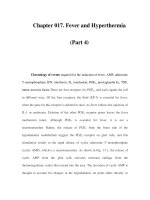
are located in the middle cranial fossa ( 734), one-third are in the posterior fossa, and 10% are found in the suprasellar region ( Fig. 4.24). Approximately one-fourth of
the middle cranial fossa cysts are bilateral and some are accompanied by hypo-genesis or compression of the temporal lobe ( 735). Additionally, they can cause a
diffuse expansion and thinning of the bones of the vault, and an elevation of the lesser wing of the sphenoid ( 573).
FIG. 4.24. Arachnoid cyst. The T1-weighted (466/16/1) axial magnetic resonance imaging demonstrates the presence of a discrete low-intensity structure ( arrow) in
the right middle fossa that displaces the anterior temporal lobe laterally. The lesion had high intensity on T2-weighted images, consistent with CSF signal
characteristics. The patient was a 3.5-year-old boy who presented with headaches and whose neurologic examination was benign. No evidence exists of a pressure
effect on the imaging study.
Although in many instances a small cyst is clinically silent, larger cysts or cysts located in the posterior fossa can produce signs and symptoms of increased
intracranial pressure. The cyst has the potential of producing hydrocephalus by increasing the resistance to CSF flow in the subarachnoid space. As the cyst
enlarges, it eventually produces extrinsic compression of the ventricular system or of the subarachnoid channels. Symptoms can begin at any time during life. Aside
from hydrocephalus, they include headaches and seizures. The relationship between headaches or seizures and the presence of an arachnoid cyst is often difficult to
establish. Hemorrhage into an arachnoid cyst can cause the sudden onset of focal neurologic signs. A subdural hematoma has been reported to originate from a
preexisting arachnoid cyst (736). Spinal cord arachnoid cysts can be intradural or less frequently extradural. They may present as space-occupying lesions with
radicular pain, progressive weakness and spasticity, scoliosis, recurrent urinary tract infections, and constipation ( 737). An accompanying neural tube defect is
common (738).
With the increased use of imaging studies, many cysts are discovered accidentally, particularly in the course of evaluating a seizure patient. Gandy and Heier believe
that removal of the cyst does not improve seizure control (659). In other instances, removal of the cyst has been associated with complete or improved seizure control
(739). Because the large majority of cysts remain constant in size, and only large cysts tend to expand, we suggest only those cysts that present as space-occupying
lesions should require surgical removal.
Achondroplasia
Achondroplasia is an abnormality of endochondral bone development transmitted as an autosomal dominant trait. It occurs in approximately 1 in 25,000 births, and
80% occur sporadically as new mutations. The gene for achondroplasia has been mapped to the tip of the short arm of chromosome 4 (4p16.3) in close proximity to
the gene for Huntington disease (740). The gene (FGFR3) codes for a tyrosine kinase transmembrane receptor for fibroblast growth factor ( 740). FGRFs are
members of a superfamily that bind fibroblast growth factors and initiate an intracellular signaling cascade. Remarkable genetic homogeneity exists, and almost all
achondroplastic subjects have the same point mutation.
Clinically, a decrease in the rate of endochondral bone formation is seen with normal membranous bone formation. The condition is characterized by dwarfism and a
variety of skeletal abnormalities. Neurologic symptoms are the result of macrocephaly, which might or might not be accompanied by hydrocephalus, and
cervicomedullary compression.
Because the cranial base is the only portion of the skull that is preformed in cartilage, its growth is selectively impaired, and a compensatory growth of the calvaria
and an increase in the vertical diameter of the skull occur. The skull base is narrowed and the petrous pyramids tower. This results in an abnormal orientation of inner
and middle ear structures (741). These growth changes produce a characteristic brachycephalic configuration and narrowing of all foramina that pass through the
base of the skull.
As Dandy (742) demonstrated in 1921 using pneumoencephalography, the ventricular system is enlarged in approximately 50% of achondroplastic individuals ( 743).
The cause for ventricular dilatation is still under debate. Some authorities have suggested that it results from a mechanical block to the flow of CSF in the area of the
foramen magnum, which is abnormally small in almost all patients with achondroplasia. This is confirmed by invasive monitoring, which has demonstrated an elevation
of intracranial pressure (743). In addition, venography of the jugular veins demonstrates stenosis at the level of the jugular foramen and a pressure gradient across
the foramen. Flodmark suggests that this phenomenon is responsible for venous hypertension and impaired CSF absorption ( 736). Yamada and colleagues have
confirmed the presence of both causes for hydrocephalus ( 744). Venous decompression at the jugular foramen, and construction of a venous bypass from the
transverse sinus to the jugular vein, have reduced ventriculomegaly, as has venous decompression at the jugular foramen ( 744,745).
Cervicomedullary compression resulting from stenosis of the foramen magnum is a serious complication, presenting at any time from infancy to adult life. In the series
of Ryken and Menezes, 3.2% of subjects with achondroplasia demonstrated symptoms or signs of progressive compression of neural structures at the level of the
foramen magnum (746). These included a variety of respiratory complications that are frequent in achondroplasia, notably apnea and respi-ratory irregularities. In the
series of Nelson and coworkers, the incidence of apnea was 28% (747). Other signs include ataxia and spastic quadriparesis. In some instances brainstem
compression can be insidious and can lead to syringobulbia, tetraplegia, and sudden infant death syndrome ( 748). Rarely, other neurologic signs are seen, including
weak cry, failure to thrive, hypersomnia, and persistent papilledema ( 748,749). Paraplegia can develop as a result of compression of the spinal cord in the thoracic
area (750). Mental retardation and seizures are not common features of achondroplasia ( 751). Although sensorineural hearing loss is common and subtle cognitive
deficits have been uniformly demonstrated, general intelligence is within normal limits in most patients.
Considerable controversy exists as to who and when to perform suboccipital decompressive surgery ( 748,752,753). On the one hand, surgery is not without risk, and
almost all achondroplastic infants who present with spasticity ultimately attain normal motor development if left alone ( 752). In addition, because the foramen magnum
grows faster than the spinal cord, the impingement of the posterior rim of the foramen magnum on the cord decreases with maturation. On the other hand, MRI and
pathologic evidence suggest deformational and traumatic changes to the spinal cord as a result of foramen magnum stenosis ( 753). We believe MRI evidence of
notching or indentation of the spinal cord at the level of the foramen magnum has little clinical significance, and that unless evidence exists of spasticity or increased
signal within the spinal cord on T2-weighted images, decompressive surgery can be deferred.
Osteopetrosis
The osteopetroses are a rare and heterogeneous group of disorders characterized by generalized bone sclerosis with thickening and increased fragility of cortical and
spongy bone. Both autosomal dominant and autosomal recessive forms have been encountered, with the former being more common. As a result of a defect in
osteoclast function bone resorption is reduced, and the skull base is thickened and the foramina are narrowed. The autosomal dominant form has a benign prognosis,
and subjects may remain asymptomatic. The recessive form is characterized by delayed psychomotor development, optic atrophy, conductive hearing loss, and facial
nerve palsy (754,755). Other neurologic complications include hydrocephalus and intracranial hemorrhage ( 755). The earlier the onset of symptoms, the more
malignant the course.
Cerebral atrophy with secondary ventricular dilatation is frequently evident. A combination of calvarial thickening and hydrocephalus explains the degree of
macrocephaly only in part. The process involves all skeletal bone and results in hematologic and bleeding disorders and in frequent fractures.
Computed tomographic scans are diagnostic. MRI often reveals delayed myelination and cerebral atrophy. Calcifications can be seen in the periventricular area and
the falx (756). The only curative treatment is early allogeneic bone marrow transplantation. Surgical decompression can stabilize cranial nerve deficits, especially
optic nerve entrapments. Visual-evoked potentials and electroretinography can be used to show the first indications of visual impairment ( 757).
A syndrome of osteopetrosis, renal tubular acidosis, and cerebral calcification is inherited as an autosomal recessive trait. Mental retardation is common, and patients
have unusual facies. The primary defect in this entity appears to be one of carbonic anhydrase II, one of two enzymes catalyzing the association of water and carbon
dioxide to form bicarbonate (758,759). Osteopetrosis also has been associated with infantile neuroaxonal dystrophy ( 760).
CONGENITAL DEFECTS OF CRANIAL NERVES AND RELATED STRUCTURES
Möbius Syndrome
Möbius syndrome, first described by Harlan in 1880 ( 761), by Chisholm in 1882 (762), and more extensively by Möbius in 1888 and 1892 (763,764), is characterized
by congenital paralysis of the facial muscles and impairment of lateral gaze.
Möbius syndrome results from diverse causes. Pathologic lesions include complete or partial absence of the facial nuclei, dysplasia of the facial musculature, and
hypoplasia of the facial nerve. The entity also has been seen in a variety of conditions involving progressive disease of muscle, anterior horn cells, or peripheral
neurons. In some instances, absence, faulty attachment, or fibrosis of the extraocular muscles is present, whereas in other cases, the brainstem nuclei showed
multiple areas of calcification and necrosis, suggesting a prenatal vascular etiology ( 765,766 and 767). The symmetric calcified lesions with chronic gliosis, including
gemistocytes, are in the tegmentum of the pons and medulla oblongata, a watershed zone of the brainstem between the territories of the paramedian penetrating
arteries and the long circumferential arteries, both branches of the basilar artery. The vascular anatomy and the histopathologic findings at birth indicate a period of
systemic hypotension in fetal life at least 4 to 6 weeks before birth ( 768). In a few cases, the electromyography points to the presence of a supranuclear lesion
(769,770).
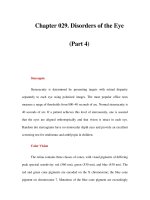
Most patients with Möbius syndrome have a variable degree of unilateral, asymmetric, or symmetric bilateral facial paralysis, with an inability to abduct the eyes
beyond the midline (771). Occasionally, the weakness is restricted to portions or quadrants of the face ( Fig. 4.25). Atrophy of the tongue, paralysis of the soft palate or
masseters, congenital clubfoot, deafness, or a mild spastic diplegia also can be pres-ent. Because of bulbar deficits, the language and communication disorder is far
greater than general intelligence would predict ( 772). Möbius syndrome is nonprogressive. In some instances, however, myotonic dystrophy or muscular dystrophy
can accompany Möbius syndrome (773).
FIG. 4.25. Möbius syndrome. As the infant cries, she demonstrates the bilateral weakness of the lower facial musculature and the marked weakness of the right upper
facial muscles. Additionally, a palsy of both external recti, tongue, and palatal musculature occurred, requiring gastrostomy feeding.
Congenital Sensorineural Deafness
Congenital deafness resulting from a lesion of the acoustic nerve can occur in isolation or in combination with a variety of anomalies. The conditions can appear
sporadically or be transmitted in a dominant, recessive, or sex-linked manner ( 774,775). In the experience of Das, a family history of deafness was elicited in 23% of
children assessed for bilateral sensorineural hearing loss. Perinatal asphyxia was believed responsible for 13%, congenital infections for 8.2%, bacterial meningitis
for 6.5%, chromosomal anomalies for 5.3%, and a variety of syndromes, notably Waardenburg syndrome, for 5.3%. In 34%, the cause was unknown ( 776). Mutations
in the connexin 26 gene are the single most common cause of genetic hearing loss (777). Other causes for profound hearing loss occurring in childhood are outlined
in Table 4.15.
In a significant proportion of patients, dermatologic manifestations accompany the hearing loss. Waardenburg syndrome, probably the most common entity of this
group, is characterized by widely spaced medial canthi, a flat nasal root, a white forelock, and heterochromia iridis. It is transmitted as a dominant trait ( 778). The
gene defective in Waardenburg syndrome ( PAX3) is located on the long arm of chromosome 2 (2q35-2q37), and normally codes for a protein termed HuP2. HuP2
protein binds DNA and the gene that codes for it is suspected to be one of a family of genes, the homeobox genes, which regulate mammalian development; in
animals, mutations in these genes result in major developmental abnormalities ( 779,780).
In another group of syndromes, congenital sensorineural deafness is associated with visual symptoms. In Usher syndrome, a condition transmitted in an autosomal
recessive manner, congenital neural hearing loss is seen in conjunction with progressive visual impairment and retinitis pigmentosa ( 774,781). Usher syndrome is the
most common of a number of conditions in which retinitis pigmentosa is combined with deafness. These are reviewed by Mills and Calver ( 781). When a sensorineural
hearing loss accompanies a neurologic disorder, it is usually in the framework of a peripheral neuropathy, and the hearing loss is progressive rather than congenital
(774).
A distinct syndrome of congenital weakness of the musculature of the face, tongue, and palate unassociated with atrophy has been termed congenital suprabulbar
paresis by Worster-Drought (782). This condition is related to the perisylvian syndrome, in which facio-lingual-masticatory diplegia results from bilaterally anterior
opercular infarctions. In the congenital form of this condition, marked feeding difficulties are accompanied by dysarthria, restricted tongue movements, and an absent
gag reflex. Intellect is relatively preserved. In the series of Kuzniecky and coworkers, seizures were documented in 87% ( 783). These tend to have their onset after
the age of 5 years, and most commonly are atonic and tonic drop attacks. Neuroimaging studies frequently disclose polymicrogyria, which has a predilection for the
opercular areas. Associated malformations, notably arthrogryposis, clubfeet, and spastic quadriparesis are not uncommon ( 782,783).
Other congenital disorders of the cranial nerves and the musculature innervated by them are presented in Table 4.14.
CHAPTER REFERENCES
1. García-Bellido A. The development of concepts on development: a dialogue with Antonio García-Bellido (Interview by Enrique Cerda-Olmedo). Int J Dev Biol 1998;42:233–236.
2. Wolpert L, Beddington R, Brocken J, et al. Principles of development, Oxford and NY: Oxford University Press, 1998.
3. Patten BM. Early embryology of the chick, 4th ed. New York: McGraw-Hill, 1951.
4. Spemann H, Mangold H. Über Induktion von Embryonalanlagen durch Implantation vonfremder Organisatoren. Wilhelm Roux Arch Entwick 1924;100: 599–638.
5. Cho KWY, Blumberg B, Steinbeisser H, De Robertis EM. Molecular nature of Spemann's organizer: the role of the Xenopus homeobox gene goosecoid. Cell 1991;67:1111–1120.
6. Wakamiya M, Rivera-Pérez JA, Baldini A, Behringer RR. Goosecoid and goosecoid-related genes in mouse embryogenesis. Cold Springs Harbor Symp Quant Biol 1997;62:145–149.
7. De Robertis EM, Kim S, Leyns L, et al. Patterning by genes expressed in Spemann's organizer. Cold Springs Harbor Symp Quant Biol 1997;62:169–175.
8. Hume CR, Dodd J. Cwnt-8C: a novel Wnt gene with a potential role in primitive streak formation and hindbrain organization. Development 1993;119:1147–1160.
9. Stein S, Kessel M. A homeobox gene involved in node, notochord and neural plate formation of chick embryos. Mech Dev 1995;49:37–48.
10. Duprat AM, Gualandris L, Kan P, et al. Review: neural induction. Arch d'Anat Microsc Morphol Exp 1987;75:211–227.
11. Tiedemann H. The molecular mechanism of neural induction: neural differentiation of Triturus ectoderm exposed to Hepes buffer. Roux's Arch Dev Biol 1986;195:399–402.
12. Fortini ME, Artavanis-Tsakonas S. Notch: neurogenesis is only part of the picture. Cell 1993;75:1245–1247.
13. Fontaine-Pérus JC, Chanconie M, Le Douarin NM, et al. Mitogenic effect of muscle on the neuroepithelium of the developing spinal cord. Development 1989; 107:413–422.
14. Fontain-Pérus J. Migration of crest-derived cells from gut: gut influences on spinal cord development. Brain Res Bull 1993;30:251–255.
15. Dickson ME, Krumlauf R, McMahon AP. Evidence for a mitogenic effect of Wnt-1 in the developing mammalian central nervous system. Development 1994;120:1453–1471.
16. DiCicco-Bloom E, Black IB. Insulin growth factors regulate the mitotic cycle in cultured rat sympathetic neuroblasts. Proc Natl Acad Sci USA 1988;85: 4066–4070.
17. Tao Y, Black LB, DiCicco-Bloom E. Neurogenesis in neonatal rat brain is regulated by peripheral injection of basic fibroblast growth factor (bFGF). J Comp Neurol 1996;376:653–663.
18. Tao Y, Black IB, DiCicco-Bloom E. In vivo neurogenesis is inhibited by neutralizing antibody to basic fibroblast growth factor. J Neurobiol 1997;33:289–296.
19. Turner DL. Weintraub H. Expression of achaete-scute homolog 3 in Xenopus embryos converts ectodermal cells to a neural fate. Genes Dev 1994;8:1434–1447.
20. Anderson DJ. A molecular switch for the neuron-glia developmental decision. Neuron 1995;15:1219–1222.
21. Hemmati-Brivantou A, Melton D. Vertebrate embryonic cells will become nerve cells unless told otherwise. Cell 1997;88:13–17.
22. Tanabe Y, Jessell TM. Diversity and pattern in the developing spinal cord. Science 1996;274:1115–11123.
23. McClure, CFW. The segmentation of the primitive vertebrate brain. J Morphol 1890;4:35–56.
24. Keynes R, Lumsden A. Segmentation and the origin of regional diversity in the vertebrate central nervous system. Neuron 1990;2:1–9.
25. McGinnis W, Krumlauf R. Homeobox genes and axial patterning. Cell 1992;68:283–302.
26. Keynes R, Krumlauf R. Hox genes and regionalization of the nervous system. Annu Rev Neurosci 1994;17:109–132.
27. Guthrie S. Patterning the hindbrain. Curr Opin Neurobiol 1996;6:41–48.
28. Wassef M, Joyner AL. Early mesencephalon/metencephalon patterning and development of the cerebellum. Perspect Dev Neurobiol 1997;5:3–16.
29. Goldowitz D, Hamre K. The cells and molecules that make a cerebellum. Trends Neurosci 1998;21:375–382.
30. Figdor MC, Stem CD. Segmental organization of embryonic diencephalon. Nature 1993;363:630–634.
31. Rubenstein JLR, Shimamura K, Martínez S. Regionalization of the prosencephalic neural plate. Ann Rev Neurosci 1998;21:445–477.
32. Rubenstein JLR, Beachy PA. Patterning of the embryonic forebrain. Curr Opin Neurobiol 1998;8:18–26.
33. Mai JK, Andrressen C, Ashwell KWS. Demarcation of prosencephalic regions by CD15-positive radial glia. Eur J Neurosci 1998;10:746–751.
34. Guthrie S, Butcher M, Lumsden A. Patterns of cell division and interkinetic nuclear migration in the chick embryo hindbrain. J Neurobiol 1991;22:742–754.
35. Joyner AL. Engrailed Wnt and Pax genes regulate midbrain-hindbrain development. Trends Genet 1996;12: 15–20.
36. Wassarman KM, Lewandoski M, Campbell K., et al. Specification of the anterior hindbrain organizer is dependent on Gbx2 gene function. Development 1997;124:2923–2934.
37. Nomes HO, Dressler GR, Knapik EW, et al. Spatially and temporally restricted expression of Pax-2 during neurogenesis. Development 1990;109:797–809.
38. Puschel AW, Westerfeld M, Dressler G. Comparative analysis of Pax-2 protein distributions during neurulation in mice and zebrafish. Mech Dev 1992;38: 197–208.
39. Rowitch DH, McMahon AP. Pax-2 expression in the murine neural plate precedes and encompasses the expression domains of Wnt-1 and En-1. Mech Dev 1995;52:3–8.
40. Rowitch DH, Danielian PS, Lee SMK, et al. Cell interactions in patterning the mammalian midbrain. Cold Springs Harbor Symp Quant Biol 1997;62:535–544.
41. Wurst W, Auerback AB, Joyner AL. Multiple developmental defects in Engrailed-1 mutant mice: an early mid-hindbrain deletion and patterning defects in forelimbs and sternum. Development
1994;120:2065–2075.
42. McMahon AP, Joyner AL, Bradley A, et al. The midbrain-hindbrain phenotype of Wnt-1-/Wnt-1- mice results from stepwise deletion of engrailed-expressing cells by 9.5 days postcoitum. Cell
1992;69:581–595.
43. Millen KJ, Hui C-C, Joyner AL. A role for En-2 and other murine homologues of Drosophila segment polarity genes in regulating position information in the developing cerebellum. Development
1995;121:3935–3945.
44. Kuemerle B, Zanjani H, Joyner A, Herrup K. Pattern deformities and cell loss in Engrailed-2 mutant mice suggest two separate patterning events during cerebellar development. J Neurosci
1997;17:7881–7889.
45. Rhinn M, Dierich A, Shawlot W, et al. Sequential roles for Otx2 in visceral endoderm and neuroectoderm for forebrain and midbrain induction and specification. Development
1989;125:845–856.
46. Acampora D, Simeone A. Understanding the roles of Otx1 and Otx2 in the control of brain mophogenesis. Trends Neurosci 1999;22:116–122.
47. Ryan AK, Blumberg B, Rodríguez-Estaban C, et al. Pitx2 determines left-right asymmetry of internal organs in vertebrates. Nature 1998;394:545–551.
47a.Zhao Q, Behringer RR, de Crombrugghe B. Prenatal folic acid treatment suppresses acrania and meroanencephaly in mice mutant for the Cart1 homeobox gene. Nat Genet 1996;13:275–283.
47b.Anderson SA, Eisenstat DD, Shi L, Rubenstein JLR. Interneuron migration from basal forebrain to neocortex: dependence on Dix genes. Science 1997;278: 474–476.
48. De Pomerai D. From gene to animal: an introduction of the molecular biology of animal development, 2nd ed. Cambridge, U.K: Cambridge University Press, 1990.
49. El-Baradi T, Pieler T. Zinc finger proteins: what we know and what we would like to know. Mech Devel 1991;35:155–169.
50. Schneider-Maunoury S, Topilko P, Seitanidou T, et al. Disruption of Krox-20 results in alteration of rhombomeres 3 and 5 in the developing hindbrain. Cell 1993;75:1199–1214.
51. Topilko P, Schneider-Maunoury S, Levi G, et al. Krox-20 controls myelination in the peripheral nervous system. Nature 1994;371:796–799.
52. Nieto MA, Sechrist J, Wilkinson DG, Bronner-Fraser M. Relationship between spatially restricted Krox-20 gene expression in branchial neural crest and segmentation in the chick embryo
hindbrain. EMBO J 1995;14:1697–1710.
52a.Faina GT, Cardini FA, D'Incerti L, et al. Familial schizencephaly associated with EMX2 mutation. Neurology 1997;48:1403–1406.
53. Wilkinson DG, Krumlauf R. Molecular approaches to the segmentation of the hindbrain. Trends Neurosci 1990;13:335–339.
54. Morriss-Kay GM, Murphy P, Hill RE, Davidson DR. Effects of retinoic acid excess on expression of Hox-2.9 and Krox-20 and on morphological segmentation in the hindbrain of mouse embryos.
EMBO J 1991;10:2985–2995.
55. Nonchev S, Maconochie M, Vesque C, et al. The conserved role of Krox-20 in directing Hox gene expression during vertebrate hindbrain segmentation. Proc Natl Acad Sci U S A
1996;93:9339–9345.
56. Cook M, Gould A, Brand N, et al. Expression of the zinc-finger gene PLZF at rhombomere boundaries in the vertebrate hindbrain. Proc Natl Acad Sci U S A 1995;92:2249–2253.
57. Ingraham HA, Chen PRP, Mangalan HJ, et al. A tissue-specific transcription factor containing a homeodomain specifies a pituitary phenotype. Cell 1988;55:519–529.
58. Xu L, Lavinsky RM, Dasen JS. Signal-specific co-activator domain requirements for Pit-1 activation. Nature 1998;395:301–306.
59. Doniach T. Basic FGF as an inducer of anteroposterior neural pattern. Cell 1995;83:1067–1070.
60. Brand-Saberi B, Ebensperger C, Wilting J, et al. The ventralizing effect of the notochord on somite differentiation in chick embryos. Anat Embryol 1993;188:239–245.
60a.Fox JW, Lamperti ED, Eksioglu YZ, et al. Mutations in filamin 1 prevent migration of cerebral cortical neurons in human periventricular heterotopia. Neuron 1998;21:1315–1325.
61. Pourquié O, Coltey M, Teillet M-A, et al. Control of dorsoventral patterning of somite derivatives by notochord and floor plate. Proc Natl Acad Sci U S A 1993;90:5242–5246.
62. Martí E, Bumcrot DA, Takada R, McMahon AP. Requirement of 19K form of Sonic hedgehog for induction of distinct ventral cell types in CNS explants. Nature 1995;375:322–325.
63. Roelink H, Porter JA, Chiang C, et al. Floor plate and motor neuron induction by different concentrations of the amino-terminal cleavage product of Sonic hedgehog autoproteolysis. Cell
1995;81:445–455.
64. Tabin CJ, McMahon AP. Recent advances in hedgehog signalling. Trends Cell Biol 1997;7:442–446.
65. Goulding M. Specifying motor neurons and their connections. Neuron 1998;21:943–946.
65a.Sasaki H, Hogan BLM. HNF-3b as a regulator of floor plate development. Cell 1994;76:103–115.
66. van Straaten HWM, Hekking JWM, Wiertz-Hoessels EJLM, et al. Effect of the notochord on the differentiation of the floor plate area in the neural tube of the chick embryo. Anat Embryol
1988;177:317–324.
67. Sarnat HB. Cerebral dysgenesis: embryology and clinical expression, New York: Oxford University Press, 1992.
68. Ericson J, Muhr J, Placzek M, et al. Sonic hedgehog induces the differentiation of ventral forebrain neurons: a common signal for ventral patterning within the neural tube. Cell
1995;81:747–756.
69. Roessler E, Belloni E, Gaudenz K, et al. Mutations in the human Sonic hedgehog gene cause holoprosencephaly. Nature Genet 1996;14:357–360.
70. Callaerts P, Halder G, Gehring WJ. Pax-6 in development and evolution. Annu Rev Neurosci 1997;20: 483–532.
70a.Brown NL, Kanekar S, Vetter ML, et al. Math5 encodes a murine basic helix-loop-helix transcription factor expressed during early stages of retinal neurogenesis. Development
1998;125:4621–4633.
70b.Tanabe Y, William C, Jessell TM. Specification of motor neuron identity by the MNR2 homeodomain protein. Cell 1998;95:67–80.
71. Stoykova A, Gruss P. Roles for Pax genes in developing and adult brain as suggested by expression patterns. J Neurosci 1994;14:1395–1412.
72. Saint-Jeannet J-P, He X, Varmus HE, Dawid IB. Regulation of dorsal fate in the neuraxis by Wnt-1 and Wnt-3a. Proc Natl Acad Sci U S A 1997;94:13713–13718.
73. Keynes R, Krumlauf R. Hox genes and regionalization of the nervous system. Annu Rev Neurosci 1994; 17:109–132.
74. Murphy P, Hill RE. Expression of the mouse labial-like homebox-containing genes, Hox-2.9 and Hox-1.6, during segmentation of the hindbrain. Development 1991; 111:61–74.
75. Boncinelli E, Somma R, Acampora D, et al. Organization of human homeobox genes. Hum Reprod 1988;3:880–886.
75a.Lee JE, Hollenberg SM, Snider I, et al. Conversion of Xenopus ectoderm into neurons by NeuroD: a basic helix-loop-helix protein. Science 1995;268:836–844.
75b.Yokoyama M, Nishi Y, Miyamoto Y, et al. Molecular cloning of a human neuroD from a neuroblastoma cell line specifically expressed in the fetal brain and adult cerebellum. Brain Res Mol Brain
Res 1996;42: 135–139.
76. Capecchi MR. Hox genes and mammalian development. Cold Springs Harbor Symp Quant Biol 1997;62: 273–281.
77. Stern CD, Foley AC. Molecular dissection of Hox gene induction and maintenance in the hindbrain. Cell 1998;94:143–145.
78. Kessel M. Reversal of axonal pathways from rhombomere 3 correlates with extra Hox expression domains. Neuron 1993;10:379–393.
79. Millen KJ, Wurst W, Herrup K, Joyner AL. Abnormal embryonic cerebellar development and patterning of postnatal foliation in two mouse Engrailed-2 mutants. Development
1994;120:695–706.
80. Mastick GS, Fan C-M, Tessier-Lavigne M, et al. Early deletion of neuromeres in Wnt-1-/- mutant mice: evaluation by morphological and molecular markers. J Comp Neurol 1996;374:246–258.
80a. Lee JE. NeuroD and neurogenesis. Dev Neurosci 1997;19:27–32.
80b. Katayama M, Mizuta I, Sakayama Y, et al. Differential expression of neuroD in primary cultures of cerebral cortical neurons. Exp Cell Res 1997;236:412–417.
81. Zec N, Rowitch DH, Bitgood MJ, Kinney HC. Expression of the homeobox-containing genes EN1 and EN2 in human fetal midgestational medulla and cerebellum. J Neuropathol Exp Neurol
1997;56:236–242.
82. Wagner M, Thaller C, Jessell T, et al. Polarizing activity and retinoid synthesis in the floor plate of the neural tube. Nature 1990;345:819–822.
83. Ruberte E, Dolle P, Chambon P, et al. Retinoid acid receptors and cellular retinoid binding proteins. II. Their differential pattern of transcription during early morphogenesis in mouse.
Development 1991;111:45–60.
84. Summerbell D, Maden M. Retinoic acid, a developmental signalling molecule. Trends Neurosci 1990; 13:142–147.
85. Momoi M, Yamagata T, Ichihashi K, et al. Expression of cellular retinoic-acid binding protein in the developing nervous system of mouse embryos. Dev Brain Res 1990;54:161–167.
86. Thaller C, Eichele G. Isolation of 3,4-didehydroretinoic acid: a novel morphogenetic signal in the chick wing bud. Nature 1990;345:815–819.
87. Brockes J. Reading the retinoid signals. Nature 1990; 345:766–768.
88. Kikuchi Y, Segawa H, Tokumoto M, et al. Ocular and cerebellar defects in zebrafish induced by overexpression of the LIM domains of the Islet-3 LIM/homeo-domain protein. Neuron
1997;18:369–382.
89. Porter FD, Drago J, Xu Y, et al. Lhx2, a LIM homeobox gene, is required for eye, forebrain, and definitive erythrocyte development. Development 1997;124:2935–2944.
90. Durston AJ, Timmermans JPM, Hage WJ, et al. Retinoic acid causes an anteroposterior transformation in the developing central nervous system. Nature 1989;340:140–144.
91. Marín-Padilla M, Marin-Padilla MT. Mophogenesis of experimentally induced Arnold-Chiari malformation. J Neurol Sci 1981;50:29–55.
91a. Morrow EM, Furukawa T, Lee JE, Cepko CL. NeuroD regulates multiple functions in the developing neural retina in rodent. Development 1998;126:23–36.
91b. Yan RT, Wang SZ. NeuroD induces photoreceptor cell overproduction in vivo and de novo generation in vitro. J Neurobiol 1998;15:485–496.
92. Marín-Padilla M. Embryology and pathology of the axial skeleton and neural dysraphic disorders. Can J Neurol Sci 1991;18:153–169.
93. Alles AJ, Sulik KK. Retinoic acid-induced spina bifida: evidence for a pathogenetic mechanism. Development 1990;108:73–81.
94. Wohl CA, Weiss S. Retinoic acid enhances neuronal proliferation and astroglial differentiation in cultures of CNS stem cell-derived precursors. J Neurobiol 1998;37:281–290.
94a. Tamimi R, Steingrimsson E, Copeland NG, et al. The NEUROD gene maps to human chromosome 2q32 and mouse chromosome 2. Genomics 1996;34:418–421.
94b. Yamimi RM, Steingrimsson E, Montgomery-Dyer K, et al. NEUROD2 and NEUROD3 genes map to human chromosomes 17q12 and 5q23-q31 and mouse chromosomes 11 and 13,
respectively. Genomics 1997;40: 355–357.
95. Meyers EN, Lewandoski M, Martin GR. An Fgf8 mutant allelic series generated by Cre- and Flp-mediated recombination. Nature Genet 1998;18:136–141.
96. Price M, Lazzaro D, Pohl T, et al. Regional expression of the homeobox gene Nkx-2.2 in the developing mammalian forebrain. Neuron 1992;8:241–255.
97. Qiu M, Shimamuira K, Sussel L, et al. Control of anteroposterior and dorsoventral domains of Nkx-6.1 gene expression relative to other Nkx genes during vertebrate CNS development. Mech
Dev 1998;72:77–88.
98. Dorsky RI, Moon RT, Raible DW. Control of neural crest cell fate by the Wnt signaling pathway. Nature 1998;396:370–373.
99. Condron BG, Patel NH, Zinn K. Engrailed controls glial/neuronal cell fate decisions at the midline of the central nervous system. Neuron 1994;13:541–554.
100. Shah NM, Marchionni MA, Isaacs I, et al. Glial growth factor restricts mammalian neural crest stem cells to a glial fate. Cell 1994;77:349–360.
100a. Ma Q, Kinter C, Anderson DJ. Identification of neurogenin—a vertebrate neuronal determination gene. Cell 1996;87:43–52.
100b. Sommer I, Ma Q, Anderson DJ. Neurogenins: a novel family of atonal-related bHLH transcription factors are putative mammalian neuronal determination genes that reveal progenitor cell
heterogeneity in the developing CNS and PNS. Mol Cell Neurosci 1996;8: 221–241.
100c. Ma Q, Chen Z, del Barco Barrantes I, et al. Neurogenin1 is essential for the determination of neuronal precursors for proximal cranial sensory ganglia. Neuron 1998;20:469–482.
101. Urbnek P, Fetka I, Meisler MH, Busslinger M. Cooperation of Pax2 and Pax5 in midbrain and cerebellum development. Proc Natl Acad Sci USA 1997;94: 5703–5708.
102. Kikuchi Y, Segawa H, Tokumoto M, et al. Ocular and cerebellar defects in zebrafish induced by overexpression of the LIM domains of the Islet-3 LIM/homeo-domain protein. Neuron
1997;18:369–382.
103. Salinas PC, Fletcher C, Copeland NG, et al. Maintenance of Wnt-3 expression in Purkinje cells of the mouse cerebellum depends on interaction with granule cells. Development
1994;120:1277–1286.
104. Lange W. Regional differences in the cytoarchitecture of the cerebellar cortex. In: Palay SL, Chan-Palay V, eds. The cerebellum: new vistas. Berlin: Springer-Verlag, 1982:93–107.
105. Korbo L, Andersen BB, Ladefoged O, Moller A. Total numbers of various cell types in rat cerebellar cortex using an unbiased stereological method. Brain Res 1993;609:262–268.
106. Goodrich LV, Mienkovi L, Higgins KM, Scott MP. Altered neural cell fates and medulloblastoma in mouse patched mutants. Science 1997;277:1109–1113.
107. Traiffort E, Charaytoniuk DA, Faure H, Ruat M. Regional distribution of Sonic hedgehog, Patched and Smoothened mRNA in the adult rat brain. J Neurochem 1998;70:1327–1330.
108. Wechsler-Reya, Scott MP. Control of neuronal precursor proliferation in the cerebellum by Sonic hedgehog. Neuron 1999;22:103–114.
109. Walter M, Reifenberger J, Summer C, et al. Mutations in the human homologue of the Drosophila segment polarity gene patched (PTCH) in sporadic basal cell carcinomas of the skin and
primitive neuroectodermal tumors of the central nervous system. Cancer Res 1997;57:2581–2585.
110. Smith JL, Schoenwolf GC. Neurulation: coming to closure. Trends Neurosci 1997;20:510–517.
111. Schoenwolf GC, Smith JL. Mechanisms of neurulation: traditional viewpoint and recent advances. Development 1990;109:243–270.
111a.McMahon JA, Takada S, Zimmerman LB, et al. Noggin-mediated antagonism of BMP signaling is required for growth and patterning of the neural tube and somite. Genes Dev
1998;12:1438–1452.
112. Alvarez I, Schoenwolf GC. Expansion of surface epithelium provides the major extrinsic force for bending of the neural plate. J Exp Zool 1992;261: 340–348.
113. Jacobson AG. Experimental analysis of the shaping of the neural plate and tube. Am Zool 1991;31:628–643.
114. Schoenwolf GC, Franks MV. Quantitative analyses of changes in cell shapes during bending of the avial neural plate. Dev Biol 1984;105:257–272.
115. Smith J, Schoenwolf GC. Notochordal induction of cell wedging in the chick neural plate and its role in neural tube formation. J Exp Zool 1989;25:49–62.
115a.Zhong W, Jiang M-M, Weinmaster G, et al. Differential expression of mammalian Numb, Numblike and Notch1 suggests distinct roles during mouse cortical neurogenesis. Development
1997;124:1887–1897.
116. Sarnat HB. Role of the human fetal ependyma. Pediatr Neurol 1992;8:163–178.
117. Aaku-Saraste E, Oback B, Hellwig A, Huttner WB. Neuro-epithelial cells downregulate their plasma membrane polarity prior to neural tube closure and neurogenesis. Mech Dev 1997;69:71–81.
118. Sausedo RA, Smith JL, Schoenwolf GC. Role of randomly oriented cell division in shaping and bending of the neural plate. J Comp Neurol 1997;381:473–488.
119. Juriloff DM, Harris JM, Tom C, et al. Normal mouse strains differ in the site of initiation of closure of the cranial neural tube. Teratology 1991;44:225–233.
120. Golden JA, Chernoff GF. Intermittent pattern of neural tube closure in two strains of mice. Teratology 1993;47:73–80.
121. Busam KJ, Roberts DJ, Golden JA. Clinical teratology counseling and consultation. Case report: two distinct anterior neural tube defects in a human fetus: evidence for an intermittent pattern of
neural tube closure. Teratology 1993;48:399–403.
122. O'Rahilly R, Müller F. Bidirectional closure of the rostal neuropore in the human embryo. Am J Anat 1989;184:259–268.
123. Lemire RJ. Variations in development of the caudal neural tube in human embryos (Horizons XIV-XXI). Teratology 1969;2:361–370.
123a.Goodrich LV, Scott MP. Hedgehog and Patched in neural development. Neuron 1998;21:1243–1257.
124. Papan C, Campos-Ortega JA. On the formation of the neural keel and neural tube in the zebrafish Danio (Brachydanio) rerio. Roux's Arch Dev Biol 1994;203: 178–186.
125. Dao AH, Netsky MG. Human tails and pseudotails. Hum Pathol 1984;15:449–453.
126. Lu FL, Wang P-J, Teng R-J, Yau K-IT. The human tail. Pediatr Neurol 1998;19:230–233.
127. James HE, Canty TG. Human tail and associated spinal anomalies. Clin Pediatr 1995;34:386–388.
128. Aruga J, Yokota N, Hashimoto M, et al. A novel zinc finger protein, Zic, is involved in neuronogenesis, especially in the cell lineage of cerebellar granule cells. J Neurochem
1994;63:1880–1890.
129. Aruga J, Minowa O, Yaginuma H, et al. Mouse Zic1 is involved in cerebellar development. J Neurosci 1998;18:284–293.
130. Ben-Arie N, Bellen HJ, Armstrong DL, et al. Math1 is essential for genesis of cerebellar granule neurons. Nature 1997;390:169–172.
131. Ericson J, Briscoe J, Rashbass P, et al. Graded Sonic hedgehog signaling and the specification of cell fate in the ventral neural tube. Cold Springs Harbor Symp Quant Biol 1997;62:451–466.
132. Fritzsch B, Nichols DH, Echelard Y, McMahon AP. Development of midbrain and anterior hindbrain ocular motoneurons in normal and Wnt-1 knockout mice. J Neurobiol 1995;27:457–469.
133. Porter JD, Baker RS. Absence of oculomotor and trochlear motorneurons leads to altered extraocular muscle development in the Wnt-1 null mutant mouse. Dev Brain Res 1997;100:121–126.
134. Tsuchida T, Ensini M, Morton SB, et al. Topographic organization of embryonic motor neurons defined by expression of LIM homeobox genes. Cell 1994;79: 957–970.
135. Appel B, Korzh V, Glasgow E, et al. Motoneuron fate specification revealed by patterned LIM homeobox gene expression in embryonic zebrafish. Development 1995;121:4117–4125.
136. Pfaff SL, Mendelsohn M, Stewart CL, et al. Requirement of LIM homeobox gene Is/1 in motor neuron generation reveals a motor neuron-dependent step in interneuron differentiation. Cell
1996;84:309–320.
137. D'Costa AP, Prevette DM, Houenou LJ, et al. Mechanisms of insulin-like growth factor regulation of programmed cell death of developing avian motoneurons. J Neurobiol 1998;36:379–394.
137a.Jeh-Ping L, Jessell TM. A role for rhoB in the delamination of neural crest cells from the dorsal neural tube. Development 1998;125:5055–5067.
138. Le Douarin N. The Neural Crest, Cambridge, UK: Cambridge University Press, 1982.
138a.Chen Z-F, Behringer RR. Twist is required in head mesenchyme for cranial neural tube morphogenesis. Genes Dev 1995;9:686–699.
139. Tan SS, Morriss-Kay GM. The development and distribution of the cranial neural crest in the rat embryo. Cell Tissue Res 1985;240:403–416.
140. Gerhart J, Kirschner M. Cells, embryos and evolution, Malden: Blackwell Science, 1997.
141. Bronner-Fraser M. Neural crest formation and migration in the developing embyro. FASEB J 1994;8:699–706.
142. Bronner-Fraser M. Origins and developmental potential of the neural crest. Exp Cell Res 1995;218:405–417.
143. Sadaghiani B, Crawford BJ, Vielkind JR. Changes in the distribution of extracellular matrix components during neural crest development in Xiphophorus spp. Embryos. Can J Zool
1994;72:1340–1353.
144. Selleck MAJ, Scherson TY, Bronner-Fraser M. Origins of neural crest diversity. Dev Biol 1993;159:1–11.
145. ElShamy WM, Linnarsson S, Lee K-F, et al. Prenatal and postnatal requirements of NT-3 for sympathetic neuroblast survival and innnervation of specific targets. Development
1996;122:491–500.
146. Sauer FC. Mitosis in the neural tube. J Comp Neurol 1935;62:377–405.
147. Nurse P. Ordering S-phase and M-phase in the cell cycle. Cell 1994;79:547–550.
148. Caviness VS Jr, Pinto-Lord MC, Evrard P. The development of laminated patterns in the mammalian neocortex. In: Connelly TG, ed. Morphogenesis and pattern formation. New York: Raven
Press, 1981:103–126.
149. Burek MJ, Oppenheim RW. Programmed cell death in the developing nervous system. Brain Pathol 1996;6:427–446.
150. Chenn A, McConnell SK. Cleavage orientation and the asymmetric inheritance of Notch1 immunoreactivity in mammalian neurogenesis. Cell 1995;82:631–641.
151. Zhong W, Feder JN, Jiang M-M, et al. Asymmetrical localization of a mammalian numb homolog during mouse cortical neuronogenesis. Neuron 1996;17:43–53.
152. Mione MC, Cavanaugh JFR, Harris B, Parnavelas JG. Cell fate specification and symmetrical/asymmetrical divisions in the developing cerebral cortex. J Neurosci 1997;17:2018–2029.
153. Bass PW. Microtubules and neuronal polarity: lessons from mitosis. Neuron 1999;22:23–31.
154. Crews L, Hunter D. Neurogenesis in the olfactory epithelium. Perspect Dev Neurobiol 1994;2:151–161.
155. Menezes JRL, Smith CM, Nelson KC, Luskin MB. The division of neuronal progenitor cells during migration in the neonatal mammalian forebrain. Mol Cell Neurosci 1995;6:496–508.
156. Kendler A, Golden JA. Progenitor cell proliferation outside the ventricular and subventricular zones during human brain development. J Neuropathol Exp Neurol 1996;55:1253–1258.
157. Hamburger V, Levi-Montalcini R. Proliferation, differentiation and degeneration in the spinal ganglia of the chick embryo under normal and experimental conditions. J Exp Zool
1949;111:457–502.
158. O'Connor TM, Wyttenback CR. Cell death in the embryonic chick spinal cord. J Cell Biol 1974;60: 448–459.
159. Okado N, Oppenheim RW. Cell death of motorneurons in the chick embryo spinal cord. J Neurosci 1984;4:1639–1652.
160. Harris AJ, McCaig CD. Motorneuron death and motor unit size during embryonic development of the rat. J Neurosci 1984;4:13–24.
161. Ferrer I, Serrano T, Soriano E. Naturally occurring cell death in the subicular complex and hippocampus in the rat during development. Neurosci Res 1990;8:60–66.
162. Diener PS, Bregman BS. Neurotrophic factors prevent the death of CNS neurons after spinal cord lesions in newborn rats. NeuroReport 1994;5:1913–1917.
163. Pittman RN, Wang S, DiBenedetto AJ, et al. A system for characterizing cellular and molecular events in programmed neuronal cell death. J Neurosci 1993;13: 3669–3680.
164. Gómez-Pinilla F, Lee JW-K, Cotman CW. Distribution of brain fibroblast growth factor in the developing brain. Neuroscience 1994;61:911–923.
165. Page KJ, Saha A, Everitt BJ. Differential activation and survival of basal forebrain neurons following infusions of excitatory amino acids: studies with the intermediate early gene c- fos. Exp
Brain Res 1993;93:412–422.
166. Lefebvre S, Burglen L, Reboullet S, et al. Identification and characterization of a spinal muscular atrophy determining gene. Cell 1995;80:155–165.
167. Roy N, Mahadevan MS, McLean M, et al. The gene for neuronal apoptosis inhibitory protein is partially deleted in individuals with spinal muscular atrophy. Cell 1995;80:167–178.
168. Homma S, Yaginuma H, Oppenheim RW. Programmed cell death during the earliest stages of spinal cord development in the chick embryo: a possible means of early phenotypic selection. J
Comp Neurol 1994;345:377–395.
169. Nurcombe V, McGrath PA, Bennett MR. Postnatal death of motor neurons during the development of the brachial spinal cord of the rat. Neurosci Lett 1981;27:249–254.
170. Duckett S, Pearse AGE. The cells of Cajal-Retzius in the developing human brain. J Anat 1968;102:183–187.
171. Marín-Padilla M. Dual origin of the mammalian neocortex and evolution of the cortical plate. Anat Embryol 1978;152:109–126.
172. Bayer SA, Altman J. Development of layer 1 and the subplate in the rat neocortex. Exp Neurol 1990;107:48–62.
173. Marín-Padilla M. Cajal-Retzius cells and the development of the neocortex. Trends Neurosci 1998;21: 64–71.
174. Ogawa M, Miyata T, Nakajima K, et al. The reeler gene-associated antigen on Cajal-Retzius neurons is a crucial molecule for laminar organization of cortical neurons. Neuron 1995;14:899–912.
175. Curran T, D'Arcangelo G. Role of reelin in the control of brain development. Brain Res Rev 1998;26:285–294.
176. Clark DC, Mizuguchi M, Antalffy B, et al. Predominant localization of the LIS family of gene products to Cajal-Retzius cells and ventricular neuroepithelium in the developing human cortex. J
Neuropathol Exp Neurol 1997;56:1044–1052.
177. Huntley GW, Jones EG. Cajal-Retzius neurons in developing monkey neocortex show immunoreactivity to calcium binding proteins. J Neurocytol 1990;19:200–219.
178. Derer P, Derer M. Cajal-Retzius cell ontogenesis and death in mouse brain visualized with horseradish peroxidase and electron microscopy. Neuroscience 1990; 32:707–717.
179. Smart IHM. Proliferative characteristics of the ependymal layer during the early development of the spinal cord in the mouse. J Anat 1972;111:365–380.
180. Sarnat HB. Regional differentiation of the human fetal ependyma: immunocytochemical markers. J Neuropathol Exp Neurol 1992;51:58–75.
181. Snow DM, Steindler DA, Silver J. Molecular and cellular characterization of the glial roof plate of the spinal cord and optic tectum: a posible role for a proteoglycan in the development of an
axon barrier. Dev Biol 1990;138:359–376.
182. Bovolentá P, Dodd J. Guidance of commissural growth cones at the floor plate in embryonic rat spinal cord. Development 1990;109:435–447.
183. Kennedy TE, Serafini T, de la Torre J, et al. Netrins are diffusible chemotropic factors for commissural axons in the embryonic spinal cord. Cell 1994;78:425–435.
184. Colamarino SA, Tessier-Lavigne M. The axonal chemoattractant netrin-1 is also a chemorepellant for trochlear motor neurons. Cell 1995;81:621–629.
185. Keynes R, Cook GMW. Axonal guidance molecules. Cell 1995;83:161–169.
186. Sarnat HB, Netsky MG. Evolution of the nervous system, 2nd ed. New York: Oxford University Press, 1981.
187. Roessmann U, Gambetti P. Astrocytes in the developing human brain: an immunohistochemical study. Acta Neuropathol 1986;70:308–313.
188. Anton ES, Cameron RS, Rakic P. Role of neuron-glial junctional domain proteins in the maintenance and termination of neuronal migration across the embryonic cerebral wall. J Neurosci
1996;16:1193–2283.
189. Zheng C, Heintz N, Hatten ME. CNS gene encoding astrotactin which supports neuronal migration along glial fibers. Science 1996;272:417–419.
190. Jouet M, Kenwrick S. Gene analysis of L1 neural cell adhesion molecule in prenatal diagnosis of hydrocephalus. Lancet 1995;345:161–162.
191. Herman J-P, Victor JC, Sanes JR. Developmentally regulated and spatially restricted antigens of radial glial cells. Dev Dynamics 1993;197:307–318.
192. Thomas LB, Gates MA, Steindler DA. Young neurons from the adult subependymal zone proliferate and migrate along an astrocyte, extracellular matrix-rich pathway. Glia 1996;17:1–14.
193. Rice DS, Sheldon M, D'Arcangelo G, et al. Disabled-1 acts downstream of Reelin in a signaling pathway that controls laminar organization in the mammalian brain. Development
1998;125:3719–3729.
194. O'Rourke NA, Dailey ME, Smith SJ, McConnell SK. Diverse migratory pathways in the developing cerebral cortex. Science 1992;258:299–302.
195. Rakic P. Radial versus tangential migration of neuronal clones in the developoing cerebral cortex. Proc Natl Acad Sci U S A 1995;92:11323–11327.
196. O'Rourke NA, Sullivan DP, Kaznowski CE, et al. Tangential migration of neurons in the developing cerebral cortex. Development 1995;121:2165–2176.
197. Wichterle H, García-Verdugo JM, Alvarez-Buylla A. Direct evidence for homotypic glia-independent neuronal migration. Neuron 1997;18:779–791.
198. Dobyns WB, Reiner O, Carrozzo R, Ledbetter DH. Lissencephaly: a human brain malformation associated with deletion of the LIS1 gene located at chromosome 17p13. JAMA
1993;270:2838–2842.
199. Lo Nigro C, Chong SS, Smith ACM, et al. Point mutations and an intragenic deletion in LIS1: the lissencephaly causitive gene in isolated lissencephaly sequence and Miller-Dieker syndrome.
Hum Mol Genet 1997;6:157–164.
200. Chong SS, Pack SD, Roschke AV, et al. A revision of the lissencephaly and Miller-Dieker syndrome critical regions in chromosome 17p13.3. Hum Mol Genet 1997;6:147–155.
201. Gleeson JG, Allen KM, Fox JW, et al. Doublecortin, a brain-specific gene mutated in human X-linked lissencephaly and double cortex syndrome, encodes a putative signaling protein. Cell
1998;92:63–72.
202. des Portes V, Pinard JM, Billuart P, et al. A novel CNS gene required for neuronal migration and involved in X-linked subcortical laminar heterotopia and lissencephaly syndrome. Cell
1998;92:51–61.
203. Eksioglu YZ, Scheffer IE, Cardena P, et al. Periventricular heterotopia: an X-linked dominant epilepsy locus causing aberrant cerebral cortical development. Neuron 1996;16:77–87.
204. Fox JW, Lamperti ED, Eksioglu YZ, et al. Mutations in filamin 1 prevent migration of cerebral cortical neurons in human periventricular heterotopia. Neuron 1998;21:1315–1325.
205. Tsuru A, Mizuguchi M, Uyemura K, et al. Immunohistochemical expression of cell adhesion molecule L1 in hemimegalencephaly. Pediatr Neurol 1997;16:45–49.
206. Ramón y Cajal, S de. Histologie du systéme nerveux central de l'homme et des vértébrés, Paris: Maloine, 1909–1911.
207. Dotti CG, Simons K. Polarizing sorting of viral glycoproteins to at the axon and dendrites of hippocampal neurons in culture. Cell 1990;62:63–72.
208. Higgins D, Burack M, Lein P, Banker G. Mechanisms of neuronal polarity. Curr Opin Neurobiol 1997;7: 599–604.
209. Bredt DS. Sorting out genes that regulate epithelial and neuronal polarity. Cell 1998;94:691–694.
210. Tessier-Lavigne M, Placzek M, Lumsden AGS, et al. Chemotropic guidance of developing axons in the mammalian central nervous system. Nature 1988; 336:775–778.
211. Oakley RA, Tosney KW. Contact-mediated mechanisms of motor axon segmentation. J Neurosci 1993;13:3773–3792.
212. Erskine L, McCaig CD. Growth cone neurotransmitter receptor activation modulates electric field-guided nerve growth. Dev Biol 1995;171:330–339.
213. Tosney KW. Somites and axon guidance. Scan Electron Microsc 1988;2:427–442.
214. Guthrie S, Pini A. Chemopulsion of developing motor axons by the floor plate. Neuron 1995;14:1117–1130.
215. Dodd J, Schuchardt A. Axon guidance: a compelling case for repelling growth cones. Cell 1995;81:471–474.
216. Tanaka E, Sabry J. Making the connection: cytoskeletal rearrangements during growth cone guidance. Cell 1995;83:171–176.
217. Yu W, Baas PW. The growth of the axon is not dependent upon net microtubule assembly at its distal tip. J Neurosci 1995;15:6827–6833.
218. Clark GD, McNeil RS, Bix GL, Swann JW. Platelet-activating factor produces neuronal growth cone collapse. NeuroReport 1995;6:2569–2575.
219. Minturn JE, Fryer HJL, Geschwing DH, et al. TOAD-64P: a gene expressed early in neuronal differentiation in the rat is related to unc-33, a C. elegans gene involved in axon outgrowth. J
Neurosci 1995;15:6757–6766.
220. Wilson SW, Placzek M, Furley AJ. Border disputes: do boundaries play a role in growth-cone guidance? Trends Neurosci 1993;16:316–323.
221. Chédodtal A, Pourquié O, Sotelo C. Initial tract formation in the brain of the chick embryo: selective expression of the BEN/SCI/DM-GRASP cell adhesion molecule. Eur J Neurosci
1995;7:193–212.
222. Hirotsune S, Takahara T, Sasaki N, et al. The reeler gene encodes a protein with an EGF-like motif expressed by pioneer neurons. Nat Genet 1995; 10:77–83.
223. Purves D, Lichtman JW. Elimination of synapses in the developing nervous system. Science 1980;210: 153–157.
224. Van Huizen F, Romijn HJ, Corner MA. Indications for a critical period for synapse elimination in developing rat cerebral cortex cultures. Dev Brain Res 1987;31:1–6.
225. Corriveau RA, Huh GS, Shatz CJ. Regulation of Class I MHC gene expression in the developing and mature CNS by neural activity. Neuron 1998;21:506–520.
226. Haydon PG, Drapeau P. From contact to connection: early events during synaptogenesis. Trends Neurosci 1995;18:196–201.
227. Koch C, Zador A. The function of dendritic spines: devices subserving biochemical rather than electrical compartmentalization. J Neurosci 1993;13:413–422.
228. Thoenen H. Neurotrophins and neuronal plasticity. Science 1995;270:593–598.
229. Greenough WT. Structural correlates of information storage in the mammalian brain: a review and hypothesis. Trends Neurosci 1984;7:229–233.
230. Lipton SA, Kater SB. Neurotransmitter regulation of neuronal outgrowth, plasticity and survival. Trends Neurosci 1989;12:265–270.
231. McAllister AK, Katz LC, Lo DC. Opposing roles for endogenous BDNF and NT-3 in regulating cortical dendritic growth. Neuron 1997;18:767–778.
232. Breder CD, Dewitt D, Kraig RP. Characterizatin of inducible cyclooxygenase in rat brain. J Comp Neurol 1995;355:296–315.
233. Kaufmann WE, Worley PF, Pegg J, et al. Cox-2: a synapatically induced enzyme is expressed by excitatory neurons at postsynaptic sites in rat cerebral cortex. Proc Natl Acad Sci U S A
1996;93:2317–2321.
234. Lerea LS, McNamara JO. Ionotropic glutamate receptor subtypes activate c- fos transcription by distinct calcium-requiring intracellular signaling pathways. Neuron 1993;10:31–41.
235. Williams JH, Errington ML, Lynch MA, et al. Arachidonic acid induces a long-term activity-dependent enhancement of synaptic transmission in the hippocampus. Nature 1989;341:739–742.
236. Shepherd GM, Greer CA. The dendritic spine: adaptations of structure and function for different types of synaptic integrations. In: Lesek R, Black M, eds. Intrinsic determinants of neuronal form
and function. New York: AR Liss, 1989:245–314.
237. Walz W. Role of glial cells in the regulation of the brain microenvironment. Progr Neurobiol 1989;33:309–333.
238. Prochianz A. Neuronal polarity: giving neurons heads and tails. Neuron 1995;15:743–746.
239. Craig AM, Banker G. Neuronal polarity. Annu Rev Neurosci 1994;17:267–310.
240. Sarnat HB, Nochlin D, Born DE. Neuronal nuclear antigen (NeuN): a marker of neuronal maturation in the early human fetal nervous system. Brain Dev 1998;20:88–94.
241. Sarnat HB, Born DE. Synaptophysin immunocytochemistry with thermal intensification: a marker of terminal axonal maturation in the human fetal nervous system. Brain Dev 1999;21:41–50.
242. Menchine M, Emeline JK, Mischel PS, et al. Tissue and cell-type specific expression of the tuberous sclerosis gene, TSC2, in human tissues. Modern Pathol 1996;9:1071–1080.
243. Wienecke R, Maize JC Jr, Reed JA, et al. Expression of the TSC2 product tuberin and its target Rap1 in normal human tissues. Am J Pathol 1997;150: 43–50.
244. van Slegtenhorst M, de Hoogt R, Hermans C, et al. Identification of the tuberous sclerosis gene TSC1 on chromosome 9q34. Science 1997;277:805–808.
245. Ali JB, Sepp T, Ward S, et al. Mutations in the TSC1 gene account for a minority of patients with tuberous sclerosis. J Med Genet 1998;35:969–972.
246. Au KS, Rodriguez JA, Finch JL, et al. Germ-line mutational analysis of the TSC2 gene in 90 tuberous sclerosis patients. Am J Hum Genet 1998;62:286–294.
247. Kwiatkowska J, Jozwiak S, Hall F, et al. Comprehensive mutational analysis of the TSC1 gene: observations on frequency of mutation, associated features, and non-penetrance. Ann Hum
Genet 1998;18:9365–9375.
248. Yakovlev PI, Lecours A-R. The myelination cycles of regional maturation of the brain. In: Minkowsky A, ed. Regional development of the brain in early life. Philadelphia: FA Davis Co,
1967:3–70.
249. Hasegawa M, Houdou S, Mito T, et al. Development of myelination in the human fetal and infant cerebrum: a myelin basic protein immunohistochemical study. Brain Dev 1992;14:1–6.
250. Colello RJ, Pott U. Signals that initiate myelination in the developing mammalian nervous system. Mol Neurobiol 1997;15:83–100.
251. Compston A, Zajicek J, Sussman J, et al. Glial lineages and myelination in the central nervous system. J Anat 1997;190:161–200.
252. Yu W-P, Collarini EJ, Pringle NP, et al. Embryonic expression of myelin genes: evidence for a focal source of oligodendrocyte precursors in the ventricular zone of the neural tube. Neuron
1994;12:1353–1362.
253. Lemke G. The molecular genetics of myelination: an update. Glia 1993;7:263–271.
254. Byravan S, Foster LM, Phan T, et al. Murine oligodendroglial cells express nerve growth factor. Proc Natl Acad Sci USA 1994;91:8812–8816.
255. Zumkeller W. The effect of insulin-like growth factors on brain myelination and their potential therapeutic application in myelination disorders. Eur J Paediatr Neurol 1997;4:91–101.
256. Ogawa-Goto K, Abe T. Gangliosides and glycosphingolipids of peripheral nervous system myelins: a minireview. Neurochem Res 1998;23:305–310.
257. Hasan SU, Sarnat HB, Auer RN. Vagal nerve maturation in the fetal lamb: an ultrastructural and morphometric study. Anat Rec 1993;237:527–537.
258. Kalter H, Warkany, J. Medical progress. Congenital malformations: etiologic factors and their role in prevention. (First of 2 parts). N Engl J Med 1983;308:424–431.
259. Adams RD, Sidman RL. Introduction to neuropathology, New York: McGraw-Hill, 1968.
260. Bird TD, Hall JG. Clinical neurogenetics: a survey of the relationship of medical genetics to clinical neurology. Neurology 1977;27:1057–1060.
261. Carter CO. Genetics of common single malformations. Br Med Bull 1976;32:21–26.
262. Opitz JM, Gilbert EF. CNS anomalies and the midline as a “developmental field.” Am J Med Genet 1982; 12:443–455.
263. Nance WE. Anencephaly and spina bifida: an etiologic hypothesis. Birth Defects Original Article Series 1971; 7:97–102.
264. Robert E, Guibaud P. Maternal valproic acid and congenital tube defects. Lancet 1982;2:937.
265. Johnson RT. Effects of viral infection on the developing nervous system. N Engl J Med 1972;287:599–604.
266. Navarrete VN, et al. Subsequent diabetes in mothers delivered of a malformed infant. Lancet 1970;2:993–994.
267. Giroud A. Causes and morphogenesis of anencephaly. In: Wolstenholme GE, O'Connor CM, eds. CIBA Foundation Symposium on Congenital Malformations. London: Churchill Livingstone,
1960.
268. Müller F, O'Rahilly R. Development of anencephaly and its variants. Am J Anat 1991;190:193–218.
269. Padget DH. Development of so-called dysraphism with embryologic evidence of clinical Arnold-Chiari and Dandy-Walker malformations. Johns Hopkins Med J 1972;130:127–165.
270. Gardner WJ, Breuer AC. Anomalies of heart, spleen, kidneys, gut and limbs may result from an overdistended neural tube: a hypothesis. Pediatrics 1980; 65:508–514.
271. Osaka K, et al. Myeloschisis in early human embryos. Child's Brain 1978;4:347–359.
272. Osaka K, et al. Myelomeningocele before birth. J Neurosurg 1978;49:711–724.
273. Toop J, Webb JN, Emery AE. Muscle differentiation in anencephaly . Dev Med Child Neurol 1973;15:164–170.
274. Garcia CA, Duncan C. Atelencephalic microcephaly. Dev Med Child Neurol 1977;19:227–232.
275. Iivanainen M, Haltia M, Lydecken K. Atelencephaly. Dev Med Child Neurol 1977;19:663–668.
276. Hendricks SK, et al. Exencephaly: clinical and ultrasonic correlation to anencephaly. Obstetr Gynecol 1988;72:898–900.
277. Naidich TP, et al. Cephaloceles and related malformations. Am J Neuroradiol 1992;13:655–690.
278. Naggan L, Macmahon B. Ethnic differences in the prevalence of anencephaly and spina bifida in Boston, Massachusetts. N Engl J Med 1967;277:1119–1123.
279. Stone DH. The declining prevalence of anencephalus and spina bifida: its nature, causes and implications. Dev Med Child Neurol 1987;29:541–546.
280. Yen IH, et al. The changing epidemiology of neural tube defects: United States 1968–1989. Amer J Dis Child 1992;146:857–861.
281. Carstairs V, Cole S. Spina bifida and anencephaly in Scotland. BMJ 1984;289:1182–1184.
282. Nakano KK. Anencephaly: a review. Dev Med Child Neurol 1973;15:383–400.
283. Fedrick J. Anencephalus in the Oxford record linkage study area. Dev Med Child Neurol 1976;18:643–656.
284. James WH. The sex ratios of anencephalics born to anencephalic-prone women. Dev Med Child Neurol 1980;22:618–622.
285. Peiper A, Hempel HC, Wunscher W. Der gampersche verbeugungsreflex. Mschr Kinderhk 1959;107:393–622.
286. Danner R, Shewman A, Sherman MP. Seizures in an atelencephalic infant: is the cortex essential for neonatal seizures? Arch Neurol 1985;42:1014–1016.
287. Brock DJ. Biochemical and cytological methods in the diagnosis of neural tube defects. Prog Med Genet 1977;2:1–37.
288. Nicolaides KH, et al. Ultrasound screening for spina bifida: cranial and cerebellar signs. Lancet 1986;2:72–74.
289. Brock DJ. a-fetoprotein and the prenatal diagnosis of central nervous system disorders: a review. Child's Brain 1976;2:1–23.
290. Laurence KM. Fetal malformations and abnormalities. Lancet 1974;2:939–942.
291. Milunsky A. Prenatal detection of neural tube defects: false positive and negative results. Pediatrics 1977;59:782–783.
292. MRC Vitamin Study Research Group. Prevention of neural tube defects: result of the Medical Research Council Vitamin Study. Lancet 1991;338:131–137.
293. Smithells RW, et al. Further experience of vitamin supplementation for prevention of neural tube defect recurrences. Lancet 1983;1:1027–1031.
294. Rhoads GG, Mills JL. Can vitamin supplements prevent neural-tube defects? Current evidence and ongoing investigations. Clin Obstet Gynecol 1986;29:569–1986.
295. Czeizel AE, Dudas I. Prevention of the first occurrence of neural-tube defects by preconceptional vitamin supplementation. N Engl J Med 1992;327:1832–1835.
296. Lemire RJ. Neural tube defects. JAMA 1988;259: 558–562.
297. Morrison K, et al. Susceptibility to spina bifida: an association study of five candidate genes. Ann Hum Genet 1998;62:379–396.
298. Gilbert JN, et al. Central nervous system anomalies associated with meningomyelocele, hydrocephalus, and the Arnold-Chiari malformation: reappraisal of theories regarding the pathogenesis
of posterior neural tube closure defects. Neurosurgery 1986;18:559–564.
299. Dekaban AS. Anencephaly in early human embryos. J Neuropathol Exp Neurol 1963;22:533–548.
300. McLone DG, Naidich TP. Developmental morphology of the subarachnoid space, brain vasculature, and contiguous structures, and the cause of the Chiari II malformation. Am J Neuroradiol
1992;13:463–482.
301. Marin-Padilla M. Embryology and pathology of axial skeletal and neural dysraphic disorders. Can J Neurol Sci 1991;18:153–169.
302. Dryden R. The fine structure of spina bifida in an untreated three-day chick embryo. Dev Med Child Neurol 1971;25[Suppl]:116–124.
303. Milunsky A, et al. Maternal heat exposure and neural tube defects. JAMA 1992;268:882–885.
304. Milunsky A. Prenatal detection of neural tube defects. VI. Experience with 20,000 pregnancies. JAMA 1980;244:2731–2735.
305. Matson DD. Neurosurgery of infancy and childhood, 2nd ed. Springfield, IL: Charles C Thomas, 1969.
306. Anderson FM, Burke BL. Anterior sacral meningocele. JAMA 1977;237:39–42.
307. Richaud J. Spinal meningeal malformations in children (without meningoceles or meningomyeloceles). Childs Nerv Syst 1988;4:79–87.
308. Chiari H. Ueber Veränderungen des Kleinhirns infolge von Hydrocephalie des Grosshirns. Dtsch Med Wochenschr 1891;17:1172–1175.
309. Chiari H. Über die Veränderungen des Kleinhirns, des Pons und der Medulla Oblongata in Folge von congenitaler Hydrocephalie des Grosshirns. Denkschrift Akad Wissenschaften in Wien
1895;63:71–116.
310. Mackenzie NG, Emery JL. Deformities of the cervical cord in children with neurospinal dysraphism. Dev Med Child Neurol 1971;25[Suppl]:58–67.
311. Emery JL, Lendon RG. Clinical implications of cord lesions in neurospinal dysraphism. Dev Med Child Neurol 1972;27[Suppl]:45–51.
312. Blaauw G. Defect in posterior arch of atlas in myelomeningocele. Dev Med Child Neurol 1971; 25[Suppl]:113–115.
313. Brunberg JA, et al. Magnetic resonance imaging of spinal dysraphism. Radiol Clin North Am 1988;26: 181–205.
314. Yakovlev PI, Wadsworth RC. Schizencephalies: a study of the congenital clefts in the cerebral mantle. J Neuropathol Exp Neurol 1946;5:116–130, 169–206.
315. Anderson FM. Occult spinal dysraphism: a series of 73 cases. Pediatrics 1975;55:826–834.
316. Carter CO, Evans KA, Till K. Spinal dysraphism: genetic relation to neural tube malformations. J Med Genet 1976;13:343–350.
317. Little BB, et al. Hereditary cranial bifidum and symmetric parietal foramina are the same entity. Am J Med Genet 1990;35:453–458.
318. Bartsch O, et al. Delineation of a contiguous gene syndrome with multiple exostoses, enlarged parietal foramina, craniofacial dysostosis, and mental retardation caused by deletions in the short
arm of chromosome 11. Am J Hum Genet 1996;58:734–742.
319. Kaplan SB, Kemp SS, Oh KS. Radiographic manifestations of congenital anomalies of the skull. Radiol Clin North Am 1991;29:195–218.
320. Jones KL. Smith's recognizable patterns of human malformation, 5th ed. Philadelphia: WB Saunders, 1997:778–779.
321. Hoving EW, Vermeij-Keers C, Mommaas-Kienhuis AM, Separation of neural and surface ectoderm after closure of the rostral neuropore. Anat Embryol 1990;182:455–463.
322. Suwanwela C, Suwanwela N. A morphological classification of sincipital encephalomeningoceles. J Neurosurg 1972;36:201–211.
323. Lorber J, Schofield JK. The prognosis of occipital encephalocele. Z Kinderchir 1979;28:347–351.
324. Simpson DA, David DJ, White J. Cephaloceles: treatment, outcome and antenatal diagnosis. Neurosurgery 1984;15:14–21.
325. Smith MT, Huntington HW. Inverse cerebellum and occipital encephalocele: a dorsal fusion uniting the Arnold-Chiari and Dandy-Walker spectrum. Neurology 1977;27:246–251.
326. Cohen MM, Lemire RM. Syndromes with cephalo-celes. Teratology 1982;25:161–172.
327. Summers MC, Donnenfeld AE. Dandy-Walker malformation in the Meckel syndrome. Am J Med Genet 1995;55:57–61.
328. Zee CS, et al. Lipomas of the corpus callosum associated with frontal dysraphism. J Comput Assist Tomogr 1981;5:201–205.
329. French BN. Midline fusion defects and defects of formation. In: Youmans JR, ed . Neurological surgery. 3rd ed. Philadelphia: WB Saunders, 1990:1081–1235.
330. Stark GD. Neonatal assessment of the child with a meningomyelocele. Arch Dis Child 1971;46:539–548.
331. Mortier W, von Bernuth H. The neural influence on muscle development in myelomeningocele: histochemical and electrodiagnostic studies. Dev Med Child Neurol 1971;25[Suppl]:82–89.
332. Laurence KM. The natural history of spina bifida cystica: detailed analysis of 407 cases. Arch Dis Child 1964;39:41–57.
333. Lorber J. Ventriculo-cardiac shunts in the first week of life. Dev Med Child Neurol 1969;20[Suppl]:13–22.
334. Hammock MK, Milhorat TH, Baron IS. Normal pressure hydrocephalus in patients with myelomeningocele. Dev Med Child Neurol 1976; 37[Suppl]:55–68.
335. el Gammal T, Mark EK, Brooks BS. MR imaging of Chiari II malformation. AJR Am J Roentgenol 1988;150:163–170.
336. Quencer RM. Intracranial CSF flow in pediatric hydrocephalus: evaluation with cine MR imaging. Am J Neuroradiol 1992;13:601–608.
337. Charney EB, et al. Management of Chiari II complications in infants with myelomeningocele. J Pediatr 1987;111:364–371.
338. Venes JL, Black KL, Latack JT. Preoperative evaluation and surgical management of the Arnold-Chiari II malformation. J Neurosurg 1986;64:363–370.
339. Bell WO, et al. Symptomatic Arnold-Chiari malformation: review of experience with 22 cases. J Neurosurg 1987;66:812–816.
340. Naik DR, Emery JL. The position of the spinal cord segments related to the vertebral bodies in children with meningomyelocele and hydrocephalus. Dev Med Child Neurol
1968;16[Suppl]:62–88.
341. Kaplan WE, McLone DG, Richards I. The urological manifestations of the tethered spinal cord. Z Kinderchir 1987;42[Suppl 1]:27–31.
342. Altman RP, Randolph JG, Lilly JR. Sacrococcygeal teratoma: American Academy of Pediatrics, Surgical Section Survey-1973. J Pediatr Surg 1974;9:389–398.
343. Smith ED. Spina bifida and the total care of spinal meningomyelocele, Springfield, IL: Charles C Thomas, 1965.
344. Stark G. Prediction of urinary continence in myelomeningocele. Dev Med Child Neurol 1971;13:388–389.
345. Stark G. The pathophysiology of the bladder in myelomeningocele and its correlation with the neurological picture. Dev Med Child Neurol 1968; 16[Suppl]:76–86.
346. Spindel MR, et al. The changing neurourologic lesion in myelodysplasia. JAMA 1987;258:1630–1633.
347. Brem AS, et al. Long-term renal risk factors in children with meningomyelocele. J Pediatr 1987;110:51–55.
348. Thomas M, Hopkins JM. A study of the renal tract from birth in children with myelomeningocele. Dev Med Child Neurol 1971;25[Suppl]:96–100.
349. Guthkelch AN. Aspects of the surgical management of myelomeningocele: a review. Dev Med Child Neurol 1986;28:525–532.
350. Sharrard WJ, Zachary RB, Lorber J. Survival and paralysis in open myelomeningocele with special reference to the time of repair of the spinal lesion. Dev Med Child Neurol
1967;11[Suppl]:35–50.
351. Sharrard WJ, et al. A controlled trial of immediate and delayed closure of spina bifida cystica. Arch Dis Child 1963;38:18–22.
352. Brocklehurst G. Spina bifida for the clinician, London: William Heinemann Med Books, 1976.
353. Lorber J. Spina bifida cystica: results of treatment of 270 consecutive cases with criteria for selection for the future. Arch Dis Child 1972;47:8548–8573.
354. Soare PL, Raimondi AJ. Intellectual and perceptual motor characteristics of treated myelomeningocele children. Am J Dis Child 1977;131:199–204.
355. Park TS, et al. Progressive spasticity and scoliosis in children with myelomeningocele. J Neurosurg 1985;62:367–375.
356. Lorber J. Selective treatment of myelomeningocele: to treat or not to treat? Pediatrics 1974;53:307–308.
357. Hunt GM. Open spina bifida: outcome for a complete cohort treated unselectively and followed into adulthood. Dev Med Child Neurol 1990;32:108–118.
358. McLone DG. Care of the neonate with a myelomeningocele. Neurosurg Clin N Am 1998;9:111–120.
359. Hobbins JC. Diagnosis and management of neuraltube defects today. N Engl J Med 1991;324:690–691.
360. McLone DG. Continuing concepts in the management of spina bifida. Pediatr Neurosurg 1992;18:254–256.
361. Luthy DA, et al. Cesarean section before the onset of labor and subsequent motor function in infants with meningomyelocele diagnosed antenatally. N Engl J Med 1991;324:662–666.
362. Tulipan N, Bruner JP. Myelomeningocele repair in utero: a report of three cases. Pediatr Neurosurg 1998; 28:177–180.
363. Hunt GM, Holmes AE. Some factors relating to intelligence in treated children with spina bifida cystica. Am J Dis Child 1976;130:823–827.
364. Bier JB, et al. Medical and social factors associated with cognitive outcome in individuals with myelomeningocele. Dev Med Child Neurol 1997;39:263–266.
365. Carmel PW. Management of the Chiari malformation in childhood. Clin Neurosurg 1983;30:385–406.
366. Laurence KM, Tew BJ. Follow-up of 65 survivors from 425 cases of spina bifida born in South Wales between 1956 and 1962. Dev Med Child Neurol 1967; 11[Suppl]:13.
367. Paul KS, et al. Arnold-Chiari malformation: review of 71 cases. J Neurosurg 1983;58:183–187.
368. Vandertop WP, et al. Surgical decompression for symptomatic Chiari II malformation in neonates with myelomeningocele. J Neurosurg 1992;77:541–544.
369. Drummond DS, et al. Postoperative neuropathic fractures in patients with myelomeningocele. Dev Med Child Neurol 1981;23:147–150.
370. Caldaerelli M, Di Rocco C, La Marca F. Treatment of hydromyelia in spina bifida . Surg Neurol 1998;50: 411–420.
371. Fernandez ET, et al. Neurogenic bladder dysfunction in children: review of pathophysiology and current management. J Pediatr 1994;124:1–7.
372. Gonzalez R. Urinary incontinence. In: Kelalis PK, et al, eds. Clinical pediatric urology. Philadelphia: WB Saunders, 1992:384–398.
373. Whitehead WE, et al. Biofeedback treatment of fecal incontinence in patients with meningomyelocele. Dev Med Child Neurol 1981;23:313–320.
374. Wald A. Use of biofeedback in treatment of fecal incontinence in patients with meningomyelocele. Pediatrics 1981;68:45–49.
375. Malone PS, Ransley PG, Kiely EM. Preliminary report: the antegrade continence enema. Lancet 1990;336:1217–1218.
376. Rekate HL. Comprehensive management of spina bifida, Boca Raton, FL: CRC Press, 1991.
377. Laurence KM. Effect of early surgery for spina bifida cystica on survival and quality of life. Lancet 1974;1:301–304.
378. Menzies RG, Parkin JM, Hey EN. Prognosis for babies with meningomyelocele and higher lumbar paraplegia at birth. Lancet 1985;2:993–997.
379. Hide DW, Williams HP, Ellis HL. The outlook for the child with a myelomeningocele for whom early surgery was considered inadvisable. Dev Med Child Neurol 1972;14:304–307.
380. McLaughlin JF, et al. Influence of prognosis on decisions regarding the care of newborns with myelodysplasia. N Engl J Med 1985;312:1589–1594.
381. Hunt GM. Spina bifida: implications for 100 children at school. Dev Med Child Neurol 1981;23:160–172.
382. Gilbertson M, et al. ASBAH-independence. Z Kinderchir Grenzgeb 1979;28:425–432.
383. Leonard CO, Freeman JM. Spina bifida: a new disease. Pediatrics 1981;68:136–137.
384. Cleland J. Contribution to the study of spina bifida, encephalocele, and anencephalus. J Anat Physiol 1883; 17:257–292.
385. Harding B, Copp AJ. Malformations. In: Graham DI, Lantos PL, eds. Greenfield's neuropathology. 6th ed. New York: Oxford University Press, 1997:417–422.
386. Marin-Padilla M, Marin-Padilla MT. Morphogenesis of experimentally induced Arnold-Chiari malformation. J Neurol Sci 1981;50:29–55.
387. Elster AD, Chen MY. Chiari I malformations: clinical and radiological reappraisal. Radiology 1992;183: 347–353.
388. Pascual J, et al. Headache in type I Chiari malformation. Neurology 1992;42:1519–1521.
389. Nohria V, Oakes WJ. Chiari I malformation: a review of 43 patients. Pediatr Neurosurg 1990–1991;16: 222–227.
390. Gardner WJ. Anatomic features common to the Arnold-Chiari and the Dandy-Walker malformations suggests a common origin. Cleve Clin Q 1959;26: 206–222.
391. Peach B. Arnold-Chiari malformation: anatomic features of 20 cases. Arch Neurol 1965;12:613–621.
392. Emery JL, MacKenzie NG. Medullocervical dislocation deformity (Chiari II deformity) related to neurospinal dysraphism (meningomeylocele). Brain 1973;96:155–162.
393. Adams RD, Schatzki R, Scoville WB. The Arnold-Chiari malformation diagnosis: demonstration by intraspinal lipiodol and successful surgical treatment. N Engl J Med 1941;225:125–131.
394. Schachenmayr W, Friede RL. Rhombencephalosynapsis: a Viennese malformation? Dev Med Child Neurol 1982;24:178–182.
395. Carter CO, et al. Spinal dysraphism: genetic relation to neural tube malformations. J Med Genet 1976;13: 343–350.
396. James CC, Lassman LP. Spinal dysraphism: spina bifida occulta, London: Butterworth, 1972.
397. Pang D, Dias MS, Ahab-Barmada M. Split cord malformation: Part I: a unified theory of embryogenesis for double spinal cord malformations. Neurosurgery 1992;31:451–480.
398. Pang D. Split cord malformation: Part II: clinical syndrome. Neurosurgery 1992;31:481–500.
399. Yamada S, Zinke DE, Sanders D. Pathophysiology of “the tethered cord syndrome.” J Neurosurg 1981;54: 494–503.
400. Dryden RJ. Duplication of the spinal cord: a discussion of the possible embryogenesis of diplomyelia. Dev Med Child Neurol 1980;22:234–243.
401. Naidich TP, et al. Congenital anomalies of the spine and spinal cord. In: Atlas SW, ed. Magnetic Resonance Imaging of the brain and spine. New York: Raven Press, 1991:902–907.
402. Bremer JL. Dorsal intestinal fistula; accessory neurenteric canal; diastematomyelia. Arch Pathol Lab Med 1952;54:132–138.
403. Guthkelch AN. Diastematomyelia with median septum. Brain 1974;97:729–742.
404. Sheptak PE, Susen AF. Diastematomyelia. Am J Dis Child 1967;113:210–213.
405. Hendrick EB. On diastematomyelia. Prog Neurol Surg 1971;4:277–288.
406. Pang D, Parrish RG. Regrowth of diastematomyelic bone spur after extradural resection: case report. J Neurosurg 1983;59:887–890.
407. Batzdorf U. Syringomyelia: concepts in diagnosis and treatment, Baltimore: Williams & Wilkins, 1990.
408. Williams B. Pathogenesis of syringomyelia. In: Batzdorf U, ed. Syringomyelia: current concepts in diagnosis and treatment. Baltimore: Williams & Wilkins, 1991:59–90.
409. Oldfield EH, et al. Pathophysiology of syringomyelia associated with Chiari I malformation of the cerebellar tonsils: implications for diagnosis and treatment. J Neurosurg 1994;80:3–15.
410. Williams B. Syringomyelia. Neurosurg Clin N Am 1990;1:653–685.
411. Hall PV, et al. Myelodysplasia and developmental scoliosis: a manifestation of syringomyelia. Spine 1976;1:48–56.
412. Alcalá H, Dodson WE. Syringobulbia as a cause of laryngeal stridor in childhood. Neurology 1975;25:875–878.
413. Powell M. Syringomyelia: how MRI aids diagnosis and management. Acta Neurochir 1988;43[Suppl]:17–21.
414. Wisoff JH. Hydromyelia: a critical review. Child Nerv Syst 1988;4:1–8.
415. Filizzolo F, et al. Foramen magnum decompression versus terminal ventriculostomy for the treatment of syringomyelia. Acta Neurochir 1988;96:96–99.
416. Logue V, Edwards MR. Syringomyelia and its surgical treatment—an analysis of 75 patients. J Neurol Neurosurg Psychiatry 1981;44:273–284.
417. Towfighi J, Housman C. Spinal cord abnormalities in caudal regression syndrome. Acta Neuropathol 1991; 81:458–466.
418. Lynch SA, Bond PM, Copp AJ, et al. A gene for autosomal dominant sacral agenesis maps to the holoprosencephaly region at 7q36. Nat Genet 1995;11:93–95.
419. Sarnat HB, Case ME, Graviss R. Sacral agenesis: neurologic and neuropathologic features. Neurology 1976;26:1124–1129.
420. Thompson IM, Kirk RM, Dale M. Sacral agenesis. Pediatrics 1974;54:236–238.
421. Mills JL, Baker L, Goldman AS. Malformations in infants of diabetic mothers occur before the seventh gestational week: implications for treatment. Diabetes 1979;28:292–293.
422. Passarge E, Lenz W. Syndrome of caudal regression in infants of diabetic mothers: observation of further cases. Pediatrics 1966;37:672–675.
423. Matson DD, Jerva MJ. Recurrent meningitis associated with congenital lumbo-sacral dermal sinus tract. J Neurosurg 1966;25:288–297.
424. Frieden IJ. Aplasia cutis congenita: a clinical review and proposal for classification. J Am Acad Dermatol 1986;14:646–660.
425. Hurst JA, Baraitser M. Johanson-Blizzard syndrome. J Med Genet 1989;26:45–48.
426. Altman NR, Naidich TP, Braffman BH. Posterior fossa malformations. Am J Neuroradiol 1992;13:691–724.
427. Pascual-Castroviejo I, et al. Primary degeneration of the granular layer of the cerebellum: a study of 14 patients and review of the literature. Neuropediatrics 1994;25:183–190.
428. Sarnat HB, Alcala H. Human cerebellar hypoplasia: a syndrome of diverse causes. Arch Neurol 1980;37: 300–305.
429. Friede RL. Developmental neuropathology, 2nd ed. Berlin: Springer Verlag, 1989:361–371.
430. Barth PG, et al. The syndrome of autosomal recessive pontocerebellar hypoplasia, microcephaly, and extrapyramidal dyskinesia (pontocerebellar hypoplasia type 2): compiled data from 10
pedigrees. Neurology 1995; 45:311–317.
431. Isaac M, Best P. Two cases of agenesis of the vermis of the cerebellum with fusion of the dentate nuclei and cerebellar hemispheres. Acta Neuropathol 1987;74: 278–280.
432. Tomiwa K, Baraitser M, Wilson J. Dominantly inherited congenital ataxia with atrophy of the vermis. Pediatr Neurol 1987;3:360–362.
433. Joubert M, et al. Familial agenesis of the cerebellar vermis. Neurology 1969;19:813–825.
434. Kendall B, et al. Joubert syndrome: a clinico-radiological study. Neuroradiology 1990;31:502–506.
435. Legge RH, et al. Periodic alternating gaze deviation in infancy. Neurology 1992;42:1740–1743.
436. Bordarier C, Aicardi J, Goutières F. Congenital hydrocephalus and eye abnormalities with severe developmental brain defects: Warburg's syndrome. Ann Neurol 1984;16:60–65.
437. Robain O, Dulac O, Lejeune J. Cerebellar hemispheric agenesis. Acta Neuropathol 1987;60:137–141.
438. Bull JS, Nixon WLP, Pratt RTC. Radiological criteria and familial occurrence of primary basilar impression. Brain 1955;78:229–247.
439. DeLong WB, Schneider RC. Surgical management of congenital spinal lesions associated with abnormalities of the cranio-spinal junction. J Neurol Neurosurg Psychiatry 1966;29:319–322.
440. Klippel M, Feil A. Anomalie de la colonne vertebrale par d'absence des vertèbres cervicales. Bull et Mém Soc Anat Paris 1912;87:185–188.
441. Gunderson CH, Solitare GB. Mirror movements in patients with Klippel-Feil syndrome. Arch Neurol 1968;18:675–679.
442. Morrison SG, Perry LW, Scott LP. Congenital brevicollis (Klippel-Feil syndrome) and cardiovascular anomalies. Am J Dis Child 1968;115:614–620.
443. Foster JB, Hudgson P, Pearce GW. The association of syringomyelia and congenital cervico-medullary anomalies: pathological evidence. Brain 1969;92: 25–34.
444. Bach C, et al. La dysostose cléido-cranienne étude de six observations: association à des manifestations neurologiques. Ann Pediatr (Paris) 1966;13:67–77.
445. Belloni E, et al. Identification of Sonic hedgehog as a candidate gene responsible for holoprosencephaly. Nat Genet 1996;14:353–356.
446. Roessler E, et al. Mutations in the human Sonic hedgehog gene cause holoprosencephaly. Nat Genet 1996;14:357–360.
447. Müller F, O'Rahilly R. Mediobasal prosencephalic defects, including holoprosencephaly and cyclopia, in relation to the development of the human forebrain. Am J Anat 1989;185:391–414.
448. Kelley RL, et al. Holoprosencephaly in RSH/Smith-Lemli-Opitz syndrome: does abnormal cholesterol metabolism affect the function of Sonic hedgehog? Am J Med Genet 1996;66:78–84.
449. Brown SA, et al. Holoprosencephaly due to mutations in ZIC2, a homologue of Drosophila odd-paired. Nat Genet 1998;20:180–183.
450. Matsunaga E, Shiota K. Holoprosencephaly in human embryos: epidemiologic study of 150 cases. Teratology 1977;16:261–272.
451. Kobori JA, Herrick MK, Urich H. Arhinencephaly: the spectrum of associated malformations. Brain 1987;110:237–260.
452. Ming PL, Goodner DM, Park TS. Cytogenetic variants in holoprosencephaly. Report of a case and review of the literature. Am J Dis Child 1976;130:864–867.
453. Barr M, Hanson JW, Currey K. Holoprosencephaly in infants of diabetic mothers. J Pediatr 1983;102:565–568.
454. Burck U. Genetic counselling in holoprosencephaly. Helv Paediatr Acta 1982;37:231–237.
455. Gurrieri F, et al. Physical mapping of the holoprosencephaly critical region on chromosome 7q36. Nat Genet 1993;3:247–251.
456. Ardinger HH, Bartley JA. Microcephaly in familial holoprosencephaly. J Craniofac Genet Dev Biol 1988;8:53–61.
457. McKusick VA. Holoprosencephaly. In: Mendelian inheritance in man. Catalogs of autosomal dominant, autosomal recessive, and X-linked phenotypes. 10th ed. Baltimore: Johns Hopkins
University Press, 1992: 1443–1444.
458. De Meyer W. The median cleft face syndrome. Neurology 1967;17:961–972.
459. De Meyer W. Classification of cerebral malformations. Birth Defects Original Article Series 1971;7:78–93.
460. Dekaban AS. Arhinencephaly. Am J Ment Defic 1948;63:428–432.
461. Sarnat HB. Role of human fetal ependyma. Pediatr Neurol 1992 ;8:163–178.
462. Jellinger K, et al. Holoprosencephaly and agenesis of the corpus callosum: frequency of associated malformations. Acta Neuropathol 1981;55:1–10.
463. De Meyer W. Median facial malformations and their implications for brain malformations. Birth Defects Original Articles Series 1975;7:155–181.
464. Winter RM. What's in a face? Nature Genet 1996;12:124–129.
465. De Meyer W. Orbital hypertelorism. In: Vinken PJ, Bruyn GW, eds. Handbook of clinical neurology. Vol. 30. Congenital malformations of the brain and skull. Part I. New York: Elsevier North
Holland, 1977:235–255.
466. De Meyer W, Zeman W, Palmer CG. Familial alobar holoprosencephaly (arhinencephaly) with median cleft lip and palate. Neurology 1963;13:913–918.
467. Aleksic S, et al. Unilateral arhinencephaly in Goldenhar Gorlin syndrome. Dev Med Child Neurol 1975; 17:498–504.
468. Merriam GR, Beitins IZ, Bode HH. Father-to-son transmission of hypogonadism with anosmia. Am J Dis Child 1977;131:1216–1219.
469. DeMorsier G. Études sur les dysgraphies cranioencéphaliques. II. Agénesie du septum lucidum avec malformation du tractus optique. La dysplasie septo-optique. Schweiz Arch Neurol Psychiat
1956;77:267–292.
470. Roessmann U, et al. Neuropathology of “septo-optic dysplasia” (de Morsier's syndrome) with immunohistochemical studies of the hypothalamus and pituitary gland. J Neuropath Exp Neurol
1987;46:597–608.
471. Masera N, et al. Diabetes insipidus with impaired osmotic regulation in septo-optic dysplasia and agenesis of the corpus callosum. Arch Dis Child 1994;70:51–53.
472. Michaud J, Mizrahi EM, Urich H. Agenesis of the vermis with fusion of the cerebellar hemispheres, septo-optic dysplasia and associated anomalies. Acta Neuropathol 1982;56:161–166.
473. Fielder AR, Levene MI, Trounce JQ. Optic nerve hypoplasia in infancy. J Roy Soc Med 1986;79:25–29.
474. Zeki SM, Dutton GN. Optic nerve hypoplasia in children. Br J Ophthalmol 1990;74:300–304.
475. Nelson M, Lessell S, Sadun AA. Optic nerve hypoplasia and maternal diabetes mellitus. Arch Neurol 1986;43:20–25.
476. Parr JH. Midline cerebral defect and Kallmann's syndrome. J Roy Soc Med 1988;81:355–356.
477. Barth P. Disorders of neuronal migration. Can J Neurol Sci 1987;14:1–16.
478. Barkovich AJ, Gressens P, Evrard P. Formation, maturation, and disorders of brain neocortex. Am J Neuroradiol 1992;13:423–446.
479. Dekaban A. Large defects in cerebral hemispheres associated with cortical dysgenesis. J Neuropathol Exp Neurol 1965;24:512–530.
480. Barkovich AJ, Kjos B. Schizencephaly: correlation of clinical findings with MR characteristics. Am J Neuroradiol 1992;13:85–94.
481. Faina GT, et al. Familial schizencephaly associated with EMX2 mutation. Neurology 1997;48:1403–1406.
482. Granata T, et al. Familial schizencephaly associated with EMX2 mutation. Neurology 1997;48:1403–1406.
483. Tardieu M, Evrard P, Lyon G. Progressive expanding congenital porencephalies: a treatable cause of progressive encephalopathy. Pediatrics 1981; 68:198–202.
484. Miller GM, et al. Schizencephaly: a clinical and CT study. Neurology 1984;34:997–1001.
485. Benda CE. Developmental disorders of mentation and cerebral palsies, New York: Grune & Stratton, 1952.
486. Steward RM, Richman DP, Caviness VS. Lissencephaly and pachygyria: an architectonic and topographical analysis. Acta Neuropathol 1975;31:1–12.
487. Barkovich AJ, Koch TK, Carrol CL. The spectrum of lissencephaly: report of 10 patients analyzed by magnetic resonance imaging. Ann Neurol 1991;30:139–146.
488. Dobyns WB, et al. Lissencephaly: a human brain malformation associated with deletion of the LIS1 gene located at chromosome 17p13. JAMA 1993;270:2838–2842.
489. Lo Nigro C, et al. Point mutations and an intragenic deletion in LIS1: the lissencephaly causative gene in isolated lissencephaly sequence and Miller-Dieker syndrome. Hum Mol Genet
1997;6:157–164.
490. Chong SS, et al. A revision of the lissencephaly and Miller-Dieker syndrome critical regions in chromosome 17p13.3. Hum Mol Genet 1997;6:147–155.
491. Clark DC, et al. Predominant localization of the LIS family of gene products to Cajal-Retzius cells and ventricular neuroepithelium in the developing human cortex. J Neuropathol Exp Neurol
1997;56:1044–1052.
492. Fogli A, et al. Intracellular levels of the LIS1 protein correlate with clinical and neuroradiological findings in patients with classical lissencephaly. Ann Neurol 1999;45:154–161.
493. Aicardi J. The agyria-pachygyria complex: a spectrum of cortical malformations. Brain Develop 1991;13: 1–8.
494. Gastaut H, et al. Lissencephaly (Agyria-pachygyria): clinical findings and serial EEG studies. Dev Med Child Neurol 1987;29:167–180.
495. Alvarez LA, et al. Miller-Dieker syndrome: a disorder affecting specific pathways of neuronal migration. Neurology 1986;36:489–493.
496. Robain O, et al. Hemimegalencephaly: a clinicopathological study of four cases. Neuropathol Appl Neurobiol 1988;14:125–135.
497. Kuzniecky R, Berkovic S, Andermann F. Focal cortical myoclonus and rolandic cortical dysplasia: clarification by magnetic resonance imaging. Ann Neurol 1988;23:317–325.
498. Dobyns WB, Stratton RF, Greenberg F. Syndromes with lissencephaly. Miller-Dieker and Norman-Roberts syndrome and isolated lissencephaly. Am J Med Genet 1984;18:509–526.
499. Haberland C, Perou M. Encephalocraniocutaneous lipomatosis. Arch Neurol 1970;22:144–155.
500. Choi BH, Kudo M. Abnormal neuronal migration and gliomatosis cerebri in epidermal nevus syndrome. Acta Neuropathol 1981;53:319–325.
501. Grunnet ML, Bale JF. Brain abnormalities in infants with Potter syndrome (oligohydramnios tetrad). Neurology 1981;31:1571–1574.
502. Shigematsu H, et al. Neuropathological and Golgi study on a case of thanatophoric dysplasia. Brain Develop 1985;17:628–632.
503. Robain O, Deonna T. Pachygyria and congenital nephrosis: disorder of migration and neuronal orientation. Acta Neuropathol 1983;60:137–141.
504. Hansen LA, Pearl GS. Isoretinoin teratogenicity: case report with neuropathological findings. Acta Neuropathol 1985;65:335–337.
505. Kuzniecky R, et al. Congenital bilateral perisylvian syndrome: study of 31 patients. Lancet 1993;341:608–612.
506. Guerrini R, et al. Bilateral parasagittal parietooccipital polymicrogyria and epilepsy. Ann Neurol 1997;41:65–73.
507. Gropman AL, et al. Pediatric congenital bilateral perisylvian syndrome: clinical and MRI features in 12 patients. Neuropediatrics 1997;28:198–203.
508. Becker PS, Dixon AM, Troncoso JC. Bilateral opercular polymicrogyria. Ann Neurol 1989;25:90–92.
509. Palmini A, et al. Focal neuronal migration disorders and intractible partial epilepsy: a study of 30 patients. Ann Neurol 1991;30:741–749.
510. Sankar R, et al. Microscopic cortical dysplasia in infantile spasms: evolution of white matter abnormalities. Am J Neuroradiol 1995;16:1265–1272.
511. Palmini A, et al. Diffuse cortical dysplasia or the “double cortex” syndrome. Neurology 1991;41:1656–1662.
512. Pinard J-M, et al. Subcortical laminar heterotopia and lissencephaly in two families: a single X-linked dominant gene. J Neurol Neurosurg Psychiatry 1994;57: 914–920.
513. Gleeson JG, Allen KM, Fox JW. Doublecortin, a brain-specific gene mutated in human X-linked lissencephaly and double cortex syndrome, encodes a putative signaling protein. Cell
1998;92:63–72.
514. Gleeson JG, et al. Characterization of mutations in the gene doublecortin in patients with double cortex syndrome. Ann Neurol 1999;45:146–153.
515. des Portes V, et al. A novel CNS gene required for neuronal migration and involved in X-linked subcortical laminar heterotopia and lissencephaly syndrome. Cell 1998;92:51–61.
516. Miura K, Watanabe K, Maeda N, et al. Magnetic resonance imaging and positron emission tomography of band heterotopia. Brain Dev 1993;15:288–290.
517. Huttenlocher PR, Taravath S, Mojtahedi S. Periventricular heterotopia and epilepsy. Neurology 1994;44:51–55.
518. Dubeau F, Tampieri D, Lee N. Periventricular and subcortical nodular heterotopia: a study of 33 patients. Brain 1995;118:1273–1287.
519. Li LM, et al. Periventricular nodular heterotopia and intractable temporal lobe epilepsy: poor outcome after temporal lobe resection. Ann Neurol 1997;41:662–668.
520. Musumeci SA, et al. A new family with periventricular nodular heterotopia and peculiar dysmorphic features. Arch Neurol 1997;54:61–64.
521. Eksioglu YZ, et al. Periventricular heterotopia: an X-linked dominant epilepsy locus causing aberrant cerebral cortical development. Neuron 1996;16:77–87.
522. Jeret JS, et al. Frequency of agenesis of the corpus callosum in the developmentally disabled population as determined by computerized tomography. Pediatr Neurosci 1986;12:101–103.
523. Zaki W. Le processus dégénératif au cours du développement du corps colleux. Arch Anat Micr Morphol Expér 1985;74:133–149.
524. Ettlinger G, et al. Agenesis of the corpus callosum: a behavioral investigation. Brain 1972;95:327–346.
525. Serur D, Jeret JS, Wisniewski K. Agenesis of the corpus callosum: clinical, neuroradiological and cytogenetic studies. Neuropediatrics 1988;19:87–91.
526. Barkovich AJ, Lyon G, Evrard P. Formation, maturation, and disorders of white matter. Am J Neuroradiol 1992;13:447–461.
527. Barkovich AJ, Kjos B. Normal postnatal development of the corpus callosum as demonstrated by MR imaging. Am J Neuroradiol 1988;9:487–491.
528. Menkes JH, Philippart M, Clark DB. Hereditary partial agenesis of the corpus callosum. Arch Neurol 1964;11:198–208.
529. Khanna S, et al. Corpus callosum agenesis and epilepsy: PET findings. Pediatr Neurol 1994;10: 221–227.
530. Graham JM, et al. FG syndrome: report of three new families with linkage to Xq12-q22.1. Am J Med Genet 1998;80:145–156.
531. Bamforth F, et al. Abnormalities of corpus callosum in patients with inherited metabolic diseases. Lancet 1988;2:451.
532. Dobyns WB. Agenesis of the corpus callosum and gyral malformations are frequent manifestations of nonketotic hyperglycinemia. Neurology 1989;39: 817–820.
533. Njiokiktjien C, Valk J, Ramaekers G. Malformation or damage of the corpus callosum? A clinical and MRI study. Brain Develop 1988;10:92–99.
534. deJong JG, et al. Agenesis of the corpus callosum, infantile spasms, ocular anomalies (Aicardi's syndrome). Neurology 1976;26:1152–1158.
535. Bertoni JM, von Loh S, Allen RJ. The Aicardi syndrome: report of 4 cases and review of the literature. Ann Neurol 1979;5:475–482.
536. Burn J, Martin N. Two retarded male cousins with odd facies, hypotonia, and severe constipation: possible examples of the X-linked FG syndrome. J Med Genet 1983;20:97–99.
537. Landman J, et al. Radiological colpocephaly: a congenital malformation or the result of intrauterine and perinatal brain damage. Brain Develop 1989;11:313–316.
538. Rakic P, Yakovlev P. Development of the corpus callosum and cavum septi in man. J Comp Neurol 1968;132:45–72.
539. Nakajima Y, et al. Ultrasonographic evaluation of cavum septi pellucidi and cavum vergae. Brain Develop 1986;8:505–508.
540. Miller ME, Kido D, Horner F. Cavum vergae: association with neurologic abnormality and diagnosis by magnetic resonance imaging. Arch Neurol 1986;43:821–823.
541. Bodensteiner JB. The saga of the septum pellucidum: a tale of unfunded clinical investigations. J Child Neurol 1995;10:227–231.
542. Pauling KJ, et al. Does selection bias determine the prevalence of the cavum septi pellucidi? Pediatr Neurol 1998;19:195–198.
543. Nellhaus G. Head circumference from birth to 18 years. Pediatrics 1968;41:106–114.
544. Sells CJ. Microcephaly in a normal school population. Pediatrics 1977;59:262–265.
545. Burton BK. Dominant inheritance of microcephaly with short stature. Clin Genet 1981;20:25–27.
546. Dorman C. Microcephaly and intelligence. Dev Med Child Neurol 1991;33:267–269.
547. Dolk H. The predictive value of microcephaly during the first year of life for mental retardation at 7 years. Dev Med Child Neurol 1991;33:974–983.
548. Book JA, Schut JW, Reed AC. A clinical and genetical study of microcephaly. Am J Ment Defic 1953;57: 637–660.
549. Merlob P, Steier D, Reisner SH. Autosomal dominant isolated (“uncomplicated”) microcephaly. J Med Genet 1988;25:750–753.
550. Connolly CJ. The fissural pattern of primate brain. Am J Phys Anthropol 1936;21:301–422.
551. Tolmie JL, et al. Microcephaly: genetic counselling and antenatal diagnosis after the birth of an affected child. Am J Med Genet 1987;27:583–594.
552. McKusick VA, et al. Chorioretinopathy with hereditary microcephaly. Arch Ophthalmol 1966;75:597–600.
553. Scott-Emuakpor A, et al. A syndrome of microcephaly and cataracts in four siblings: a new genetic syndrome? Am J Dis Child 1977;131:167–169.
554. Miller RW, Blot WJ. Small head after in utero exposure to atomic radiation. Lancet 1972;2:784–787.
555. Dekaban A. Abnormalities in children exposed to x-radiation during various stages of gestation: tentative timetable of radiation injury to the human fetus, Part I. J Nucl Med 1968;9:471–477.
556. Wood JW, Johnson KG, Omori Y. In utero exposure to the Hiroshima atomic bomb: an evaluation of head size and mental retardation 20 years later. Pediatrics 1967;39:385–392.
557. Amatuzzi R. Hazards to the human fetus from ionizing radiation at diagnostic dose levels: review of the literature. Perinatol Neonatal 1980;4:23–30.
558. Steinlin M, et al. Contribution of magnetic resonance imaging on the evaluation of microcephaly. Neuropediatrics 1991;22:184–189.
559. Brennan TL, Funk SG, Frothinghom TE. Disporportionate intrauterine head growth and developmental outcome. Dev Med Child Neurol 1985;27:746–750.
560. Lipper E, et al. Determinants of neurobehavioral outcome in low-birth-weight infants. Pediatrics 1981;67:502–505.
561. Virchow R. Ueber den Cretinismus, namentlich in Franken, and über pathologische Schädelformen. Verh Phys Med Ges (Würzburg) 1851;2:230–271.
562. David JD, Poswillo D, Simpson D. The craniosynostoses, Berlin: Springer-Verlag, 1982.
563. Cohen MM Jr. Craniosynostosis update 1987. Am J Med Genet 1988;4[Suppl]:99–148.
564. Park EA, Powers GF. Acrocephaly and scaphocephaly with symmetrically distributed malformations of the extremities: A study of the so-called acrocephalosyndactylism. Am J Dis Child
1920;20:235–315.
565. Moloney DM, et al. Prevalence of Pro250Arg mutation of fibroblast growth factor receptor 3 in coronal craniosynostosis. Lancet 1997;349:1059–1062.
566. Passos-Bueno MR, et al. Description of a new mutation and characterization of FGFR1, FGFR2, and FGFR3 mutations among Brazilian patients with syndromic craniosynostoses. Am J Med
Genet 1998;78:237–241.
567. Graham JM, Badura RJ, Smith DW. Coronal cranio-stenosis: fetal head constraint as one possible cause. Pediatrics 1980;65:995–999.
568. Babler WJ, et al. Skull growth after coronal suturectomy, periostectomy, and dural transection. J Neurosurg 1982;56:529–535.
569. Menking M, et al. Premature craniosynostosis associated with hyperthyroidism in 4 children with reference to 5 further cases in the literature. Mschr Kinderheilk 1972;120:106–110.
570. Comings DE, Papazian C, Schoene HR. Conradi's disease: Chondrodystrophia calcificans congenita, congenital stippled epiphyses. J Pediatr 1968;72:63–69.
571. Reilly BJ, Leeming JM, Fraser D. Craniosynostosis in the rachitic spectrum. J Pediatr 1964;64:396–405.
572. Cohen MM Jr. Perspectives on craniosynostosis. West J Med 1980;132:507–513.
573. Till K. Paediatric neurosurgery, Oxford: Blackwell Scientific Publications, 1975.
574. Freeman JM, Borkowf S. Craniostenosis: review of the literature and report of 34 cases. Pediatrics 1962; 30:57–70.
575. Müke R. Neue gesichtspunkte zur pathogenese und therapie der kraniosynostose. Acta Neurochir 1972; 26:191–250, 293–326.
576. Fishman MA, et al. The concurrence of hydrocephalus and craniosynostosis. J Neurosurg 1971;34:621–629.
577. Cinalli G, et al. Chronic tonsillar herniation in Crouzon's and Apert's syndromes: the role of premature synostosis of the lambdoid suture. J Neurosurg 1995;83:575–582.
578. Francis PM, et al. Chronic tonsillar herniation and Crouzon's syndrome. Pediatr Neurosurg 1992;18:202–206.
579. Steinberger D, et al. The mutations of FGFR2-associated craniosynostoses are clustered in five structural elements of immunoglobulin-like domain III of the receptor. Hum Genet
1998;102:145–150.
580. Schaefer F, et al. Novel mutation in the FGFR2 gene at the same codon as the Crouzon syndrome mutations in a severe Pfeiffer syndrome type 2 case. Am J Med Genet 1998;75:252–255.
581. Park WJ, et al. Analysis of phenotypic features and FGFR2 mutations in Apert syndrome. Am J Hum Genet 1995;57:321–328.
582. Cohen MM, Kreiborg S. The central nervous system in the Apert syndrome. Am J Med Genet 1990;35:36–45.
582a.Ladda RL, et al. Craniosynostosis associated with limb reduction malformations and cleft lip/palate: a distinct syndrome. Pediatrics 1978;61:12–15.
583. Menard RM, David DJ. Unilateral lambdoid synostosis: morphological characteristics. J Craniofac Surg 1998;9:240–246.
584. Lee FA, McComb JG. The breech head. (Personal communication.)
585. Dohn DF. Surgical treatment of unilateral coronal craniosynostosis (plagiocephaly): report of three cases. Clev Clin Quart 1963;30:47–54.
586. Rekate HL. Occipital plagiocephaly: a critical review of the literature. J Neurosurg 1998;89:24–30.
587. McComb JG, Withers GJ, Davis RL. Cortical damage from Zenker's solution applied to the dura mater. Neurosurgery 1981;6:68–71.
588. Anderson FM. Treatment of coronal and metopic synostosis: 107 cases. Neurosurgery 1981;8:143–149.
589. Marsh JL, Schwartz HG. The surgical correction of coronal and metopic craniosynostoses. J Neurosurg 1983;59:245–251.
590. Shillito J, Matson DD. Craniosynostosis: a review of 519 surgical patients. Pediatrics 1968;41:829–853.
591. Popich GA, Smith DW. Fontanels: range of normal size. J Pediatr 1972;80:749–752.
592. Aisenson MR. Closing of the anterior fontanelle. Pediatrics 1950;6:223–226.
593. Gooskens RHJM. Megalencephaly: a subtype of macrocephaly, Wijk bij Duurstede: Drukwerkverzorging ADDIX, 1987.
594. Saul RA, et al. Mental retardation in the Bannayan syndrome. Pediatrics 1982;69:642–644.
595. Powell BR, Budden SS, Buist NRM. Dominantly inherited megalencephaly, muscle weakness, and myoliposis: a carnitine-deficient myopathy within the spectrum of the Ruvalcaba-Myhre-Smith
syndrome. J Pediatr 1993;123:70–75.
596. Cohen MM. Bannayan-Riley-Ruvalcaba syndrome: renaming three formerly recognized syndromes as one etiologic entity. Am J Med Genet 1990;35:291.
597. Smith RD. Abnormal head circumference in learning-disabled children. Dev Med Child Neurol 1981;23: 626–632.
598. Wilson SAK. Megalencephaly. J Neurol Psychopathol 1934;14:173–186.
599. Lorber J, Priestley BL. Children with large heads: a practical approach to diagnosis in 557 children with special reference to 109 children with megalencephaly. Dev Med Child Neurol
1981;23:494–504.
600. Sandler AD, et al. Neurodevelopmental dysfunction among nonreferred children with idiopathic megalencephaly. J Pediatr 1997;31:320–324.
601. Fishman RA. Cerebrospinal fluid in diseases of the nervous system, 2nd ed. Philadelphia: WB Saunders, 1992.
602. Davson H, Welch K, Segal MB. Physiology and pathophysiology of the cerebrospinal fluid. Edinburgh/New York: Churchill Livingstone, 1987.
603. Müller F, O'Rahilly R. The human brain at stages 18-20, including the choroid plexuses and the amygdaloid and septal nuclei. Anat Embryol 1990;182:285–306.
604. Shuangshoti S, Netsky MG. Histogenesis of choroid plexus in man. Am J Anat 1966;118:283–316.
605. Dooling EC, Chi Je G, Gilles FH. Ependymal changes in the human fetal brain. Ann Neurol 1977;1:535–541.
606. McComb JG. Cerebrospinal fluid physiology of the developing fetus. Am J Neuroradiol 1992;13:595–599.
607. Rottenberg DA, Howieson J, Deck MDF. The rate of CSF formation in man: preliminary observations on metrizamide washout as a measure of CSF bulk flow. Ann Neurol 1977;2:503–510.
608. Masserman JH. Cerebrospinal hydrodynamics. IV. Clinical experimental studies. Arch Neurol Psychiatr 1934;32:523–553.
609. Cutler RWP, et al. Formation and absorption of cerebrospinal fluid in man. Brain 1968;91:707–720.
610. Lorenzo AV, Page LK, Watters GV. Relationship between cerebrospinal fluid formation, absorption and pressure in human hydrocephalus. Brain 1970;93:679–692.
611. Rubin LL, Staddon JM. The cell biology of the blood-brain barrier. Annu Rev Neurosci 1999;22:11–28.
612. Greitz D. Cerebrospinal fluid circulation and associated intracranial dynamics: a radiologic investigation using MR imaging and radionuclide cisternography. Acta Radiol 1993;34[Suppl
386]:1–23.
613. Welch K, Friedman V. The cerebrospinal fluid valves. Brain 1960;83:454–469.
614. Upton ML, Weller RO. The morphology of cerebrospinal fluid drainage pathways in human arachnoid granulations. J Neurosurg 1985;63:867–875.
615. Mann JD, et al. Regulation of intracranial pressure in rat, dog, and man. Ann Neurol 1978;3:156–165.
616. James AE, et al. The effect of cerebrospinal fluid pressure on the size of the drainage pathways. Neurology 1976;26:659–663.
617. Shabo AL, Maxwell DS. The morphology of the arachnoid villi: a light and electronmicroscopic study in the monkey. J Neurosurg 1968;29:451–463.
618. McComb JG, et al. Cerebrospinal fluid drainage as influenced by ventricular pressure in the rabbit. J Neurosurg 1982;56:790–797.
619. McComb JG. Recent research into the nature of cerebrospinal fluid formation and absorption. J Neurosurg 1983;59:369–383.
620. James AE Jr, et al. An alternative pathway of cerebrospinal fluid absorption in communicating hydrocephalus: transependymal movement. Radiology 1974; 111:143–146.
621. Hiratsuka H, et al. Evaluation of periventricular hypodensity in experimental hydrocephalus by metrizamide CT ventriculography. J Neurosurg 1982;56:235–240.
622. Zervas NT, et al. Cerebrospinal fluid may nourish cerebral vessels through pathways in the adventitia that may be analogous to systemic vasa vasorum. J Neurosurg 1982;56:475–481.
623. Nabeshima S, et al. Junctions in the meninges and marginal glia. J Comp Neurol 1975;164:127–170.
624. Alvarez LA, Maytal J, Shinnar S. Idiopathic external hydrocephalus: natural history and relationship to benign familial macrocephaly. Pediatrics 1986;77: 901–907.
625. Andersson H, Elfverson J, Svendson P. External hydrocephalus in infants. Childs Brain 1984;11:398–402.
626. Lorenzo AV, Bresnan MJ, Barlow CF. Cerebrospinal fluid absorption deficit in normal pressure hydrocephalus. Arch Neurol 1974;30:387–393.
627. Murthy VS, Deshpande DH. The central canal of the filum terminale in communicating hydrocephalus. J Neurosurg 1980;53:528–532.
628. Bering EA. Circulation of the cerebrospinal fluid: demonstration of the choroid plexuses as the generator of the force of flow of fluid and ventricular enlargement. J Neurosurg 1962;19:405–413.
629. Weller RO, Wisniewski H. Histological and ultrastructural changes with experimental hydrocephalus in adult rabbits. Brain 1969;92:819–828.
630. Milhorat TH, et al. Structural, ultrastructural and permeability changes in the ependyma and surrounding brain favoring equilibration in progressive hydrocephalus. Arch Neurol
1970;22:397–407.
631. Weller RO, Shulman K. Infantile hydrocephalus: clinical, histological, and ultrastructural study of brain damage. J Neurosurg 1972;36:255–267.
632. Bannister CM, Chapman SA. Ventricular ependyma of normal and hydrocephalic subjects: a scanning electronmicroscopic study. Dev Med Child Neurol 1980; 22:725–735.
633. Levin VA, et al. Physiological studies on the development of obstructive hydrocephalus in the monkey. Neurology 1971;21:238–246.
634. Hochwald GM, et al. Experimental hydrocephalus: changes in cerebrospinal fluid dynamics as a function of time. Arch Neurol 1972;26:120–129.
635. Shulman K, Marmarou A. Pressure-volume considerations in infantile hydrocephalus. Dev Med Child Neurol 1971;25[Suppl]:90–95.
636. Hayden PW, Shurtleff DB, Foltz EL. Ventricular fluid pressure recordings in hydrocephalic patients. Arch Neurol 1970;23:147–154.
637. Hill A, Volpe JJ. Decrease in pulsatile flow in the anterior cerebral arteries in infantile hydrocephalus. Pediatrics 1982;69:4–7.
638. Milhorat TH, et al. Choroid plexus papilloma. I. Proof of cerebral spinal fluid overproduction. Child's Brain 1976;2:273–289.
639. Eisenberg HM, McComb JG, Lorenzo AV. Cerebrospinal fluid overproduction and hydrocephalus associated with choroid plexus papilloma. J Neurosurg 1974;40:381–385.
640. Adams C, Johnston WP, Nevin NC. Family study of congenital hydrocephalus. Dev Med Child Neurol 1982;24:493–498.
641. Emery JL, Staschak MC. The size and form of the cerebral aqueduct in children. Brain 1972;95:591–598.
642. Woollam DHM, Millen JW. Anatomical considerations in the pathology of stenosis of the cerebral aqueduct. Brain 1953;76:104–112.
643. Russell DS. Observations on the pathology of hydrocephalus. Medical Research Council Special Report Series. No. 265. London: His Majesty's Stationery Office, 1949:138.
644. McMillan JJ, Williams B. Aqueduct stenosis: case review and discussion. J Neurol Neurosurg Psychiatry 1977;40:521–532.
645. Edwards JH, Norman RM, Roberts JM. Sex-linked hydrocephalus: report of a family with 15 affected members. Arch Dis Child 1961;36:481–485.
646. Jouet M, Kenwrick S. Gene analysis of L1 neural cell adhesion molecule in prenatal diagnosis of hydrocephalus. Lancet 1995;345:161–162.
647. Yamasaki M, Thompson P, Lemmon V. CRASH syndrome: Mutations in L1CAM correlate with severity of the disease. Neuropediatrics 1997;28:175–178.
648. Graf WD, Born DE, Sarnat HB. The pachygyria-polymicrogyria spectrum of cortical dysplasia in X-linked hydrocephalus. Eur J Pediatr Surg 1999 (in press).
649. Sarnat HB. Ependymal reactions to injury: a review. J Neuropathol Exp Neurol 1995;54:1–15.
650. Dandy WE, Blackfan KD. Internal hydrocephalus: an experimental, clinical and pathological study. Am J Dis Child 1914;8:406–482.
651. Hart NM, Malamud N, Ellis WG. The Dandy-Walker syndrome: a clinicopathological study based on 28 cases. Neurology 1972;22:771–780.
652. Tal Y, et al. Dandy-Walker syndrome: analysis of 21 cases. Dev Med Child Neurol 1980;22:189–201.
653. Hirsh JF, et al. The Dandy-Walker malformation: a review of 40 cases. J Neurosurg 1984;61:515–522.
654. Cartwright MJ, Eisenberg MB, Page LK. Posterior fossa arachnoid cyst presenting with an isolated twelfth nerve paresis: case report and review of the literature. Clin Neurol Neurosurg
1991;93:69–72.
655. Maria BL, et al. Dandy-Walker syndrome revisited. Pedatr Neurosci 1987;13:45–51.
656. McLaurin RL, Crone KR. Dandy-Walker malformation. In: Wilkins RH, Rengachary SS, eds. Neurosurgery, 2nd ed. New York: McGraw-Hill, 1996:3669–3672.
657. Curless RG, et al. Magnetic resonance demonstration of intracranial CSF flow in children. Neurology 1992;42:377–381.
658. Gilles FH, Shillito J. Infantile hydrocephalus: retrocerebellar subdural hematoma. J Pediatr 1970;76: 529–537.
659. Gandy SE, Heier LA. Clinical and magnetic resonance features of primary intracranial arachnoid cysts. Ann Neurol 1987;21:342–348.
660. Rosman NP, Shands KN. Hydrocephalus caused by increased intracranial venous pressure: a clinicopathological study. Ann Neurol 1978;3:445–450.
661. Beck DJK, Russell DS. Experiments on thrombosis of the superior longitudinal sinus. J Neurosurg 1946;3:337–347.
662. Gordon N. Normal pressure hydrocephalus and arrested hydrocephalus. Dev Med Child Neurol 1977; 19:540–543.
663. Hakim S, et al. The physics of the cranial cavity, hydrocephalus and normal pressure hydrocephalus: mechanical interpretation and mathematical model. Surg Neurol 1976;5:187–210.
664. Mori K, Mima T. To what extent has the pathophysiology of normal pressure hydrocephalus been clarified? Crit Rev Neurosurg 1998;8:232–243.
665. Bret P, Chazal J. Chronic (“normal pressure”) hydrocephalus in childhoodand adolescence: a review of 16 cases and reappraisal of the syndrome. Childs Nerv Syst 1995;11:687–691.
666. Milhorat TH. Hydrocephalus and the cerebrospinal fluid. Baltimore: Williams & Wilkins, 1972:137, 170.
667. Rogers M, Kaplan AM, Ben-Ora A. Fetal hydrocephalus. Use of ultrasound in diagnosis and management. Perinatol Neonatol 1980;4(6):31–34.
668. Johnson ML, et al. Fetal hydrocephalus: diagnosis and management. Semin Perinatol 1983;7:83–89.
669. Renier D, et al. Prenatal hydrocephalus: outcome and prognosis. Child's Nerv Syst 1988;4:213–222.
670. Glick PL, et al. Management of ventriculomegaly in the fetus. J Pediatr 1984;105:97–105.
671. Laurence KM. The pathology of hydrocephalus. Ann Roy Coll Surg Engl 1959;24:388–401.
672. Rubin RC. The effect of the severe hydrocephalus on size and number of brain cells. Dev Med Child Neurol 1972;27[Suppl]:117–120.
673. Tomasovic JA, Nellhaus G, Moe PG. The Bobble-head doll syndrome: an early sign of hydrocephalus. Two new cases and review of the literature. Dev Med Child Neurol 1975;17:777–783.
674. Hier DB, Wiehl AC. Chronic hydrocephalus associated with short stature and growth hormone deficiency: case report. Ann Neurol 1977;2:246–248.
675. Kim CS, Bennett DR, Roberts TS. Primary amenorrhea secondary to noncommunicating hydrocephalus. Neurology 1969;19:533–535.
676. Fiedler R, Krieger DT. Endocrine disturbances in patients with congenital aqueductal stenosis. Acta Endocrinol 1975;80:1–13.
677. Miller E, Sethi L. The effect of hydrocephalus on perception. Dev Med Child Neurol 1971;25[Suppl]:77–81.
678. Hagberg B, Sjörgen I. The chronic brain syndrome of infantile hydrocephalus: a follow-up study of 63 spontaneously arrested cases. Am J Dis Child 1966;112: 189–196.
679. Sher PK, Brown SB. A longitudinal study of head growth in preterm infants. I. Normal rates of head growth. Dev Med Child Neurol 1975;17:705–710.
680. Fujimura M, Seryu J. Velocity of head growth during the perinatal period. Arch Dis Child 1977;52:105–112.
681. Fujimura M. Factors which influence the timing of maximum growth rate of the head in low birthweight infants. Arch Dis Child 1977;52:113–117.
682. Babson SG, Benda GI. Growth graphs for the clinical assessment of infants of varying gestational age. J Pediatr 1976;89:814–820.
683. Dodge PR, Porter P. Demonstration of intracranial pathology by transillumination. Arch Neurol 1961; 5:594–605.
684. Horbar JD, et al. Ultrasound detection of changing ventricular size in posthemorrhagic hydrocephalus. Pediatrics 1980;66:674–678.
685. Afschrift M, et al. Ventricular taps in the neonate under ultrasonic guidance: technical note. J Neurosurg 1983;59:1100–1101.
686. Newman GC, et al. Dandy-Walker syndrome diagnosed in utero by ultrasonography. Neurology 1982;32: 180–184.
687. Britton J, et al. MRI and hydrocephalus in childhood. Neuroradiology 1988;30:310–314.
688. Atlas SW, Mark AS, Fram EK. Aqueductal stenosis: evaluation with gradient-echo rapid MR imaging. Radiology 1988;169:449–453.
689. Barkovich AJ, Edwards MSB. Applications of neuroimaging in hydrocephalus. Pediatr Neurosurg 1992;18:65–83.
690. Nitz WR, et al. Flow dynamics of cerebrospinal fluid: assessment with phase-contrast velocity MR imaging performed with retrospective cardiac gating. Radiology 1992;183:395–405.
691. Ohara S, et al. MR imaging of CSF pulsatory flow and its relation to intracranial pressure. J Neurosurg 1988;69:675–682.
692. Schroth G, Klose U. Cerebrospinal fluid flow. III. Pathological cerebrospinal fluid pulsations. Neuroradiology 1992;35:16–24.
693. Quencer RM. Intracranial CSF flow in pediatric hydrocephalus: evaluation with cine-MR imaging. Am J Neuroradiol 1992;13:601–608.
694. Goumnerova LC, Frim DM. Treatment of hydrocephalus with third ventriculocisternostomy: outcome and CSF flow patterns. Pediatr Neurosurg 1997;27:149–152.
695. DiChiro G, Ashburn WL, Briner WH. Technetium Tc 99m serum albumin for cisternography. Arch Neurol 1968;19:218–227.
696. McCullough DC, et al. Prognostic criteria for cerebrospinal fluid shunting from isotope cisternography in communicating hydrocephalus. Neurology 1970; 20:594–598.
697. McLone DG, Aronyk KE. An approach to the management of arrested and compensated hydrocephalus. Pedatr Neurosurg 1993;19:101–103.
698. Schain RJ. Carbonic anhydrase inhibitors in chronic infantile hydrocephalus. Am J Dis Child 1969;117: 621–625.
699. Mealey J Jr, Barker DT. Failure of oral acetazolamide to advert hydrocephalus in infants with myelomeningocele. J Pediatr 1968;72:257–259.
700. Putnam TJ. Treatment of hydrocephalus by endoscopic coagulation of choroid plexuses: description of a new instrument and preliminary report of results. N Engl J Med 1934;210:1373–1376.
701. Cinalli G, et al. Failure of third ventriculostomy in the treatment of aqueductal stenosis in children. J Neurosurg 1999;90:448–454.
702. Matson DD. A new operation for the treatment of communicating hydrocephalus: report of a case secondary to generalized meningitis. J Neurosurg 1949;6:238–247.
703. Post EM. Shunt systems. In: Wilkins RH, Rengachary SS, eds. Neurosurgery, 2nd ed. New York: McGraw-Hill, 1996:3645–3653.
704. McLaurin RL. Ventricular shunts: complications and results. In: Section of Pediatric Neurosurgery, American Association of Neurological Surgeons, eds. Pediatric Neurosurgery. Surgery of the
Developing Nervous System. 2nd ed. New York: Grune & Stratton, 1989: 219–229.
705. Forslund M, Bjerre I, Jeppson JO. Cerebrospinal fluid protein in shunt-treated hydrocephalic children. Dev Med Child Neurol 1976;18:784–790.
706. Bayston R. Hydrocephalus shunt infections, London: Chapman and Hall, 1989.
707. Haines SJ, Walters BC. Antibiotic prophylaxis for cerebrospinal fluid shunts: a metanalysis. Neurosurgery 1994;34:87–92.
708. Meirovitch J, et al. Cerebrospinal fluid shunt infections in children. Pediatr Infect Dis J 1987;6:921–924.
709. Pople IK, Bayston R, Hayward RD. Infection of cerebrospinal fluid shunts in infants: a study of etiologic factors. J Neurosurg 1992;77:29–36.
710. Forward KR, Fewer HD, Stiver HG. Cerebrospinal fluid shunt infections: a review of 35 infections in 32 patients. J Neurosurg 1983;59:389–394.
711. Ronan A, Hogg GG, King GL. Cerebrospinal fluid shunt infections in children. Pediatr Infect Dis J 1995;14:782–786.
712. Vinchon M, et al. Cerebrospinal fluid eosinophilia in shunt infections. Neuropediatrics 1992;23:235–240.
713. Davidson RI. Peritoneal bypass in the treatment of hydrocephalus: historical review and abdominal complications. J Neurol Neurosurg Psychiatry 1976;39:640–646.
714. McComb JG. Comments. In: Abud-Dulu K, et al. Colonic complications of ventriculo-peritoneal shunts. Neurosurgery 1983;13:167–169.
715. Copeland GP, Foy PM, Shaw MD. The incidence of epilepsy after ventricular shunting operations. Surg Neurol 1982;17:279–281.
716. Laws ER, Niedermeyer E. EEG findings in hydrocephalic patients with shunt procedures. Electroenceph Clin Neurophysiol 1970;29:325.
716a.Baskin JJ, Manwaring KH. Rekate HL Ventricular shunt removal: the ultimate treatment of the slit ventricle syndrome J Neurosurg 1998;88:478–484.
717. Epstein FJ. Increased intracranial pressure in hydrocephalic children with functioning shunts: a complication of shunt dependency. Concepts Pediatr Neurosurg 1983;4:119–130.
718. Rekate HL. Classification of slit-ventricle syndromes using intracranial pressure monitoring. Pediatr Neurosurg 1993;19:15–20.
719. Saukkonen A, et al. Electroencephalographic findings and epilepsy in the slit ventricle syndrome of shunt treated hydrocephalic children. Child's Nerv Syst 1988;4:344–347.
720. Laurence KM, Coates S. Natural history of hydrocephalus: detailed analysis of 182 unoperated cases. Arch Dis Child 1962;37:345–362.
721. Laurence KM. Neurologic and intellectual sequelae of hydrocephalus. Arch Neurol 1969;20:73–81.
722. Riva D, et al. Intelligence outcome in children with shunted hydrocephalus. Child's Nerv Syst 1994;10: 70–73.
723. Hirsch JF. Consensus: long-term outcome in hydrocephalus. Child's Nerv Syst 1994;10:64–69.
724. Hemmer R, Böhm B. Once a shunt, always a shunt. Dev Med Child Neurol 1976;18[Suppl]:69–73.
725. Hayden PW, Shurtleff DB, Stuntz TJ. A longitudinal study of shunt function in 360 patients with hydrocephalus. Dev Med Child Neurol 1983;25:334–337.
726. Lorber J, Pucholt V. When is a shunt no longer necessary? An investigation of 300 patients with hydrocephalus and myelomeningocele: 11–22-year follow-up. Z Kinderchir 1981;34:327–329.
727. Lindenberg R, Swanson PD. “Infantile hydrancephaly.” A report of five cases of infarction of both cerebral hemispheres in infancy. Brain 1967;90:839–850.
728. Fowler M, et al. Congenital hydrocephalus-hydrancephaly in five siblings with autopsy studies: a new disease. Dev Med Child Neurol 1972;14:173–188.
729. McElfresh AE, Arey JB. Generalized cytomegalic inclusion disease. J Pediatr 1957 51:146–156.
730. Myers RE. Brain pathology following fetal vascular occlusion: an experimental study. Invest Ophthalmol 1969;8:41–50.
731. Hockey A. Proliferative vasculopathy and an hydrancephalic-hydrocephalic syndrome: a neuropathological study of two siblings. Dev Med Child Neurol 1983; 25:232–239.
732. Lott IT, McPherson DL, Starr A. Cerebral cortical contributions to sensory evoked potentials: hydran-encephaly. Electroenceph Clin Neurophysiol 1986;64: 218–223.
733. Hoffman J, Liss L. “Hydranencephaly.” A case report with autopsy findings in a 7-year-old girl. Acta Paediatr Scand 1969;58:297–300.
734. Wang PJ, et al. Intracranial arachnoid cysts in children: related signs and associated anomalies. Pediatr Neurol 1998;19:100–104
735. Robertson SJ, Wolpert SM, Runge VM. MR imaging of middle cranial fossa arachnoid cysts: temporal lobe agenesis syndrome revisited. Am J Neuroradiol 1989;10:1007–1010.
736. Flodmark O. Neuroradiology of selected disorders of the meninges, calvarium and venous sinuses. Am J Neuroradiol 1992;13:483–491.
737. Aithala GJ, et al. Spinal arachnoid cyst with weakness in the limbs and abdominal pain. Pediatr Neurol 1999;20:155–156.
738. Rabb CH, et al. Spinal arachnoid cysts in the pediatric age group: an association with neural tube defects. J Neurosurg 1992;77:369–372,
739. Sato K, et al. Middle fossa arachnoid cyst: clinical, neuroradiological and surgical features. Child's Brain 1983;10:301–316.
740. Horton WA. Molecular genetic basis of the human chondrodysplasias. Endocrin Metab Clin N Amer 1996;25:683–697.
741. Shohat M, et al. Hearing loss and temporal bone structure in achondroplasia . Am J Med Genet 1993;45: 548–551.
742. Dandy WE. Hydrocephalus in chondrodystrophy. Bull Johns Hopkins Hosp 1921;32:5–10.
743. Reid CS, et al. Cervicomedullary compression in young patients with achondroplasia: value of comprehensive neurologic and respiratory evaluation. J Pediatr 1987;110:522–530.
744. Yamada Y, et al. Surgical management of cervicomedullary compression in achondroplasia. Child's Nerv Syst 1996;12:737–741.
745. Lundar T, Bakke SJ, Nornes H. Hydrocephalus in an achondroplastic child treated by venous decompression at the jugular foramen: case report. J Neurosurg 1990;73:138–140.
746. Ryken TC, Menezes AH. Cervicomedullary compression in achondroplasia. J Neurosurg 1994;81:43–48.
747. Nelson FW, et al. Neurological basis of respiratory complications in achondroplasia. Ann Neurol 1988;24:89–93.
748. Pauli RM, et al. Prospective assessment of risks for cervicomedullary-junction compression in infants with achondroplasia. Am J Hum Genet 1995;56:732–744.
749. Landau K, Gloor BP. Therapy-resistant papilledema in achondroplasia. J Neuro Opthalmol 1994;14:24–28.
750. Hahn YS, et al. Paraplegia resulting from thoracolumbar stenosis in a 7-month-old achondroplastic dwarf. Pediatr Neurosci 1989;15:39–43.
751. Rogers JG, Perry MA, Rosenberg LA. I.Q. measurements in children with skeletal dysplasia. Pediatrics 1979;63:894–897.
752. Rimoin DL. Cervicomedullary junction compression in infants with achondroplasia: when to perform neurosurgical decompression. Am J Hum Genet 1995;56: 824–827.
753. Pauli RM. Surgical intervention in achondroplasia. Am J Hum Genet 1995;56:1501–1502.
754. Elster AD, et al. Cranial imaging in autosomal recessive osteopetrosis. Part II. Skull base and brain. Radiology 1992;183:137–144.
755. Lehman RAW, et al. Neurological complications of infantile osteopetrosis. Ann Neurol 1977;2:378–384.
756. Patel PJ, et al. Osteopetrosis: brain ultrasound and computed tomography findings. Eur J Pediatr 1992;151:827–828.
757. Thompson DA, et al. Early VEP and ERG evidence of visual dysfunction in autosomal recessive osteopetrosis. Neuropediatrics 1998; 29: 137–144.
758. Ohlsson A, et al. Carbonic anhydrase II deficiency syndrome: recessive osteopetrosis with renal tubular acidosis and cerebral calcification. Pediatrics 1986;77:371–381.
759. Al Rajeh S, et al. The syndromes of osteopetrosis, renal acidosis and cerebral calcification in two sisters. Neuropediatrics 1988;19:162–165.
760. Rees H, et al. Association of infantile neuroaxonal dystrophy and osteopetrosis: a rare autosomal recessive disorder. Pediatr Neurosurg 1995;22:321–327.
761. Harlan GC. Congenital paralysis of both abducens and both facial neres. Trans Am Ophth Soc 1880;3: 216–218.
762. Chisholm JJ. Congenital Paralysis of the sixth and seventh pairs of cranial nerves in an adult. Arch Opthalmol 1882;11:323–325.
763. Möbius PJ. Uber angeborene doppelseitige Abducens-Facialis-Lähmung. München Med Wschr 1888;35: 91,108.
764. Möbius PJ. Ueber infantilen Kernschwund. München Med Wschr 1892;39:17, 41, 55.
765. Thakkar N, et al. Möbius syndrome due to brain stem tegmental necrosis. Arch Neurol 1977;34:124–126.
766. Singh B, et al. Möbius syndrome with basal ganglia calcification. Acta Neurol Scand 1992;85:436–438.
767. O'Neill F, et al. Möbius syndrome: evidence for a vascular etiology. J Child Neurol 1993;8:260–265.
768. Sarnat HB. Ischemic brainstem syndromes of the fetus and neonate. Eur J Paediatr Neurol 1999 (in press).
769. Richter RB. Unilateral congenital hypoplasia of the facial nucleus. J Neuropathol Exp Neurol 1960;19: 33–41.
770. Pitner SE, Edwards JE, McCormick WF. Observations of pathology of Möbius syndrome. J Neurol Neurosurg Psychiatry 1965;28:362–374.
771. Henderson JL. The congenital facial diplegia syndrome: clinical features, pathology and etiology—a review. Brain 1939;62:381–403.
772. Meyerson MD, Foushee DR. Speech, language and hearing in Möbius syndrome. Dev Med Child Neurol 1978;20:357–365.
773. Sudarshan A, Goldie WD. The spectrum of congenital facial diplegia (Möbius syndrome). Pediatr Neurol 1985;1:180–185.
774. Konigsmark BW. Hereditary deafness in man. N Engl J Med 1969;281:713–720, 774–778, 827–832.
775. Steele MW. Genetics of congenital deafness. Pediatr Clin North Am 1981;28:973–980.
776. Das VK. Aetiology of bilateral sensorineural hearing impairment in children: a 10 year study. Arch Dis Child 1996;74:8–12.
777. Cohn ES, et al. Clinical studies of families with hearing loss attributable to mutations in the connexin 26gene (GJB2/DFNB1). Pediatrics 1999;103:546–550.
778. Waardenburg PJ. A new syndrome combining developmental anomalies of the eyelids, eyebrows and nose root with pigmentary defects of the iris and head hair and with congenital deafness.
Am J Hum Genet 1951;3:195–253.
779. Baldwin CT, et al. An exonic mutation in the HuP2 paired domain gene causes Waardenburg's syndrome. Nature 1992;355:637–638.
780. DeStefano AL, et al. Correlation between Waardenburg syndrome phenotype and genotype in a population of individuals with identified PAX3 mutations. Hum Genet 1998;102:499–506.
781. Mills RP, Calver DM. Retinitis pigmentosa and deafness. J Roy Soc Med 1987;80:17–20.
782. Worster-Drought C. Suprabulbar paresis. Dev Med Child Neurol 1974[Suppl 30].
783. Kuzniecky R, Andermann F, Guerrini R. The epileptic spectrum in the congenital bilateral perisylvian syndrome. CBPS Multicenter Collaborative Study. Neurology 1994;44:379–385.
784. Fraser GR. The causes of profound deafness in childhood. In: Worstenholme GE, Knight J, eds. Sensorineural hearing loss. A CIBA Foundation Symposium. London: J. & A. Churchill,
1970:5–40.
785. Parker N. Congenital deafness due to a sex-linked recessive gene. Ann Hum Genet 1958;10:196–200.
786. Jervell A, Lange-Nielsen F. Congenital deaf-mutism, functional heart disease with prolongation of the -Q-T interval and sudden death. Am Heart J 1957; 54:59–68.
787. Wildervanck LS. Hereditary malformations of the ear in three generations. Marginal pits, preauricular appendages, malformations of the auricle and conductive deafness. Acta Otolaryngol
1962;54:553–560.
788. Falls HF, Kruse WT, Cotterman CW. Three cases of Marcus Gunn phenomenon in two generations. Am J Ophthalmol 1949;32(Part 2):53–59.
789. Duane A. Congenital deficiency of abduction, associated with impairment of abduction retraction movements, contractions of the palpebral fissure and oblique movements of the eye. Arch
Ophthalmol 1905; 34:133–159.
790. Margalith D, et al. Clinical spectrum of congenital optic nerve hypoplasia: review of 51 patients. Dev Med Child Neurol 1984;26:311–322.
791. Skarf B, Hoyt CS. Optic nerve hypoplasia in children: association with anomalies of the endocrine and CNS. Arch Ophthalmol 1984;102:62–67.
792. Dickinson JT, Srisomboon P, Kamerer DB. Congenital anomaly of the facial nerve. Arch Otolaryngol 1968;88:357–359.
793. Nelson KB, Eng GD. Congenital hypoplasia of the depressor anguli oris muscle: differentiation from congenital facial palsy. J Pediatr 1972;81:16–20.
794. Pagon RA, et al. Coloboma, congenital heart disease, and choanal atresia with multiple anomalies: CHARGE association. J Pediatr 1981;99:223–227.
Chapter 5 Perinatal Asphyxia and Trauma
Child Neurology
Chapter 5
Perinatal Asphyxia and Trauma
John H. Menkes and
R
Harvey B. Sarnat
Departments of Neurology and Pediatrics, University of California, Los Angeles, UCLA School of Medicine, and Department of Pediatric Neurology, Cedars-Sinai Medical Center, Los Angeles,
California 90048; and
R
Departments of Pediatric Neurology and Neuropathology, University of Washington School of Medicine, and Children's Hospital and Regional Medical Center, Seattle,
Washington 98105
Craniocerebral Trauma
Cephalhematoma and Subgaleal Hematoma
Skull Fractures
Traumatic Intracranial Hemorrhage
Perinatal Asphyxia
Pathogenesis
Pathology
Multicystic Encephalomalacia
Periventricular Leukomalacia
Parasagittal Cerebral Injury
Ulegyria
Abnormalities of Basal Ganglia
Abnormalities of Cerebellum, Brainstem, and Pons
Pontosubicular Degeneration
Infarcts
Porencephaly
Intracranial Hemorrhage
Primary and Secondary Malformations of the Central Nervous System
Clinical Manifestations of Cerebral Perinatal Injuries
Spastic Quadriparesis
Spastic Diplegia
Spastic Hemiparesis
Extrapyramidal Cerebral Palsies
Hypotonic (Atonic) Cerebral Palsy
Cerebellar Cerebral Palsy
Mixed Forms of Cerebral Palsy
Diagnosis
Treatment
Prognosis
Perinatal Injury to the Spinal Cord
Pathogenesis and Pathology
Clinical Manifestations
Diagnosis
Treatment and Prognosis
Perinatal Injuries of Cranial Nerves
Facial Nerve
Other Cranial Nerves
Perinatal Injuries of Peripheral Nerves
Brachial Plexus
Other Peripheral Nerves
Idiopathic Torticollis
Chapter References
“The name 'cerebral palsy' is thus nothing other than an invented word, the product of our nosographic classification, a label which we attach to a group of clinical cases: it should not be
defined, rather it should be explained by reference to these clinical cases.”
Sigmund Freud, Infantile Cerebral Palsy, Alfred Hoelder, Vienna 1897, p. 3.
Although more than 100 years have elapsed since the publication of Little's classic paper linking abnormal parturition, difficult labor, premature birth, and asphyxia
neonatorum with a “spastic rigidity of the limbs” (1), the pathogenesis of cerebral birth injuries is far from completely understood. This is not because of lack of
interest. The evolution and ultimate neurologic picture of cerebral palsy (i.e., the various syndromes of a persistent but not necessarily unchanging disorder of
movement and posture resulting from a nonprogressive lesion of the brain acquired during development) have been recorded in innumerable papers. These include
Little's 1843 paper on spastic diplegia and Cazauvielh's 1827 monograph on congenital hemiplegia ( 2), both also containing descriptions of childhood dyskinesia.
Investigations into the causes of cerebral palsy have taken various approaches. Prospective and retrospective studies have attempted to link the various neurologic
abnormalities to specific disorders of gestation or the perinatal period. Pathologic studies of the brain have produced careful descriptions of various cerebral
abnormalities in patients with nonprogressive neurologic disorders and have led to attempts, often highly speculative, to formulate their causes. A third line of
investigation has been to induce perinatal injuries in experimental animals and to correlate the subsequent pathologic and clinical pictures with those observed in
children. These approaches have been supplemented by neuroimaging studies conducted during the perinatal period and in later life. Images have been correlated
with neurologic or developmental outcome or with the pathologic examination of the brain.
The various investigations have demonstrated that a given clinical neurologic deficit can be caused by a cerebral malformation of gestational origin, by destructive
processes of antenatal, perinatal, or early postnatal onset affecting a previously healthy brain, or by the various processes acting in concert. Developmental
anomalies are discussed in Chapter 4 and intrauterine infections in Chapter 6. This chapter considers perinatal trauma, perinatal asphyxia, and the neurologic
complications of prematurity.
The reader is referred to the excellent texts by Friede ( 3) on developmental neuropathology, by Rorke (4) on perinatal injuries, and by Volpe (5) on neonatal
neurology. Additionally, the proceedings of a 1986 conference on perinatal events and brain damage are of considerable interest in that they provide the reader with a
European point of view (6).
CRANIOCEREBRAL TRAUMA
Mechanical trauma to the central or peripheral nervous system is probably the insult that is understood best. Trauma to the fetal head can produce extracerebral
lesions, notably molding of the head, caput succedaneum, subgaleal hemorrhage, and cephalhematoma.
The fetal head is often asymmetric owing to intrauterine or intravaginal pressure. The sutures override one another, the fontanelles are small or obliterated, and the
tissues overlying the skull can be soft because of caput succedaneum. A caput usually appears at the vertex and is commonly accompanied by marked molding of the
head. The hemorrhage and edema are situated between the skin and the aponeurosis. When the hemorrhage is beneath the aponeurosis, it is termed a subgaleal
hemorrhage. As it does in a caput, blood crosses suture lines, but bleeding can continue after birth, and at times the blood loss is quite extensive.
Cephalhematoma and Subgaleal Hematoma
Cephalhematoma is a usually benign hemorrhage between the periosteum of the skull (pericranium) and the calvarium. It results from direct physical trauma or from
the differential between intrauterine and extrauterine pressure. Vaginal delivery is not necessarily a prerequisite for its occurrence; it has been encountered in infants
born by cesarean section. Neonatal cephalhematoma occurs in from 1.5% to 2.5% of deliveries. Approximately 15% occur bilaterally. A linear skull fracture is seen in
5% of unilateral and in 18% of bilateral cephalhematoma ( 7). A depressed skull fracture may underlie a minority of cephalhematomas and cannot be detected with
certainty by palpation on physical examination, so that skull radiography may be indicated in infants with cephalhematoma and neurologic symptoms or signs. Routine
ultrasound examination does not detect this lesion. Less commonly, a hematoma lies between the galea of the scalp and the periosteum. The subperiosteal
hematoma is sharply delineated by the suture lines, whereas the subgaleal hematoma is not so limited and, therefore, is more diffuse. The hematoma is usually
absorbed within 34 weeks, and aspiration, which can allow the introduction of infection, is contraindicated. On rare occasions, the scalp swelling is caused, not by a
hematoma, but by cerebrospinal fluid (CSF) that leaked from the subarachnoid compartment via a dural tear and a skull fracture. Swelling from CSF does not usually
disappear in 4 weeks, and diagnosis by aspiration becomes necessary, followed by operative repair, to avoid a growing fracture. Although occasionally a
subperiosteal hematoma calcifies (Fig. 5.1), it should cause little concern, because calcium deposits are usually reabsorbed before the end of the first year, leaving
no residual asymmetry.
FIG. 5.1. Calcified cephalhematoma. (Courtesy of Dr. Franklin G. Moser, Division of Neuroradiology, Cedars-Sinai Medical Center, Los Angeles.)
Management of a cephalhematoma is fundamentally nonoperative. Underlying skull fractures do not create a management problem and need no specific therapy
unless a significant depression of bone fragments occurs.
Large cephalhematomas can result in anemia or, more often, in hyperbilirubinemia owing to absorption of hemoglobin breakdown products ( 8). With the advent of the
vacuum extractors, there has been an increased occurrence of subgaleal hematomas. In the experience of Chadwick and his group, 89% of neonates who had
experienced a subgaleal hematoma had a vacuum extractor applied to their head at some time in the course of delivery (8a). Intracranial hemorrhage, skull fracture,
and cerebral edema can complicate a subgaleal hematoma, as can hypovolemia, coagulopathy and jaundice, the latter consequences of extensive blood loss ( 9).
Skull Fractures
The skull of the newborn is poorly mineralized and extremely pliable. These factors permit considerable distortion of the head without injury to the skull itself.
Nevertheless, a variety of skull fractures can be seen in the newborn. These can be incurred in utero, during labor, or secondary to the application of forceps.
The most common fracture is linear and is localized to the parietal or frontal regions. When no displacement is present, the fracture should heal spontaneously, and
no treatment is indicated.
A depressed skull fracture can result from pressure of the head against the pelvis. Also, incorrect application of the obstetric forceps is often held responsible for the
small, ping-pong ball depression.
Traumatic Intracranial Hemorrhage
Mechanical trauma to the infant's brain during delivery can induce lacerations in the tentorium or cerebral falx with subsequent subdural hemorrhage. With improved
obstetric techniques, these injuries have become relatively uncommon, generally occurring only in large full-term infants delivered through an inadequate birth canal.
In the series of Gröntoft published in 1953 ( 10), two-thirds of infants with tentorial lacerations weighed more than 4,500 g. Similar lesions can be seen in the
premature infant (11).
Compression of the head along its occipitofrontal diameter, resulting in vertical molding, can occur with vertex presentations, whereas compression of the skull
between the vault and the base, resulting in an anteroposterior elongation, is likely to be the outcome of face and brow presentations. Tears of the falx and tentorium
can be caused by both forms of overstretch. In particular, the use of vacuum extraction can produce vertical stress on the cranium, with tentorial tears ( 12). Such
hemorrhages are extremely common; in the series of Avrahami and colleagues published in 1993, they could be demonstrated by computed tomography (CT) in
everyone of ten infants delivered by vacuum extraction (13). Most of these are minor and inconsequential. In stretch injuries, damage usually occurs where the falx
joins the anterior edge of the tentorium and the hemorrhage is usually infratentorial. Tears and thromboses of the dural sinuses and of the larger cerebral veins,
including the vein of Galen, are accompanied commonly by subdural hemorrhages. These can be major and potentially fatal, or minor and clinically unrecognizable.
The hemorrhages are mainly localized to the base of the brain; when the tears extend to involve the straight sinus and the vein of Galen, hemorrhages expand into
the posterior fossa. The latter are poorly tolerated and can be rapidly fatal ( 14). Rarely, they can develop in utero, the consequence of motor vehicle accidents or
other nonpenetrating trauma. In utero intracranial hemorrhage caused by unknown causes has been seen in infants born to Pacific Island mothers ( 15).
Overriding of the parietal bones occasionally produces a laceration of the superior sagittal sinus and a major fatal hemorrhage. Tearing of the superficial cerebral
veins is probably relatively common. The subsequent hemorrhage results in a thin layer of blood over the cerebral convexity. Bleeding is often unilateral and usually is
accompanied by a subarachnoid hemorrhage. This form of hemorrhage usually results in minimal or no clinical signs. Because the superficial cerebral veins of the
premature infant are still underdeveloped, this hemorrhage is limited to full-term infants ( 16).
Subdural hemorrhage within the posterior fossa is being increasingly recognized by neuroimaging studies. The hemorrhage can be the result of a tentorial laceration
or a traumatic separation of the cartilagenous joint between the squamous and lateral portions of the occiput in the course of delivery ( 11). Symptoms typically appear
after a lag period of 12 hours to 4 days ( 17). They are relatively nonspecific and differ little from those seen with intracranial hemorrhage or hypoxic-ischemic
encephalopathy (HIE). They include decreased responsiveness, apnea, bradycardia, opisthotonus, and seizures ( 18). As the subdural hematoma enlarges, the fourth
ventricle is displaced forward and soon becomes obstructed, producing signs of increased intracranial pressure. Posterior fossa hemorrhage can be accompanied by
intraventricular hemorrhage (IVH) or an intracerebellar hematoma ( 19). An intracerebral hemorrhage is a less common result of craniocerebral trauma. It is usually
seen in conjunction with a major subdural or epidural hemorrhage (5).
Gross traumatic lesions to the brainstem are uncommon. Like spinal cord injuries, they are most likely to occur in the course of breech deliveries. Injury results from
traction on the fetal neck during labor or delivery, with the force of excessive flexion, hyperextension, or torsion of the spine being transmitted upward. A compression
injury can ensue, with the medulla being drawn into the foramen magnum. Other instances involve laceration of the cerebellar peduncles accompanied by local
brainstem hemorrhage. Generally, death occurs during the course of labor or soon after birth as a consequence of damage to the vital medullary centers ( 20).
Spinal cord injuries are discussed in the section dealing with perinatal injuries to the spinal cord.
PERINATAL ASPHYXIA
Although in the past mechanical damage to the brain contributed significantly to mortality during the neonatal period and to subsequent persistent neurologic deficits,
mortality and neurologic deficits are now more commonly the consequences of developmental anomalies and asphyxia, acting singly or in concert. Many definitions
exist for the term perinatal. In the context of this chapter, we restrict it to the period extending from the onset of labor to the end of the first week of postnatal life.
No generally accepted definition exists for asphyxia (21). It can be inferred on the basis of indirect clinical markers: depressed Apgar scores, cord blood acidosis, or
clinical signs in the neonate caused by HIE. From the physiologic viewpoint, asphyxia is a condition in which the brain is subjected not only to hypoxia, but also to
ischemia and hypercarbia, which, in turn, can lead to cerebral edema and various circulatory disturbances ( 5). The incidence of postasphyxial encephalopathy in
Leicester, England, from 1980 to 1984 was 6 in 1,000 full-term infants, with 1 in 1,000 infants dying or experiencing severe neurologic deficits as a consequence of
the asphyxial insult (22,23). Asphyxia can occur at one or more times during intrauterine and extrauterine life. In the large series of asphyxiated infants studied by
Brown and associates, the insult was believed to have occurred primarily antepartum in 51%, intrapartum in 40%, and postpartum in 9% ( 24). Low and coworkers who
studied autopsies on perinatal deaths, found the insult to be antepartum in 10%, antepartum and intrapartum in 40%, intrapartum in 16%, and in the neonatal period in
34% (25). Comparable figures are presented by Volpe (5).
Pathogenesis
There are two facets to the pathogenesis of asphyxial brain damage: the pathophysiologic changes ensuing from asphyxia, and the mechanisms of hypoxic-ischemic
cell damage and death within the central nervous system (CNS). The two aspects have been reviewed extensively ( 26,27,28 and 29).
Alterations in cerebral blood flow induced by asphyxia are of primary importance in understanding the genesis of birth injuries ( Fig. 5.2). Following the onset of
asphyxia, cardiac output is redistributed so that a large proportion enters the brain. This results in a 30% to 175% increase in cerebral blood flow. The increase in
cerebral blood flow is induced locally by a reduction in cerebrovascular resistance, and systemically by hypertension. The severity and the speed of onset of the
asphyxial insult determine the cerebrovascular response ( 30). When asphyxia is severe and develops rapidly, cerebral blood flow decreases rather than increases,
probably due to by increased cerebrovascular resistance. When the hypoxic-ischemic insult is prolonged, these homeostatic mechanisms fail, cardiac output
decreases, and systemic hypotension develops with reduced cerebral blood flow ( 31) (see Fig. 5.2).
FIG. 5.2. Interrelationships between perinatal asphyxia, alterations in cerebral blood flow, and brain damage. In addition to the mechanisms depicted, acidosis can
induce focal or generalized cerebral edema, which reduces cerebral blood flow. (From Volpe JJ. Neurology of the newborn, 3rd ed. Philadelphia: Saunders, 1995.
With permission.)
Normal brain vasculature can compensate for the decreased cerebral perfusion by rapid dilation of the smaller vessels, so that cerebral blood flow is maintained
relatively constant as long as blood pressure is kept within the normal range. The constancy of cerebral blood flow in the face of fluctuations in systemic blood
pressure is termed autoregulation. The large cerebral blood vessels are believed to be more important for cerebral autoregulation in the neonate than the arterioles,
with the response to changes in blood pressure being endothelium-dependent ( 32). A number of chemicals have been implicated in the control of cerebral arterial
tone (33). Nitric oxide, by acting on the calcium-activated potassium channel of vascular endothelium, induces vascular dilatation. Endothelin-1 and prostanoids
mediate vasoconstriction (32). Hypoxia, hypercarbia, and hypoglycemia all impair cerebral autoregulation. When autoregulation becomes defective as a result of
hypoxia, cerebral arterioles fail to respond to changes in perfusion pressure and carbon dioxide concentrations, resulting in a pressure-passive cerebral blood flow. It
is clear that in a clinical setting multiple factors can act in concert to cause cerebral vessels to become unresponsive to systemic blood pressure ( 34). Although in the
preterm infant the lower limits of autoregulation are very close to the mean systemic arterial pressure, adequate cerebral perfusion can be maintained as long as the
mean arterial blood pressure ranges between 24 and 39 mm Hg (35). When hypotension exceeds these lower limits, the preterm infant is unable to compensate for
the drop in blood pressure. With the arteriolar system unable to respond to decreased perfusion pressure by vasodilation, there is a striking reduction in cerebral
blood flow (35).
Asphyxial brain injury is similar regardless whether the brain has incurred a global asphyxial insult as occurs in perinatal asphyxia, hypo-perfusion as occurs after
cardiac arrest (see Chapter 15), or focal ischemia as occurs after a vascular occlusion (see Chapter 12).
The mechanisms for brain damage in asphyxial brain injury are still not completely clear. Volpe, in reviewing the physiologic aspects of asphyxial injury, suggests that
the loss of vascular autoregulation coupled with hypotension reduces cerebral blood flow to the point of producing tissue necrosis and subsequent cerebral edema
(5). Combined clinical and imaging studies by Lupton and associates, in which intracranial pressure of asphyxiated term infants was correlated with their CT scan,
corroborate Volpe's view that tissue necrosis precedes cerebral edema, rather than vice versa, with maximum abnormalities being seen between 36 and 72 hours
after the insult (36). Nevertheless, it is still likely that tissue swelling can to some extent further restrict cerebral blood flow and cause secondary edema. The earliest
phase of cerebral edema probably reflects a cytotoxic component, whereas a vasogenic component characterizes the edema that accompanies extensive tissue injury
(37). In asphyxiated newborns, increased intracranial pressure after perinatal asphyxia is a relatively uncommon complication; in the series of Lupton and coworkers it
was encountered in only 22% of asphyxiated infants (36).
Over the last few years increasing attention has been paid to the molecular and cellular aspects of cell death within the nervous system. Two distinct phases are
recognized (38). The first phase occurs during the insult and the immediate period of reperfusion and reoxygenation. The second phase evolves after a period of
some hours and extends for at least 72 hours. During the first phase, asphyxia rapidly results in the conversion of nicotinamide adenine dinucleotide (NAD) to
reduced NADH. As the energy demands fail to be met, there is a shift from aerobic to anaerobic metabolism, causing acceleration of glycolysis and increased lactate
production. In experimental animals, brain lactate increases within 3 minutes of induction of asphyxia ( 39). At the same time, the concentration of tricarboxylic acid
cycle intermediates decreases, and the production of high-energy phosphates diminishes. These changes result in a rapid fall in phosphocreatine and a slower
reduction in brain adenosine triphosphate concentrations. With reduction of adenosine triphosphate levels the various ion pumps, notably the Na
+
- K
+
pump, the
most important transporter for maintaining high intracellular concentrations of potassium and low intracellular concentrations of sodium, becomes inoperative. As ion
pump function is lost, the neuronal membrane begins to change. Some neurons, such as the CA1 and the CA3 hippocampal neurons, hyperpolarize, whereas others,
such as the dentate granule cells, depolarize. If anoxia persists, all cells undergo a rapid and marked depolarization with complete loss of membrane potential.
Decreases in intracellular and extracellular pH precede changes in membrane potential as hypoxia induces production of lactate and intracellular acidosis. The
decrease in extracellular pH is believed to be the consequence of extrusion of intracellular hydrogen ions or intracellular lactate, or both. At the time when the
neuronal membrane potential is abolished, there are a number of striking ionic changes. They include an influx into cells of sodium, chloride, and calcium and an
efflux of potassium. Intracellular calcium rises, extracellular potassium increases markedly, and extracellular calcium, sodium, and chloride decrease. Notably, there is
a massive increase in the extracellular concentration of glutamate a consequence of increased release of the neurotransmitter and impaired reuptake ( 40). The result
is an excessive stimulation of the neuronal excitatory receptors. Several other mechanisms also have been implicated in the increase of excitatory amino acids. These
have been reviewed by Martin and coworkers (41).
The aforementioned changes in lactate and high-energy phosphates can be documented in the asphyxiated infant by proton and
31
P magnetic resonance
spectroscopy. These studies show an early increase in lactate ( 42). A decrease in phosphocreatine during the initial insult is reversed on resuscitation, but is followed
by a slow secondary decline some 24 hours later. Intracellular pH and other indices of cellular energy status frequently remain normal for the first day of life ( 43,44).
Among the numerous factors responsible for asphyxial neuronal damage, excitotoxicity has received the most attention and is probably the most important. During
hypoxia, the increase in extracellular glutamate results in overstimulation of glutamate receptors and cell death ( 45). How excitotoxins induce cell death is unclear.
Rothman and Olney have proposed that prolonged neuronal depolarization induces both a rapid and a slowly evolving cell death ( 46,47). Rapid cell death is caused
by an excessive influx of sodium through glutamate-gated ion channels. This leads to the entry of chloride into neurons. The increased intracellular chloride induces
further cation influx to maintain electroneutrality, and the chloride and cation entry draws water into cells with ultimate osmotic lysis. Most important, calcium influx
occurs. A sustained increase in intracellular calcium induces a “toxic cascade” whose end result is cell death by necrosis ( 28,48). The cascade includes the activation
of a variety of catabolic enzymes, notably phospholipases, proteases, and endonucleases, with the consequent disruption of membrane phospholipids ( 48).
Additionally, the activation of phospholipase A
2
and the release of polyunsaturated fatty acids, notably arachidonic acid, might stimulate tissue damage from the
formation of free radicals (48). Calcium also activates nitric oxide synthase, and the increased formation of nitric oxide could contribute to cell death. A large number
of strategies, aimed at blocking the postasphyxial events that lead to neuronal damage have been proposed, but at this point in time none have had any significant
clinical application.
The second phase of asphyxial injury and cell death is marked by an inappropriate induction of apoptosis. Gluckman and coworkers believe that this stage can be
recognized clinically by the appearance of postasphyxial seizures ( 38). The factors that promote postasphyxial apoptosis are under intense investigation. They are
believed to include free radicals, increased expression and enhanced concentrations of inflammatory cytokines, and alterations in the concentrations or the response
to endogenous growth factors (49). The observation that neurotropins, such as brain-derived neurotrophic factor, act as neuroprotectors after a hypoxic insult provides
evidence for the importance of neurotropins in mediating postasphyxial brain injury ( 50). The activation of caspase, a key effector of apoptotic death, has been
documented in animal models of perinatal asphyxia (51). Asphyxia also induces both rapid and delayed changes in the transcription of several genes, notably c- fos,
c-jun, and some of the heat shock proteins (52,53 and 54). These substances are believed to have an important influence on the extent of apoptosis ( 53). Choi has
proposed that a single insult might trigger both excitotoxic necrosis and apoptosis, with the severity and duration of the insult determining which death pathway
predominates (48). The contributions of hypoglycemia, and intracellular acidosis, whether caused by accumulation of lactic acid or products of adenosine triphosphate
(ATP) hydrolysis, to the extent and severity of asphyxial brain damage have still not been resolved ( 55).
With reoxygenation, cerebral perfusion rebounds. The role of postischemic impairment of microvascular perfusion ( no-reflow phenomenon) and luxury perfusion in the
genesis of tissue damage in the asphyxiated human neonate is still unclear ( 53).
A more extensive review of the role of excitotoxins in mediating hypoxic brain damage is beyond the scope of this text. The reader is referred to reviews by Monaghan
and coworkers (56), Auer and Siesjö (57), and Choi and Rothman (58).
Pathology
Whatever the biochemical and physiologic mechanisms for brain damage, the relative resistance of the neonate's brain to hypoxia has been known for some time.
Probably this phenomenon reflects a slower overall cerebral metabolism and smaller energy demands by the brain of the neonate compared with that of the adult.
Total metabolism of the brain of a newborn mouse is approximately 10% that of the adult mouse's brain, and glycolysis also proceeds at a much slower rate ( 59). The
cardiovascular system's relative resistance to hypoxic injury also can be operative.
The factors that determine the selective vulnerability of certain neuronal populations are still incompletely understood. In part, regional distribution of injury reflects the
vascular supply to the brain, with the injury being maximal in the border zones between the major cerebral arteries. In addition, the topography of neuronal death is
probably related to the density of excitatory receptors, with cells having a high density of glutamate receptors being the most vulnerable to asphyxia. Variations in the
subunit composition of the receptor and changes in the expression of glutamate receptors with maturation may influence the sensitivity of neuronal to perinatal
asphyxia. Thus, the increased expression of glutamate receptors in the globus pallidus during the perinatal period could be responsible for the susceptibility of this
area to asphyxial damage (60). The striatal GABA-ergic medium-sized inhibitory neurons also are sensitive to asphyxia, whereas neurons containing the enzyme NAD
diaphorase are resistant (61,62).
It is this combination of vascular and metabolic factors that results in the various distinct pathologic lesions that have been well described by classic pathologists over
the course of the last century.
Multicystic Encephalomalacia
The neonatal brain responds to infarction differently than the mature brain. Rather than forming dense gliotic scars, pseudocysts are the usual long-term residual
lesions. The reasons for the formation of cysts in the newborn brain are that areas of infarction tend to be relatively larger than in the adult because collateral
circulation is less well developed and because the ability of neonatal brain to mobilize reactive gliosis is limited. The number of astrocytes per volume of neonatal
brain is approximately one-sixth that of the adult brain in both gray and white matter, hence the response to injury is not nearly as effective, and the glial cells present
are only able to form thin septa without neurons, compartmentalizing the empty space after macrophages have cleared away the necrotic tissue. These glial septa
create the multiple pseudocysts of multicystic encephalomalacia. They are pseudocysts rather than true cysts because they are not lined by an epithelium, as are
ependymal cysts. Multicystic encephalomalacia is therefore the end result of extensive cerebral infarcts.
When the primate fetus is subjected to acute total asphyxia, a reproducible pattern of brain disorders ensues ( 63,64). This pattern includes bilaterally symmetric
lesions in the thalamus and in a number of brainstem nuclei, notably the nuclei of the inferior colliculi, superior olive, and lateral lemniscus. The neurons of the
cerebral cortex, particularly the hippocampus, are especially vulnerable, as are the Purkinje cells of the cerebellum ( 4,65).
Soon after the initial insult, the first changes observed using electron microscopy are in the neuronal mitochondria, the internal structure of which becomes swollen
and disrupted (66). Gradual widespread transneuronal degeneration follows. With progressively longer periods of total asphyxia, the destructive changes in the
thalamus become more extensive, and damage begins to appear in the putamen and in the deeper layers of the cortex. In its extreme form, asphyxiated animals show
an extensive cystic degeneration of both cortex and white matter. Connective tissue replaces the damaged areas in the forebrain, but a relative lack of cellular
reaction occurs in the central nuclear areas ( 64).
This experimentally produced picture resembles cystic encephalomalacia (central porencephaly, cystic degeneration) of the human brain, a condition characterized by
the formation of cystic cavities in white matter (Fig. 5.3). When small, the cysts are trabeculated and do not communicate with the ventricular system. In their most
extensive form they can involve both hemispheres, leaving only small remnants of cortical tissue. The cavities are generally believed to be the products of insufficient
glial reaction, perhaps the result of cerebral immaturity, or to reflect the sudden and massive tissue damage caused by the aforementioned circulatory or anoxic
events. Lyen and associates have suggested that, in some instances, fetal viral encephalitis can induce a similar pathologic picture ( 67). The relationship of cystic
degeneration to neonatal asphyxia was already established by Little ( 1), and infants surviving this type of insult usually develop a severe form of spastic
quadriparesis.
FIG. 5.3. Cystic encephalomalacia. A: Coronal section of the brain. The parenchyma of both hemispheres is replaced by a honeycomb of fine cavities. B: Coronal
sonogram. Moderate ventriculomegaly and numerous poorly defined anechoic areas in periventricular parenchyma and in basal ganglia are visible ( arrowheads). (V,
lateral ventricles; T, temporal horn of lateral ventricle.) Ultrasonography was performed at 7 months of age, autopsy at 16 months. The infant had a history of seizures
and profound developmental retardation. In this instance, the most likely cause for the condition appeared to have been a cytomegalovirus infection. C: Gross view of
coronal section of the cerebral hemispheres in a case of extreme cystic encephalomalacia. The basal ganglia are mostly destroyed. The thalamus is pale and
bilaterally sclerotic. The hippocampal formation, on the other hand, has a normal appearance, as does the adjacent temporal lobe cortex. This condition was the
consequence of perinatal asphyxia. (A and B, from Stannard MW, Jimenez JF. Sonographic recognition of multiple cystic encephalomalacia. AJNR Am J Neuroradiol
1983;4:11. With permission. C, courtesy of Dr. Hideo H. Itabashi, Department of Pathology, Los Angeles County Harbor Medical Center.)
The pathologic differentiation between cystic degeneration and hydranencephaly is discussed in Chapter 4.
Periventricular Leukomalacia
The distribution of cerebral lesions induced by acute total asphyxia rarely reproduces the distribution of lesions found in infants who have survived partial but
prolonged asphyxia. When prolonged partial asphyxia is induced experimentally, primates develop high carbon dioxide partial pressure (pCO
2
) levels and mixed
metabolic and respiratory acidosis (64,68). These are usually accompanied by marked brain swelling, which compresses the small blood vessels of the cerebral
parenchyma. The resultant increase in vascular resistance superimposed on the systemic alterations leads to various focal cerebral circulatory lesions whose location
is governed in part by vascular patterns and in part by the gestational age of the fetus at the time of the asphyxial insult ( 69,70).
One lesion that occurs with particular frequency in the premature infant is periventricular leukomalacia (PVL) ( Fig. 5.4 and Fig. 5.5). First delineated by Banker and
Larroche (71), this condition consists of a bilateral, fairly symmetric necrosis having a periventricular distribution. The two most common sites are at the level of the
occipital radiation and in the white matter around the foramen of Monro ( 72,73). In addition, there can be diffuse cerebral white matter necrosis that usually spares the
gyral cores (74). Preterm infants of 22 to 30 weeks' gestation tend to experience more widespread and confluent periventricular necrosis, whereas older premature
infants exhibit more focal necrosis (75). Beta-amyloid precursor protein is deposited in axons around the zones of necrosis in PVL and in the neurons of the adjacent
cerebral cortex (75).
FIG. 5.4. Periventricular leukomalacia. Semicircular areas of malacia surround both lateral ventricles. (From Cooke RE. The biologic basis of pediatric practice. New
York: McGraw-Hill, 1968. With permission.)
FIG. 5.5. Evolution of cystic periventricular encephalomalacia. Ultrasound, coronal views. A: At 4 days of age, there are focal echodense areas bilaterally in the
periventricular white matter (arrows). B: At 9 days of age, the bilateral periventricular echogenicity is more clearly evident ( arrows). C: At 23 days of age, early cystic
changes are seen bilaterally in the periventricular region ( arrows). These are more severe on the right. D: At 1 month of age, multiple periventricular cystic changes
are seen (arrows). This boy was the 1,445-g product of a 30-week twin pregnancy. His neonatal course was complicated by recurrent apnea and bradycardia. A septic
work-up was negative. Neurologic examination was unremarkable but for jerky movements of the extremities. At 5 months of age, this youngster has spastic diplegia
most severe in the trunk and lower extremities. (Courtesy of Dr. Nancy Niparko, Cedars-Sinai Medical Center, Los Angeles.)
The evolution of PVL has been studied by neuropathologic and neuroimaging methods. Within 6 to 12 hours of the suspected insult, coagulation necrosis occurs in
the affected areas, accompanied by proliferation of astrocytes and microglia, loss of ependyma, and, in some cases, subcortical degeneration. Focal axonal disruption
and death of oligodendroglia are some of the earliest signs of injury, with the developing oligodendroglia being especially vulnerable.
The pathogenesis of PVL remains uncertain and is most likely to be multifactorial. Four major factors are believed to be operative. The first is a failure in perfusion of
the periventricular region. The distribution of PVL suggests inadequate circulatory perfusion and infarction of the vascular border zones “watershed areas” between
the territories supplied by the ventriculopedal penetrating branches of the anterior, middle, and posterior cerebral arteries, and ventriculofugal arteries extending
outward from striatal and choroidal branches (72,76). Nelson and colleagues, however, have questioned this widely accepted theory ( 77). They performed anatomic
studies of the microvasculature of premature and term infants and were unable to find ventriculofugal arteries or the vascular border zone between ventriculofugal and
ventriculopedal arteries previously described by de Reuck and colleagues ( 72) and Takashima and Tanaka (77).
A second factor in the pathogenesis of PVL is derived from the observation by Doppler ultrasound studies that cerebral vascular autoregulation is impaired in a
substantial proportion of premature infants, with a propensity for pressure-passive circulation ( 78). Loss of autoregulation is particularly common in preterm infants
who have experienced hypoxic ischemic events (78). Because even in healthy preterm infants white matter has an extremely low perfusion, the vulnerability of the
periventricular watershed region to ischemia becomes readily explicable ( 34,79). Experimental work has demonstrated that hypotension induced by exsanguination or
by administration of endotoxin results in reduced perfusion of periventricular white matter and occipital white matter. By contrast, these measures do not induce any
significant reduction in blood flow to the cerebral cortex or to the deep gray matter nuclei ( 80). In substantiation of the clinical importance of impaired autoregulation in
the induction of PVL, Volpe demonstrated that the subset of premature infants with pressure-passive cerebral circulation have an extremely high incidence of PVL
(81).
A third factor in the pathogenesis of PVL pertains to the intrinsic vulnerability to excitatory neurotransmitters, such as glutamate, and to attack by free radicals of the
early differentiating oligodendroglia (i.e., cells at a developmental stage before the acquisition of myelin) ( 82). This vulnerability may be the consequence of a lack of
such antioxidant enzymes as catalase and glutathione peroxidase during a period when oligodendroglia undergo rapid iron acquisition ( 81,83).
Finally, an increasing amount of clinical and experimental evidence shows that cytokines play an important role in the induction of white matter damage. The
administration of interferon-a2a to term infants as treatment for hemangiomas has resulted in spastic diplegia and delayed myelination. In some instances, diplegia
did not resolve with discontinuation of cytokine therapy ( 84). Retrospective assays of neonatal blood have shown that pre-term and term children with spastic diplegia
had higher blood levels of various cytokines, including interferon-a, interferon-g, interleukin 6 (IL-6), IL-8, and tumor necrosis factor (TNF)–a, than did control children
(85,86). In the study of Grether and colleagues, serum interferon levels were elevated in 78% of children with spastic diplegia, but only in 20% of children with
hemiparesis, and in 42% of children who developed quadriparesis ( 86). It appears likely that cytokines such as interferon-a, interferon-g, tumor necrosis factor-a, IL-6,
or IL-8 might damage white matter by leading to hypotension, or by inducing ischemia through intravascular coagulation. Cytokines also could have a direct adverse
effect on developing oligodendroglia or induce the product of other cytokines such as platelet-activating factor, which can further damage cells ( 87).
From a clinical point of view, spastic diplegia is the most common and most consistent sequel of PVL. It is nearly always bilateral although often asymmetric in
severity. Because of the propensity of the periventricular necrotizing lesions to appear earliest and most prominently around the occipital horns of the lateral
ventricles, optic radiation fibers may be involved and sometimes also result in cortical visual impairment.
A number of adverse perinatal events correlate with the development of PVL. Most important, PVL tends to occur in the larger premature infant, with the highest
incidence by both neuropathologic and ultrasound criteria being between 27 and 30 weeks' gestational age ( 88,89) (Table 5.1). In the more recent French series of
Baud and coworkers, published in 1999, the highest incidence (12.9%) was however seen in infants whose gestational age was 24 to 26 weeks ( 90). Other notable
risk factors include prenatal factors such as premature, prolonged, or both premature and prolonged rupture of membranes, chorioamnionitis, and intrauterine
infections (88,89,91). Several perinatal and postnatal factors also appear to be of importance. These include a low Apgar score, prolonged need for ventilatory
assistance, hypercarbia, and recurrent episodes of apnea and bradycardia ( 92,93). In many instances, however, infants in whom PVL evolved have had a relatively
benign postnatal course (89). Although systemic hypotension has been suggested as an important pathogenetic factor, several studies have failed to show an
association between hypotension and PVL (94). In part, this lack of documentation reflects the lack of direct and continuous blood pressure recordings, or it might
indicate that PVL results from a discrepancy between the metabolic requirements of periventricular white matter and its perfusion ( 95). We also should stress that in
infants less than 31 weeks' gestation, relatively small reductions in systemic blood pressure (less than 30 mm Hg) for 1 hour or longer suffice to induce cerebral
infarcts (96). This is particularly true in those whose autoregulation is defective or has been disrupted by asphyxia. As a rule, the less mature the periventricular
vasculature, the less significant the clinical complications that accompany the evolution of PVL.
TABLE 5.1. Incidence of periventricular leukomalacia according to gestational age
Several observational studies have reported that both maternal preeclampsia, and the prenatal administration of magnesium resulted in a lower incidence of spastic
diplegia, and by inference of PVL in very-low-birth-weight infants ( 97,98 and 99). These observations could not be confirmed in a controlled, retrospective study ( 100),
and randomized clinical trials will be necessary to determine the effectiveness of magnesium.
Newer imaging techniques such as ultrasonography and magnetic resonance imaging (MRI) permit the following of the evolution of PVL. In the series of infants
autopsied by Iida and colleagues (74), the prenatal onset of PVL was observed in 20% of stillborn infants, and in 16.4% of infants who died by 3 days of age. These
findings have been confirmed by ultrasound studies showing the presence of cystic PVL as early as the third day of life ( 74,101). The evolution of PVL can be followed
by ultrasonography. During the first week of life transient hyperechoic periventricular areas are frequent and probably represent a persistent germinal matrix ( 102).
Persistent echogenic foci are pathologic, however. They too are seen during the first week of postnatal life. Within 1 to 3 weeks they are replaced with echolucent,
cystic foci (cystic leukomalacia) (see Fig. 5.5C and Fig. 5.5D). As the intracystic fluid becomes resorbed, these cysts disappear and are replaced by gliosis ( 103). PVL
can be accompanied by cystic lesions in the subcortical white matter and by delayed myelination ( 104). In some instances, gliosis becomes interspersed with areas of
microcalcification (105). Calcification is more likely when lesions are not extensive. Periventricular echodensities can reflect several neuropathologic entities aside
from PVL. They also are observed in hemorrhagic infarctions such as are seen in association with IVH and in ischemic edema ( 106).
PVL becomes hemorrhagic in up to 25% of infants (107), mostly a consequence of a hemorrhage into the ischemic area, the outcome of subsequent reperfusion ( 5).
Parasagittal Cerebral Injury
The most common site of brain damage in the term newborn is the cortex. Infarctions in this area are secondary to arterial or venous stasis and thromboses. One
common pattern for the distribution of lesions, termed arterial “border zone” or “watershed lesions”, usually results from a sudden decrease in systolic blood pressure
and cerebral perfusion. Experimental studies have confirmed that the parasagittal cortex is the earliest and most severely damaged on prolonged asphyxia, with the
amount of damage increasing geometrically with increasing duration of asphyxia ( 108). The lesions characteristically involve the territory supplied by the most
peripheral branches of the three large cerebral arteries ( Fig. 5.6) (109). Damage is maximal in the posterior parietal-occipital region, becoming less marked in the
more anterior portions of the cortex. The lesions in the affected area can be located in the cortex or in the white matter. When gray matter is affected, damage usually
involves the portions around the depth of the sulci. In part, this distribution can reflect the effect of cerebral edema on the drainage of the cortical veins, and, in part, it
can be the consequence of the impoverished vascular supply of this area in the healthy human newborn.
FIG. 5.6. Watershed pattern in a 10-year-old child with history of prolonged labor and spastic quadriparesis. Symmetric atrophy is seen in border zones of anterior,
middle, and posterior cerebral arteries. (From Lindenberg R. Compression of brain arteries as pathogenetic factor for tissue necrosis and their areas of predilection. J
Neuropathol Exp Neurol 1955;14:223. With permission.)
Ulegyria
Lesions involving damage to the deeper portions of gray matter have been termed ulegyria (mantle sclerosis, lobar sclerosis, nodular cortical sclerosis) ( 3). A common
abnormality, ulegyria accounts for approximately one-third of clinical defects caused by circulatory disorders during the neonatal period ( 109). Its characteristic feature
is the localized destruction of the lower parts of the wall of the convolution, with relative sparing of the crown. This produces a “mushroom” gyrus ( Fig. 5.7). The
margins of affected gray matter often contain abnormally dense aggregates of myelinated fibers, whereas adjacent white matter shows a considerable amount of
myelin loss and compensatory gliosis (110). Later on, coarse bundles of abnormally oriented, heavily myelinated fibers traverse the gliotic tissue, and the myelin
sheaths enclose astrocytic processes as well as axons, similar to status marmoratus of the basal ganglia ( 111). Laminar necrosis of layer 3 often accompanies
ulegyria.
FIG. 5.7. A: Lobar sclerosis (ulegyria) in a 5-year-old child, mentally retarded and spastic since infancy. Sclerosis and distortion of the frontoparietal convolutions of
the cerebrum are present. B: Coronal section of same brain showing shrunken and gliotic convolutions. The sulci are deepened and widened (Holzer's stain for
myelin fibers). (From Towbin A. The pathology of cerebral palsy. Springfield, IL: Charles C Thomas, 1960. With permission.)
Ulegyria can be extensive or so restricted that the gross appearance of the brain is normal. It most commonly occurs in major arterial watershed border zones, in a
parasagittal distribution in the venous drainage territory of the superior sagittal sinus, or within the territory of a major cerebral artery with partial occlusion ( 112). The
perisulcal topography of the necrosis is related to reduced perfusion as a consequence of impaired sulcal venous drainage, resulting from the compression of the
stems of veins that ascend between gyri that have become edematous from previous hypoxic episodes (113). When ulegyria is widespread, an associated cystic
defect in the subcortical white matter (porencephalic cyst) and dilatation of the lateral ventricles often occur. The meninges overlying the affected area are thickened
and the small arteries occasionally can show calcifications in the elastica. Less often, ulegyria involves the cerebellum.
Abnormalities of Basal Ganglia
Abnormalities within the basal ganglia are seen in the majority of patients subjected to perinatal asphyxia (84% in the series of Christensen and Melchior) ( 114). One
common lesion seen in this area has been termed status marmoratus. This picture was described first by Anton (115) in 1893, and later by the Vogts (116).
Fundamentally, the pathologic picture is one of glial scarring corresponding to the areas of tissue destruction. It is characterized by a gross shrinkage of the striatum,
particularly the globus pallidus, associated with defects in myelination. Although in some cases myelinated nerve fibers, probably of astrocytic origin ( 111), are found
in coarse networks resembling the veining of marble (hence the name of the condition, status marmoratus) ( Fig. 5.8), in other cases the principal pattern is one of a
symmetric demyelination (status dysmyelinatus). Hypermyelination and demyelination probably represent different responses to the same insult. Hypermyelination
probably results from oligodendrocytes becoming activated to produce excessive myelin and, lacking enough axons to ensheath, they envelop astrocytic processes.
The number of nerve cells in the affected areas is usually conspicuously reduced, with the smaller neurons in the putamen and caudate nucleus appearing more
vulnerable. Cystic changes within the basal ganglia were stressed by Denny-Brown but are rarely extensive ( 117). Although the abnormalities within the basal ganglia
are often the most striking, a variety of associated cortical lesions can be detected in most instances.
FIG. 5.8. Status marmoratus of basal ganglia. (From Merritt HH. A textbook of neurology, 6th ed. Philadelphia: Lea & Febiger, 1979. With permission.)
It has been our experience, as well as that of others, that this condition is the result of an acute, severe hypoxic insult ( 118). As a rule, the asphyxia is not as
prolonged as that which results in multicystic encephalomalacia, a condition in which there is also extensive damage to the cerebral cortex and white matter. The
reason for the selective vulnerability of the basal ganglia to asphyxia is not fully understood. Given that these regions have a higher baseline oxygen consumption
than other regions of the brain, as has been evidenced by positron emission tomography (PET) ( 119), other factors also must be operative to account for the
particular vulnerability of the putamen and anterior thalamus ( 120). Experimental data have for a long time led one to suspect that the vulnerability of these regions is
determined by the patterns of neurotransmitter receptors (60,121). In infants who suffer basal ganglia necrosis secondary to asphyxia, neuronal immunoreactivity to
the various glutamate receptors was consistently decreased, with the areas of decreased immune reactivity corresponding to the damaged regions ( 122). These
observations point to a primary role of glutamate excitotoxicity in basal ganglia neuronal damage, with the type of cell death induced by asphyxia (necrosis or
apoptosis) being determined by the differential glutamate receptor phenotypes, neuronal maturity, and the severity and duration of asphyxia ( 123). Because
astrocytes and oligodendroglia also express glutamate receptors, these cells may participate in basal ganglia injury ( 123). The importance of basal ganglia dopamine
receptors in the expression and manifestations of asphyxial injury is still unclear ( 124).
The Myers model of “partial” versus “total” asphyxia, resulting in different sites of the principal lesions in neonatal monkeys, cannot be extrapolated to the human
condition. This is because the monkey brain is more mature at birth, in terms not only of structure, synaptic organization, and myelination, but also in terms of better
autoregulation of cerebral blood flow. In addition, the monkey brain is considerably smaller than the human brain, with a shorter distance for blood flow to reach
terminal perfusion, and blood vessels are narrower than in the human neonate. Human cerebral arterioles do not acquire their muscular walls until near term, an
anatomic prerequisite for autoregulatory function in cerebral blood flow, whereas the monkey already has mature cerebral vasculature for several weeks before birth.
Abnormalities of Cerebellum, Brainstem, and Pons
Occasionally, the major structural alterations resulting from perinatal injury are localized to the cerebellum. In the majority of instances, the involvement is diffuse, with
widespread disappearance of the cellular elements of the cerebellar cortex, notably the Purkinje cells, and the dentate nucleus ( 3,125). As with the periventricular
germinal matrix around the lateral ventricles, the external granular layer of the cerebellum is vulnerable to spontaneous hemorrhage, especially in preterm infants of
young gestational age. However, in the vast majority of infants, selective cerebellar involvement is not the consequence of asphyxia ( 126).
In general, the human neonatal brainstem appears to be more resistant to ischemic and hypoxic insults than the cerebral cortex, but it is not invulnerable and
sometimes lesions are more prominent in the brainstem than in supratentorial structures. The lesions are usually symmetric and involve both gray matter nuclei and
adjacent white matter tracts, but the gray matter is more focally involved, perhaps because of its higher metabolic rate. Lesions may involve the inferior or superior
colliculi almost selectively (3), or infarction may occur in the central core of the brainstem or selectively in the periaqueductal gray matter ( 3,112,127,128). Such
multiple deep microinfarcts at times extend rostrally to involve deep supratentorial structures such as the thalamus and corpus striatum ( 129). Because of the
microscopic size of many of the deep infarcts, particularly those of the brainstem, they are difficult to identify on neuroimaging.
Infarction in the nuclei of the brainstem and thalami, induced in experimental animals by total asphyxia of 10 to 25 minutes' duration, also can be seen in asphyxiated
human infants with a history of acute profound asphyxial damage (130). Additionally, transient compression of the vertebral arteries in the course of rotation or
hyperextension of the infant's head during delivery can be a cause for circulatory lesions of the brainstem ( 131,132).
One pattern of brainstem infarction is of particular importance because of its profound clinical implications. Bilateral symmetric or asymmetric infarcts of the pontine
and medullary tegmentum may occur in midfetal to late fetal life and appear at birth as zones of necrosis surrounded by gliosis and calcification ( 133). This site is a
watershed zone of the brainstem, between the territories of the segmental paramedian penetrating arteries and the long circumferential arteries. These vessels are
branches of the basilar artery, hence vulnerable to even brief periods of systemic hypotension in utero. Should such lesions involve the abducens nucleus and the
intramedullary loop of facial nerve around the abducens nucleus at the pontomedullary junction, an acquired form of Möbius syndrome may result. Tegmental infarcts
often extend longitudinally in the pons and medulla to involve the tractus solitarius and nuclei solitarius and parasolitarius. The rostral fourth of these structures are
gustatory in function, but the remainder is the neuroanatomic equivalent of the pneumotaxic center of neurophysiologists, and its involvement in ischemic lesions can
result in loss of central respiratory drive, recurrent apnea, or neurogenic respiratory failure.
Pontosubicular Degeneration
Pontosubicular degeneration in isolation or accompanied by widespread cerebral damage has been described in premature and term infants ( 3,130,131). It is not a
rare entity. It has been demonstrated in up to 59% of infants born before 38 weeks' gestation who died in the first month of postnatal life, suggesting it to be the most
common cerebral lesion of preterm neonates, exceeding even germinal matrix hemorrhages (4,134,135 and 136).
The condition represents a unique topology of pathologic neuronal apoptosis in the fetal and neonatal brain, following hypoxia or ischemia. As its descriptive name
implies, it selectively involves relay nuclei of the corticopontocerebellar pathway in the basis pontis and the subiculum, a transitional cortex between the three-layered
hippocampus and the six-layered hippocampal gyrus. Although it may coexist with other hypoxic lesions in the cortex, thalamus, and cerebellum, these other regions
are disproportionately less severely involved than the pontine nuclei and subiculum ( 112,134,137). Usually, there are no lesions in the tegmentum of the pons,
although in rare instances symmetric microinfarcts have been described ( 133,138). Focal white matter infarcts also occur occasionally.
This distribution of infarcts is generally seen in infants older than 29 weeks' gestation, most commonly in infants of 32 to 36 weeks' gestation. The pattern can also
occur in term infants and has occasionally been encountered in adult brains ( 139). The reason that pontosubicular degeneration is not better recognized by clinicians
is that it is difficult to demonstrate during life and remains essentially a postmortem neuropathologic diagnosis.
The combination of infarcts in the basis pontis and the subiculum is difficult to explain, as these regions do not share common afferent or efferent fiber connections,
use different neurotransmitters, have morphologically different types of neurons, arise from different embryologic primordia, and belong to different functional systems
of the brain.
The pathogenesis of pontosubicular degeneration is poorly understood. Hyperoxemia in the presence of acidosis and hypoxia is described in some infants,
suggesting that oxygen toxicity plays a role (135), but these cases represent only a small minority. Pontine neurons exhibiting karyorrhexis are immunoreactive to
ferritin if accompanied by spongy changes and gliosis, suggesting that iron may be released to the damaged pontine neurons ( 140). In situ DNA fragmentation studies
indicate apoptosis rather than frank necrosis as the mechanism of cellular death ( 141).
Pontosubicular degeneration has been described in stillborn fetuses, indicating that the lesions may result from intrauterine fetal distress and are not necessarily
acquired intrapartum or postpartum (142,143). Sarnat has provided details of the histologic progression of changes in the degenerating neurons ( 112).
The spinal cord is also vulnerable to hypoxic-ischemic injuries, and damage to anterior horn cells results from hypoperfusion of the watershed area between the
vascular distribution of the anterior spinal and the dorsal spinal arteries ( 144). The resultant hypotonia is generally attributed to cerebral injury, but electromyography
(EMG) can demonstrate a lower motor neuron injury.
Infarcts
An infarct, the consequence of a focal or generalized disorder of cerebral circulation that occurs during the antenatal or early postnatal period and acts in isolation, is
a relatively rare cause of brain damage. In the series of autopsies studied by Barmada and colleagues, arterial infarcts were seen in 5.4%, and infarcts of venous
origin were found in 2.4% (145). Most commonly, the infarct is in the distribution of the middle cerebral artery. It is presumed to be the result of embolization arising
from placental infarcts or of thrombosis caused by vascular maldevelopment, sepsis, or, as in the case of a twin to a macerated fetus, the exchange of thromboplastic
material from the dead infant (3). In the series of Fujimoto and colleagues, 22% followed perinatal asphyxia; their onset was in the first 3 days of life ( 146). Infarcts can
be asymptomatic, or present with convulsions. They are less common in the premature than the term infant, and compared with the term infant the premature infant
has a better prognosis with regard to neurologic residua ( 147).
With increased use of neuroimaging studies, dural sinus thrombosis can be demonstrated as a consequence of severe perinatal asphyxia. The condition is also seen
in a variety of other conditions which predispose the infant to a hypercoagulable state ( 147a,147b).
Porencephaly
A porencephalic cyst is a large intraparenchymal cyst that communicates with the ventricular system. Loculated cysts entirely within the subcortical white matter are
not porencephalic and are pseudocysts rather than true cysts because they lack an epithelial lining. Porencephalic cysts are partly, though usually sparsely, covered
by ependyma. Porencephaly results from infarction in the territory of a major artery, usually the middle cerebral artery, although at times it may be a sequel to a grade
4 IVH that extends the ventricle lumen into the empty parenchymal space left by the reabsorption of the hematoma. It is not a watershed infarct.
Porencephalic cysts often appear to communicate also with the overlying subarachnoid space, and such a communication may be reported by radiologists, but careful
neuropathologic examination demonstrates that a thin pial membrane and sometimes arachnoidal tissue separate the porencephalic and subarachnoid compartments.
The pia derives its vascular supply from meningeal rather than cerebral vessels. This membrane is too thin to resolve by CT or MRI, but is important during life in
terms of fluid shifts and CSF flow.
The cerebral cortex immediately surrounding a porencephalic cyst often appears to be polymicrogyric. This is secondary to ischemia and atrophy of immature gyri and
should not be misconstrued as a primary dysgenesis. These small gyri are gliotic and have extensive neuronal loss.
Porencephaly is usually limited to one hemisphere, and the clinical correlates are spastic hemiplegia, hemisensory deficits, and often hemianopia. Porencephalic
cysts following grade 4 IVH are visualized in survivors 10 days to 8 weeks after the event ( 148). Although it is not a progressive lesion and does not obstruct the flow
of CSF in the chronic phase, the porencephalic cyst occasionally enlarges and causes symptoms of intracranial hypertension. The reason for this phenomenon is the
pulsation of the choroid plexus, which may induce a “waterhammer effect” because the force of the pulsations is transmitted to a larger surface area and the
resistance to stretch is therefore less. The choroid plexus does not derive its blood supply from the middle cerebral artery, hence it usually survives the infarct. In
some cases, a ventriculoperitoneal shunt may be required to prevent further enlargement of the porencephalic cyst, encroachment on functional brain, and midline
shift.
Intracranial Hemorrhage
Whereas mechanical trauma can be responsible for a subdural hemorrhage and, less commonly, a primary subarachnoid hemorrhage, it plays a relatively
unimportant role in the evolution of periventricular (IVH), the most common form of neonatal intracranial hemorrhage ( Table 5.2) (5,149,150). The various grades of
hemorrhage are depicted in Figure 5.9, Figure 5.10, and Figure 5.11.
TABLE 5.2. Major types of neonatal intracranial hemorrhage and usual clinical setting
FIG. 5.9. Intraventricular hemorrhage, grade 2. Focal hemorrhage of the germinal matrix breaking through the ependyma and causing focal intraventricular
hemorrhage that does not extend throughout or dilate the ventricular system. The coronal section of this brain of a 32-week premature infant shows hemorrhage into
the left temporal horn and local dilatation of that horn, but blood does not extend into the frontal horn or across the midline, and the rest of the ventricular system is
not dilated. In the dorsolateral periventricular region of the involved temporal horn, hemorrhage is seen in the parenchyma, but confined to that zone.
FIG. 5.10. A: Intraventricular hemorrhage, grade 3. More extensive intraventricular hemorrhage than in Figure 5.2, in a 28-week premature infant. Hemorrhage
extends throughout the ventricular system and causes dilatation of the lateral ventricles bilaterally and the third ventricle; the original site of origin of the hemorrhage
may be difficult to identify, but periventricular leukomalacia is seen around both frontal horns and the left side of the third ventricle. B: Blood from the lateral and third
ventricles is seen in the subarachnoid space at the base of the brain, having exuded through the aqueduct and fourth ventricle and out the foramina of Luschka and
Magendie during the course of cerebrospinal fluid flow.
FIG. 5.11. Coronal section of brain of an infant of 36-weeks' gestation showing symmetric hemorrhagic infarction destroying the thalami. Periventricular focal
hemorrhage is seen in the inferior walls of the frontal horns of the lateral ventricles, but the ventricular system is not dilated and contains no blood.
The site of the bleeding that results in an IVH is determined by the maturity of the infant. In the premature infant, bleeding originates in the capillaries of the
subependymal germinal matrix, usually over the body of the caudate nucleus ( 150). With increasing maturation the germinal matrix involutes, so that in the term infant
the choroid plexus becomes the principal site of the hemorrhage (93,150). Although Hayden and coworkers encountered IVH in 4.6% of term neonates (151), its
incidence increases markedly with decreasing maturity, so that when ultrasonography is performed on infants with birth weights less than 1,500 g, a hemorrhage can
be documented in as many as 50%. A high-grade hemorrhage is more common in this group than in infants with birth weights more than 1,500 g ( 152,153,154). At
times, even premature infants of advanced gestational age may have extensive hemorrhages that can destroy the thalamus or basal ganglia ( Fig. 5.11).
The pathogenesis of IVH is not completely understood. The predisposition of the premature infant to IVH in part is caused by the presence of a highly vascularized
subependymal germinal matrix, to which a major portion of the blood supply of the immature cerebrum is directed. Furthermore, the capillaries of the premature infant
have less basement membrane than those of the mature brain. Finally, abnormalities in the autoregulation of arterioles in premature and distressed term infants
impair the infants' response to hypoxia and hypercarbia and thus permit transmission of arterial pressure fluctuations to the fragile periventricular capillary bed.
Prenatal, as well as perinatal and postnatal, factors have been implicated in the evolution of IVH. As a rule, IVH that develops in the first 12 hours of life is associated
with variables relating to labor and delivery, whereas IVH that starts later is associated with postpartum variables. Premature rupture of membranes and maternal
chorioamnionitis increase the risk for the condition, suggesting that cytokines play a role in its evolution ( 154,155).
In most infants, acute fluctuations in cerebral blood flow and an impaired cerebral vascular autoregulation are more important than prenatal factors in the evolution of
an IVH. Clinical studies have shown that infants with intact cerebrovascular autoregulation are at low risk for IVH. In contrast, a variety of adverse factors that disrupt
autoregulation are associated with a high risk of IVH. These include low Apgar scores, respiratory distress, artificial ventilation, the presence of a patent ductus
arteriosus, and the various complications of perinatal and postnatal asphyxia ( 154,155 and 156). An elevation of venous pressure also has been implicated. Such an
elevation can occur in the course of labor and delivery, or it can accompany positive-pressure ventilation, pneumothorax, hypoxic-ischemic myocardial failure, or
hyperosmolality induced by administration of excess sodium bicarbonate (5). The importance of pneumothorax caused by positive-pressure ventilation in producing
IVH was stressed by McCord and coworkers, who were able to reduce the incidence of respiratory distress syndrome by treatment with surfactant ( 157). This
treatment was accompanied by a reduction of the incidence of IVH from 87% to 14%. Other surfactant trials have not been as convincing ( 156), and Leviton and
colleagues conclude that the contribution of respiratory distress syndrome and pneumothorax to the evolution of IVH is relatively small ( 158). Other neonatal risk
factors that predispose to the evolution of IVH include clotting disorders, and in the past, exposure to benzyl alcohol ( 159).
In the premature infant, the hemorrhage does not occur at the time of delivery, but tends to commence later, most commonly 24 to 48 hours after a major asphyxial
insult, be it at the time of birth or subsequently (154,160). In the experience of Ment and associates, 74% of hemorrhages were detected by ultrasonography within 30
hours after birth (161) (Fig. 5.12). In the series of Trounce and colleagues, 15% of infants developed an IVH after 2 weeks of age ( 154). In some infants bleeding can
be a slow process, rather than a sudden event (162). The extent of the hemorrhage can range from a slight oozing to a massive intraventricular bleed with an
associated asymmetric periventricular hemorrhagic infarction and an extension of the blood into the subarachnoid space of the posterior fossa (see Fig. 5.9 and Fig.
5.10) (163).
FIG. 5.12. Intraventricular hemorrhage in a 1,400-g infant of 30 weeks' gestation who suffered birth asphyxia. Coronal ultrasonographic scan reveals moderate
hydrocephalus and a large subependymal hemorrhage (SH) in the wall of the right lateral ventricle. (LV, lateral ventricles; V4, fourth ventricle.) (From Babcock DS,
Han BK. The accuracy of high-resolution, real-time ultrasonography of the head in infancy. Radiology 1981;139:665. With permission.)
Blood usually clears rapidly from the intraventricular and subarachnoid spaces. In fact, hemosiderin deposition, a reliable and permanent neuropathologic marker of
old hemorrhage in the adult brain, is found rarely in children's brains after an IVH. Despite the resolution of the fresh blood, brain injury is a relatively common result
of IVH. In part, injury can be the result of an antecedent asphyxial injury that predisposed the infant to the bleeding. Other factors also are operative. As demonstrated
by PET, cerebral blood flow becomes abolished in the area of an intraparenchymal hematoma and is reduced twofold to threefold over the entire affected hemisphere
(164). How a hemorrhage induces such widespread vasospasm is unclear. Like the vasospasm encountered in older children and adults after a subarachnoid
hemorrhage induced by the rupture of an aneurysm (see Chapter 12), the vasospasm could be related to the presence of high concentrations of blood in the CSF
(165). Vasospasm may well have its major effect on the middle cerebral artery; as judged from the pulsatility index in the anterior cerebral artery, Perlman and Volpe
have found no consistent effect of an IVH on flow in the anterior cerebral artery ( 166).
In addition to the vascular changes, metabolic alterations are responsible for subsequent neurologic abnormalities. Cerebral glucose metabolism is markedly reduced
(167), and, as determined by MR spectroscopy, the brain phosphocreatine concentration is reduced for several weeks after the hemorrhage ( 168).
A fan-shaped hemorrhagic infarct, visualized by ultrasonography as an intracerebral periventricular, echodense lesion, is not unusual and can be demonstrated in
approximately 15% of infants with IVH and in approximately one-third of those who harbor a severe hemorrhage ( 5,169) (Fig. 5.13). It is marked by a large region of
hemorrhagic necrosis in the periventricular white matter at the point where the medullary veins become confluent and join the terminal vein in the subependymal
region. The necrosis is usually markedly asymmetric; it is unilateral in the majority of instances ( 81). Approximately 80% of cases are accompanied by a large IVH,
and in the past, the infarction was mistakenly described as a parenchymal extension of the IVH. Periventricular hemorrhagic infarction is believed to result from the
IVH compressing and obstructing the terminal veins and interfering with their drainage ( 170). The periventricular hemorrhagic infarct produces tissue destruction and
formation of cystic cavities and is associated with a poor functional outcome. Porencephalic cysts can develop in survivors and are visualized in 10 days to 8 weeks
after the event (171).
FIG. 5.13. Intracerebral hemorrhage in a neonate. Coronal ultrasonographic scan. The arrow indicates the presence of the hematoma. Displacement of the ventricular
system occurred. (Courtesy of Dr. Eric E. Sauerbrei, Kingston General Hospital, Kingston, Ontario, Canada.)
Progressive ventricular dilatation is a common sequel to IVH ( Fig. 5.14). Evolving 1 to 3 weeks after the hemorrhage, it is caused by a fibrotic reaction that obliterates
the subarachnoid spaces and induces ventricular dilatation with or without increased intracranial pressure (normal pressure hydrocephalus) ( 5). The factors
responsible for normal pressure hydrocephalus in the neonate are poorly understood. Hill and Volpe attribute at least some of the brain damage in posthemorrhagic
hydrocephalus to stretching or compression of the anterior cerebral artery, either by ventricular dilatation or by increased intracranial pressure with a consequent
impairment of blood flow (172).
FIG. 5.14. Posthemorrhagic hydrocephalus in a 2-week-old premature infant. The lateral ventricles and the third ventricle are dilated; the fourth ventricle is not
visualized. The increased echogenicity of the ventricular wall indicates posthemorrhagic hydrocephalus, as distinct from hydrocephalus resulting from a malformation,
in which the hyperecho ring is absent. (Courtesy of Dr. W. Donald Shields, Division of Pediatric Neurology, University of California, Los Angeles.)
When an IVH occurs in term infants, it generally emanates from the choroid plexus, less frequently from the subependymal germinal matrix. The major causes are
trauma and perinatal asphyxia (5). In the experience of Volpe, approximately one-half of term newborns with IVH have experienced difficult deliveries. The experience
of Palmer and Donn is similar (173). In approximately 25%, the IVH is of unknown etiology. One cause that probably accounts for a large number of these cases is a
cryptic hemangioma of the choroid plexus, well demonstrated at autopsy (174,175). The lesions range in size from thin-walled cavernous angiomas to fully formed
arteriovenous malformations. They are sometimes difficult to show by imaging, in part because the hemorrhage may destroy the original vascular malformation or
obscure its remains (175). Unruptured angiomas of the choroid villi are not uncommon as incidental findings at autopsy and must be distinguished from simple
vascular congestion. One of us (H.S.) has found them to be more frequent in subjects with cerebral malformations or chromosomal abnormalities.
Term infants with IVH tend to become symptomatic at a later age, often not until the fourth week of life. Irritability, changes in alertness, and seizures are common
presenting symptoms. In the series of Palmer and Donn, seizures were the first symptom in 69%; 23% of infants presented with apnea ( 173).
Extension of intracranial hemorrhage to the vertebral canal has received relatively little attention in the United States. Both epidural and subdural bleeding have been
encountered, the former being far more common. Epidural bleeding is associated not only with intracranial hemorrhage owing to asphyxia, but also with traumatic birth
injuries (176,177). Although these hemorrhages either are asymptomatic or induce deficits that are obscured by the more obvious symptoms of an intracranial
hemorrhage, diagnosis by MRI of the spinal cord is now possible and was accomplished in one of our cases.
Primary and Secondary Malformations of the Central Nervous System
In addition to direct trauma, asphyxia, and circulatory disturbances, malformations of the CNS play an important part in the genesis of the lesions of perinatal asphyxia
and trauma. Little doubt exists that in the premature infant, for instance, both faulty maturation of the nervous system and a greater vulnerability to perinatal trauma
and asphyxia are responsible for the high incidence of neurologic deficits ( Table 5.3) (178). The relative frequency of prenatal and perinatal brain lesions in
moderately and severely retarded individuals can be determined from autopsy studies such as those by Freytag and Lindenberg ( 109) (Table 5.4).
TABLE 5.3. Neuropathology in premature and full-term infants with cerebral palsy
TABLE 5.4. Frequency of brain lesions
Ischemic, hypoxic, and traumatic insults of the fetal brain in the second and third trimesters can induce malformations that are not primary defects of genetic
programming. Because development is incomplete, lesions that interrupt or alter radial glial fibers, for example, can prevent further neuroblast and glioblast migration
before the process is complete (Fig. 5.15) and may result in focal dysplasias of cortical lamination and in deep heterotopia of neurons arrested in migration. The
abnormal synaptic relations that result from the abnormal anatomic positions of neurons may become a basis for later epilepsy.
FIG. 5.15. Drawing of a coronal section of the cerebral hemisphere of a preterm infant to illustrate three possible sites where ischemic-hypoxic lesions might disrupt
radial glial cells or their fibers to interfere with neuroblast and glioblast migration and thus cause secondary, acquired malformations as focal cortical dysplasias and
subcortical heterotopia of incompletely migrated cells: ( 1) In the periventricular region, periventricular leukomalacia or grade 1 or 2 germinal matrix hemorrhage; ( 2) in
the deep subcortical white matter, as either ischemic or hemorrhagic infarction; and ( 3) at the pial surface, where injury might cause retraction of radial glial end-feet.
Examples of insults at the pial surface include contusions of the brain at delivery, subarachnoid hemorrhage, and neonatal meningitis. (From Sarnat HB. Cerebral
dysgenesis. Embryology and clinical expression. New York: Oxford University Press, 1992. With permission.)
Clinical Manifestations of Cerebral Perinatal Injuries
This section describes the clinical appearance of the neonate who has been subjected to perinatal asphyxia or trauma. It also traces the evolution of the spastic and
extrapyramidal deficits and concludes with a discussion of the various syndromes of cerebral palsy, acknowledging that in many instances cerebral malformations
play an etiologic role equaling or surpassing that of perinatal asphyxia and trauma.
The interested reader is referred to the pioneering studies by Paine on the evolution of tone and postural reflexes in neurologically damaged neonates ( 179,180).
Neonatal Period
The degree of a newborn's functional abnormality secondary to asphyxia incurred during labor and delivery depends on the severity, timing, and duration of the insult.
(See the Introduction for a description of the essentials of a neurologic examination of the infant or small child.)
After birth, the infant subjected to perinatal asphyxia shows certain alterations in alertness, muscle tone, and respiration. These important clinical features of HIE were
first graded by Sarnat and Sarnat (181) (Table 5.5). Several other grading schemes subsequently have been devised. In essence, they are similar to that of Sarnat
and Sarnat; however, some schemes place infants with repetitive or prolonged seizures into grade 3, rather than grade 2 ( 21). The Sarnats specifically excluded
seizures as a criterion for grading acute encephalopathy because of poor correlation with outcome in their experience and because some of the most severely
involved infants in stage 3 do not have seizures because the cerebral cortex is so severely impaired that it can no longer generate epileptic activity. They did include
electroencephalographic (EEG) criteria, however, as a measure of cerebral function rather than of paroxysmal activity. They also emphasized the importance of
autonomic features, with sympathomimetic effects in stage 1 and strong parasympathetic (i.e., vagal) effects in stage 2.
TABLE 5.5. Clinical features of hypoxic-ischemic encephalopathy
In the experience of Levene and coworkers, 3.9 in 1,000 newborn term infants develop grade 1 HIE, 1.1 in 1,000 grade 2 HIE, and 1.0 in 1,000 develop grade 3 HIE
(182).
Infants with grade 1 HIE are irritable with some degree of feeding difficulty, and are in a hyperalert state, in which their eyes are open with a “worried” facial
appearance and a decreased frequency of blinking. They seem hungry and respond excessively to stimulation. Tremulousness, especially when provoked by abrupt
changes of limb position or tactile stimulation, can resemble seizures. Mild degrees of hypotonia can be documented by a head lag and a lack of the normal biceps
flexor tone in the traction response from the supine position. The Landau reflex is often abnormal, in that the infant's body tends to collapse into an inverted U shape.
A greater hypoxic insult results in the evolution of grade 2 HIE. Infants are lethargic or obtunded with delayed or incomplete responses to stimuli. Focal or multifocal
seizures are common. Severely asphyxiated infants develop clinical signs of grade 3 HIE. The infant is markedly hypotonic. The sucking and swallowing reflexes are
absent, producing difficulties in feeding. Palmar and plantar grasps are weak, the Moro reflex can be absent, and the placing and stepping reactions are impossible to
elicit.
A variety of respiratory abnormalities can be encountered. These include a failure to initiate breathing after birth, which suggests a hypoxic depression of the
respiratory reflex within the brainstem. Tachypnea or dyspnea in the absence of pulmonary or cardiac disease also suggests a neurologic abnormality. Periodic bouts
of apnea are normal in the smaller premature infant. In larger infants, periodic apnea can result from a depression of the respiratory reflex or can indicate a seizure
disorder (see Chapter 13).
The Apgar score has been used to measure the severity of the initial insult. Although a depressed score at 1 and 5 minutes implies the possibility of a hypoxic insult,
the value of the score in terms of indicating asphyxia is limited. For instance, in the experience of Sykes and coworkers, only 20% of infants with a 5-minute Apgar
score of less than 7 had an umbilical artery pH below 7.10 ( 183). However, virtually all of the infants of 35 to 36 weeks' gestation with a cord pH under 7.25 had a
5-minute Apgar score of 7 or more. In preterm infants the Apgar score is of even less value, and the more premature the infant, the more likely that the Apgar score
will be low in the presence of a normal cord pH (184). The extended Apgar scores (Apgar scores taken at 10 and 20 minutes of life), however, are valuable in
predicting neurologic outcome, in that the likelihood of ensuing cerebral palsy increases significantly once the Apgar score remains under 3 for 10 minutes or longer
(185).
After 12 to 48 hours, the clinical picture of the previously hypotonic or flaccid infant (grade 3) can change to that of grade 2 or 1.5 ( 24). The infant becomes jittery, the
cry is shrill and monotonous, the Moro reflex becomes exaggerated, and the infant has an increased startle response to sound. The deep tendon reflexes become
hyperactive, and an increased extensor tone develops. Seizures can appear at this time. These signs of cerebral irritation also are noted in an infant who has
experienced a major intracranial hemorrhage. In the series of Brown and associates (24), 24% of infants who were subjected to perinatal hypoxia demonstrated
hypotonia progressing to extensor hypertonus. In the experience of DeSouza and Richards, this clinical course has an ominous prognosis, for none of the infants
following it were ultimately free of neurologic deficits ( 186). In our experience, the greater the delay in emerging from grade 3 HIE, the worse the ultimate prognosis.
In other instances [24% in the series of Brown and associates ( 24)], an infant who has sustained perinatal asphyxia exhibits hypertonia and rigidity during the
neonatal period. The clinical picture of spasticity in the neonate is modified by the immaturity of some of the higher centers. In the spastic infant, the deep tendon
reflexes are not exaggerated, but can be depressed as a result of muscular rigidity. Hyperreflexia becomes evident only during the second half of the first year of life.
A more reliable physical sign indicating spasticity is the presence of a sustained tonic neck response, which indicates a tonic neck pattern that can be imposed on the
infant for an almost indefinite time and that the infant cannot break down. Such a response is never normal (see Fig. I.5). Spastic hemiparesis is manifest during the
neonatal period in only 10% of infants ( 187), usually by a reduction of spontaneous movements or by excessive fisting in the upper extremity. Obvious paralyses
during the neonatal period are rarely caused by cerebral damage; rather, they suggest a peripheral nerve or spinal cord lesion.
The evolution of IVH can go unrecognized clinically in more than 50% of infants ( 188,189). The remainder can have a sudden, sometimes catastrophic deterioration
highlighted by alterations in consciousness, abnormalities of eye movements, and respiratory irregularities. Deterioration can continue over several hours, then stop,
only to resume hours or days later (5). The presence of a full fontanel is noted in approximately one-third of asphyxiated infants ( 24). It can be the consequence of a
massive intracranial hemorrhage, cerebral edema, or, less often, an acute subdural hemorrhage, the result of concomitant cerebral trauma.
Seizures secondary to perinatal asphyxia usually occur after 12 hours of age. However, when asphyxia is acute and profound, as can occur when a cord prolapse
occurs, seizures can begin much earlier, and as a rule their onset cannot be used to time the asphyxial episode. The characterization and classification of seizures
has been facilitated by the development of time-synchronized video and EEG/polygraphic monitoring. Generalized tonic-clonic convulsions are rare in the newborn.
More often, one observes unifocal or multifocal clonic movements that tend to move from one part of the body to another. Generalized slow myoclonic jerks are
another common neonatal seizure. EEG abnormalities do not accompany some behaviors previously considered to be neonatal seizures. These include a high
percentage of tonic seizures, particularly those manifest by transient opisthotonos, and the various unusual forms of seizure activity (“subtle seizures”). Subtle
seizures include paroxysmal blinking, changes in vasomotor tone, nystagmus, chewing, swallowing, or pedaling or swimming movements. These seizures probably
represent primitive brainstem and spinal motor patterns, released from the normal tonic inhibition of forebrain structures ( 190). Apnea is not observed as the sole
seizure manifestation. Seizures resulting from birth trauma or perinatal asphyxia often cease spontaneously within a few days or weeks, or they become relatively
easy to control with adequate dosages of anticonvulsants. The topic of neonatal seizures is taken up in greater detail in Chapter 13.
Evolution of Motor Patterns
Infants who have suffered perinatal asphyxia experience various sequential changes of muscle tone and an abnormal evolution of postural reflexes.
Most often, there is a gradual change from the generalized hypotonia in the newborn period to spasticity in later life. In these subjects, the earliest sign of spasticity is
the presence of increased resistance on passive supination of the forearm, or on flexion and extension of the ankle or knee. In spastic diplegia, this abnormal stretch
reflex is first evident in the lower extremities and is often accompanied by the appearance of extension and scissoring in vertical suspension (see Fig. I.6), the late
appearance or asymmetry of the placing response, a crossed adductor reflex that persists beyond 8 months of age, and the increased mobilization of extensor tone in
the supporting reaction (180). In spastic hemiplegia, abnormalities first become apparent in the upper extremity. When five infants with unilateral hemispheric lesions
detected by routine imaging studies during the first week of life were subjected to regular neurologic examinations, no abnormalities could be detected until 3 months
of age, when one of the infants showed an asymmetric popliteal angle. Between 3 and 6 months of age the signs were subtle and usually consisted of asymmetric
kicking in vertical suspension, which was seen in three infants by 6 months of age. Hand preference became apparent between 3 and 9 months of age ( 191). The
more severe the hemiplegia, the earlier the abnormalities make their appearance. Other signs of hemiparesis include inequalities of muscle tone, asymmetry of fisting,
and inequalities of the parachute reaction (see Fig. I.7). In many instances, parents also note poor feeding and frequent regurgitation.
Ingram has observed a remarkably constant sequence of neurologic manifestations in the progression from hypotonia to spasticity ( 192). The hypotonic stage lasts
from 6 weeks to 17 months or longer. In general, the longer its duration, the more severely handicapped the child.
In a significant percentage of children, 1.3% in the series of Skatvedt ( 193), but 20% of the group with cerebral palsy at the Southbury Training School, Southbury, CT
(194), the hypotonic state persists beyond the second or third year of life and, accordingly, the condition is designated as hypotonic (atonic) cerebral palsy, a term first
proposed by Förster in 1910 (195). The differential diagnosis between hypotonic cerebral palsy and abnormalities in muscle function is discussed more fully in
Chapter 14.
A stage of intermittent dystonia often becomes apparent when the infant is first able to hold up his or her head. At that time, abrupt changes in position, particularly
extension of the head, elicit a response that is similar to extensor decerebrate rigidity. Probably, the frequency with which this intermediate dystonic stage is observed
is a function of the care with which neurologic observations are performed. In the majority of children, dystonic episodes are present from 2 to 12 months of age.
Ultimately, as rigidity appears, episodes become less frequent and more difficult to elicit. Transient dystonic posturing, notably torticollis or opisthotonos, has been
associated with maternal use of cocaine (196).
In a smaller number of children with cerebral palsy, a transition occurs from the diffuse hypotonia seen in the neonatal period to an extrapyramidal form of cerebral
palsy. Although a characteristic feature of the motor activity of the healthy premature and full-term infant is the presence of choreoathetoid movements of the hands
and feet, the fully developed clinical picture of dyskinesia is not usually apparent until the second year of life ( Table 5.6) (190). Until then, the neurologic picture is
marked by persistent hypotonia accompanied by a retention of the immature postural reflexes. In particular, the tonic neck reflex, the righting response, and the Moro
reflex are retained for longer periods in infants with extrapyramidal cerebral palsy than in those in whom a spastic picture predominates ( 180). In general, the earliest
evidence of extrapyramidal disease is observed in the posturing of the fingers when the infant reaches for an object ( Fig. 5.16). This can be noted as early as 9
months of age, and, as a rule, the early appearance of extrapyramidal movements indicates that the ultimate disability will be mild. In the child with dyskinesia,
dystonic posturing can be elicited by sudden changes in the position of the trunk or limbs, particularly by extension of the head. Characteristically, when an infant with
early extrapyramidal disease is placed with support in the sitting position, the infant resists passive flexion of the neck and tends to retroflex the back and shoulders.
TABLE 5.6. Evolution of athetosis in infants with extrapyramidal cerebral palsy
FIG. 5.16. Athetotic posture of the hand in an infant. The child is attempting to reach a proffered object. (From Cooke RE. The biologic basis of pediatric practice. New
York: McGraw-Hill, 1968. With permission.)
Every physician examining infants suspected of having sustained a cerebral birth injury has encountered a group of patients who appear to have clear-cut neurologic
signs in early infancy but who, on subsequent examinations, have lost all of their motor dysfunction ( 197). Many of these have not escaped brain damage; follow-up
studies show them to have delayed milestones, a high incidence of mental retardation (22%), abnormalities of extraocular movements (22%), and afebrile seizures
(4.4%) (197). Still, approximately one-third appear normal, or, at worst, demonstrate mild perceptual handicaps or hyperkinetic behavior patterns ( 198).
Childhood
With the decline in neonatal mortality, the prevalence of cerebral palsy has risen significantly in most Western countries. In Northern California it was 1.23 in 1,000
3-year-old children born between 1983 and 1985 (199). In this group of children, 53% of children with cerebral palsy had birth weights of 2,500 g or less, and 28% had
birth weights less than 1,500 g. In Sweden, 2.17 in 1,000 children born between 1979 and 1982 were diagnosed as suffering from cerebral palsy; 43% of these
children were preterm (200). The increase in the incidence of cerebral palsy as a consequence of a decrease in neonatal mortality also has been reported from
Australia, England, and Ireland (201).
In older children, the manifestations of cerebral birth injuries are so varied that it is difficult to devise an adequate scheme of classification. Yet the differences in
cause, clinical picture, and prognosis require that cerebral palsy be subdivided into various entities based on the clinical picture. In this chapter, the following system
is used:
Spastic cerebral palsy
Spastic quadriparesis
Spastic diplegia
Spastic hemiparesis
Extrapyramidal cerebral palsy
Hypotonic cerebral palsy
Mixed and atypical forms
Table 5.7 shows the incidence of some of these forms of cerebral palsy. As many as one-seventh of children show a mixture of clear-cut pyramidal and extrapyramidal
signs, and almost every child with spastic diplegia is found to have spastic quadriparesis on careful examination of the upper extremities. A number of authors have
distinguished an ataxic form of cerebral palsy (192,193). In Skatvedt's series, published in 1958, this condition accounted for approximately 7% of children with
cerebral palsy (193). Subsequent series do not distinguish this form of cerebral palsy. In most instances, neuropathologic studies on patients with cerebellar signs
have revealed malformations of the cerebellum, often accompanied by even more conspicuous malformations of the cerebral hemispheres ( 125,126,131). Conversely,
most patients with histologically verified lesions of the cerebellum attributable to perinatal trauma or asphyxia did not show cerebellar signs during their lifetime ( 111).
Although Crothers and Paine (187) distinguished a condition termed spastic monoplegia (Table 5.7), this entity is probably rare. Most children fitting this designation
have spastic hemiparesis revealed on subsequent examinations (187).
TABLE 5.7. Incidence of various forms of cerebral palsy
Spastic Quadriparesis
The heading Spastic Quadriparesis includes a group of children whose appearance corresponds to the description of spastic rigidity as given by Little ( 1) and
contains some of the most severely damaged patients. In the Northern California series, spastic quadriparesis accounted for 22% of all children with cerebral palsy
who were examined (202). This form of cerebral palsy was seen in 10% of Swedish children with cerebral palsy born at term, and 4% of children with cerebral palsy
born preterm (200).
Although a mixed etiology exists for this form of cerebral palsy, abnormalities in delivery, particularly a prolonged second stage of labor, precipitate delivery, or fetal
distress, are common causes. These abnormalities accounted for some 30% of cases in a 1989 Swedish series, whereas prenatal factors were thought to be
responsible for some 55% (203). In the Australian series, intrapartum events were believed to be responsible in some 20% of children. It was higher in those children
who had an associated athetosis (204).
In his classic pathologic studies, Benda noted the frequent occurrence of an extensive cystic degeneration of the brain (polycystic encephalomalacia,
polyporencephaly) (see Fig. 5.3) (205). Other cerebral abnormalities included destructive cortical and subcortical lesions, PVL, and a variety of developmental
malformations or residua of intrauterine infections.
Neuroimaging studies support the clinical and pathologic impressions of a multiplicity of causes for this form of cerebral palsy. MRI studies on term infants with spastic
quadriparesis demonstrate a mixture of polycystic encephalomalacia, parasagittal cortical lesions, and a variety of developmental abnormalities such as
polymicrogyria and schizencephaly. It is of note that in a series of 26 term infants with spastic quadriparesis subjected to MRI, 12 (46%) demonstrated PVL ( 206). In
term infants with spastic quadriparesis who suffered perinatal asphyxia, parasagittal cortical lesions, polycystic encephalomalacia, and basal ganglia lesions were the
most common lesions (206). Several other studies, as well as our own clinical experience, corroborate these findings ( 207).
Patients with spastic quadriparesis demonstrate a generalized increase in muscle tone and rigidity of the limbs on both flexion and extension. In the experience of one
of us (J.H.M.), the right side is more severely affected in the majority of children. In the most extreme form of spastic quadriparesis, the child is stiff and assumes a
posture of decerebrate rigidity. Generally, impairment of motor function is more severe in the upper extremities. Few voluntary movements are present, and vasomotor
changes in the extremities are common. Most children have pseudobulbar signs, with difficulties in swallowing and recurrent aspiration of food material. Optic atrophy
and grand mal seizures are noted in approximately one-half of patients ( 192).
Intellectual impairment is severe in nearly all instances, and no child in Ingram's series was considered to be educable ( 192).
Spastic Diplegia
As defined by Freud, who coined the term diplegia, this condition is characterized by bilateral spasticity, with greater involvement of the legs than arms ( 208).
Although the term diplegia inaccurately describes the clinical findings, we continue to use it for the sake of convenience. In the experience of Ford, spastic diplegia
was the most common form of cerebral palsy encountered at Johns Hopkins Hospital (209). As is evident from Table 5.7, the incidence of spastic diplegia has
increased in the decades between the 1950s and the 1990s. In Sweden, this form of cerebral palsy accounts for 18.7% of cerebral palsy in term infants and 71.3% of
cases of cerebral palsy in preterm infants (210). The experience in Northern California is somewhat similar. Spastic diplegia is seen in 48% of those with birth weights
less than 1,500 g, as contrasted with an incidence of 23% in children with cerebral palsy with birth weights of 1,500 to 2,499 g, and 28% of those with birth weights
more than 2,500 g (202). In other studies, the frequency of prematurity is equally striking. In the classic series of Ingram, 44% of children with spastic diplegia had a
birth weight of 2,500 g or less (192). Conversely, 81% of premature infants who develop the cerebral palsy syndrome have spastic diplegia ( 210).
Full-term and premature infants appear to differ with respect to the cause of this condition as determined by pathologic examination or neuroimaging studies. In
premature infants, the most common finding is PVL (see Fig. 5.4). It was present in nearly all premature infants with spastic diplegia subjected to MRI ( 206,207,211).
Most commonly, periventricular high-intensity areas are seen on T2-weighted images ( Fig. 5.17). These are most marked in the white matter adjacent to the trigones
and the bodies of the ventricles, and at times can be asymmetric. Additionally, a marked loss of periventricular white matter is seen, most striking in the trigonal region
with compensatory ventricular dilatation (212). The distribution of the periventricular high-intensity areas corresponds to the anatomic distribution of PVL. The location
of the white matter lesions produces an interruption of the downward course of the pyramidal fibers from the cortical leg area as they traverse the internal capsule,
which explains the predominant involvement of the lower extremities. Lesions of the internal capsule and the thalamus also are noted on MRI. These usually are seen
in the more severely affected children (213). Yokochi observed a paroxysmal downward deviation of the eyes in a large proportion of children with thalamic lesions
(213).