Digital histology an interactive cd atlas with review text phần 4 potx
Bạn đang xem bản rút gọn của tài liệu. Xem và tải ngay bản đầy đủ của tài liệu tại đây (375.89 KB, 22 trang )
7.
Muscle Tissue 69
Skeletal muscle
Connective tissue components of
skeletal muscle
Endomysium
Epimysium
Fascicle
Perimysium
Skeletal muscle fiber
A band
Cross-striations
I band
Myofibrils
Nucleus
Sarcolemma
Sarcomere
Z-line
Motor end plate
Myelinated axons
Presynaptic terminals
Proprioceptors
Extrafusal muscle fibers
Intrafusal muscle fibers
Muscle spindle
Sensory axons
Cardiac muscle
Cardiac muscle fibers
A band
Cross-striations
Glycogen granules
H band
I band
Intercalated disc
M band
Mitochondria
Myofibrils
Myofilaments
Nucleus
Sarcomere
Z-lines
Capillaries
Connective tissue
Smooth muscle
Autonomic ganglion
Small intestine
Inner circular layer of smooth
muscle
Outer longitudinal layer of
smooth muscle
Mucosa
Smooth muscle fascicle
Smooth muscle fibers
Nucleus
Sarcolemma
Spindle shape
Autonomic neuron cell body
᭹
Multiunit smooth muscle
᭜
Richly innervated
᭜
Specialized for precise, graded contraction (e.g., iris of the eye)
Structures Identified in this Section
General Considerations
➢ Nervous tissue is highly specialized to employ modifications in
membrane electrical potentials to relay signals throughout the body.
Neurons form intricate circuits that (1) relay sensory information
from the internal and external environments; (2) integrate informa-
tion among millions of neurons; and (3) transmit effector signals to
muscles and glands.
➢ Anatomical subdivisions of nervous tissue
᭹
Central nervous system (CNS)
᭜
Brain
᭜
Spinal cord
᭹
Peripheral nervous system (PNS)
᭜
Nerves
᭜
Ganglia (singular, ganglion)
Cells of Nervous Tissue
➢ Neurons
᭹
Functional units of the nervous system; receive, process, store, and
transmit information to and from other neurons, muscle cells, or
glands
CHAPTER
8
Nervous Tissue
71
Digital Histology: An Interactive CD Atlas with Review Text, by Alice S. Pakurar and
John W. Bigbee
ISBN 0-471-64982-1 Copyright © 2004 John Wiley & Sons, Inc.
᭹
Composed of a cell body, dendrites, axon and its terminal arboriza-
tion, and synapses
᭹
Form complex and highly integrated circuits
➢ Supportive cells
᭹
Outnumber neurons 10:1
᭹
Provide metabolic and structural support for neurons, insulation
(myelin sheath), homeostasis, and phagocytic functions
᭹
Comprised of astrocytes, oligodendrocytes, microglia, and
ependymal cells in the CNS; comprised of Schwann cells in the
PNS
Structure of a “Typical” Neuron
➢ Cell body (soma, perikaryon)
᭹
Nucleus
᭜
Large, spherical, usually centrally located in the soma
᭜
Highly euchromatic with a large, prominent nucleolus
᭹
Cytoplasm
᭜
Well-developed cytoskeleton
Intermediate filaments (neurofilaments). 8–10nm in diameter
Microtubules. 18–20nm in diameter
᭜
Abundant rough endoplasmic reticulum and polysomes (Nissl
substance)
᭜
Well-developed Golgi apparatus
᭜
Numerous mitochondria
➢ Dendrite(s)
᭹
Usually multiple and highly branched at acute angles
᭹
May possess spines to increase surface area for synaptic contact
᭹
Collectively, form the majority of the receptive field of a neuron;
conduct impulses toward the cell body
᭹
Organelles
᭜
Microtubules and neurofilaments
᭜
Rough endoplasmic reticulum and polysomes
᭜
Smooth endoplasmic reticulum
᭜
Mitochondria
➢ Axon
᭹
Usually only one per neuron
72
Digital Histology
᭹
Generally of smaller caliber and longer than dendrites
᭹
Branches at right angles, fewer branches than dendrites
᭹
Organelles
᭜
Microtubules and neurofilaments
᭜
Lacks rough endoplasmic reticulum and polysomes
᭜
Smooth endoplasmic reticulum
᭜
Mitochondria
᭹
Axon hillock. Region of the cell body where axon originates
᭜
Devoid of rough endoplasmic reticulum
᭜
Continuous with initial segment of the axon that is a highly elec-
trically excitable zone for initiation of nervous impulse
᭹
Usually ensheathed by supporting cells
᭹
Transmits impulses away from the cell body to
᭜
Neurons
᭜
Effector structures. Muscle and glands
᭹
Terminates in a swelling, the terminal bouton, which is the presy-
naptic element of a synapse
8.
Nervous Tissue 73
FIGURE 8.1. Types of neurons based on shape.
Type of Neurons by Shape and Function
➢ Multipolar neuron. Most numerous and structurally diverse type
᭹
Efferent. Motor or integrative function
᭹
Found throughout the CNS and in autonomic ganglia in the PNS
➢ Pseudounipolar neuron
᭹
Afferent. Sensory function
᭹
Found in selected areas of the CNS and in sensory ganglia of
cranial nerves and spinal nerves (dorsal root ganglia)
➢ Bipolar neuron
᭹
Afferent. Sensory function
᭹
Found associated with organs of special sense (retina of the eye,
olfactory epithelium, vestibular and cochlear ganglia of the inner
ear)
᭹
Developmental stage for all neurons
Arrangement of Neuronal Cell Bodies and
Their Processes
➢ In both CNS and PNS, cell bodies are found in clusters or layers and
axons travel in bundles. These groupings are based on common func-
tions and/or common connections.
74
Digital Histology
Group of cell bodies Bundle of processes
Central nervous system Nucleus or cortex Tract
(gray matter) (white matter)
Peripheral nervous system Ganglion Nerve
Synapse
➢ The function of the synapse is to alter the membrane potential of the
postsynaptic target cell to either facilitate or inhibit the likelihood of
the stimulus to be propagated by the postsynaptic cell. Most neurons
receive thousands of synaptic contacts, both stimulatory and
inhibitory, and the algebraic sum of these inputs determines whether
the postsynaptic cell will depolarize.
➢ Classified according to postsynaptic target
᭹
Axodendritic. Most common
᭹
Axosomatic
᭹
Axoaxonic. Mostly occur at presynaptic terminals
᭹
Neuromuscular junction
➢ Structure of the synapse
᭹
Presynaptic component
᭜
Distal end of the axon branches, each branch terminating in a
swelling or button called the terminal bouton.
᭜
Bouton contains synaptic vesicles/granules, which contain neuro-
transmitters and numerous mitochondria.
᭹
Synaptic gap/cleft. Separation (20–30nm) between pre- and postsy-
naptic cells.
᭹
Postsynaptic component
᭜
Formed by the membrane of the postsynaptic neuron or muscle
cell and contains receptors for neurotransmitters
᭜
Membrane shows a postsynaptic density or thickening on its cyto-
plasmic side.
᭹
Bouton en passant. “Bouton-like” swellings along the length of an
axon, allows a single axon to contact many distant cells. Common
in smooth muscle innervation.
The Reflex Arc
➢ The reflex arc is the simplest neuronal circuit and includes each of
the elements discussed above. These circuits provide rapid, stereo-
typed reactions to help maintain homeostasis. To begin the reflex, a
pseudounipolar, sensory neuron is activated by a receptor. The axon
carries an afferent signal from the skin into the spinal cord where it
synapses on a multipolar association neuron or interneuron. The
interneuron signals a multipolar, motor neuron whose axon then
carries an efferent signal to skeletal muscle to initiate contraction.
Supportive Cells
➢ Supporting cells of the CNS (neuroglial cells); outnumber neurons
10:1
᭹
Astrocytes
᭜
Stellate morphology
᭜
Types
Fibrous astrocytes in white matter
Protoplasmic astrocytes in gray matter
8.
Nervous Tissue 75
᭜
Functions
Physical support
Transport nutrients
Maintain ionic homeostasis
Take up neurotransmitters
Form glial scars (gliosis)
᭹
Oligodendrocytes
᭜
Present in white and gray matter
᭜
Interfascicular oligodendrocytes are located in the white matter of
the CNS, where they produce the myelin sheath.
᭹
Ependymal cells. Line ventricles
᭹
Microglia
᭜
Not a true neuroglial cell; derived from mesoderm whereas neu-
roglial cells, as well as neurons, are derived from ectoderm
᭜
Highly phagocytic cells
➢ Supporting cells of the PNS. Schwann cells
᭹
Satellite Schwann cells surround cell bodies in ganglia
76
Digital Histology
FIGURE 8.2. The reflex arc.
᭹
Ensheathing Schwann cells
᭜
Surround unmyelinated axons. Numerous axons indent the
Schwann cell cytoplasm and are ensheathed only by a single
wrapping of plasma membrane.
᭜
Produce the myelin sheath around axons
Myelin Sheath
➢ The myelin sheath is formed by the plasma membrane of supporting
cells wrapping around the axon. The sheath consists of multilamel-
lar, lipid-rich segments produced by Schwann cells in the PNS and
oligodendrocytes in the CNS.
➢ Functions
᭹
Increases speed of conduction (saltatory conduction)
᭹
Insulates the axon
➢ Similar structure in CNS and PNS with some differences in protein
composition
➢ Organization
᭹
Internode. Single myelin segment
᭹
Paranode. Ends of each internode where they attach to the axon
᭹
Node of Ranvier. Specialized region of the axon between myelin
internodes where depolarization occurs
➢ In the PNS, each Schwann cell associates with only one axon and
forms a single internode of myelin.
➢ In the CNS, each oligodendrocyte associates with many (40–50) axons
(i.e. each oligodendrocyte forms multiple internodes on different
axons).
Connective Tissue Investments of Nervous Tissue
➢ Peripheral nervous system
᭹
Endoneurium. Delicate connective tissue surrounding Schwann
cells; includes the basal lamina secreted by Schwann cells as well
as reticular fibers
᭹
Perineurium. Dense tissue surrounding groups of axons and their
surrounding Schwann cells, forming fascicles; forms the blood-
nerve barrier
᭹
Epineurium. Dense connective tissue surrounding fascicles and the
entire nerve
8.
Nervous Tissue 77
78
Digital Histology
Glial cells
Astrocyte, protoplasmic
Astrocyte, fibrous
Astrocyte nuclei
Astrocytic end feet
Microglial cell nuclei
Myelin sheath
Oligodendrocyte nuclei
Oligodendrocyte, satellite
Oligodendrocyte, interfascicular
Grey matter
Meninges
Arachnoid
Dura mater
Pia mater
Subarachnoid space
Subdural space
Neuron Types
Bipolar neurons
Central axons
Peripheral axons
Cochlear branch of cranial nerve
VIII
Organ of Corti
Bone
Multipolar neurons
Axon
Axon hillock
Cell body
Dendrite
Nissl substance
Nucleolus
Nucleus
Types
➢ Central nervous system
᭹
Meninges
᭜
Pia mater
Thin membrane lying directly on the surface of the brain and
spinal cord
Accompanies larger blood vessels into the brain and spinal
cord
᭜
Arachnoid membrane
Separated from pia mater by connective tissue trabeculae
Encloses the subarachnoid space, which contains blood vessels
and the cerebrospinal fluid (CSF) produced by the cells of the
choroid plexus
Together with pia mater, constitute the leptomeninges; inflam-
mation of these membranes produces meningitis
᭜
Dura mater
Outermost of the meninges
Dense connective tissue that includes the periosteum of the
skull
Structures Identified in This Section
8.
Nervous Tissue 79
Autonomic ganglion
Purkinje cell (neuron)
Purkinje cell body
Purkinje cell dendrites
Dendritic spines
Pyramidal neuron
Apical dendrites
Pseudounipolar neurons
Axons
Dorsal root ganglion
Myelin
Satellite Schwann cells
Peripheral nerve
Adipose tissue
Axon
Basal lamina
Blood vessels
Connective tissue
Duct of sweat glands
Endoneurium
Epineurium
Microtubules
Muscle tissue
Myelin lamella
Myelin sheath
Nerve fascicle
Neurofilaments
Node of Ranvier
Paranodal loops
Paranodal region
Perineurium
Schwann cell nucleus
Schwann cell process
Unmyelinated axons
Receptors
Axon
Meissner’s corpuscle
Muscle spindle
Skeletal muscle fibers
Modified skeletal muscle fibers
Capsule
Sensory axon
Pacinian corpuscle
Perineurial cells
Spinal cord
Spinal nerve roots
Synapses
Motor end plate
Skeletal muscle
Axons
CNS synapse
Terminal bouton
Synaptic vesicles (Neurotrans-
mitter vesicles)
Mitochondria
Synaptic cleft
Postsynaptic cell
Postsynaptic density
Dendrite
Dendritic spine
White matter
Lumen
➢The cavity or channel within a hollow organ.
Membranes
➢ Definition. A layer of epithelium and its underlying connective tissue
that covers a surface of the body. A membrane lines almost all sur-
faces of the body.
➢ Types of membranes
᭹
Cutaneous membrane or skin
᭜
Covers the exterior surface of the body.
᭜
Composition
Stratified squamous keratinized epithelium called epidermis
Two layers of connective tissue, loose connective tissue and
dense irregular connective tissue, called the dermis
᭜
May possess hair follicles, sweat glands, and sebaceous glands
᭹
Mucous membrane or mucosa
᭜
Lines all interior lumens of organs that open to the exterior, such
as stomach, uterus, and trachea.
CHAPTER
9
Concepts and Terminology
81
Digital Histology: An Interactive CD Atlas with Review Text, by Alice S. Pakurar and
John W. Bigbee
ISBN 0-471-64982-1 Copyright © 2004 John Wiley & Sons, Inc.
᭜
Composition
Epithelium varies depending on the location.
The lamina propria, composed of loose connective tissue, lies
beneath the epithelium.
Muscularis mucosae, a layer of smooth muscle, is frequently,
but not always, present in a mucous membrane.
᭜
Many mucous membranes are associated with mucus-secreting
cells or glands that lubricate the surface of the membrane.
᭹
Serous membrane or serosa
᭜
Location
Lines the perimeter of all internal body cavities that do not
open to the exterior, such as peritoneal cavity (called peri-
toneum), pleural cavity (called pleura), and pericardial cavity
(called pericardium). This lining constitutes the parietal layer
of these cavities.
Covers and forms the outer layer of any organs that protrude
into those body cavities, such as stomach and jejunum (peri-
toneum), lungs (pleura), and heart (pericardium). This
covering constitutes the visceral layer of these cavities.
᭜
Composition
Simple squamous epithelium called mesothelium
Loose connective tissue
82
Digital Histology
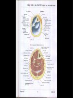
FIGURE 9.1. Cross-section through the abdomen, illustrating epithelial membranes.
9.
Concepts and Terminology 83
Lumen
Membranes
Adventitia
Connective tissue of serosa
Cutaneous membrane
Dermis
Epidermis
Epithelium
Keratin
Lamina propria
Lumen
Mesothelium
Mucosa
Mucous membrane
Muscularis externa
Muscularis mucosae
Peritoneal space
Serosa
Serous membrane
Simple squamous epithelium
Submucosa
Cortex and medulla
Cortex
Medulla
Stroma and parenchyma
Parenchyma
Stroma
Cortex vs. Medulla
➢ Components
᭹
Cortex. The outer region or portion of some organs, such as kidney,
lymph nodes, and adrenal glands; surrounds the internal medulla
᭹
Medulla. The center portion of some organs; surrounded by a
cortex
➢ Differentiation of these two subdivisions is a result of their different
components, origins and/or functions.
Stroma vs. Parenchyma
➢ Stroma. The supporting framework of an organ, usually composed of
connective tissue
➢ Parenchyma. The cells and tissues of an organ that perform the func-
tion of the organ; composed of epithelium, muscle, nerve and, some-
times, connective tissue
Structures Identified in This Section
General Considerations
➢ Continuous tubular system for transporting blood, carrying oxygen,
carbon dioxide, hormones, nutrients, and wastes
➢ Components of the circulatory system
᭹
Heart. Highly modified, muscular blood vessel specialized for
pumping the blood. Composed of two atria and two ventricles.
᭹
Closed circuit of vessels. The vessels are listed below in the order that
blood would follow as it leaves the heart.
᭜
Elastic arteries (e.g., aorta and pulmonary arteries)
᭜
Muscular arteries (remaining named arteries)
᭜
Small arteries and arterioles
᭜
Capillaries
᭜
Venules and small veins
᭜
Medium veins (most named veins)
᭜
Large veins (e.g., venae cavae return blood to the heart)
➢ Circuitry of the circulatory system
᭹
Pulmonary circulation
᭜
Circuit of blood between the heart and lungs
CHAPTER
10
Cardiovascular System
85
Digital Histology: An Interactive CD Atlas with Review Text, by Alice S. Pakurar and
John W. Bigbee
ISBN 0-471-64982-1 Copyright © 2004 John Wiley & Sons, Inc.
᭜
Blood leaves the right ventricle of the heart through pulmonary
arteries and proceeds through a series of smaller arteries to
supply pulmonary capillaries in the lungs. Blood returns through
a series of increasingly larger veins to the pulmonary veins to the
left atrium.
᭜
Functions for exchange of carbon dioxide and oxygen between
the blood and atmosphere
᭹
Systemic circulation
᭜
Circuit that distributes blood from the heart to the body
tissues
᭜
Blood leaves the left ventricle of the heart through the aorta and
proceeds through a series of smaller arteries to supply systemic
capillaries throughout the body. Blood returns through a series
of increasingly larger veins via the superior and inferior venae
cavae to the right atrium.
᭜
Functions for exchange of carbon dioxide and oxygen, and
nutrients and metabolic wastes between the blood and tissues;
distribution of hormones.
᭹
Lymphatic circulation. Consists of a system of blind-ended lymph
vessels positioned throughout the body, which return tissue fluid
to the venous circulation.
Basic Structural Organization
➢ The walls of the entire cardiovascular system, consists of three con-
centric layers or tunics that are continuous between both the heart
and vessels. The constituents and thickness of these layers vary
depending on the mechanical and metabolic functions of the vessel.
➢ Inner tunic
᭹
In the heart, this layer is called the endocardium; in vessels it is
termed the tunica intima.
᭹
Composition
᭜
Simple squamous epithelium (endothelium)
᭜
Varying amounts and types of connective tissue
᭜
In the largest vessels, longitudinally oriented smooth muscle
may be present in the connective tissue layer.
➢ Middle tunic
᭹
In the heart this layer is composed of cardiac muscle and is called
the myocardium.
86
Digital Histology
᭹
In vessels this layer is composed of circularly oriented smooth
muscle or smooth muscle plus connective tissue and is called the
tunica media.
➢ Outer tunic
᭹
In the heart, this layer consists of a serous membrane, called the
epicardium (visceral pericardium) composed of connective tissue
covered with a simple squamous epithelium (mesothelium).
᭹
In vessels, this layer is called the tunica adventitia and is composed
of connective tissue; variable amount of longitudinally arranged
smooth muscle is present in this layer in the largest veins.
᭹
Possesses blood vessels that supply the wall of the heart or larger
blood vessels
᭜
Coronary blood vessels. Supply the heart wall
᭜
Vasa vasorum. Consists of a system of small blood vessels that
supply the outer wall of larger vessels
Arteries
➢ General considerations
᭹
Carry blood away from the heart and toward capillary beds
᭹
Have thicker walls and smaller lumens than veins of similar size
᭹
Tunica media is the predominate tunic.
᭹
Cross-sectional outlines are more circular in arteries than in veins.
10.
Cardiovascular System 87
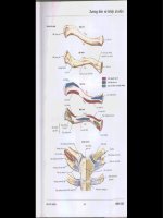
FIGURE 10.1. Structure of a muscular artery.
᭹
Types
᭜
Elastic (large) arteries (aorta, pulmonary arteries)
Internal elastic lamina is present but difficult to distinguish.
Tunica media is composed of fenestrated sheets of elastic
tissue (elastic lamellae) and smooth muscle.
Passively maintain blood pressure by distension and recoil of
the elastic sheets
᭜
Muscular (medium, distributing)
Tunica media is composed of smooth muscle.
Internal elastic lamina. Single, fenestrated, elastic sheet; lies
internal to the smooth muscle of the tunica media.
External elastic laminae. Multiple elastic sheets; lie external to
the smooth muscle of the tunica media
Regulate blood pressure and blood distribution by contrac-
tion and relaxation of smooth muscle in the tunica media
᭜
Small arteries and arterioles
Less than 200 microns in diameter
Small arteries have an internal elastic lamina and up to eight
layers of smooth muscle in the tunica media.
Arterioles usually lack an internal elastic lamina and have one
to two layers of smooth muscle in the tunica media.
Arterioles are the vessels that regulate blood pressure and
deliver blood under low pressure to capillaries.
Capillaries
➢ General considerations
᭹
Function to exchange oxygen and carbon dioxide and nutrients
and metabolic wastes between blood and cells
᭹
Lumen is approximately 8 microns in diameter, thus only large
enough for RBCs to move through in a single row.
᭹
Composed of the endothelium (simple squamous epithelium) and
its underlying basal lamina
➢ Types
᭹
Continuous capillaries
᭜
Most common
᭜
Endothelium is continuous (i.e., has no pores)
88
Digital Histology
᭹
Fenestrated capillaries
᭜
Endothelium contains pores that may or may not be spanned
by a diaphragm. If present, the diaphragm is thinner than two
apposed plasma membranes.
᭜
Pores with diaphragms are common in capillaries in the
endocrine organs and portions of the digestive tract. Pores
lacking diaphragms are uniquely present in the glomerular cap-
illaries of the kidney.
᭜
Pores facilitate diffusion across the endothelium
᭹
Discontinuous sinusoidal capillaries
᭜
Larger diameter and slower blood flow than in other capillaries
᭜
Endothelium has large pores that are not closed by a diaphragm.
᭜
Gaps are present between adjacent endothelial cells.
᭜
Partial or no basal lamina present.
᭜
Prominent in spleen and liver
Veins
➢ General considerations
᭹
Return blood from capillary beds to the heart
᭹
Have thinner walls and larger lumens than arteries of similar size;
cross-sectional outlines are more irregular
᭹
Tunica adventitia is the predominate tunic.
᭹
Larger veins possess valves, that are extensions of the tunica intima
that serve to prevent back-flow of blood.
᭹
Types
᭜
Venules and small veins
Tunica media is absent in venules. Smooth muscle fibers
appear in the tunica media as venules progress to small veins.
High endothelial venules. Venules in which the endothelium is
simple cuboidal; facilitate movement of cells from the blood
into the surrounding tissues (diapedesis). This type of venule
is found in many of the lymphatic tissues.
᭜
Medium veins. Smooth muscle forms a more definitive and con-
tinuous tunica media; most named veins are in this category.
᭜
Large veins, includes superior and inferior venae cavae; have
well-developed, longitudinally oriented smooth muscle in the
tunica adventitia in addition to the smooth muscle in the tunica
media.
10.
Cardiovascular System 89
Heart
➢ Develops by a vessel folding back on itself to produce four chambers
in the adult. Two upper chambers, atria (singular, atrium), receive
blood from the body and lungs; two ventricles pump blood out of the
heart.
➢ Impulse conducting system. Formed of specialized cardiac muscle
fibers that initiate and coordinate the contraction of the heart
᭹
Sinoatrial (SA) node in the right atrium is the electrical pacemaker
that initiates the impuse.
᭹
Fibers spread the impulse throughout the atria as well as transfer-
ring it to the atrioventricular node.
᭹
The atrioventricular (AV) node is located in the interatrial septum.
᭹
An atrioventricular bundle extends from the AV node in the septum
membranaceum and bifurcates into right and left bundle branches that
lie beneath the endocardium on both sides of the interventricular
septum.
᭹
Purkinje fibers, modified, enlarged cardiac muscle fibers leave the
bundle branches to innervate the myocardium.
➢ Tunics
᭹
Endocardium
᭜
Homologous to the tunica intima of vessels
90
Digital Histology
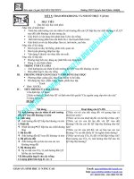
FIGURE 10.2. Diagram of a frontal section of the heart (RA, right atrium; LA, left
atrium; RV, right ventricle; LV, left ventricle).
10.
Cardiovascular System 91
Blood cells
Neutrophil (polymorphonuclear,
PMN)
Eosinophil
Basophil
Lymphocyte
Monocyte
Platelets
Heart
Cardiac skeleton and valves
Annulus fibrosus
Aortic (seminlunar) valve
Atrioventricular valve
Septum membranaceum
Chambers
Left atrium
᭜
Consists of an endothelium (simple squamous epithelium) plus
underlying connective tissue
᭜
Cardiac valves. Folds of the endocardium
Semilunar valves at the base of the aortic and pulmonary
trunks prevent backflow of blood into the heart.
Atrioventricular valves (bicuspid and tricuspid) prevent backflow
of blood from the ventricles into the atria.
᭹
Myocardium
᭜
Composed of cardiac muscle
᭜
Fibers insert on components of the cardiac skeleton.
᭜
Thickest layer of the heart
᭜
Variation in thickness depends on the function of each chamber;
thicker in ventricles than atria and thicker in left ventricle than
right ventricle
᭹
Epicardium (visceral pericardium)
᭜
Serous membrane on the surface of the myocardium
᭜
Consists of a simple squamous epithelium and a loose connec-
tive tissue, with adipocytes, adjacent to the myocardium.
᭜
Coronary blood vessels are located in the connective tissue.
᭹
Cardiac skeleton. Thickened regions of dense connective tissue that
provide support for heart valves and serve as insertion of cardiac
muscle fibers
᭜
Annuli fibrosi are connective tissue rings that surround and sta-
bilize each valve.
᭜
Septum membranceum is a connective tissue partition forming
the upper portion of the interventricular septum; this con-
nective tissue also separates the left ventricle from the right
atrium.
Structures Identified in This Section
92
Digital Histology
Left ventricle
Right atrium
Right ventricle
Conducting system
Atrioventricular bundle (of
His)
Purkinje fibers
Right and left bundle branches
Wall
Cardiac muscle fibers
Endocardium
Epicardium
Interatrial septum
Intercalated discs
Interventricular septum
Myocardium
Vessels
Structures
Elastic lamellae
Endothelium
External elastic laminae
Internal elastic lamina
Pericyte
Smooth muscle cells
(longitudinal and circular)
Subendothelium of tunica
intima
Tunica adventitia
Tunica intima
Tunica media
Vasa vasorum
Types
Arteriole
Elastic artery (aorta)
High endothelial venule
Large vein (inferior vena cava)
Muscular artery
Small/medium vein
Transitional artery
Venule
Capillaries (blood)
Continuous
Fenestrated (with endothelial
cell fenestrae and
diaphragms)
Discontinuous sinusoidal
capillary
Lymphatic capillaries
Lymphatic vessels
Functions of Skin
➢ Protection against physical abrasion, chemical irritants, pathogens,
UV radiation, and dessication
➢ Thermoregulation
➢ Reception of pressure and touch sensations
➢ Production of vitamin D
➢ Excretion
Components of the Integument
➢ Epidermis. Stratified squamous keratinized epithelium
➢ Dermis. Composed of two layers of connective tissue containing
blood vessels, nerves, sensory receptors, and sweat and sebaceous
glands. Beneath the dermis is a layer of loose connective and adipose
tissues that forms the superficial fascia of gross anatomy termed the
hypodermis. This layer is considered along with the skin, though tech-
nically it is not part of the integument.
CHAPTER
11
Skin
93
Digital Histology: An Interactive CD Atlas with Review Text, by Alice S. Pakurar and
John W. Bigbee
ISBN 0-471-64982-1 Copyright © 2004 John Wiley & Sons, Inc.