Báo cáo y học: " Draft genome sequence of the Daphnia pathogen Octosporea bayeri: insights into the gene content of a large microsporidian genome and a model for host-parasite interactions" pps
Bạn đang xem bản rút gọn của tài liệu. Xem và tải ngay bản đầy đủ của tài liệu tại đây (232.09 KB, 12 trang )
Genome Biology 2009, 10:R106
Open Access
2009Corradiet al.Volume 10, Issue 10, Article R106
Research
Draft genome sequence of the Daphnia pathogen Octosporea bayeri:
insights into the gene content of a large microsporidian genome and
a model for host-parasite interactions
Nicolas Corradi
*
, Karen L Haag
†‡
, Jean-François Pombert
*
, Dieter Ebert
¤
†
and Patrick J Keeling
¤
*
Addresses:
*
Canadian Institute for Advanced Research, The Biodiversity Research Centre, University of British Columbia, University
Boulevard, Vancouver, BC, V6T 1Z4, Canada.
†
Universität Basel, Zoologisches Institut, Evolutionsbiologie, Vesalgasse, CH-4051 Basel,
Switzerland.
‡
Department of Genetics, UFRGS, Porto Alegre, RS 91501-970, Brazil.
¤ These authors contributed equally to this work.
Correspondence: Patrick J Keeling. Email:
© 2009 Corradi et al.; licensee BioMed Central Ltd.
This is an open access article distributed under the terms of the Creative Commons Attribution License ( which
permits unrestricted use, distribution, and reproduction in any medium, provided the original work is properly cited.
The Octosporea bayeri genome sequence<p>The draft genome sequence of Octosporea bayeri, a microsporidian pathogen of Daphnia, provides insights into the content and evo-lution of a large microsporidian genome</p>
Abstract
Background: The highly compacted 2.9-Mb genome of Encephalitozoon cuniculi placed the
microsporidia in the spotlight, encoding a mere 2,000 proteins and a highly reduced suite of
biochemical pathways. This extreme level of reduction is not universal across the microsporidia,
with genomes known to vary up to sixfold in size, suggesting that some genomes may harbor a gene
content that is not as reduced as that of Enc. cuniculi. In this study, we present an in-depth survey
of the large genome of Octosporea bayeri, a pathogen of Daphnia magna, with an estimated genome
size of 24 Mb, in order to shed light on the organization and content of a large microsporidian
genome.
Results: Using Illumina sequencing, 898 Mb of O. bayeri genome sequence was generated, resulting
in 13.3 Mb of unique sequence. We annotated a total of 2,174 genes, of which 893 encodes proteins
with assigned function. The gene density of the O. bayeri genome is very low on average, but also
highly uneven, so gene-dense regions also occur. The data presented here suggest that the O. bayeri
proteome is well represented in this analysis and is more complex that that of Enc. cuniculi.
Functional annotation of O. bayeri proteins suggests that this species might be less biochemically
dependent on its host for its metabolism than its more reduced relatives.
Conclusions: The combination of the data presented here, together with the imminent annotated
genome of Daphnia magna, will provide a wealth of genetic and genomic tools to study host-parasite
interactions in an interesting model for pathogenesis.
Published: 6 October 2009
Genome Biology 2009, 10:R106 (doi:10.1186/gb-2009-10-10-r106)
Received: 9 July 2009
Revised: 2 September 2009
Accepted: 6 October 2009
The electronic version of this article is the complete one and can be
found online at /> Genome Biology 2009, Volume 10, Issue 10, Article R106 Corradi et al. R106.2
Genome Biology 2009, 10:R106
Background
Microsporidia are extremely successful, highly adapted obli-
gate intracellular parasites known to infect a wide range of
animals, such as arthropods, fish, and mammals, including
humans [1,2]. These parasites are characterized by the pres-
ence of a highly specialized host invasion apparatus called the
polar tube (or polar filament), which is used to penetrate and
infect new host cells. Microsporidian cells significantly differ
from other eukaryotes, as they lack conventional mitochon-
dria and Golgi apparatus and harbor 70S instead of 80S
ribosomes [3-5]. These features were once taken to suggest
that microsporidia represent a very ancient eukaryotic line-
age [6-11], but recent advances in cell biology, genome
sequencing, and phylogenetic reconstruction have all shown
that all these apparently primitive features instead reflect an
extreme state of reduction, perhaps a result of their obligate
intracellular parasitic lifestyle. Instead, it is now widely
acknowledged that microsporidia are, in fact, related to fungi,
and have relict mitochondria (called mitosomes) [12], degen-
erated eukaryote-like ribosomal RNA subunits [13], and
reduced genes and genomes [14-24].
The extremely reduced nature of microsporidian genomes
has attracted attention since they were first noted at the end
of the 1990s [13], culminating in 2001 with the completion of
the first microsporidian genome from the mammalian para-
site Encephalitozoon cuniculi [25]. The Enc. cuniculi genome
is extremely small, at only 2.9 Mb, and the 2,000 genes it
encodes provided the first compelling evidence for a strong
correlation between obligate intracellular parasitism and the
loss of metabolically important genes in eukaryotes. Meta-
bolic capabilities are indeed significantly reduced in Enc.
cuniculi, and genes required for de novo biosynthesis of
purine and pyrimidine nucleotides or those involved in the
tricarboxylic acid cycle, fatty acid beta-oxidation, respiratory
electron-transport chain and the F
0
F
1
-ATPase complex are
completely absent from its genome. The reduction of several
metabolic pathways in Enc. cuniculi implied that these para-
sites might be extremely dependent on their host for obtain-
ing most of their metabolites and energy. For example, it has
been indeed recently demonstrated that this parasite and its
mitosomes both import ATP from its host via specific trans-
porters [26,27].
In addition to a significant reduction in its metabolic capabil-
ities, the genome of Enc. cuniculi is also very compact. Its
genes are reduced in size and separated by remarkably short
intergenic regions. This extreme compaction has impacted
the process of transcription so that in the microsporidia Enc.
cuniculi and Antonospora locustae a significant part of their
mRNA transcripts has been found to overlap between adja-
cent genes [28-30]. Genome reduction has also apparently
affected the rate of gene rearrangement, as conservation of
gene order is strikingly high among microsporidia compared
to what has been reported for other eukaryotes [31,32].
Since the completion of the Enc. cuniculi genome, new
genomic data from other microsporidian parasites have been
limited to two in-depth genome surveys from Enterocytozoon
bieneusi and Nosema ceranae [33,34], a smaller survey from
A. locustae [32] and some very small surveys from various
other species [35-38]. The deeper-sampled genomes of Ent.
bieneusi and A. locustae show many similarities with that of
Enc. cuniculi - all three genomes are compact and contain
roughly the same number of genes and pathways - but this is
perhaps not surprising because all three genomes are also rel-
atively small (ranging from 2.9 to 6 Mb) and might not, there-
fore, represent all microsporidian genomes adequately.
So how do larger microsporidian genomes compare with
smaller ones? Does their large size reflect the presence of
more genes and pathways or do they harbor the same genes
but separated by much larger intergenic regions? These ques-
tions have been partly addressed with genome surveys from
Spraguea lophii [35], Vittaforma cornea [36], Edhazardia
aedis, and Brachiola algerae [37,38], but because of their
very low sequence coverage no conclusion can be drawn
about their overall gene content and evolution. In the present
study, we provide a 37× sequence coverage of the large
genome of the microsporidian Octosporea bayeri. O. bayeri
is a parasite of the freshwater planktonic crusteacean Daph-
nia magna [39]. Other Daphnia species have never been
found to be infected. The parasite is both horizontally and
vertically transmitted [40]. Vertical transmission occurs with
100% efficiency to the asexual (parthenogenetic) eggs of the
host and with somewhat reduced efficiency to the sexual eggs.
Horizontal transmission occurs after the host cadaver decom-
poses and environmental spores are released. Infection fol-
lows ingestion of spores by the filter feeding host. The
parasite reduces host survival and fecundity. Its geographic
distribution is limited to rock pool D. magna
populations
along the baltic Sea in Finland and Sweden [39] and a single
report from the Czech Republic.
From our sequence survey, over 13 Mb of unique O. bayeri
sequence data have been assembled and 2,174 ORFs have
been identified, providing an excellent framework to charac-
terize the overall gene content and structure of a large micro-
sporidian genome, to compare it with its more reduced
relatives and to increase the availability of genetic markers
from this latter species. Consistent with small surveys from
microsporidia with large genomes, the gene density of the O.
bayeri genome is generally low but also highly variable. Most
of the genes known in the Enc. cuniculi genome are also found
in O. bayeri, but a number of other genes are also found that
are apparently absent in other microsporidia. The functional
distribution of the proteins significantly differed between O.
bayeri and its more reduced relatives, suggesting the meta-
bolic capacity and host dependency within the group is also
variable. The wealth of genomic data from this parasite cou-
pled with the annotation of the Daphnia genome should fur-
Genome Biology 2009, Volume 10, Issue 10, Article R106 Corradi et al. R106.3
Genome Biology 2009, 10:R106
ther increase the interest for this model of host-parasite
interactions [41].
Results
Gene content of the O. bayeri genome
Approximately 898 Mb of DNA sequence was obtained from
shotgun and paired-end 35-bp reads with the Illumina
Genome Analyzer™, resulting in an estimated 34.2 to 37.2×
coverage of the O. bayeri genome, which has been estimated
to 24 Mb based on total number of bases sequenced divided
by the average coverage. This calculation does not take into
account the fact that some assembled contigs might represent
several identical regions in the reference genome, and that
unassembled reads might represent DNA sequences from
other sources (that is, contaminants). Reads were assembled
into 41,804 contigs representing a total of 13.3 Mb of
sequence data (26% G+C), with only 20 contigs displaying
evidence of contamination. The length of contigs averaged
320 bp (100 bp to a maximum of 8 kb). The small size of most
contigs resulted in the incompleteness of most ORFs identi-
fied in this study and, on average, incomplete ORFs were
found to encode 60% of the amino acids of their respective
eukaryotic homologs. This explains why the complete (or
almost complete) O. bayeri proteome has been identified
within an assembly that is almost half the size of the esti-
mated genome.
A total of four rRNA genes, 37 tRNAs and 2,174 predicted pro-
tein-coding ORFs were identified (Table 1). Of the O. bayeri
ORFs, 1,405 were found to have homologs in the Enc. cuniculi
genome, representing about 70% of its annotated genes [25]
(Additional data file 1). Over 93% of Enc. cuniculi proteins
with assigned functions and 53% of its hypothetical proteins
had clear homologs in the O. bayeri genome [25,33]. Over
25% of Enc. cuniculi homologs identified are full length, while
others were slightly truncated in the carboxy-terminal or
amino-terminal regions, or both. Another 80 ORFs were
identified that were found to have homologs in other organ-
isms, but not Enc. cuniculi, 72 of which could be assigned to a
functional category (Additional data file 2), the majority of
which have highest similarities with fungal homologs, sug-
gesting that they are ancestral within the lineage and not
recently introduced into the O. bayeri genome. The remain-
ing 689 O. bayeri putative ORFs (of at least 200 amino acids)
returned no significant hits in BLAST homology searches
against the National Center for Biotechnology Information
(NCBI) non-redundant database. However, 25 of these
showed significant similarities with hypothetical proteins
from the A. locustae database, indicating that O. bayeri and
A. locustae share a number of hypothetical proteins that are
Table 1
General characteristics of O. bayeri and other microsporidian genomes
General characteristics O. bayeri Enc. cuniculi Ent. Bieneusi
Number of chromosomes NA 11 6
Genome size (Mb) 24.2* 2.9 6
Assembled Mb 13.3 2.5 3.86
Genome coverage (%) 55
†
86 64
G+C content (%) 26 47 25
Gene density 1 per 4,593 bases
‡
1 per 1,025 bases 1 per 1,148 bases
Mean intergenic region (bp) 429
§
129 127
Presence of overlapping genes No Yes Yes
Number of SSU-LSU rRNA genes 2
¶
22 Unkown
¥
Number of 5S rRNA genes 2
¶
3Unkown
¥
Number of tRNAs 37 46 46
Number of tRNA synthetases 21 21 21
Number of tRNA introns (size in bp) 1 (50) 2 (16, 42) 2 (13, 30)
Number of splicesomal introns (size in bp) 6 (24-33) 13 (23-52) 19 (36-306)
Number of predicted ORFs 2,174
#
1,997 3,804**
Number of ORFs assigned to functional categories 894 (41%) 884 (44%) 669 (39%)
Mean size of CDS (bp) 1,056
††
1,017
††
1,002
††
*The genome size has been estimated using total number of bases sequenced divided by the average coverage.
†
Based on the 24.2-Mb estimated
genome size.
‡
Based on the 200 largest contigs.
§
Based on contigs (n = 23) in which two or more ORFs of at least 100 amino acids have been
identified.
¶
Only two contigs harboring an SSU-5.8S-LSU gene array have been identified in the O. bayeri genome survey.
¥
Based on [33].
#
Includes
ORFs with assigned functions, homologs of Enc. cuniculi hypothetical proteins, and hypothetical proteins of at least 200 amino acids identified in the
O. bayeri genome. **The Ent. bieneusi genome has been subjected to several segmental duplications and the number of ORFs identified in that study
includes a very large number of duplicates [33]. This number should, therefore, not be taken into account to determine the haploid coding capacity
of this species.
††
Based on 95 and 63 complete Enc. cuniculi and Ent. bieneusi orthologs, respectively. CDS, coding sequence.
Genome Biology 2009, Volume 10, Issue 10, Article R106 Corradi et al. R106.4
Genome Biology 2009, 10:R106
absent in Enc. cuniculi and Ent. bieneusi. It is also important
to note that a large proportion of microsporidian hypothetical
proteins have been found to be smaller than 200 amino acids
[25,31-33], so the actual number of ORFs could be over 25%
higher than what we report here, perhaps in the range of, or
higher than, what has been recently reported for N. ceranae
[34].
Functional categories represented in O. bayeri
All identified O. bayeri ORFs were assigned to the 11 func-
tional categories listed in [25,33] (Figure 1; Additional data
file 3). Such comparison is currently unavailable for N. cera-
nae [34]. O. bayeri ORFs are well distributed among the func-
tional categories, yet display differences when compared to
Enc. cuniculi and Ent. bieneusi. Specifically, five categories
(metabolism, energy production, cell growth and DNA syn-
thesis, transcription and protein destination) are more repre-
sented in O. bayeri than in Enc. cuniculi and Ent. bieneusi,
whereas four other categories (transport facilitation, intracel-
lular transport, cellular organization - biogenesis, and cell
rescue) are reduced in number in O. bayeri. Within each
functional category, several pathways stood out as being par-
ticularly different among the three species. For instance,
genes involved in lipid and fatty acid metabolism and glyco-
sylation were better represented in O. bayeri (37 and 12 pro-
teins, respectively) than either Enc. cuniculi (29 and 7
proteins) or Ent. bieneusi (8 and 5 proteins), while proteins
involved in the translocation of various substrates across
membranes are underrepresented in O. bayeri (Figure 2).
Finally, in contrast to what has been reported for other spe-
cies with smaller genomes [33,34], no evidence for gene or
segmental genome duplication events has been identified in
the present survey.
Phylogeny of O. bayeri and evolution of the ATP
transporters in the microsporidia
O. bayeri was put into a phylogenetic context by comparing
the amino acid sequences from its newly identified alpha- and
beta-tubulins with those of other microsporidia (Figure 3a).
Our tree is consistent with the most recently reported using
the same amino acid sequences [42]. Specifically, Nosema
and Encephalitozoon are sisters to one another, as are Anton-
ospora and Brachiola. The remaining species all branch more
deeply, and O. bayeri is in this tree basal to all other micro-
sporidian species from which large genome sequence data are
presently available. Only a single ATP transporter protein was
identified in O. bayeri, and phylogenetic analyses of all pres-
ently known microsporidian members of this family show the
O. bayeri protein clustering with strong support at the base of
a clade including Antonospora and Brachiola homologues,
all of which are sister to the Encephalitozoon/Enterocyto-
zoon/Nosema clade (Figure 3b). This is not consistent with
the rRNA tree, and might represent a mis-rooting of either
tree, or ancient paralogy of the ATP transporters.
O. bayeri introns
Only 13 introns have been annotated in the Enc. cuniculi
genome at present, and we identified a total of 6 introns in the
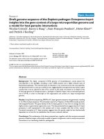
Distribution of O. bayeri (blue), Enc. cuniculi (yellow) and Ent. bieneusi (red) proteins among functional categoriesFigure 1
Distribution of O. bayeri (blue), Enc. cuniculi (yellow) and Ent. bieneusi (red) proteins among functional categories. The ordinate represents
the number of ORFs assigned to the corresponding category. Each of the O. bayeri proteins was assigned to only one of eleven functional categories listed
in [25,33]. The corresponding gene list is presented in the online version of this manuscript (Additional data file 3). *Based on a 4× sequence coverage
[33].
O. bayeri
Enc. cuniculi
Ent. bieneusi
0
50
100
150
200
Energy
Metabolism
Cell
growth,
division
and DNA
synthesis
Transcription
Protein
synthesis
Protein
destination
Transport
facilitation
Intracellular
Transport
Cellular
organisation -
Biogenesis
Communi-
cation -
Signal
transduc-
tion
Cell rescue,
defense,
death and
aging
*
Genome Biology 2009, Volume 10, Issue 10, Article R106 Corradi et al. R106.5
Genome Biology 2009, 10:R106
present survey, all of which are homologous to introns
reported in Enc. cuniculi ribosomal protein genes (L19, L27a,
L37a, L37, L39, S26) [25]. All the O. bayeri introns identified
here are located within or close to the start codon, which is
consistent with the introns in Enc. cuniculi [25], Saccharo-
myces cerevisiae [43] and cryptomonad nucleomorphs [44].
The retention of the majority of these introns leads to frame-
shifts and termination codons, while their removal leads to a
complete ORF that is highly conserved with homologs from
other eukaryotes. The intron sequences are available with the
online version of this paper (Additional data file 4).
O. bayeri-specific large amino acid insertions
A number of large insertions ranging from 15 to 57 amino
acids were identified in 14 conserved proteins in O. bayeri (O-
sialoglycoprotein endopeptidase, 3-hydroxy-3-methylglu-
taryl CoA reductase, 3-ketoacyl CoA thiolase, -trehalase
precursor, choline phosphate cytidyltranferase, transcription
factor of the E2F/DP family, tubulin -chain, kinesin-like pro-
tein, pyruvate dehydrogenase E1 component subunit , repli-
cation factor C, T complex protein 1 subunit, threonyl tRNA
synthetase, and translation elongation factor 2). These inser-
tions are all in-frame and in most cases are surrounded by
highly conserved amino acid motifs, although they are not
generally located within functionally important domains
(Additional data file 5). RT-PCR confirmed that none of these
inserts are removed from mRNA and so do not represent spli-
ceosomal introns (data not shown). Similar insertions have
been previously reported in the parasites Plasmodium
berghei and Toxoplasma gondii [45,46].
Length of O. bayeri proteins
The majority of O. bayeri proteins were found to be larger
than homologs from Enc. cuniculi (69%) and Ent. bieneusi
(65%) (Figure 4). However, the opposite trend was identified
when O. bayeri genes were compared with other fungal line-
ages, in which case the majority of O. bayeri proteins (75% on
average) were found to be smaller than homologs from the
other fungal lineages, even when the fungal species compared
had a smaller genome than O. bayeri. The difference in the
number of amino acids was found to be significantly larger
between O. bayeri and other fungal lineages (14% smaller on
average) than between O. bayeri and other microsporidia (3%
larger on average) (Figure 4).
Gene density and synteny
Gene density and synteny in O. bayeri were examined by
annotating all ORFs of at least 100 amino acids on the 200
largest contigs (average length of 2,795 bp). In more than half
of these contigs, no putative ORF could be identified. One
contig was found to harbor three putative ORFs, whereas 72
and 22 contigs harbored one or two recognizable ORFs,
respectively. No correlation between the length of the contigs
and the number of ORFs could be identified (Figure 5a).
Based on these contigs, gene density was calculated to be 1
gene every 4,593 bases. However, when two or more ORFs
were identified on the same contig the average intergenic
region was calculated to be only 429 bp, suggesting the gene
density is highly variable across the genome. Conservation in
gene order could be identified in only two cases, representing
8% of all the gene pairs identified (Figure 5b).
Repeated elements
The large amount of small, non-coding DNA sequences iden-
tified in this study could reflect the presence of highly
repeated sequences in the O. bayeri genome. This possibility
was investigated by measuring the sequence coverage of each
contig and identifying a possible correlation with their length.
As suspected, the contigs with highest coverage are also the
smallest. Specifically, all contigs with a coverage over 200×
are smaller than 300 bp, suggesting these are highly repeti-
tive (Additional data file 6).
The presence of repeated elements was also investigated
among all contigs. A total of 74 O. bayeri contigs harbor DNA
segments homologous to known fungal repeated elements
(Additional data file 7). The Mariner, Gypsy and Copia classes
of repeated elements are the most frequently observed in O.
bayeri. The O. bayeri contigs also display DNA strings that
are repeated in tandem, with strings repeated at least twice
identified in 1,345 contigs (data not shown). However, these
tandem repeats are usually short and rarely exceed ten con-
secutive repeated strings. Putative stem-loop structures with
Examples of sub-functional categories showing sharp differences in distribution between O. bayeri (blue), Enc. cuniculi (yellow) and Ent. bieneusi (red) proteinsFigure 2
Examples of sub-functional categories showing sharp differences
in distribution between O. bayeri (blue), Enc. cuniculi (yellow) and
Ent. bieneusi (red) proteins. (a) Functional sub-categories more highly
represented in O. bayeri than in Enc. cuniculi and Ent. bieneusi. (b)
Functional sub-categories less represented in O. bayeri than in Enc. cuniculi
and Ent. bieneusi. *Based on a 4× sequence coverage [33] (that is, almost
10 times lower than the present genome draft), suggesting a number of
these transporters may yet be identified in the Ent. bieneusi genome
survey.
0
5
10
15
20
25
30
35
40
Glycosylation Lipid fatty
Biosynthesis
ADP/ATP
Transporters
ABC
Transporters
(a) (b)
O. bayeri
Enc. cuniculi
Ent. bieneusi
*
Genome Biology 2009, Volume 10, Issue 10, Article R106 Corradi et al. R106.6
Genome Biology 2009, 10:R106
AT-rich palindromic stems have been identified in a number
of contigs, although the primary sequences of these potential
structures, aside from their biased nucleotide composition,
do not appear to be repeated per se.
Discussion
Architecture of a large microsporidian genome
The currently available microsporidian genomes best repre-
sent the lower limits in the spectrum of genome sizes, not only
for Eukaryotes as a whole, but also microsporidia. The single
exception to this is N. ceranea, whose genome is more inter-
mediate in size, but our knowledge of microsporidian
genomes is still strongly biased, which might hinder the elu-
cidation of the evolution of this poorly understood group. Our
present survey of the O. bayeri genome is the first deep sur-
vey of a larger microsporidian genome, and estimates from
sequence coverage suggest it may even be the largest known
microsporidian genome (at 24 MB). What accounts for this
variation in genome size and which features of microsporid-
ian genomes have to be reconsidered after adding a genome
from the other end of the genome size spectrum? There are
several answers to these questions.
Phylogenetic relationships of microsporidia and their ATP transportersFigure 3
Phylogenetic relationships of microsporidia and their ATP transporters. (a) Phylogenetic reconstruction of the microsporidian phylogeny based
on available - and -tubulin amino acid sequences and gains of ATP and ABC transporters. Known genome sizes and number of transporters are shown.
Ent. bieneusi tubulins cluster as a sister group to the clade including Encephalitozoon and Nosema species; this position is represented by a black square. (b)
Evolution of the ATP transporter family based on available amino acid sequences from a range of microsporidian parasites. 1, Putative ancestral duplication
of ATP transporters within the microsporidia following lateral gene transfer from prokaryotes. 2, A putative secondary gene duplication occurred in the
more diverged genera, Nosema, Enterocytozoon and Encephalitozoon. 3, Supported lineage including all three diverged genera. 4, Species-specific duplication
of an ATP transporter. *Data from NC, JFP et al., unpublished.
0.05
2.5Mb
2.3Mb
2.9Mb
413
12
8
4
1
≥3*
<10Mb
15Mb
5.4Mb
<20Mb
6.2Mb
24Mb
19.5Mb
?
??
?
4*
4*
?
?
?
?
?
?
?
?
?
?
?
?
0
0
?
?
?
?
?
20Mb
?
~8.9 (?)
Concatenated α- and β-tubulin microsporidian phylogeny
Reported genome size
# of ADP/ATP
Transporters
# of Proteins
with "ABC" motifs
Nosema bombycis
Nosema ceranae
Encephalitozoon cuniculi
Encephalitozoon intestinalis
Encephalitozoon hellem
Antonospora locustae
Brachiola algerae
Microsporidia sp. AMVB
Edhazardia aedis
Glugea plecoglossi
Trachipleistophora hominis
Spraguea lophii
Octosporea bayeri
Conidiobolus coronatus
Entomophaga maimaiga
100
100
100
100
100
98
-
99
100
100
Large set
of transporters
Reduced set of transporters
(a)
Chlamydophila pneumoniae
Chlamydophila abortus
0.2
Nosema ceranae
Nosema ceranae
Nosema ceranae
Nosema ceranae
Antonospora locustae
Antonospora locustae
Antonospora locustae
Octosporea bayeri
Enterocytozoon bieneusi
Paranosema grylli
100
100
99
100
97
100
96
100
100
79
81
76
82
100
Enterocytozoon bieneusi
Enterocytozoon bieneusi
Enterocytozoon bieneusi
Encephalitozoon cuniculi
Encephalitozoon cuniculi
Encephalitozoon cuniculi
Encephalitozoon cuniculi
ATP transporters Clade II
Diverged ATP transporters?
ATP transporters Clade I
Ancestral ATP transporters?
1
2
74
3
(b)
Genome Biology 2009, Volume 10, Issue 10, Article R106 Corradi et al. R106.7
Genome Biology 2009, 10:R106
Genomes might be larger due to the presence of more genes,
which could be due to whole or partial genome duplications,
repetitive sequences, expansion of gene families, or the reten-
tion of a greater diversity of genes in general. They might also
have about the same complement of genes but have larger
intergenic regions, more or larger introns, more transposable
elements, and so on. Previous small-scale surveys of micro-
sporidia with larger genomes have demonstrated a higher
proportion of non-coding DNA, but reveal nothing about the
overall organization of the genome because the fragments
sampled were small and only a tiny fraction of the genome
was characterized in any one case [35,36,38]. The data pre-
sented here provide additional evidence that large micro-
sporidian genomes have a very low gene density, in this case
up to a fivefold decrease compared to species with smaller
genomes, but also provide information on the organization
and structure of a large genome in this group. [25,32-34].
First, gene density is not homogeneous across the genome,
but is instead a sum of long stretches (5.5 kb) of non-coding
sequences, as well as regions where genes are separated by
only 45 bp, which is even shorter than most intergenic regions
found in Enc. cuniculi, Ent. bieneusi and A. locustae. Second,
it now seems obvious that gene density alone accounts for
most of the variation in genome size between different micro-
sporidian species, although we did find numerous genes in O.
bayeri that are absent in Enc. cuniculi (see below).
Smaller microsporidian genomes have also been noted as
sharing a high conservation of gene order across distantly
related species, which has been attributed to compaction
[31,32,34]. Despite the overall low gene density, we found 8%
of all annotated gene pairs (equating to 2 out of 24 gene pairs)
were conserved in order between O. bayeri and Enc. cuniculi.
This is not very different to what is found in other micro-
sporidia [31,32], and close to the expectation for closely
related fungi [47]. It is interesting that both cases described
here involve pairs of genes that are unusually close to one
another (423 and 15 bp apart). This may reflect the role of
compaction in conservation of gene order, but it might also be
a sampling bias since closely spaced genes are more likely to
Differences in gene length among microsporidia and their fungal relativesFigure 4
Differences in gene length among microsporidia and their fungal
relatives. (a) Comparison of the length (in amino acids) of O. bayeri
proteins to orthologs from Enc. cuniculi, Ent. bieneusi, S. cerevisiae, U.
maydis, B. dendrobatidis and R. oryzae. In general, O. bayeri proteins are
longer than microsporidian orthologues, but shorter than fungal
orthologues. Vertical arrows indicate the average reduction or increase in
protein size compared to O. bayeri. (b) Specific examples of length
variation between orthologs from O. bayeri, Enc. cuniculi, Ent. bieneusi and S.
cerevisiae.
Same
Larger
Smaller
0
200
400
600
800
1000
1200
O. bayeri
E. cuniculi
E. bieneusi
S. cerevisiae
Protein length (in amino acids)
26S
protea-
some beta
subunit
RPL11 Thymidylate
kinase
Peptide
chain
release
factor
subunit 1
Ribo-
nucleoside
diphos-
phate
reductase
small chain
Tubulin
gamma
chain
Gamma
glutamyl
transpep-
tidase
DNA-
directed
RNA
poly-
merase
III
subunit 2
(130kDa)
Zinc protein
(ECU02_
0310)
(a)
(b)
S. cerevisiae
n = 50
~ 15%
U. maydis
n = 49
~ 21%
B. dendrobatidis
n = 52
~ 10%
R. oryzae
n = 51
~ 11%
E. cuniculi
n = 95
~ 2%
E. bieneusi
n = 63
~ 4%
Variation in gene density across the O. bayeri genomeFigure 5
Variation in gene density across the O. bayeri genome. (a)
Identification and distribution of ORFs (of at least 100 amino acids) among
the largest O. bayeri contigs. Only the 100 largest contigs are shown here
for convenience. Yellow dots represent contigs in which no ORF could be
annotated. Blue and red arrows and dots represent contigs harboring two
or one ORF, respectively. (b) Two cases of gene order conservation
between O. bayeri and Enc. cuniculi.
O. bayeri
E. cuniculi
Ecu02_1420 Ecu02_1430
(570bp) (813bp)
71bp
Ecu02_1420 Ecu02_1430
(791bp)(incomplete on 5’)
423bp
Ecu06_0350 Ecu06_0360
(743bp) (824bp)
62bp
Ecu06_0360Ecu06_0350
(incomplete on 5’)(399bp)
45bp
(b)
(a)
0 20 40 60 80 100 120
1000
2000
3000
4000
5000
6000
7000
8000
Length (in bp)
Contig
Chromosome 2
Contig 6939
Chromosome 6
Contig 4605
Genome Biology 2009, Volume 10, Issue 10, Article R106 Corradi et al. R106.8
Genome Biology 2009, 10:R106
be found on the same contig in our survey, which is based on
contigs, rather than a complete genome.
The large size of the O. bayeri genome does not reflect exten-
sive and segmental gene duplication. However, numerous
non-coding and small genomic repetitions could have played
a role in its expansion. The origin of these repetitive regions
is difficult to assess without a better genome assembly.
Because these do not encode known functional proteins, nor
harbor potential ORFs, however, it is possible that these rep-
resent telomeric and sub-telomeric regions of the O. bayeri
genome. If this is the case, genome size variation in micro-
sporidia could also be a consequence of variation in the size of
telomeres. This prediction is supported by the recent acquisi-
tion in our laboratory of genome data from other, much
smaller genomes, showing that the vast majority of unassem-
bled Illumina™ reads belong indeed to telomeric regions
(NC, JFP and PJK, unpublished).
Length of microsporidian genes and size of the protein
network
Microsporidian proteins are known to be shorter in general
than orthologs in other organisms, a characteristic that has
been attributed to the reduction in gene content and, by
extension, protein networks in these cells [25,48]. In keeping
with this, the majority of the O. bayeri proteins are shorter
than orthologs from S. cerevisiae (approximately 5,570
genes, size 12 Mb), Ustilago maydis (approximately 6,500
genes, 20 Mb genome), Batrachytridium dendrobatidis
(approximately 8,700 genes, 24 Mb genome) and Rhizopus
oryzae (approximately 17,459 genes, 35 Mb genome). Inter-
estingly, however, O. bayeri proteins are also larger than
orthologues found in Enc. cuniculi and Ent. bieneusi. Conse-
quently, the O. bayeri genome provides additional evidence
that microsporidian proteins are shorter than their homologs
from other fungal phyla, but also that their size correlates bet-
ter with the coding capacity rather than the size of the genome
in which they are found.
Evidence for the progressive loss of ancestral genes
throughout the evolution of the microsporidian lineage
Prior to this study, the vast majority of genes with predicted
functions found in diverse microsporidia were also found in
Enc. cuniculi [32-36,38]. Three exceptions were found in A.
locustae [49-51] and a single one was found in Ent. bieneusi
[33]. This suggested that all members of this group share a
common core set of genes that have been retained after mas-
sive gene losses occurred in their ancestor, resulting in only a
small degree of variability in gene content. This prediction
was based, however, on a very low coverage for two large
microsporidian genomes [38]. The O. bayeri genome and its
evolutionary position within the group suggest that perhaps
early microsporidians possessed many more genes with pre-
dicted functions than previously thought. It now seems likely
that there was a large reduction in the ancestral proteome fol-
lowing the origin of microsporidia, but this was also followed
by lineage-specific reductions and expansions in some
branches of the microsporidian tree. The total number of
ORFs identified in O. bayeri also suggests an overall coding
capacity that is at least 10% larger than that of Enc. cuniculi.
This is a conservative estimate based on the annotation of O.
bayeri hypothetical proteins of at least 200 amino acids.
Since it is known that Enc. cuniculi proteins shorter than 200
amino acids make up over a quarter of its total coding capac-
ity [25], the overall coding capacity of O. bayeri is almost cer-
tainly greater still. It has been suggested that both N. ceranae
and Enc. bieneusi genomes contain genes that are absent in
Enc. cuniculi; however, the novel sequences in these genomes
are apparently all hypothetical ORFs or transposable-like ele-
ments, and not genes with predicted functions. In these cases
we cannot rule out that these are rapidly evolving genes with
unrecognized homologues in other microsporidian genomes,
or in some cases are not functional genes at all. In contrast,
the genome of O. bayeri contains at least 80 genes with pre-
dicted functions and recognizable homologues in other
organisms, but which are absent in Enc. cuniculi. This con-
firms that the proteome complexity of the ancestral micro-
sporidian was greater than that seen in Enc. cuniculi (and
other current taxa for which genome level surveys have been
conducted to date), and suggests that further genome
sequencing, especially of putatively deep-branching taxa,
should reveal still more genes previously unseen in micro-
sporidia. It is also formally possible that many genes were
acquired relatively recently in the lineage leading to O. bayeri
by lateral gene transfer, which has indeed been observed in
other microsporidia [49]. However, this does not seem likely
for all these genes given the rarity of transferred genes in
other microsporidia, and especially given that the most highly
conserved cases are all notably similar to homologs in fungi,
suggesting they are more likely ancestral to the micro-
sporidia. This implies that much more proteome diversity
awaits discovery as more microsporidian genomes are char-
acterized.
Functional importance of O. bayeri proteins absent in
other microsporidia
Perhaps the most intriguing finding of the present study is the
identification of 80 O. bayeri proteins sharing homology with
eukaryotes but not with Enc. cuniculi. Not surprisingly, these
include eight transposable elements, some of which showed a
high similarity to those reported from Nosema bombycis
[52]. Transposable elements are absent in the most reduced
microsporidian genomes [25,33], but are commonly reported
in the ones that are larger and less compact [34,37,38,52], so
in this case our study simply corroborates previous findings.
The remainder of these eukaryotic proteins stood out for
being involved in important functional processes. In total, 14
are involved in transcriptional processes, including RNA
polymerases or proteins involved in the transcription of
tRNAs, while 19 are part of different metabolic pathways such
as the metabolism of fatty acids and lipids and nucleotide
Genome Biology 2009, Volume 10, Issue 10, Article R106 Corradi et al. R106.9
Genome Biology 2009, 10:R106
metabolism. A whole set of proteins involved in the modifica-
tion of proteins and three cation transporters are also present
in O. bayeri but absent in Enc. cuniculi. The identification of
these eukaryotic proteins is important as it shows that the O.
bayeri proteome is more complex than that of Enc. cuniculi
or Ent. bieneusi. Moreover, most of these proteins have high-
est similarities with homolgs from fungal lineages, suggesting
they arose through common descent rather than by their
recent incorporation into the genome by lateral gene transfer.
Do O. bayeri protein categories reflect a lesser host
dependency?
Aside from the set of O. bayeri proteins that are absent in
Enc. cuniculi, the overall number of proteins with assigned
functions is generally similar in the two genomes. This does
not imply that both species encode the same set of identifiable
eukaryotic homologs and, indeed, we observed several differ-
ences in the functional distribution of their proteins. For
instance, genes involved in lipid and fatty acid metabolism
are at least 25% more common in O. bayeri than in Enc.
cuniculi or Ent. bieneusi. Similarly, O. bayeri harbors two
additional genes involved in energy production compared to
Enc. cuniculi, a trehalose synthase and an alternative oxi-
dase.O. bayeri also harbors almost twice the number of pro-
teins involved in glycosylation compared to Enc. cuniculi,
suggesting a greater capacity to modify proteins, and perhaps
the presence of a less simplified endoplasmic reticulum and
Golgi apparatus compared to other microsporidia [3].
The presence of a larger number of genes for metabolic and
energy generating proteins in O. bayeri does not by itself nec-
essarily mean that this species is less dependent on its host for
energy than are other microsporidia; however, we also
observed a marked underrepresentation of proteins involved
in stealing metabolites from the host. At the extreme, only
one-quarter of the ATP transporters present in other micro-
sporidia and around half of the Enc. cuniculi homologs of
amino acid and sugar transporters were found in O. bayeri.
Octosporea also appears to harbor a reduced set of ABC
transporters compared to both Enc. cuniculi and Ent.
bieneusi. Taken together, this implies that O. bayeri has a
broader metabolic repertoire than other microsporidia while
at the same time a reduced capability to derive metabolic
products and energy from its host, both of which suggest it is
less host-dependent than other microsporidia with smaller
genomes.
The phylogenetic placement of O. bayeri is also consistent
with the idea that host dependency evolved hand in hand with
reduction in genome size and hyper-adaptation for intracellu-
lar parasitism. Indeed, O. bayeri clusters at a basal position in
the microsporidian phylogeny, in the proximity of other spe-
cies characterized by large genomes, and the only ATP trans-
porter identified from this species was also found to be a basal
representative of the gene family. If both phylogenies depict
the correct evolutionary relationships within the micro-
sporidia, then the ancestral genome of microsporidia was
almost certainly large, complex, and encoded few transport-
ers. Certainly, genome surveys of other basal representatives
of the group such as Glugea plecoglossi or Trachipleisto-
phora hominis would provide decisive evidence in support or
against the evolution of reduced microsporidian genomes
from larger and complex relatives. This certainly warrants
further need for investigating the genomics of these highly
adapted and successful parasites.
Conclusions
Not all microsporida are characterized by small and highly
reduced genomes. Here we demonstrate that the proteome
complexity can vary greatly across the different species of the
group, and that a larger genome size could be a good predictor
of increased genomic complexity and reduced host depend-
ency in microsporidia.
Since a microsporidian genome has now been surveyed with
454™ (N. ceranae [34]) and Illumina™ sequencing technol-
ogy (this study), it might be interesting to compare the
results. The 454™ de novo genome assembly of N. ceranae
[34] resulted in lower overall sequence coverage, but an
assembly of larger contigs, on average, due to the longer
sequence reads. However, the Illumina™ methodology used
to survey O. bayeri required substantially less high molecular
weight DNA - in our case only 100 ng of sheared DNA. The
downside of very short reads (35 bp) was mostly offset by the
deep sequence coverage, allowing a detailed analysis of the
coding capacity of the O. bayeri genome, but not of its struc-
ture (for example, conservation of gene order). Moreover, the
small quantity of DNA required opens the door to genomic
analyses from a broad range of uncultivatable organisms
from which only a handful of contaminant-free DNA can be
extracted.
Finally, an important goal of the present study was to gather
a large amount of genome sequence information from O. bay-
eri so that it may complement the soon-to-be annotated
genome of its exclusive host, the crustacean D. magna. These
two species represent an excellent and well-recognized model
to study host-parasite interactions [41]. The complementary
nature of both genomic datasets will therefore form a great
study system and provide a unique opportunity to further
expand this specific field of evolutionary ecology into the
post-genomic era
Materials and methods
DNA and RNA extraction and DNA sequencing
Total RNA and genomic DNA from O. bayeri (isolate OER 3-
3 from the Island Oeren in the Tvärminne archipelago, south-
western Finland) were obtained from purified spores isolated
from a laboratory culture of infected D. magna hosts (Univer-
sity of Basel, Switzerland). A total of 100 ng of genomic DNA
Genome Biology 2009, Volume 10, Issue 10, Article R106 Corradi et al. R106.10
Genome Biology 2009, 10:R106
was sequenced with single and paired-end 35-bp reads on the
Illumina™ Genome Analyzer from Solexa (San Diego, CA,
USA) by FASTERIS SA (Geneva, Switzerland). Reads were
assembled using EDENA version 2.1.1, Velvet version 0.6.03
and ELAND version GAPipeline-1.0rc4 programs. This whole
genome shotgun project has been deposited at GenBank
under project accession [GenBank:ACSZ00000000
]. The
version described in this paper is the first version [Gen-
bank:ACSZ01000000
].
Identification of O. bayeri homologs present in the Enc.
cuniculi genome
The O. bayeri homologs that are present in the Enc. cuniculi
genome were identified by BLAST homology searches [53]
against the complete Enc. cuniculi genome using the NCBI
BLASTALL suite. First, TBLASTX searches were performed
under a cutoff E-value (E 1E-10) against our local Enc.
cuniculi database, then the Enc. cuniculi genes that were not
found in O. bayeri were searched for using TBLASTX against
the O. bayeri contigs. The O. bayeri tRNAs and tRNA introns
identified using tRNAscan-SE and default parameters [54]
were searched for in the Enc. cuniculi genome manually.
Identification of O. bayeri eukaryotic homologs that are
absent in Enc. cuniculi
The contigs sharing no similarities in TBLASTX searches (E >
1E-3) with the Enc. cuniculi genome have been annotated for
potential ORFs using the program GETORF from the
EMBOSS package [55]. Eukaryotic homologs were identified
by BLASTP searches (E 1E-10) against a local copy of the
NCBI non-redundant database using the NCBI BLASTALL
suite. Following the BLASTP procedure, TBLASTX searches
on contigs harboring ORFs that retrieved significant BLASTP
hits were performed for further validation. The resulting
ORFs were assigned to functional categories using the Kyoto
Encyclopedia of Genes and Genomes (KEGG) [56], Pfam [57],
and UniProt [58] databases (Additional data file 2).
Identification of putative O. bayeri-specific hypothetical
proteins
ORFs of at least 200 amino acids that did not retrieve signifi-
cant homology in BLAST searches against the Enc. cuniculi
genome or the NCBI non-redundant database were queried
against the genome survey of A. locustae [59] using TBLASTP
searches (E 1E-10) to identify potential hypothetical pro-
teins of microsporidian origin. Potential functions for these
ORFs were also searched for using the KEGG [56], Pfam [57],
and UniProt [58] databases. ORFs of at least 200 amino acids
that showed no homology in any of these searches were con-
sidered O. bayeri-specific putative proteins.
Phylogenetic reconstruction
A total of 13 - and -tubulin amino acid sequences have been
identified from a range of microsporidian species and used to
reconstruct their phylogenetic relationships, as they repre-
sent the most conserved and widely sampled proteins within
the group and have been successfully used in the past for sim-
ilar purposes [42]. Ent. bieneusi tubulins have been discarded
from the present phylogeny because of their extreme amino
acid divergence, resulting in its unsupported positioning
within the tree and in an overall reduction in the statistical
support for all other phylogenetic clades. Two zygomycetes
have been used as outgroups as this phylum has been pro-
posed to represent the most recent fungal common ancestor
of microsporidia [20,22]. The - and -tubulin amino acid
sequences were aligned using Muscle v3.7 [60] and the most
conserved regions selected using Gblocks 0.91b [61]. The
microsporidia phylogeny was reconstructed using concate-
nated - and -tubulin amino acid sequences and MrBayes v
3.1.2 [62] with six General Time Reversible (GTR) types of
substitutions, Dayoff acid substitution model and invariable
plus gamma rate variations across sites. The Markov chain
Monte Carlo search was run for 10,000 generations, sampling
the Markov chain every 10 generations, and 250 were dis-
carded as 'burn-in'. The relationships among microsporidia
ATP transporters were studied in parallel using amino acid
sequences retrieved from public databases and the parame-
ters explained above.
Introns, gene density, and gene length
The O. bayeri ORFs with assigned functions were screened
for potential frameshit mutations caused by the potential
presence of introns, with introns previously reported in Enc.
cuniculi [25] searched for manually. Gene density in the O.
bayeri genome was determined by annotating ORFs of at
least 100 amino acids along the 200 largest contigs used in
this study. A number of complete O. bayeri proteins have
been compared against orthologs from Enc. cuniculi, Ent.
bieneusi, S. cerevisiae, Neurospora crassa, U. maydis, B.
dendrobatidis and R. oryzae to identify the presence of sig-
nificant differences in gene length. O. bayeri-specific inserts
in otherwise highly conserved proteins were screened for by
visual inspection of BLAST search results, compared with
orthologs using MEGA 4 [63], and their presence in mRNAs
confirmed by RT-PCR. Locations of the O. bayeri-specific
inserts on the corresponding protein three-dimensional
structures were determined using SwissPDB-viewer and
QuickPDB from the RSCB Protein Data Bank for available
structures.
Repeated elements
DNA regions in the O. bayeri contigs showing homology with
fungal repeated elements were identified with CENSOR [64]
from the Genetic Information Research Institute webserver.
Repeated elements arrayed in tandem in the O. bayeri contigs
were determined with Tandem Repeat Finder 4.03 [65] using
a match/mismatch/indel ratio of 2/7/7 and a minimum score
of 50. Putative stem-loop structures in the O. bayeri contigs
were screened for with PALINDROME from the EMBOSS
package using a minimum stem length of 10 and a maximum
loop length of 4.
Genome Biology 2009, Volume 10, Issue 10, Article R106 Corradi et al. R106.11
Genome Biology 2009, 10:R106
Abbreviations
GTR: General Time Reversible; KEGG: Kyoto Encyclopedia of
Genes and Genomes; NCBI: National Center for Biotechnol-
ogy Information; ORF: open reading frame.
Authors' contributions
NC conceived the study, performed molecular and bioinfor-
matics analyses, contributed major scientific ideas and
drafted the manuscript. KLH cultured O. bayeri strains and
provided DNA and RNA samples required for sequencing.
JFP performed bioinformatics analyses and drafted the man-
uscript. DE provided the raw sequence data on which all pre-
sented analyses have been performed and drafted the
manuscript. PJK contributed to scientific ideas presented
here and in conceiving the study, and drafted the manuscript.
Additional data files
The following additional data are available with the online
version of this paper: a table listing Enc. cuniculi predicted
genes and the putative counterparts we identified in O. bayeri
(Additional data file 1); a table listing the 80 O. bayeri pro-
teins with assigned functions and motifs that are absent in
Enc. cuniculi (Additional data file 2); a table listing O. bayeri
ORFs and their assignment to functional categories (accord-
ing to [25]) (Additional data file 3); the sequences of the six
introns identified in O. bayeri (Additional data file 4); a figure
showing three examples of large gene inserts we identified in
otherwise conserved eukaryotic proteins (Additional data file
5); a graphical representation of the number of contigs used
in this study and their respective sequence coverage (Addi-
tional data file 6); list of a number of repetitive elements we
identified in the O. bayeri genome (Additional data file 7).
Additional data file 1Enc. cuniculi predicted genes and the putative counterparts identi-fied in O. bayeriEnc. cuniculi predicted genes and the putative counterparts identi-fied in O. bayeri.Click here for fileAdditional data file 2The 80 O. bayeri proteins with assigned functions and motifs that are absent in Enc. cuniculiThe 80 O. bayeri proteins with assigned functions and motifs that are absent in Enc. cuniculi.Click here for fileAdditional data file 3O. bayeri ORFs and their assignment to functional categories (according to [25])O. bayeri ORFs and their assignment to functional categories (according to [25]).Click here for fileAdditional data file 4Sequences of the six introns identified in O. bayeriSequences of the six introns identified in O. bayeri.Click here for fileAdditional data file 5Three examples of large gene inserts identified in otherwise con-served eukaryotic proteinsThree examples of large gene inserts we identified in otherwise conserved eukaryotic proteins.Click here for fileAdditional data file 6The number of contigs used in this study and their respective sequence coverageThe number of contigs used in this study and their respective sequence coverage.Click here for fileAdditional data file 7Repetitive elements we identified in the O. bayeri genomeRepetitive elements we identified in the O. bayeri genome.Click here for file
Acknowledgements
This work was supported by Canadian Institute of Health Research (CIHR)
operating MOP (MOP-42517) to PJK and the Swiss National Foundation to
DE. PJK is a Fellow of the Canadian Institute for Advanced Research
(CIFAR) and a Senior Scholar of the Michael Smith Foundation for Health
Research (MSFHR). NC is a Scholar of the Canadian Institute for Advanced
Research (CIFAR) and a senior postdoctoral fellow of the Swiss National
Science Foundation (PA00P3_124166). JFP is the recipient of the Fonds
Québécois de la Recherche sur la Nature et les Technologies (FQRNT)/
Génome Québec Louis-Berlinguet Postdoctoral Fellowship. KLH's work in
Basel was supported by a Brazilian fellowship from CNPq, process
#201401/2007-0. We thank Hilary Morrison and acknowledge the Jose-
phine Bay Paul Center for Comparative Molecular Biology and Evolution
for the use of data included in the Antonospora locustae Genome Project
funded by NSF award number 0135272. We would like to thank Erick James
and two anonymous reviewers for their important comments on previous
versions of the manuscript, Renny Lee for his help in identifying O. bayeri
introns, Sylvia Doan for her help in annotating O. bayeri full length ORFs and
Laurent Farinelli (FASTERIS SA, Switzerland) for sequencing.
References
1. Becnel JJ, Andreadis TG: Microsporidia in insects. In The Micro-
sporidia and Microsporidiosis Edited by: Witter M, Weiss LM. Washing-
ton, DC: American Society of Microbiology Press; 1999:447-501.
2. Larsson JIR: Identification of microsporidia. Acta Protozoologica
1999, 38:161-197.
3. Vávra J, Larsson JIR: Structure of the microsporidia. In The Micro-
sporidia and Microsporidiosis Edited by: Wittner M, Weiss LM. Wash-
ington, DC: ASM Press; 1999:7-84.
4. Ishihara R, Hayashi YJ: Some properties of ribosomes from the
sporoplasm of Nosema bombycis. Invert Pathol 1968, 11:377-385.
5. Curgy JJ, Vávra J, Vivarès CP: Presence of ribosomal RNAs with
prokaryotic properties in Microsporidia, eukaryotic organ-
isms. Biol Cell 1980, 38:49-51.
6. Brown JR, Doolittle WF: Root of the universal tree of life based
on ancient aminoacyl-tRNA synthetase gene duplications.
Proc Natl Acad Sci USA 1995, 92:2441-2445.
7. Cavalier-Smith T: Eukaryote kingdoms: seven or nine? Biosys-
tems 1981, 14:461-481.
8. Cavalier-Smith T: Eukaryotes with no mitochondria. Nature
1987, 326:332-333.
9. Kamaishi T, Hashimoto T, Nakamura Y, Masuda Y, Nakamura F,
Okamoto K, Shimizu M, Hasegawa M: Complete nucleotide
sequences of the genes encoding translation elongation fac-
tors 1 alpha and 2 from a microsporidian parasite, Glugea
plecoglossi: implications for the deepest branching of eukary-
otes. J Biochem 1996, 120:1095-1103.
10. Kamaishi T, Hashimoto T, Nakamura Y, Nakamura F, Murata S,
Okada N, Okamoto K, Shimizu M, Hasegawa M: Protein phylogeny
of translation elongation factor EF-1 alpha suggests micro-
sporidians are extremely ancient eukaryotes. J Mol Evol 1996,
42:257-263.
11. Vossbrinck CR, Maddox JV, Friedman S, Debrunner-Vossbrinck BA,
Woese CR: Ribosomal RNA sequence suggests microsporidia
are extremely ancient eukaryotes. Nature 1987, 326:411-414.
12. Williams BA, Hirt RP, Lucocq JM, Embley TM:
A mitochondrial
remnant in the microsporidian Trachipleistophora hominis.
Nature 2002, 418:865-869.
13. Biderre C, Peyretaillade E, Duffieux F, Peyret P, Metenier G, Vivares
C: The rDNA unit of Encephalitozoon cuniculi (Micro-
sporidia): complete 23S sequence and copy number. J
Eukaryot Microbiol 1997, 44:76S.
14. Brown JR, Doolittle WF: Gene descent, duplication, and hori-
zontal transfer in the evolution of glutamyl- and glutaminyl-
tRNA synthetases. J Mol Evol 1999, 49:485-495.
15. Cavalier-Smith T: Only six kingdoms of life. Proc Biol Sci 2004,
271:1251-1262.
16. Corradi N, Keeling PJ: Microsporidia: a journey through radical
taxonomical revisions. Fungal Biol Rev 2009. doi: 10.1016/
j.fbr.2009.05.001.
17. Edlind TD, Li J, Visvesvara GS, Vodkin MH, McLaughlin GL, Katiyar
SK: Phylogenetic analysis of beta-tubulin sequences from
amitochondrial protozoa. Mol Phylogenet Evol 1996, 5:359-367.
18. Fast NM, Logsdon JM Jr, Doolittle WF: Phylogenetic analysis of
the TATA box binding protein (TBP) gene from Nosema
locustae: evidence for a microsporidia-fungi relationship and
spliceosomal intron loss. Mol Biol Evol 1999, 16:1415-1419.
19. Hirt RP, Logsdon JM Jr, Healy B, Dorey MW, Doolittle WF, Embley
TM: Microsporidia are related to Fungi: evidence from the
largest subunit of RNA polymerase II and other proteins.
Proc Natl Acad Sci USA 1999, 96:580-585.
20. Keeling PJ: Congruent evidence from alpha-tubulin and beta-
tubulin gene phylogenies for a zygomycete origin of micro-
sporidia. Fungal Genet Biol 2003, 38:298-309.
21. Keeling PJ, Luker MA, Palmer JD: Evidence from beta-tubulin
phylogeny that microsporidia evolved from within the fungi.
Mol Biol Evol 2000, 17:23-31.
22. Lee SC, Corradi N, Byrnes EJ 3rd, Torres-Martinez S, Dietrich FS,
Keeling PJ, Heitman J:
Microsporidia evolved from ancestral
sexual fungi. Curr Biol 2008, 18:1675-1679.
23. Thomarat F, Vivares CP, Gouy M: Phylogenetic analysis of the
complete genome sequence of Encephalitozoon cuniculi sup-
ports the fungal origin of microsporidia and reveals a high
frequency of fast-evolving genes. J Mol Evol 2004, 59:780-791.
24. Peer Y Van de, Ben Ali A, Meyer A: Microsporidia: accumulating
molecular evidence that a group of amitochondriate and sus-
pectedly primitive eukaryotes are just curious fungi. Gene
2000, 246:1-8.
25. Katinka MD, Duprat S, Cornillot E, Metenier G, Thomarat F, Prensier
G, Barbe V, Peyretaillade E, Brottier P, Wincker P, Delbac F, El Ala-
houi H, Peyret P, Saurin W, Gouy M, Weissenbach J, Vivarés CP:
Genome sequence and gene compaction of the eukaryote
parasite Encephalitozoon cuniculi. Nature 2001, 414:450-453.
Genome Biology 2009, Volume 10, Issue 10, Article R106 Corradi et al. R106.12
Genome Biology 2009, 10:R106
26. Tsaousis AD, Kunji ER, Goldberg AV, Lucocq JM, Hirt RP, Embley TM:
A novel route for ATP acquisition by the remnant mitochon-
dria of Encephalitozoon cuniculi. Nature 2008, 453:553-556.
27. Williams BA, Haferkamp I, Keeling PJ: An ADP/ATP-specific mito-
chondrial carrier protein in the microsporidian Antonospora
locustae. J Mol Biol 2008, 375:1249-1257.
28. Corradi N, Burri L, Keeling PJ: mRNA processing in Antonospora
locustae spores. Mol Genet Genomics 2008, 280:565-574.
29. Corradi N, Gangaeva A, Keeling PJ: Comparative profiling of
overlapping transcription in the compacted genomes of
microsporidia Antonospora locustae and Encephalitozoon
cuniculi. Genomics 2008, 91:388-393.
30. Williams BA, Slamovits CH, Patron NJ, Fast NM, Keeling PJ: A high
frequency of overlapping gene expression in compacted
eukaryotic genomes. Proc Natl Acad Sci USA 2005,
102:10936-10941.
31. Corradi N, Akiyoshi DE, Morrison HG, Feng X, Weiss LM, Tzipori S,
Keeling PJ: Patterns of genome evolution among the micro-
sporidian parasites Encephalitozoon cuniculi, Antonospora
locustae and Enterocytozoon bieneusi. PLoS ONE 2007, 2:e1277.
32. Slamovits CH, Fast NM, Law JS, Keeling PJ: Genome compaction
and stability in microsporidian intracellular parasites. Curr
Biol 2004, 14:891-896.
33. Akiyoshi DE, Morrison HG, Lei S, Feng X, Zhang Q, Corradi N, May-
anja H, Tumwine JK, Keeling PJ, Weiss LM, Tzipori S:
Genomic sur-
vey of the non-cultivatable opportunistic human pathogen,
Enterocytozoon bieneusi. PLoS Pathog 2009, 5:e1000261.
34. Cornman RS, Chen YP, Schatz MC, Street C, Zhao Y, Desany B,
Egholm M, Hutchison S, Pettis JS, Lipkin WI, Evans JD: Genomic
analyses of the microsporidian Nosema ceranae, an emergent
pathogen of honey bees. PLoS Pathog 2009, 5:e1000466.
35. Hinkle G, Morrison HG, Sogin ML: Genes coding for reverse
transcriptase, DNA-directed RNA polymerase, and chitin
synthase from the microsporidian Spraguea lophii. Biol Bull
1997, 193:250-251.
36. Mittleider D, Green LC, Mann VH, Michael SF, Didier ES, Brindley PJ:
Sequence survey of the genome of the opportunistic micro-
sporidian pathogen, Vittaforma corneae. J Eukaryot Microbiol
2002, 49:393-401.
37. Gill EE, Becnel JJ, Fast NM: ESTs from the microsporidian Edhaz-
ardia aedis. BMC Genomics 2008, 9:296.
38. Williams BA, Lee RC, Becnel JJ, Weiss LM, Fast NM, Keeling PJ:
Genome sequence surveys of Brachiola algerae and Edhaz-
ardia aedis reveal microsporidia with low gene densities. BMC
Genomics 2008, 9:200.
39. Ebert D: Ecology, Epidemiology, and Evolution of Parasitism in Daphnia
[Internet] [ />Bethesda (MD): National Library of Medicine (US), National Center
for Biotechnology Information
40. Vizoso DB, Lass S, Ebert D: Different mechanisms of transmis-
sion of the microsporidium Octosporea bayeri: a cocktail of
solutions for the problem of parasite permanence. Parasitology
2005, 130:501-509.
41. Ebert D: Host-parasite coevolution: insights from the Daph-
nia-parasite model system. Curr Opin Microbiol 2008, 11:290-301.
42. Lee RC, Williams BA, Brown AM, Adamson ML, Keeling PJ, Fast NM:
Alpha- and beta-tubulin phylogenies support a close relation-
ship between the microsporidia
Brachiola algerae and Anton-
ospora locustae. J Eukaryot Microbiol 2008, 55:388-392.
43. Spingola M, Grate L, Haussler D, Ares M Jr: Genome-wide bioin-
formatic and molecular analysis of introns in Saccharomyces
cerevisiae. RNA 1999, 5:221-234.
44. Douglas S, Zauner S, Fraunholz M, Beaton M, Penny S, Deng LT, Wu
X, Reith M, Cavalier-Smith T, Maier UG: The highly reduced
genome of an enslaved algal nucleus. Nature 2001,
410:1091-1096.
45. Clarke JL, Sodeinde O, Mason PJ: A unique insertion in Plasmo-
dium berghei glucose-6-phosphate dehydrogenase-6-phos-
phogluconolactonase: evolutionary and functional studies.
Mol Biochem Parasitol 2003, 127:1-8.
46. Yang S, Parmley SF: Toxoplasma gondii expresses two distinct
lactate dehydrogenase homologous genes during its life
cycle in intermediate hosts. Gene 1997, 184:1-12.
47. Seoighe C, Federspiel N, Jones T, Hansen N, Bivolarovic V, Surzycki
R, Tamse R, Komp C, Huizar L, Davis RW, Scherer S, Tait E, Shaw DJ,
Harris D, Murphy L, Oliver K, Taylor K, Rajandream MA, Barrell BG,
Wolfe KH: Prevalence of small inversions in yeast gene order
evolution. Proc Natl Acad Sci USA 2000, 97:14433-14437.
48. Zhang J: Protein-length distributions for the three domains of
life. Trends Genet 2000, 16:107-109.
49. Fast NM, Law JS, Williams BA, Keeling PJ: Bacterial catalase in the
microsporidian Nosema locustae: implications for micro-
sporidian metabolism and genome evolution. Eukaryot Cell
2003, 2:1069-1075.
50. Slamovits CH, Keeling PJ: Class II photolyase in a microsporid-
ian intracellular parasite. J Mol Biol 2004, 341:713-721.
51. Williams BA, Keeling PJ: Microsporidian mitochondrial proteins:
expression in Antonospora locustae spores and identification
of genes coding for two further proteins. J Eukaryot Microbiol
2005, 52:271-276.
52. Xu J, Pan G, Fang L, Li J, Tian X, Li T, Zhou Z, Xiang Z: The varying
microsporidian genome: existence of long-terminal repeat
retrotransposon in domesticated silkworm parasite Nosema
bombycis. Int J Parasitol 2006, 36:1049-1056.
53. Altschul SF, Gish W, Miller W, Myers EW, Lipman DJ: Basic local
alignment search tool. J Mol Biol 1990, 215:403-410.
54. Lowe TM, Eddy SR: tRNAscan-SE: a program for improved
detection of transfer RNA genes in genomic sequence.
Nucleic Acids Res 1997, 25:955-964.
55. Rice P, Longden I, Bleasby A: EMBOSS: the European Molecular
Biology Open Software Suite. Trends Genet 2000, 16:276-277.
56. Ogata H, Goto S, Sato K, Fujibuchi W, Bono H, Kanehisa M: KEGG:
Kyoto Encyclopedia of Genes and Genomes. Nucleic Acids Res
1999, 27:29-34.
57. Finn RD, Tate J, Mistry J, Coggill PC, Sammut SJ, Hotz HR, Ceric G,
Forslund K, Eddy SR, Sonnhammer EL, Bateman A: The Pfam pro-
tein families database. Nucleic Acids Res 2008, 36:D281-288.
58. Apweiler R, Bairoch A, Wu CH, Barker WC, Boeckmann B, Ferro S,
Gasteiger E, Huang H, Lopez R, Magrane M, Martin MJ, Natale DA,
O'Donovan C, Redaschi N, Yeh LSL: UniProt: the Universal Pro-
tein knowledgebase. Nucleic Acids Res 2004, 32:115-119.
59. Antonospora locustae Genome Database [ />perl/site/antonospora01]
60. Edgar RC: MUSCLE: multiple sequence alignment with high
accuracy and high throughput. Nucleic Acids Res 2004,
32:1792-1797.
61. Talavera G, Castresana J: Improvement of phylogenies after
removing divergent and ambiguously aligned blocks from
protein sequence alignments. Syst Biol 2007, 56:564-577.
62. Ronquist F, Huelsenbeck JP: MrBayes 3: Bayesian phylogenetic
inference under mixed models. Bioinformatics 2003,
19:1572-1574.
63. Tamura K, Dudley J, Nei M, Kumar S:
MEGA4: Molecular Evolu-
tionary Genetics Analysis (MEGA) software version 4.0. Mol
Biol Evol 2007, 24:1596-1599.
64. Kohany O, Gentles AJ, Hankus L, Jurka J: Annotation, submission
and screening of repetitive elements in Repbase: Rep-
baseSubmitter and Censor. BMC bioinformatics 2006, 7:474.
65. Benson G: Tandem repeats finder: a program to analyze DNA
sequences. Nucleic Acids Res 1999, 27:573-580.