Báo cáo y học: " Tracing the evolution of tissue identity with microRNAs" ppt
Bạn đang xem bản rút gọn của tài liệu. Xem và tải ngay bản đầy đủ của tài liệu tại đây (334.3 KB, 4 trang )
Animal evolution has fascinated biologists for centuries
and, despite tremendous progress in our understanding
of the evolutionary process, it still keeps many of its
mysteries secret. Initially, morphological and develop-
mental studies were performed to reconstruct the road
that animal evolution has followed. With the coming of
age of molecular biology, comparative single- and multiple-
gene analyses contributed to the further unraveling of
evolutionary relationships within the animal kingdom.
Although these studies resulted in the separation of the
main phyla and taxa, the occurrence of convergent
evolution, secondary loss of characters, poor knowledge
of several animal groups at key positions and the
presence of slow- and fast-evolving genomes complicated
the reconstruction of the exact evolutionary paths.
Over the past decade, it has become clear that the
appearance of more complex organisms during animal
evolution was driven by an increase in the complexity of
gene regulatory mechanisms [1] at both a transcriptional
and a post-transcriptional level [2]. Intriguingly, mecha-
nisms of post-transcriptional gene regulation by non-
coding RNAs were already present early on in the
evolution of the Metazoa [3]. In particular, microRNAs
(miRNAs) have been suggested to have a major role in
evolutionary changes of body structure, as the number of
miRNA genes correlates strikingly with the morpho-
logical complexity of organisms [4-6]. miRNAs are small
21 to 23 nucleotide non-coding RNAs that regulate gene
expression by binding to specific target mRNAs, leading
to their translational inhibition and/or degradation.
Given that miRNAs control gene expression in a wide
range of biological processes, including developmental
timing, cell proliferation and differentiation, it is feasible
that alterations in spatio-temporal expression of miRNAs
during evolution could result in significant changes in
physiology and morphology between different taxa.
Novel miRNAs continuously evolve in animal genomes
[7]. Once integrated into a gene regulatory network,
miRNAs are strongly conserved and not susceptible to
significant secondary loss. As such, miRNA studies
partially overcome the limitations faced by morpho-
logical, developmental and protein comparison
approaches, such as parallel evolution, convergence and
missing data. ese appealing characters rapidly attracted
the attention of evolutionary biologists, and miRNAs
became a promising tool for reconstructing animal
evolution.
The coming age of miRNAs in evolutionary studies
In a recent study, Christodoulou et al. [8] have begun to
assess the correlation between expression patterns of
ancient miRNAs and body-plan evolution in Bilateria. e
Bilateria mainly consists of protostomes and deutero-
stomes, which are collectively called nephro zoans, plus a
few basal phyla, such as acoels, nemerto dermatids and
chaetognaths (Figure 1). In their compara tive approach,
Christodoulou et al. [8] focused on miRNAs conserved
between the two major superphyla within the Bilateria -
protostomes (for example, arthropods, nematodes and
molluscs) and deuterostomes (for example, vertebrates
and echinoderms). e authors hypothesized that any
specific localization shared between protostomes and
deuterostomes should reflect an ancient specificity of
that miRNA in their last common ancestor. To address
this question, they used the annelids Platynereis dumerilii
and Capitella sp. (new representatives of the under-
studied lophotrochozoan protostomes) and the sea
urchin Strongylocentrotus purpuratus (basal represen ta-
tive of the deuterostomes), with the cnidarian Nemato-
stella vectensis as an outgroup for the Bilateria.
Initially, the authors [8] performed deep sequencing of
the small RNA repertoire to identify the conserved
Abstract
Comparison of microRNA expression identied tissues
present in the last common ancestor of Bilaterians and
put evolution of microRNAs in the context of tissue
evolution.
© 2010 BioMed Central Ltd
Tracing the evolution of tissue identity with
microRNAs
Katrien De Mulder and Eugene Berezikov*
R E S E A RC H H IG H L I GH T
*Correspondence:
Hubrecht Institute and University Medical Center Utrecht, Uppsalalaan 8, 3584CT
Utrecht, The Netherlands
De Mulder and Berezikov Genome Biology 2010, 11:111
/>© 2010 BioMed Central Ltd
bilaterian miRNAs, and found, in accordance with recent
studies [3-6], 34 miRNA families common to protostomes
and deuterostomes. Subsequently, they investigated in
detail the spatio-temporal localization profile of these
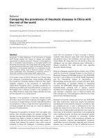
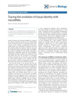
Figure 1. Phylogenetic relationships between major taxonomic phyla according to [9] and reconstruction of ancestral tissue types
based on conserved miRNA expression patterns. NLCA, BLCA and ELCA: the Nephrozoan, Bilaterian and Eumetazoan last common ancestor,
respectively. The summary for the BLCA is preliminary owing to the absence of a sequenced acoel genome and miRNA expression data.
Representatives of the taxa used in the study of Christodoulou et al. [8] are in bold.
NLCA
Foregut
miR-100:miR-125; let-7; miR-10;
miR-31; miR-278
Motile cilia
miR-29; miR-34; miR-92
Neurosecretory brain cells
miR-7; miR-137; miR-153
Sensory brain tissue
miR-9; mir-9*
Body musculature
miR-1; mir-22; miR-133
General CNS
miR-71; miR-124; miR-184;
miR-190; miR-219
Sensory organs
miR-8; miR-183; miR-263;
miR-252; miR-2001
Gut
miR-216; miR-283
Other
miR-315; miR-281; miR-210;
miR-33
BLCA
mir-100; mir-31; mir-34;
mir-92; mir-124
ELCA
Cells surrounding
digestive opening
miR-100
Vertebrata
(eg mouse, human, zebrafish)
Tunicata (Urochordata)
(eg Ciona intestinalis)
Cephalochordata
Echinodermata
(eg Strongylocentrotus purpuratus)
Arthropoda
(eg Drosophila melanogaster)
Nematoda
(eg Caenorhabditis elegans)
Mollusca
Annelida
(eg Platynereis dumerilii,
Capitella sp.)
Platyhelminthes
(eg Schmidtea mediterranea,
Macrostomum lignano)
Acoela
(eg Isodiametra pulchra)
Cnidaria
(eg Nematostella vectensis)
Sponges
Deuterostomes
EcdysozoaLophotrochozoa
Protostomes
Nephrozoans
Bilateria (triploblasts)
De Mulder and Berezikov Genome Biology 2010, 11:111
/>Page 2 of 4
conserved miRNAs in Platynereis using whole mount in
situ hybridization and found that expression patterns of
these miRNAs are highly specific for certain tissues and
cell types and are strongly conserved throughout
bilaterian evolution.
is comparison allowed Christodoulou and colleagues
[8] to reconstruct the minimal set of cell types and tissues
that existed in the last common ancestor of nephrozoans
(Figure1). is ancestor is predicted to have had neuro-
secretory cells along its mouth (characterized by the
expres sion of miR-100, miR-125 and let-7) and motile
ciliated cells (miR-29
+
miR-34
+
miR-92
+
). In addition, the
nephrozoan ancestor would have had a miR-1
+
miR-22
+
miR-133
+
body musculature, a miR-12
+
miR-216
+
miR-283
+
gut and miR-9
+
miR-9*
+
cells related to sensory
information processing. Finally, the nephrozoan ancestor
is predicted to have had a miR-124
+
central nervous
system, which would be connected with a miR-8
+
miR-183
+
miR-263
+
peripheral sensory tissue, and to be
already equipped with neurosecretory cells in a primitive
brain (miR-7
+
miR-137
+
miR-153
+
).
Implications and new directions
Innovation at the post-transcriptional gene regulatory
level through expansion of the miRNA repertoire has
previously been suggested as one of the driving forces
behind the evolution of animal complexity [3-7]. It is not
clear, however, how exactly novel miRNAs evolve and
what roles they have in the establishment of tissue
identity. According to the model of transcriptional control
of new miRNA genes suggested by Chen and Rajewsky
[2], newly emerging miRNAs initially should be expressed
at low levels and in specific tissues in order to minimize
deleterious off-targeting effects and to allow natural
selection to eliminate these slightly deleterious targets
over time. Subsequently, miRNA expression levels can be
increased and tissue-specificity relaxed [2]. Now, with the
discovery of Christodoulou et al. [8] that ancient miRNAs
were expressed in specific cell types of the protostome-
deuterostome ancestor and in many cases assumed
broader expression patterns later in evolution, this model
of miRNA emergence gains additional solid experimental
support.
As shown by Christodoulou et al. [8], comparison of
the miRNA repertoire between different taxa can
significantly contribute to the hypothetical reconstruc-
tion of the ancestral body plan: by a detailed examination
in which tissues/cell types conserved miRNAs evolved,
the authors [8] were able to create a hypothetical picture
of an ancestor at a key phylogenetic position for which
we have no fossils. Although the appearance of the last
common ancestor of deuterostomes and protostomes
still remains elusive, the authors [8] elucidated the
differentiated cell repertoire from this ancestor and, by
doing so, unequivocally established miRNAs as a power-
ful new tool for reconstructing ancient animal body plans
at important evolutionary nodes. Further investigation of
miRNA repertoires and expression patterns in additional
taxa might give fundamental clues about unknown nodes
within the animal tree and resolve some phylogenetic
uncertainties.
For example, one of the frequently disputed questions
is the phylogenetic position of Acoelomorpha (which
includes the flatworm-like acoels and nemertodermatids).
Acoels were originally grouped within the phylum
Platyhelminthes but have recently been placed at a key
position at the base of the Bilateria on the basis of new
molecular data [9] (Figure 1). Earlier studies revealed that
the highly conserved miRNA let-7, which is present in all
other Bilaterians, is absent in acoels, indicating that
acoels might have branched off earlier from the last
common ancestor of protostomes and deuterostomes. In
addition, although acoels are believed to primitively lack
a real brain, having instead a simple ‘commissural’ brain
characterized by transverse fiber accumulation in the
head, without classical ganglionic cell mass [10],
Christodoulou et al. [8] suggest that nervous system
centralization was already present before the split
between protostomes and deuterostomes. erefore, a
detailed analysis of the acoel miRNA repertoire and their
corresponding expression patterns might help to further
reveal how evolution at the base of the Bilateria took
place and whether or not the urbilaterian - the last
common ancestor of acoels and nephrozoans - had
complex tissues.
Conservation of sequence and expression patterns
suggests that the core functions of ancient miRNAs also
remained conserved through evolution. What are these
core functions? From data from other animal models,
Christodoulou et al. [8] speculate that some miRNAs,
such as miR-100 and let-7, could have roles in develop-
mental timing. However, only few miRNA genes are
known to work as developmental switches, and, perhaps
surprisingly, the majority of miRNAs are in fact not
essential for initial establishment of tissue identity but
seem to be important for the maintenance of cells in
differentiated states. It is likely, then, that miRNAs
facilitate evolution of complexity by stabilizing existing
and newly emerging regulatory circuits and transcrip-
tional programs. Elucidating the principle components of
miRNA-containing networks that were present at the
dawn of animal evolution and tracing the acquisition of
new miRNA circuitry through evolution is the next great
evo-devo challenge in the miRNA field.
Acknowledgments
We thank Bernhard Egger and Turan Demircan for fruitful discussions.
Published: 30 March 2010
De Mulder and Berezikov Genome Biology 2010, 11:111
/>Page 3 of 4
References
1. Levine M, Tjian R: Transcription regulation and animal diversity. Nature
2003, 424:147-151.
2. Chen K, Rajewsky N: The evolution of gene regulation by transcription
factors and microRNAs. Nat Rev Genet 2007, 8:93-103.
3. Grimson A, Srivastava M, Fahey B, Woodcroft BJ, Chiang HR, King N, Degnan
BM, Rokhsar DS, Bartel DP: Early origins and evolution of microRNAs and
Piwi-interacting RNAs in animals. Nature 2008, 455:1193-1197.
4. Sempere LF, Cole CN, McPeek MA, Peterson KJ: The phylogenetic
distribution of metazoan microRNAs: insights into evolutionary
complexity and constraint. J Exp Zool B Mol Dev Evol 2006, 306:575-588.
5. Prochnik SE, Rokhsar DS, Aboobaker AA: Evidence for a microRNA
expansion in the bilaterian ancestor. Dev Genes Evol 2007, 217:73-77.
6. Heimberg AM, Sempere LF, Moy VN, Donoghue PC, Peterson KJ: MicroRNAs
and the advent of vertebrate morphological complexity. Proc Natl Acad Sci
USA 2008, 105:2946-2950.
7. Berezikov E, Thuemmler F, van Laake LW, Kondova I, Bontrop R, Cuppen E,
Plasterk RH: Diversity of microRNAs in human and chimpanzee brain. Nat
Genet 2006, 38:1375-1377.
8. Christodoulou F, Raible F, Tomer R, Simakov O, Trachana K, Klaus S, Snyman H,
Hannon GJ, Bork P, Arendt D: Ancient animal microRNAs and the evolution
of tissue identity. Nature 2010, 463:1084-1048.
9. Egger B, Steinke D, Tarui H, De Mulder K, Arendt D, Borgonie G , Funayama N,
Gschwentner R, Hartenstein V, Hobmayer B, Hooge M, Hrouda M, Ishida S,
Kobayashi C, Kuales G, Nishimura O, Pster D, Rieger R, Salvenmoser W, Smith
J, Technau U, Tyler S, Agata K, Salzburger W, Ladurner P: To be or not to be a
flatworm: the acoel controversy. PLoS ONE 2009, 4:e5502.
10. Raikova OI, Reuter M, Kotikova EA, Gustafsson MKS: A commissural brain!
The pattern of 5-HT immunoreactivity in Acoela (Plathelminthes).
Zoomorphology 1998, 118:69-77.
doi:10.1186/gb-2010-11-3-111
Cite this article as: De Mulder K, Berezikov E: Tracing the evolution of tissue
identity with microRNAs. Genome Biology 2010, 11:111.
De Mulder and Berezikov Genome Biology 2010, 11:111
/>Page 4 of 4