Báo cáo y học: " High-resolution transcription atlas of the mitotic cell cycle in budding yeast" pptx
Bạn đang xem bản rút gọn của tài liệu. Xem và tải ngay bản đầy đủ của tài liệu tại đây (3.45 MB, 11 trang )
RESEA R C H Open Access
High-resolution transcription atlas of the mitotic
cell cycle in budding yeast
Marina V Granovskaia
1†
, Lars J Jensen
1,2†
, Matthew E Ritchie
3,4†
, Joern Toedling
5
, Ye Ning
6
, Peer Bork
1
,
Wolfgang Huber
1,5
, Lars M Steinmetz
1*
Abstract
Background: Extensive transcription of non-coding RNAs has been detected in eukaryotic genomes and is
thought to constitute an additional layer in the regulatio n of gene expression. Despite this role, their transcription
through the cell cycle has not been studied; genome-wide approaches have only focused on protein-coding
genes. To explore the complex transcriptome architecture underlying the budding yeast cell cycle, we used 8 bp
tiling arrays to generate a 5 minute-resolution, strand-specific expression atlas of the whole genome.
Results: We discovered 523 antisense transcripts, of which 80 cycle or are located opposite periodically expressed
mRNAs, 135 unannotated intergenic non-coding RNAs, of which 11 cycle, and 109 cell-cycle-regulated protein-
coding genes that had not previously been shown to cycle. We detected periodic expression coupling of sense
and antisense transcript pairs, including antisense transcripts opposite of key cell-cycle regulators, like FAR1 and
TAF2.
Conclusions: Our dataset presents the most comprehensive resource to date on gene expression during the
budding yeast cell cycle. It reveals periodic expression of both protein-coding and non-coding RNA and profiles
the expression of non-annotated RNAs throughout the cell cycle for the first time. This data enables hypothesis-
driven mechanistic studies concerning the functions of non-coding RNAs.
Background
Genome-wide transcriptome analyses in humans [1-5],
mouse [6], Drosophila melanogaster [7 ,8], Arabidopsis
thaliana [9], and fission and budding yeast [10-12] have
provided evidence for widespread expression of non-
coding RNAs (ncRNAs) from intergenic as well as
protein-coding regions (for example, antisense or
intron-derived transcripts). ncRNAs have been
implicated in regulation of chromatin structure, DNA
methylation, transcription,translation,aswellasRNA
silencing and stability [2,13-15].
Extensive transcription of intergenic regions and the
antis ense strands of hundreds of annotated protein-cod-
ing genes occurs in budding yeast, despite it lacking ves-
tiges of the protein machinery required for microRNA
or sm all interfering RNA processing [11,16-18]. It is not
clear to what extent these RNAs are functional [19], but
several have been shown to regulate transcription, acting
through either transcriptional interference or epigenetic
modifications. Examples of transcriptional interference
are SRG1, a ncRNA transcribed in cis across the promo-
ter of SER3 [20,21], and the antisense transcript of
IME4 [22], whereas the antisense transcripts of PHO5
[23], PHO84 [24], transposable element Ty1 [25] and
GAL10-ncRNA [26] function through epigenetic modifi-
cation. For most newly discovered ncRNAs, the biolog i-
cal roles and mechanisms of action rema in unknown.
To unravel the functions of ncRNAs in yeast, it is infor-
mative to characterize them in the context of a robustly
regulated and well-understood cellular process, such as
the mitotic cell cycle, in which regulatory roles of
ncRNAs have not been studied extensively.
The cell cycle orchestrates virtually all cellular pro-
cesses - metabolism, protein synthesis, secretion, DNA
replication, organelle biogenesis, cytoskeletal dynamics
and chromosome segregation [27] - and diverse regula-
tory events depend on the maintenance of its periodi-
city. Between 400 and 800 periodically expressed
* Correspondence:
† Contributed equally
1
EMBL - European Molecular Biology Laboratory, Department of Genome
Biology, Meyerhofstr. 1, D-69117 Heidelberg, Germany
Granovskaia et al. Genome Biology 2010, 11:R24
/>© 2010 Granovskaia et al.; licensee BioMed Central Lt d. This is an open access article distributed under the terms of the Creative
Commons Attribution License ( which pe rmits unrestricted use, distribution, and
reproduction in any medium, provided the original work is properly cited.
protein-coding genes have been identified in the mitotic
cell cycle and the genomic binding sites of transcription
factors that control phase-specific expression of these
genes have been mapped in genome-wide location ana-
lyses [28-30]. In addition to transcriptional regulatio n,
strict timing of cell-cycle progression is ensured by
post-translationa l regulation. This includes post-tra nsla-
tional modifications, targeted protein degradation and
indirect regulation via interactions with cell-cycle-regu-
lated proteins [31].
To investigate the gl obal cell cycle regulation of all
transcripts, we measured high-resolution, strand-specific
tiling microarray profiles of RNA expression during the
Saccharomyces cerevisiae cell c ycle. In contrast to pre-
viou s studies [29,30], which only interrogated annotated
features within the genome without resolving strand
specificity, the fine spatial and temporal resolution of
our dataset enabled us to look at the whole transcrip-
tome on both strands, including non-coding RNAs
(both away from coding genes and in antisense posi-
tion), complex transcription architecture of protein-cod-
ing genes, alternative transcription start and
polyadenylation sites, splicing, and differential regulation
of sense and antisense transcripts. Our data reveal cell-
cycle-regulated non-coding genes, complex expression
coupling between sense and antisense transcripts, as
well as over 100 protein-coding genes that were not pre-
viously known to cycle.
Results and discussion
Detecting periodic transcripts
We monitored genome-wide cell-cycle-regulated exp res-
sion at 5-minute intervals for up to three cell division
cycles, using whole-genome tiling arrays [11]. The array
is unique in interrogating every base pair in the geno me
on average six times and providing an 8-bp resolution
for strand-specific p robes. Two independent synchroni-
zation methods were used in order to obtain synchro-
nous cultures (see Materials and metho ds; Additional
file 1). Late G1 phase arrest wa s induced by exposure of
bar1 cells to alpha factor, and by raising the tempera-
ture to 38°C for temperature-sensitive cdc28-13 mutant
cells. Expression profiles for all genomic regions are
provided in a database that is searchable by gene symbol
or chromosomal coordinate [32].
To identify all transcribed sequences, we segmented
along-chromosome expression profiles, applying an
adaptation of the method described by Huber et al. [33]
(see Materials and methods). In addition to protein-cod-
ing transcripts and inf ras tructur al RNAs, we registe red
abundant expression of unannotated non-coding RNAs
(Additional file 2). These unannotated expressed fea-
tures comprise 523 antisense transcripts opposite pro-
tein-coding regions and 135 intergenic transcripts
(Additional file 3). The length distribution of ORFs in
these unannotated transcripts is within the range that is
expected by chance. Hence, we find no evidence for the
unannotated transcripts to be protein coding.
The average segment levels from each time-point were
analyzed for periodic e xpression by two different com-
putational methods [34,35], as well as by visual inspec-
tion. The aim of this combination of methods was
accurate and sensitive detection of cell-cycle-regulated
transcripts (see Materials and methods). In order to vali-
date our approach, we compared our gene list of peri-
odic protein-coding genes to a benchmark set that
comprised all known cell-cycle-regulated genes identi-
fied in single-gene experiments [35,36]. Our individual
cdc28 and alpha-factor datasets were each better than
most of the available ones [28-30] (Additional file 4).
Furthermore, our combined list of periodic protein-cod-
ing genes, despite being based on just two experimental
datasets, performed almost as well in identifying the
benchmark set of genes a s that of Gauthier et al.[37],
which integrated all available genomic datasets of cell-
cycle-regulated genes perfo rmed to dat e (Add itional file
4). Thus, our dataset and analysis method reproduced
the previous data on cycling protein-coding genes.
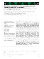
Altogether, 598 periodic mRNAs, 37 cycling antisense
RNAs, and 11 cycling intergenic transcripts were ide nti-
fied and ranked according to their peak time of expres-
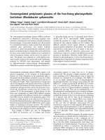
sion (Figure 1; Additional file 5). Non-coding periodic
transcripts were expressed in all cell-cycle phases
(Figure 2; see Additional f ile 6 for the determination of
the boundaries of the cell cycle phases). Overall, the
peak times of antisense periodic expression were consis-
tent with the waves of expression of periodic protein-
coding genes [38]. To charac terize the newly discovered
periodic ncRNAs, we overlapped them with regions of
conserved R NA secondary structure [39]. Despite their
cell-cycle-regulated expression, the unannotated inter-
genic and antisense ncRNAs had little s econdary struc-
ture (Additional file 6). Conversely, infrastructural
ncRNAs,comprisingtRNAs,rRNAs,smallnuclearand
small nucleolar RNAs, were highly structured but were
not periodically expressed.
Cell-cycle-regulated expression of unannotated non-
coding RNAs
Studies in mammalian cells have suggested that anti-
sense RNAs could regulate gene expression of their
sense counterparts, whereby sense and antisense tran-
scripts often exhibit expression correlat ion patterns
[40,41] and overlap in opposite directionality [42]. We
thus analyzed antisense RNAs in the context of the
sense-antisensepairs(SAPs)ofwhichtheyareapart.
We categorized the pairs into four classes based on
their expression coupling: 13 periodic antisense
Granovskaia et al. Genome Biology 2010, 11:R24
/>Page 2 of 11
Figure 1 Gene expression profiles ordere d by expression peak times. CDC28 and alpha -factor panels show the expression profiles for all
identified cell cycle-regulated genes, including 598 protein-coding genes, 37 unannotated antisense transcripts and 11 intergenic transcripts,
ordered by their peak times. Profiles for annotated ORFs are graded in blue; all non-coding RNAs are graded in red. Each column of the two
time-course panels represents a single experimental 5-minute time-point. The scales on the left display the relative duration and number of
transcripts expressed in each phase. Key cell-cycle-regulated genes are indicated on the right side. In each row, white and dark blue (or dark red
for the non-coding RNAs) represent the minimum and maximum expression levels, respectively, of the corresponding transcript. Intermediate
values are shown by colors that scale linearly over the range.
Granovskaia et al. Genome Biology 2010, 11:R24
/>Page 3 of 11
transcripts opposite periodic sense transcripts; 24 peri-
odic antisense transcripts opposite non-periodic sense
transcripts; 43 non-periodic antisense transcripts oppo-
site periodic sense transcripts; and 443 non-periodic
antisense and sense transcript pairs (Additional file 7).
The 13 periodic antisense transcripts opposite periodic
sense transcripts wer e further subdivided based on the
relative timing of expression of the sense and antisense
transcripts. Considering the absolute difference between
their expression peak times, two pairs (ALK1 and HSL1)
cycle in-phase, whereas seven (CTF4, FAR1, HMS2,
TAF2, TIP1, YNL300W and YPL162C) show anti-corre-
lated e xpression (Additional file 8). Expres sion profiles
of the other four SAPs (PRY3, YLR050C, YMR253C and
YPL230W) had phase shifts between 0 and π.
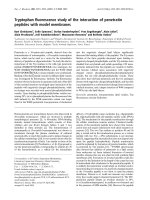
Remarkably, several genes encoding important cell
cycle regulators fall within the categories listed above
(Figure 3a-c). Among them, FAR1 is important for ma t-
ing pheromone-induced growth arrest and, together
with cyclins CLN2 and CLN3, plays one of the key roles
in the G1/S transition [43]. FAR1 is expressed at the M/
G1 transition and needs to be shut down in late G1 for
the cell to pass the G1/S checkpo int. Its antisense RNA
peaks starting f rom the lat e G1 pha se and thro ughout
the G1/S transition, when Far1 protein should not be
present. TAF2, which is involved in transcription initia-
tion, is expressed in late M and early G1 phase; its anti-
sense transcript peaks in late G1 and further into S
phase. The sense and antisense transcripts of CTF4,
which shapes and maintains chromatin structure to
ensure the passage through the S-phase checkpoint [44],
are expressed in an a nti-correlated m anner, peaking in
the G1/S and G2/M transitions, respectively. The CTF4
sense transcript appears to be transcribed from a
bidirectional promoter shared with the antisense tran-
script of the neighboring gene, MSS18 (Additional files
6 and 9). Together these expression patterns suggest
that some of the antisense transcripts may play a role in
cell-cycle regulation.
We analyzed Gene Ontology (GO) categories of genes
overlapped by antisense transcripts. Most of the protein-
coding messages opposite the 37 periodic antisense tran-
scripts (13 + 24) fall into GO categories linked with the
process of cell division, includ ing cell wall and or ganelle
organization and biosynthesis, regulation of transcrip-
tion, signal transduction and protein modification, car-
bohydrate metabolic processes, and cell cycle
(Additional file 10). Surprisingly, 15 of the 37 sense
transcripts are of unknown function. We carried out a
similar analysis for the 43 non-periodic antisense tran-
scripts opposite periodic sense transcripts. As expected,
most of these cycling sense messages fall into cell-cycle-
related GO categories, including genes involved in bud
site selection and polarization (BUD9, GIC1), daughter
cell separation from the mother (DSE2, CTS1), cell wall
proteins, a nd so on (Additional file 7). Analysis of GO
categories for the r emaining 443 non-periodic SAPs did
not show enrichment in any parti cular ca tegory,
although almost a quarter of the genes have unknown
function (Additional file 11).
We observed a statistically significant correlation (P <
0.002; 5 × 4 contingen cy table; c
2
test) between the
overlap patterns of the sense and antisense transcripts
and the relationship of their expression profiles (Addi-
tional file 12). Altogether we distinguished five types of
overlap within a given SAP: antisense transcript contains
the transcribed message of its sense counterpart; the
antisense transcript is contained within the sense
Figure 2 Gene expression profiles for all identified cell-cycle-regulated ncRNAs ordered by their expression peak times. Each column of
the CDC28 and alpha-factor time-course panels represents a single experimental 5-minute time-point. The scales on the left display the relative
duration and number of transcripts expressed in each phase. In each row, white and dark red represent the minimum and maximum expression
levels, respectively, of the corresponding transcript. Intermediate values are shown by colors that scale linearly over the range.
Granovskaia et al. Genome Biology 2010, 11:R24
/>Page 4 of 11
transcript; the antisens e transcri pt overlaps either the 3’
or the 5’ end of its sense partner; and the antisense
transcript overlaps two distinct sense transcripts. The
following patterns of overlap were over-represented
compared to what was expected by chance. In 8 out of
13 periodic antisense transcripts opposite periodic sense
transcripts, the antisense transcript is mainly contained
within the protein-coding message; 2 of these 8 cycle in-
phase, and 6 display opposite-phase expression. For 5 of
24 SAPs in which only the antisense transcript cycles,
the antisense transcript contains the complete sense
message, and for another 5, it overlaps 2 sense tran-
scripts. In 15 of the 43 pairs in which only the sense
message is cell cycle regulated, the antisense transcript
overlaps the 5’ end of the mRNA and in many cases
extends further upstream.
To inves tigat e sense and antisense expression in more
detail, we also searched for putative TF binding sites
(Additional file 6) and supported these predictions with
the existing ChIP-chip data. TF binding site analyses are
inhere ntly non-stra nd-specific; however, our data on the
temporal expression of the sense and antisense tran-
scripts yield clues to the regulation of strand-specific
expression. For example, ChIP-c hip data and our motif
analysis for FAR1 suggest binding of both the M-phase
TF Mcm1 [45] and the G1/S TF SBF [46] within the
region spanned by 600 bases before and after the tran-
script. This evidence for SBF regulation of FAR1 contra-
dicts the timing of expression of the sense tr anscript
since FAR1 is expressed at the M/G1 transition and
needs to be shut down in late G1. Our data show late-
G1-specific expr ession of the FAR1 antisense transcript,
thus providing a putative explanation for the presence
of the TF binding site for SBF. Overall, our analyses
indicate that the cycling unannotated transcripts have
binding sites for the same set of TFs that drive sense
transcription during the cell cycle (Additional file 6).
Altogether, 135 unannotated intergenic transcripts
were detected in our dataset. Of these, 11 oscillate with
mitotic progression (Additional files 5; Additional file
13c). As for the antisense transcripts, their peak in
expression follows the waves of excitation in mitotic
Figure 3 Expression for sense and antisense transcripts. Heatmaps of expression for sense and antisense transcripts of (a) FAR1, (b) TAF2, (c)
CTF4, (d) SPS100 and (e) YLR050C. Each horizontal line represents a single experimental time-point. The unit of the time axis (vertical) is minutes.
The horizontal axis in the center of each panel represents genomic coordinates, and annotated coding genes are indicated by blue boxes. The
heatmap in the upper half of each panel represents signal on the Watson strand, the one in the lower half signal on the Crick strand. The
horizontal orange lines separate alpha-factor (above the line) and Cdc28 (below the line) experimental datasets. Vertical red lines show the
segment boundaries.
Granovskaia et al. Genome Biology 2010, 11:R24
/>Page 5 of 11
progression observed for protein-coding genes [38]. To
elucidate the role of these intergenic transcripts in cell
cycle regulation, deletion strains for 10 of the 11 unan-
notated periodic transcripts were generated i n both
strain backgrounds. Growth curves of the deletion
strains did not show significant lagging in cell doubling
time after asynchronous growth in rich media for 28
hours at 30°C and 37°C. Lack of phenotype is consistent
with our previous observations for the unannotated
intergenic transcripts detected from asynchronous cul-
ture [11]. This suggests that their deletion phenotypes
have more subtle e ffects than those of many protein-
coding genes.
Cell cycle-regulated protein-coding genes
Previous studies have identified a large number of anno-
tated periodic t ranscripts. Compared to the integrated
dataset of Gauthier et al. [37], our list contains 223
additional periodic protein-coding genes, of which 109
were also not identified by Pramila et al. [29] and Spell-
man et al. [30] (Figure 4; Additional file 14). Only 3 of
the 109 have been shown to be periodically expressed in
small scale experiments [47]. GOslim analysis [48]
showed that the biological function is unknown for 35
of these 109 genes, whereas 41 perform functions
directly or indirectly associated with the regulation of
the cell cyc le, such as organelle organization and bio-
genesis, cytoskeleton organization and biogenesis, ribo-
some biogenesis and assembly, and so on (Additional
file 15).
Of the 598 periodically expressed protein-coding genes,
just 7 contain an intron according to the Saccharomyces
Genome Database anno tation: CIN2, MOB1, PMI40,
RFA2, SRC1, TUB1,andUSV1. This is due to the fact
that many of the budding yeast introns reside within
genes that encode ribosomal proteins [48]. In addition,
none of the introns in periodically expressed genes show
signs of phase-specific splicing; hence, in contrast to
meiosis in budding and fission yeast [49,50], we see no
evidence for a regulatory role of splicing in the mitotic
cell cycle of budding yeast.
Conclusions
Our data pro vide 5-minute resolution strand-specific
profiles of temporal expression during the mitotic cell
cycle of S. cerevisiae, monitored for more than three
complete cell divisions. The result ing atlas for the first
time comprehensively maps the expression of non-anno-
tated regions transcribed in mitotic circuitry, measures
the expression coupling of protein-coding and non-cod-
ing transcript pairs and reveals strand specificity of tran-
scription regulation. Furthermore, it unravels complex
architectures o f the mitotic transcript ome, such as spli-
cing and alternative transcription start and polyadenyla-
tion sites, and extends the set of previously reported
cell-cycle-regulated genes by 109 protein-coding genes.
The abundance of antisense expression across the gen-
ome raises the question of whether it represent s oppo r-
tunistic ‘ ripples of transcription’ through active
chromatin regions, or whether it is a regulated overlap
between the transcripts [51]. An evolutionary analysis of
genes with overlapping antisense partners across a num-
ber of eukaryo tic genomes has indicated that the sense-
antisense arrangement is more highly conserved than
expected if it were random ‘leakage’ of the transcription
machinery [52].
Regulatory roles for a few antisense transcripts have
been documented in yeast [20-25], yet it is still debated
what proportion of ncRNAs are functional [19]. Our
dataset reveals that most cycling antisense transcripts
are located opposite genes with cell-cycle-related func-
tions. Antisense transcripts may regulate the corre-
sponding functional sense transcripts through several
molecular mechanisms, which can be speculated from
the mutual expression pattern of the two transcripts
[53]. For example, transcriptional interference or anti-
sense-dependent inhibitory chromatin remodeling may
give rise to the anti-correlated expression of sense and
antisense transcripts, as is observed fo r more than half
ofthe13periodicSAPs.Forthe24caseswherethe
antisense transcript cycles while the sense transcript is
stably expressed, the periodic antisense transcript may
putatively mask the sense transcript, thereby conferring
periodic regulation at the level of translation. Through
the same mechanism, the 43 stably expressed antisense
transcripts may dampen st ochastic fluctuation of sense
Figure 4 Venn diagram displays the overlap of our list of
identified cell cycle-regulated protein-coding genes with the
lists determined by the previous studies of Gauthier et al. [37],
Pramila et al. [29], and Spellman et al. [30]. The overlap shows
that we find an additional 223 genes not identified by Gauthier et
al., among which 109 are unique to our dataset and were not
previously defined by the other studies.
Granovskaia et al. Genome Biology 2010, 11:R24
/>Page 6 of 11
messages by setting a threshold above which the sense
expression must rise [53]. Alternatively, stably expressed
antisense transcripts co uld mediate activatory chromatin
remo deling that maintains the chromosomal region in a
transcriptionally activatable/repressible state and thereby
facilitate expression regulation of the periodic sense
transcript. Indeed, more than one-third of the 43 stably
expressed antisense opposite cell-cycle-regulated
mRNAs overlap with the 5’ UTRs. Altoget her, the
sense-antisense expression coupling may help to narrow
down molecular mechanisms through which a specific
antise nse transcript exerts its function. Our high-resolu-
tion, unbias ed expression atlas of the budd ing yeast cell
cycle is thus a resource with which to unravel a poten-
tial additional level of the cell cycle regulatory circuit, as
well as to study the periodic expression of protein-cod-
ing transcripts at a fine temporal and spatial resolution.
The dataset provides a link between genomic
approaches a nd hypothesis-driven mechanistic research
with regard to the functions of ncRNAs.
Materials and methods
Yeast strains and cell cycle synchronization
W101 (50 ml; MATa ade2-1 trp1-1 leu2-3, 112 his3-11,
15 ura3 can1-100 [psi1]) background temperature-sensi-
tive cdc28-13 mutant S. cerevisiae strain K3445
(YNN553) was grown for approximately 8 to 10 hours
in rich yeast-extract/peptone/dextrose (YPD) in a shak-
ing water bath at 25°C and diluted in 3 × 1.6 liter cul-
tures for overnight growth in an air incubator at 25°C.
The following morning the cultures of OD600 approxi-
mately 0.2 were mixed together, distributed into 45 ×
100 ml samples and arrested in late G1 at START by
shifting the temperature from 25°C to 38°C. After 3.5
hours, the cells were transferred back to permissive tem-
perature to re-initiate cell division and samples were
collected every 5 minutes for 215 minutes (equal to
more than two complete cell cycles). The cultures were
centrifuged and snap-frozen in liquid nitrogen. The
degree of synchrony was monitored by assessing the
number of budding cells and measuring the bud size
(Additional file 1). Nuclear position was determined by
Hoechst staining with fluorescence microscopy (Addi-
tional file 16).
To arrest bar1 strain DBY8724 (MATa GAL2 ura3
bar1::URA3)[30]inG1atSTART,alpha-factorphero-
mone was added to a final concentration of 600 ng/ml.
After 2 hours of arrest, cells were released by washing
and recovered in fresh preconditioned medium to facili-
tate initiation of mitosis. Samples were collected every 5
minutes for 200 minutes (equal to three cell cycles).
The degree o f synchrony was monitored by assessing
the number of budding cells.Nuclearpositionwas
determined by Hoechst staining with fluorescence
microscopy.
Total RNA extraction, poly(A)-RNA enrichment, cDNA
synthesis and labeling
Total RNA was isolated from the culture correspond-
ing to each time-point by the standard hot phenol
method [11]. Poly(A)-RNA was enr iched from 1 mg of
total RNA by a single passage through the Oligotex
Oligo-dT Column (Qiagen, Hilden, Germany). Poly(A)-
RNA was treated with RNase-free DNaseI (Ambion’ s
Turbo DNA-free Kit, Foster City, CA, USA) for 25
minutes at 37°C according to the manufacturer’ s
instructions and subsequently reverse transcribed to
single-stranded cDNA for microarray hybridization.
Each 200 μl reverse transcription reaction was carried
out in duplicate and comprised 6 μgofpoly(A)-RNA,
3 μg random hexamers (RH6), 1 μl of 6 mg/ml Actino-
mycin D (ActD), 0.4 mM dNTPs containing dUTP
(dTTP:dUTP = 4:1), 40 μl 5× first strand synthesis buf-
fer (Invitrogen, Karlsruhe, Germany), 20 μl0.1M
dithiothreitol (Invitrogen), and 1,600 units of Super-
Script II (Invitrogen). The synthesis wa s carried out at
42°C for 1 h and 10 minutes, followed by reverse tran-
scriptase inactivation at 70°C for 10 minutes. Poly(A)-
RNAandRNAinheteroduplexwithcDNAwere
digestedbyamixtureof3μlofRNAseA/Tcocktail
(Ambion) and 3 μl of RNAseH (Invitrogen) for 15
minutes at 37°C followed by inactivation of the
enzymes for 15 minutes at 70°C. Replicate cDNA s am-
ples were further applied to the Affy Clean-up column
(Affymetrix, Santa Clara, CA, USA), eluted together in
30 μlDEPC-H
2
O and quantified. Purified cDNA (3.3
μg of each 5-minute time-point sample) was fragmen-
ted and labeled with WT Terminal Labeling Kit (Affy-
metrix) according to the manufacturer’s instructions
and then hybridized to tiling arrays.
Genomic DNA preparation
For DNA hybridization, both strains were grown in YPD
media overnight to saturation in three biological repli-
cates and whole-genomic DNA wa s purified using the
Genomic DNA Kit (Qiagen). Genomic DNA (10 μg) was
digested to 25 to 100 base fragments with 0.2 U of
DNaseI (Invitrogen) in 1× One-Phor-All buffer (Phar-
macia, Munich, Germany) contain ing 1.5 mM CoCl
2
(Roche, M annheim, Germany) for 3.5 minutes at 37°C.
After DNaseI inactivation by boiling for 10 minutes, the
sample was 3’ end-labeled in the same buffer by the
addition of 1.5 μl of Terminal Transferase (25 u nits/μl;
Roche) and 1.5 μl 10 mM biotin-N6-ddA TP (Molecular
Probes, Karlsruhe, Germany) for 2 hours at 37°C, and
hybridized to the tiling array.
Granovskaia et al. Genome Biology 2010, 11:R24
/>Page 7 of 11
Array design
The array was designed in collaboration with Affymetrix
(PN 520055), as described in David et al.[11].Probe
sequences were aligned to the genome sequence of S.
cerevisiae strain S288c (Saccharomyces Genome Data-
base of 7 August 2005). Per fect match probes were
further analyzed.
Probe normalization and segmentation
The log-base 2 perfect match (PM) probe intensities
from each array were background corrected and cali-
brated using the DNA reference normalization method
described in Huber et al.[33],whichwasappliedsepa-
rately to both datasets, cdc28 and alpha-factor.
To determine the transcript boundaries in the com-
bined dataset, a piece-wise constant model was fitted to
the normalized intensities of the unique probes ordered
by genomic coordinates. The basic model described in
Huber et. al. [33] was mod ified to a llow time-point-
dependent levels. The normalized intensities (z
jk
)were
modeled as:
ztjt
jk sk jk S S
for
1
1
where μ
sk
is the array-specific level of the s-th seg-
ment, ε
jk
are the residuals, j =1,2,.,n indexes the
probes in ascending order along the chromosome, k
indexes the time-point (array), t
2
,., t
S
parameterize the
segment boundaries (t
1
=1andt
S+1
= n+1) and S is
the total number of segments. Model 1 was applied
separately to each strand of each chrom osome. For each
chromosome, S was chosen such that the average seg-
ment length was 1,250 nucleotide s. Change-point s were
estimated using a dynamic programming algorithm
implemented in the tilingArray package [33].
After segmentation, the average of the probe signals
within the segment boundaries was calculated for each
time point. A table of segment levels is available from
the supplementary materials webpage [32].
To estimate a threshold for expression, the average
level over both datasets was calculated for each segment.
Segments not overlapping annotat ed, transcribed fea-
tures were used to estimat e the background level as fol-
lows. A normal distribution was fit in order to
determine a threshold at which the estimated false dis-
covery rate was 0.1% [11]. For the mean of the nor mal
distribution, we used the midpoint of the shorth (the
shortest interval that covers half of the values), for the
variance, the empirical variance of the lowest 99.9% of
the data. Segments whose level fell below this thresho ld
were considered not expressed.
Segments were then assigned to different categories
depending on how they overlapped with annotated
features as de scribed in David et al. [11], with the dif-
ference of re-naming the unannotated isolated features
to the unannotated intergenic. Expression values for
each annotated feature were calculated as weighted
averages of the overlapping segments on the same
strand.
Detection of periodic genes
We used a combination of three approaches to identify
periodically expressed s egments and annotated features
based on the cdc28 and alpha-factor datasets: the
method of Ahdesmaki et al. [34], which calculates P-
value s for a robust nonparametr ic vers ion of Fisher’sg-
test [54,55], the permutation-based method of de Lich-
tenberg et al. [35], which scores genes based on both
the magnitude of regulation and the periodicity of pro-
file, and by systematic visual inspection. For the two
computational methods, score cutoffs were determined
based on comparison with existing benchmark sets of
113 known cycl ing genes i dentified in single-gene stu-
dies [47]. A combined list of cycling t ranscripts was
compiled that contains all transcripts identified as
cycling b y at least two of the three methods. The peak
time of expression for each transcript was calculated as
percentage of the cell cycle duration as previously
described [35]. To determine the length of the cell cy cle
in each experiment, the period length was optimized to
fit the expression profiles for selected genes from the
benchmark set.
Analysis of protein-coding potential
To test if the ncRNAs are likely to be novel protein- cod-
ing genes, we extracted all ORFs within unannotated
antisense and intergenic transcripts and compared their
length distributions to what would be expected by
chance. The length of an ORF was defined as the distance
between a stop codon and the most upstream ATG
codon. Two separate background distributions were used
for antisense and intergenic transcripts, to take into
account that these two types of ncRNAs have different
sequence properties (k-mer frequencies), because the for-
mer are located opposite of protein-coding genes whereas
the latter are located within intergenic regions. For anti-
sense transcripts, a set of sequences with the same length
distribution was sampled from the genomic regio ns
opposite other protein-cod ing genes. Opposite genomic
regions with matched length distribution and seque nce
properties were used as a background for the unanno-
tated intergenic RNAs. The ORF length distributions
observed for the antisense and intergenic transcripts
were not statistically significantly different from their
respective background distributions according to the Kol-
mogorov-Smirnov test.
Granovskaia et al. Genome Biology 2010, 11:R24
/>Page 8 of 11
Transcription factor binding sites analysis
We used the TAMO suite [56] to identify the TFs that
preferentially bind to regulatory regions of periodic non-
coding transcripts. We systematically searched for bind-
ing motifs that were significantly overrepresented for
the region, spanning from -600 bp upstream up to +600
bp downstream of 37 periodic unannotated antisense
and 11 intergenic transcripts of interest, relative to a
background set composed of all transcripts detected in
the alpha-factor experiment. A benchmark set com-
prised 113 genes whose transcription was reported as
cell cycle regulated in single-gene studies previously
[47], whereas the lowest scoring 252 non-periodic anti-
sense transcripts from the alpha-factor induced arrest
dataset serve d as a negative control. We also performed
de novo motif discovery on these sequences, using the
combination of methods contained in the TAMO soft-
ware suite. This analysis revealed no significantly overre-
presented sequence m otifs. We then searched for the
putative TF binding sites that matched the position-spe-
cific score matrices from MacIsaac [57,58].
Analysis of RNA secondary structure conservation
We investigated the overlap between transcripts and
genomic regions with conserved secondary structure
[39]. We used Steigele et al.’s [39] regions for cutoff 0.5.
The regions wer e remapped to the current genome
assembly using Exonerate (requiring 100% identity). The
regions are strand-specific and overlap with these
regions was also considered in a strand-specific way.
Deletion strains of the periodic unannotated intergenic
transcripts
We generated deletion strains with the help of PCR-
based technology as described on the Stanford Yeast
Deletion webpage [59] using a set of up- and down-
stream primers flanking the defined periodic unanno-
tated sequence listed in Additional file 5. The growth of
deletion strains was monitored in liquid media using
GENios automatic microplate readers (TECAN).
Additional file 1: A table providing control data on the synchronous
division of the yeast cells. Excel sheet 1 contains a table of the number
and percentage of budded cells and dividing nuclei over time with the
progression of the cell cycle; sheet 2 contains a chart of these data.
Additional file 2: A figure showing categories of expressed segments.
The pie chart shows the categories and the numbers of all identified
transcribed segments. The unassigned categories encompass the
segments that did not meet filter criteria and were excluded from further
analyses [11]; correspondingly, the filtered categories are those that did
pass the filtering criteria.
Additional file 3: A table listing antisense and novel intergenic
transcripts identified in our study. Excel sheet 1 is a table of all 523
antisense transcripts, characterized by their genomic position, length and
overlapping sense feature; sheet 2 is a table of all 135 unannotated
intergenic transcripts, categorized by genomic position and length.
Cycling intergenic transcripts are highlighted in sheet 2.
Additional file 4: A figure showing a comparison of our dataset with
the published datasets on the cell cycle in yeast. Three ROC-like plots
compare: (a) our combined dataset with that of Gauthier et al. [37]; (b)
our cdc28 dataset with the other Cdc28 datasets of Spellman et al. [30]
and Cho et al. [28]; (c) our alpha-factor dataset with the existing alpha-
factor datasets of Spellman et al. [30] and Pramila et al. [29]. The fraction
of the B1 benchmark set genes identified by the various datasets is
plotted as a function of gene rank. (a) Comparison of the method of de
Lichtenberg et al. applied to our data (red line) with the comprehensive
integrated dataset of Gauthier et al. (black line) [35]. The cross indicates
our combined list, obtained by the combination of two computational
methods of analyses, and curated manually. (b) Compariso n of Cdc28
datasets. (c) Comparison of alpha factor-induced growth arrest datasets.
The color code displays: light brown, Cho et al.; green, Spellman et al.;
cyan and blue, Pramila et al.; black, Gauthier et al.; red, this study. The
dotted line indicates random selection of genes.
Additional file 5: A table listing periodic protein-coding genes,
antisense and unannotated intergenic transcripts. Excel sheet 1 lists 598
periodic ORFs identified in our dataset, sheet 2 lists 37 cycling antisense
transcripts, and sheet 3 lists 11 periodic unannotated intergenic
transcripts.
Additional file 6: A Word document providing supplemental data. The
file provides additional information on the following sections: 1,
Determination of the boundaries of the cell cycle phases; 2, Conservation
analysis of non-coding RNAs; 3, Analysis of upstream regulatory elem ents
for periodic unannotated transcripts; 4, UTR lengths; 5, Divergently
transcribed periodic transcripts.
Additional file 7: A table listing the categories of 37 periodic and 43
non-periodic antisense transcripts. Excel table sheet 1 lists 37 periodic
antisense transcripts and sheet 2 lists 43 non-periodic antisense
transcripts, each characterized by genomic position, length, overlapping
sense feature, function of the opposite sense counterpart according to
the Saccharomyces Genome Database, and peak time of expression
(cycling 37 antisense transcripts only).
Additional file 8: A figure showing a comparison of the relative timing
of expression within 13 periodic SAPs. We calculated the peak-time
difference for the periodic sense and antisense transcripts within each of
the 13 cycling SAPs for the alpha-factor and Cdc28 experiments
separately. A difference of 0 corresponds to in-phase expression, whereas
a difference of 50 corresponds to opposite-phase expression (180 degree
phase shift). We observe a good correlation between the two
experiments. The shape of the symbol shows how the sense-antisense
counterparts overlap.
Additional file 9: A table listing pairs of pairs of divergent transcripts
from a bidirectional promoter. Each transcript in a pair is characterized
by the genomic location, category and gene name.
Additional file 10: A figure showing GO categories of the ORFs
opposite cell-cycle-regulated antisense transcripts. The x-axis displays the
number of genes and the y-axis shows the names of GO categories.
Additional file 11: A figure showing GO categories of 443 non-periodic
ORFs opposite non-periodic antisense transcripts. The x-axis displays the
number of genes and the y-axis shows the names of GO categories.
Additional file 12: A contingency table for sense-antisense transcript
overlap.
Additional file 13: A figure showing heatmaps of bi-directional
expression of neighboring cell cycle-regulated genes that share
transcription regulatory elements. (a) Two neighboring ORFs: TEL2 and
ESP1. (b) ORF and an antisense transcript of the upstream protein-coding
gene: SPT21 and antisense counterpart of YMR178W.
(c) ORF and cycling
unannotated intergenic transcript: MCD1and upstream cycling novel
transcript. The heatmap plot is explained in the caption of Figure 3.
Additional file 14: A table listing the 109 periodic ORFs identified in our
study.
Additional file 15: A figure showing GO categories of 109 periodic ORFs
unique to our dataset. The x-axis displays the number of genes and the
y-axis shows the names of GO categories.
Granovskaia et al. Genome Biology 2010, 11:R24
/>Page 9 of 11
Additional file 16: A figure showing Hoechst nuclear staining of
dividing cdc28-ts mutant cells. Control data displaying synchronous
division of the yeast cells along with the cell cycle progression. Each
image represents a gallery of approximately 10 to 20 representative cells
that were chosen, for the respective time-point, from different fields of
view. Criteria of choice were sharpness of the image and visibility of the
bud; besides these, we aimed for random selection.
Abbreviations
ChIP: chromatin immunoprecipitation; GO: Gene Ontology; ncRNA: non-
coding RNA; ORF: open reading frame; SAP: sense-antisense pair; TF:
transcription factor; UTR: untranslated region.
Acknowledgements
We thank Sandra Clauder-Muenster for technical assistance, Vladimir Benes
and Tomi Baehr-Ivacevic from EMBL GeneCore Facility for technical advi ce,
Yury Belyaev and Arne Seitz from EMBL-ALMF for help with image
processing. This work was supported by grants to LMS from the National
Institutes of Health and the Deutsche Forschungsgemeinschaft, to WH from
the Human Frontier Science Program and to PB by the Bundesministerium
fuer Bildung und Forschung (Nationales Genomforschungsnetz
Foerderkennzeichen 01GS08169.)
Author details
1
EMBL - European Molecular Biology Laboratory, Department of Genome
Biology, Meyerhofstr. 1, D-69117 Heidelberg, Germany.
2
Novo Nordisk
Foundation Center for Protein Research, Faculty of Health Sciences,
University of Copenhagen, Blegdamsvej 3b, 2200 Copenhagen N, Denmark.
3
Department of Oncology, University of Cambridge, CRUK Cambridge
Research Institute, Li Ka Shing Centre, Robinson Way, Cambridge, CB2 0RE,
UK.
4
Bioinformatics Division, The Walter and Eliza Hall Institute of Medical
Research, 1G Royal Parade, Parkville, Victoria 3052, Australia.
5
EMBL -
European Bioinformatics Institute, Welcome Trust Genome Campus, Hinxton,
Cambridge, CB10 1SD, UK.
6
Plant Biochemistry Lab, Faculty of Life Sciences,
University of Copenhagen, Thorvaldsensvej 40, 1871 Frederiksberg C,
Denmark.
Authors’ contributions
MVG and LMS designed research; MVG performed research; YN contributed
to research; MVG, MER, LJJ, JT, WH and LMS analyzed data; MVG, LJJ, MER,
WH and LMS wrote the paper; WH, PB and LMS supervised research. The
authors declare that they have no conflict of interest.
Received: 16 September 2009 Revised: 21 December 2009
Accepted: 1 March 2010 Published: 1 March 2010
References
1. Kampa D, Cheng J, Kapranov P, Yamanaka M, Brubaker S, Cawley S,
Drenkow J, Piccolboni A, Bekiranov S, Helt G, Tammana H, Gingeras TR:
Novel RNAs identified from an in-depth analysis of the transcriptome of
human chromosomes 21 and 22. Genome Res 2004, 14:331-342.
2. Kapranov P, Cheng J, Dike S, Nix DA, Duttagupta R, Willingham AT,
Stadler PF, Hertel J, Hackermuller J, Hofacker IL, Bell I, Cheung E, Drenkow J,
Dumais E, Patel S, Helt G, Ganesh M, Ghosh S, Piccolboni A,
Sementchenko V, Tammana H, Gingeras TR: RNA maps reveal new RNA
classes and a possible function for pervasive transcription. Science 2007,
316:1484-1488.
3. Penn SG, Rank DR, Hanzel DK, Barker DL: Mining the human genome
using microarrays of open reading frames. Nat Genet 2000, 26:315-318.
4. Schadt EE, Edwards SW, GuhaThakurta D, Holder D, Ying L, Svetnik V,
Leonardson A, Hart KW, Russell A, Li G, Cavet G, Castle J, McDonagh P,
Kan Z, Chen R, Kasarskis A, Margarint M, Caceres RM, Johnson JM,
Armour CD, Garrett-Engele PW, Tsinoremas NF, Shoemaker DD: A
comprehensive transcript index of the human genome generated using
microarrays and computational approaches. Genome Biol 2004, 5:R73.
5. Yelin R, Dahary D, Sorek R, Levanon EY, Goldstein O, Shoshan A, Diber A,
Biton S, Tamir Y, Khosravi R, Nemzer S, Pinner E, Walach S, Bernstein J,
Savitsky K, Rotman G: Widespread occurrence of antisense transcription
in the human genome. Nat Biotechnol 2003, 21:379-386.
6. Kiyosawa H, Yamanaka I, Osato N, Kondo S, Hayashizaki Y: Antisense
transcripts with FANTOM2 clone set and their implications for gene
regulation. Genome Res 2003, 13:1324-1334.
7. Hild M, Beckmann B, Haas SA, Koch B, Solovyev V, Busold C, Fellenberg K,
Boutros M, Vingron M, Sauer F, Hoheisel JD, Paro R: An integrated gene
annotation and transcriptional profiling approach towards the full gene
content of the Drosophila genome. Genome Biol 2003, 5:R3.
8. Stolc V, Gauhar Z, Mason C, Halasz G, van Batenburg MF, Rifkin SA, Hua S,
Herreman T, Tongprasit W, Barbano PE, Bussemaker HJ, White KP: A gene
expression map for the euchromatic genome of Drosophila
melanogaster. Science 2004, 306:655-660.
9. Yamada K, Lim J, Dale JM, Chen H, Shinn P, Palm CJ, Southwick AM,
Wu HC, Kim C, Nguyen M, Pham P, Cheuk R, Karlin-Newmann G, Liu SX,
Lam B, Sakano H, Wu T, Yu G, Miranda M, Quach HL, Tripp M, Chang CH,
Lee JM, Toriumi M, Chan MM, Tang CC, Onodera CS, Deng JM, Akiyama K,
Ansari Y, et al: Empirical analysis of transcriptional activity in the
Arabidopsis genome. Science 2003, 302:842-846.
10. Wilhelm BT, Marguerat S, Watt S, Schubert F, Wood V, Goodhead I,
Penkett CJ, Rogers J, Bahler J: Dynamic repertoire of a eukaryotic
transcriptome surveyed at single-nucleotide resolution Nature 2008,
453:1239-1243.
11. David L, Huber W, Granovskaia M, Toedling J, Palm CJ, Bofkin L, Jones T,
Davis RW, Steinmetz LM: A high-resolution map of transcription in the
yeast genome. Proc Natl Acad Sci USA 2006, 103:5320-5325.
12. Dutrow N, Nix DA, Holt D, Milash B, Dalley B, Westbroek E, Parnell TJ,
Cairns BR: Dynamic transcriptome of Schizosaccharomyces pombe shown
by RNA-DNA hybrid mapping. Nat Genet 2008, 40:977-986.
13. Mattick JS, Makunin IV: Non-coding RNA. Hum Mol Genet 2006, 15(Spec No
1):R17-29.
14. Mattick JS, Gagen MJ: The evolution of controlled multitasked gene
networks: the role of introns and other noncoding RNAs in the
development of complex organisms. Mol Biol Evol 2001, 18:1611-1630.
15. Wassenegger M: RNA-directed DNA methylation. Plant Mol Biol 2000,
43:203-220.
16. Miura F, Kawaguchi N, Sese J, Toyoda A, Hattori M, Morishita S, Ito T: A
large-scale full-length cDNA analysis to explore the budding yeast
transcriptome. Proc Natl Acad Sci USA 2006, 103:17846-17851.
17. Nagalakshmi U, Wang Z, Waern K, Shou C, Raha D, Gerstein M, Snyder M:
The transcriptional landscape of the yeast genome defined by RNA
sequencing. Science 2008, 320:1344-1349.
18. Samanta MP, Tongprasit W, Sethi H, Chin CS, Stolc V: Global identification
of noncoding RNAs in Saccharomyces cerevisiae by modulating an
essential RNA processing pathway. Proc Natl Acad Sci USA 2006,
103:4192-4197.
19. Struhl K: Transcriptional noise and the fidelity of initiation by RNA
polymerase II. Nat Struct Mol Biol 2007, 14:103-105.
20. Martens JA, Laprade L, Winston F: Intergenic transcription is required to
repress the Saccharomyces cerevisiae SER3 gene. Nature 2004,
429:571-574.
21. Martens JA, Wu PY, Winston F: Regulation of an intergenic transcript
controls adjacent gene transcription in Saccharomyces cerevisiae. Genes
Dev 2005, 19:2695-2704.
22. Hongay CF, Grisafi PL, Galitski T, Fink GR: Antisense transcription controls
cell fate in Saccharomyces cerevisiae. Cell 2006, 127:735-745.
23. Uhler JP, Hertel C, Svejstrup JQ: A role for noncoding transcription in
activation of the yeast PHO5 gene. Proc Natl Acad Sci USA 2007,
104:8011-8016.
24. Camblong J, Iglesias N, Fickentscher C, Dieppois G, Stutz F: Antisense RNA
stabilization induces transcriptional gene silencing via histone
deacetylation in S. cerevisiae. Cell 2007, 131:706-717.
25. Berretta J, Pinskaya M, Morillon A: A cryptic unstable transcript mediates
transcriptional trans-silencing of the Ty1 retrotransposon in S. cerevisiae.
Genes Dev 2008, 22:615-626.
Granovskaia et al. Genome Biology 2010, 11:R24
/>Page 10 of 11
26. Houseley J, Rubbi L, Grunstein M, Tollervey D, Vogelauer M: A ncRNA
modulates histone modification and mRNA induction in the yeast GAL
gene cluster. Mol Cell 2008, 32:685-695.
27. Tyers M: Cell cycle goes global. Curr Opin Cell Biol 2004, 16:602-613.
28. Cho RJ, Campbell MJ, Winzeler EA, Steinmetz L, Conway A, Wodicka L,
Wolfsberg TG, Gabrielian AE, Landsman D, Lockhart DJ, Davis RW: A
genome-wide transcriptional analysis of the mitotic cell cycle. Mol Cell
1998, 2:65-73.
29. Pramila T, Wu W, Miles S, Noble WS, Breeden LL: The Forkhead
transcription factor Hcm1 regulates chromosome segregation genes and
fills the S-phase gap in the transcriptional circuitry of the cell cycle.
Genes Dev 2006, 20:2266-2278.
30. Spellman PT, Sherlock G, Zhang MQ, Iyer VR, Anders K, Eisen MB, Brown PO,
Botstein D, Futcher B: Comprehensive identification of cell cycle-
regulated genes of the yeast Saccharomyces cerevisiae by microarray
hybridization. Mol Biol Cell 1998, 9:3273-3297.
31. de Lichtenberg U, Jensen TS, Brunak S, Bork P, Jensen LJ: Evolution of cell
cycle control: same molecular machines, different regulation. Cell Cycle
2007, 6:1819-1825.
32. Tiling Array Data for Saccharomyces cerevisiae Cell Cycle Experiment.
/>33. Huber W, Toedling J, Steinmetz LM: Transcript mapping with high-density
oligonucleotide tiling arrays. Bioinformatics 2006, 22:1963-1970.
34. Ahdesmaki M, Lahdesmaki H, Pearson R, Huttunen H, Yli-Harja O: Robust
detection of periodic time series measured from biological systems. BMC
Bioinformatics 2005, 6:117.
35. de Lichtenberg U, Jensen LJ, Fausboll A, Jensen TS, Bork P, Brunak S:
Comparison of computational methods for the identification of cell
cycle-regulated genes. Bioinformatics 2005, 21:1164-1171.
36. de Lichtenberg U, Jensen LJ, Brunak S, Bork P: Dynamic complex
formation during the yeast cell cycle. Science 2005, 307:724-727.
37. Gauthier NP, Larsen ME, Wernersson R, de Lichtenberg U, Jensen LJ,
Brunak S, Jensen TS: Cyclebase.org - a comprehensive multi-organism
online database of cell-cycle experiments. Nucleic Acids Res 2008, 36:
D854-859.
38. Lovrics A, Csikasz-Nagy A, Zsely IG, Zador J, Turanyi T, Novak B: Time scale
and dimension analysis of a budding yeast cell cycle model. BMC
Bioinformatics 2006, 7:494.
39. Steigele S, Huber W, Stocsits C, Stadler PF, Nieselt K: Comparative analysis
of structured RNAs in S. cerevisiae indicates a multitude of different
functions. BMC Biol 2007, 5:25.
40. Havilio M, Levanon EY, Lerman G, Kupiec M, Eisenberg E: Evidence for
abundant transcription of non-coding regions in the Saccharomyces
cerevisiae genome. BMC Genomics 2005, 6:93.
41. Lehner B, Williams G, Campbell RD, Sanderson CM: Antisense transcripts in
the human genome. Trends Genet 2002, 18:63-65.
42. Shendure J, Church GM: Computational discovery of sense-antisense
transcription in the human and mouse genomes. Genome Biol 2002, 3:
RESEARCH0044.
43. Vanoni M, Rossi RL, Querin L, Zinzalla V, Alberghina L: Glucose modulation
of cell size in yeast. Biochem Soc Trans 2005, 33:294-296.
44. Warren CD, Eckley DM, Lee MS, Hanna JS, Hughes A, Peyser B, Jie C,
Irizarry R, Spencer FA: S-phase checkpoint genes safeguard high-fidelity
sister chromatid cohesion. Mol Biol Cell 2004, 15:1724-1735.
45. Simon I, Barnett J, Hannett N, Harbison CT, Rinaldi NJ, Volkert TL, Wyrick JJ,
Zeitlinger J, Gifford DK, Jaakkola TS, Young RA: Serial regulation of
transcriptional regulators in the yeast cell cycle. Cell 2001, 106:697-708.
46. Workman CT, Mak HC, McCuine S, Tagne JB, Agarwal M, Ozier O, Begley TJ,
Samson LD, Ideker T: A systems approach to mapping DNA damage
response pathways. Science 2006, 312:1054-1059.
47. Johansson D, Lindgren P, Berglund A: A multivariate approach applied to
microarray data for identification of genes with cell cycle-coupled
transcription. Bioinformatics 2003, 19:467-473.
48. Hong EL, Balakrishnan R, Dong Q, Christie KR, Park J, Binkley G,
Costanzo MC, Dwight SS, Engel SR, Fisk DG, Hirschman JE, Hitz BC,
Krieger CJ, Livstone MS, Miyasato SR, Nash RS, Oughtred R, Skrzypek MS,
Weng S, Wong ED, Zhu KK, Dolinski K, Botstein D, Cherry JM: Gene
Ontology annotations at SGD: new data sources and annotation
methods. Nucleic Acids Res 2008, 36:D577-581.
49. Juneau K, Palm C, Miranda M, Davis RW: High-density yeast-tiling array
reveals previously undiscovered introns and extensive regulation of
meiotic splicing. Proc Natl Acad Sci USA 2007, 104:1522-1527.
50. Bahler J: Cell-cycle control of gene expression in budding and fission
yeast. Annu Rev Genet 2005, 39:69-94.
51. Ebisuya M, Yamamoto T, Nakajima M, Nishida E: Ripples from
neighbouring transcription. Nat Cell Biol 2008, 10:1106-1113.
52. Dahary D, Elroy-Stein O, Sorek R: Naturally occurring antisense:
transcriptional leakage or real overlap? Genome Res
2005, 15:364-368.
53. Lapidot M, Pilpel Y: Genome-wide natural antisense transcription:
coupling its regulation to its different regulatory mechanisms. EMBO Rep
2006, 7:1216-1222.
54. Ueda HR, Chen W, Adachi A, Wakamatsu H, Hayashi S, Takasugi T,
Nagano M, Nakahama K, Suzuki Y, Sugano S, Iino M, Shigeyoshi Y,
Hashimoto S: A transcription factor response element for gene
expression during circadian night. Nature 2002, 418:534-539.
55. Wichert S, Fokianos K, Strimmer K: Identifying periodically expressed
transcripts in microarray time series data. Bioinformatics 2004, 20:5-20.
56. Gordon DB, Nekludova L, McCallum S, Fraenkel E: TAMO: a flexible, object-
oriented framework for analyzing transcriptional regulation using DNA-
sequence motifs. Bioinformatics 2005, 21:3164-3165.
57. Harbison CT, Gordon DB, Lee TI, Rinaldi NJ, Macisaac KD, Danford TW,
Hannett NM, Tagne JB, Reynolds DB, Yoo J, Jennings EG, Zeitlinger J,
Pokholok DK, Kellis M, Rolfe PA, Takusagawa KT, Lander ES, Gifford DK,
Fraenkel E, Young RA: Transcriptional regulatory code of a eukaryotic
genome. Nature 2004, 431:99-104.
58. MacIsaac KD, Wang T, Gordon DB, Gifford DK, Stormo GD, Fraenkel E: An
improved map of conserved regulatory sites for Saccharomyces
cerevisiae. BMC Bioinformatics 2006, 7:113.
59. Yeast Deletion Webpage. [ />yeast_deletion_project/deletions3.html].
doi:10.1186/gb-2010-11-3-r24
Cite this article as: Granovskaia et al.: High-resolution transcription atlas
of the mitotic cell cycle in budding yeast. Genome Biology 2010 11:R24.
Submit your next manuscript to BioMed Central
and take full advantage of:
• Convenient online submission
• Thorough peer review
• No space constraints or color figure charges
• Immediate publication on acceptance
• Inclusion in PubMed, CAS, Scopus and Google Scholar
• Research which is freely available for redistribution
Submit your manuscript at
www.biomedcentral.com/submit
Granovskaia et al. Genome Biology 2010, 11:R24
/>Page 11 of 11