Báo cáo y học: "miRTRAP, a computational method for the systematic identification of miRNAs from high throughput sequencing data" pptx
Bạn đang xem bản rút gọn của tài liệu. Xem và tải ngay bản đầy đủ của tài liệu tại đây (1.31 MB, 12 trang )
Hendrix et al. Genome Biology 2010, 11:R39
/>Open Access
METHOD
BioMed Central
© 2010 Hendrix et al.; licensee BioMed Central Ltd. This is an open access article distributed under the terms of the Creative Commons
Attribution License ( which permits unrestricted use, distribution, and reproduction in
any medium, provided the original work is properly cited.
Method
miRTRAP, a computational method for the
systematic identification of miRNAs from high
throughput sequencing data
David Hendrix*, Michael Levine and Weiyang Shi*
miRTRAPA novel method for prediction of miRs from deep sequencing data. Its utility is demon-strated when applied to Ciona data.
Abstract
MicroRNAs (miRs) have been broadly implicated in animal development and disease. We developed a novel
computational strategy for the systematic, whole-genome identification of miRs from high throughput sequencing
information. This method, miRTRAP, incorporates the mechanisms of miR biogenesis and includes additional criteria
regarding the prevalence and quality of small RNAs arising from the antisense strand and neighboring loci. This
program was applied to the simple chordate Ciona intestinalis and identified nearly 400 putative miR loci.
Background
microRNAs (miRNAs/miRs) are small regulatory RNAs
present throughout the Eukarya [1-3]. They modulate
diverse biological processes, including embryonic devel-
opment, tissue differentiation, and tumorigenesis. miRs
inhibit translation and promote mRNA degradation via
sequence-specific binding to the 3' UTR regions of
mRNAs [2]. They are produced from hairpin precursors
(pri-miRNAs) that are sequentially processed by Drosha/
DGCR8 and Dicer to generate one or more 19- to 23-
nucleotide RNAs. The most abundant product is referred
to as miR, while the less abundant sequence produced
from the opposite arm of the hairpin is called miR*. In
addition, it has been observed that some miRNA loci can
produce up to two additional products immediately adja-
cent to the miR and miR* sequences, which are called
miRNA offset RNAs (moRs) [4,5].
The comprehensive identification of the complete set of
miRs is complicated by their small size, which limits sim-
ple cross-species comparisons based on sequence homol-
ogy. Moreover, de novo computational miRNA prediction
methods rely heavily on known miRNAs and are not
always effective for characterizing novel genomes. Recent
advances in high throughput sequencing technology pro-
vide an opportunity for the systematic identification of
every miRNA gene in a genome. Here we present such a
system for the computational identification of miRNA
genes from deep sequencing data and apply it to datasets
collected from different developmental stages of the sim-
ple chordate Ciona intestinalis. This approach predicted
over 300 novel Ciona miRNAs and revealed the molecu-
lar phylogeny of miRNA families in the chordate lineage.
This method was also used to identify novel miR loci in
the extensively characterized genome of Drosophila mel-
anogaster.
Results
A computational approach to identify miRNAs from high-
throughput sequencing data
The comprehensive identification of the full repertoire of
miRNAs in a given organism is of general interest. Early
bioinformatics approaches used machine learning and
pattern recognition to predict miRNA loci de novo from
whole genome sequences [6]. These methods correctly
identified a number of miRs but also led to a high failure
rate. Recent progress of high-throughput sequencing has
enabled systematic cloning and identification of miRNAs.
However, it is sometimes difficult to distinguish miRNAs
from other small RNAs such as endogenous small inter-
fering RNAs (siRNAs), Piwi-interacting RNAs (piRNAs),
and mRNA degradation products. Current methods
approach this problem by identifying miR-specific struc-
tures and sequence features, such as hairpin stability and
* Correspondence: ,
Department of Molecular and Cell Biology, Division of Genetics, Genomics and
Development, Center for Integrative Genomics, University of California,
Berkeley, 142 LSA#3200, Berkeley, CA 94720-3200, USA
Full list of author information is available at the end of the article
Hendrix et al. Genome Biology 2010, 11:R39
/>Page 2 of 12
base-pairing frequencies [7,8]. Such features are then
applied to either whole genome scan windows (de novo
prediction) or sequencing read windows (small RNA
library deep sequencing) to predict the likelihood of a
candidate locus being an authentic miR [9]. These meth-
ods have two major shortcomings. First, they are often
too stringent to handle sequencing errors and natural
variations in spliced products. Consequently they pro-
duce high false negative rates and perform poorly on
novel genomes. Second, many genomic sequences resem-
ble miR hairpin structures, and additional information is
required to eliminate such false positives.
Here, we describe a new method for the discovery of
novel miRNA genes. A computational approach called
miRTRAP (miRNA Tests for Read Analysis and Predic-
tion) was developed for the systematic prediction of miR-
NAs from high-throughput sequence data. In contrast to
most current methods, miRTRAP utilizes a system of
binary decisions based on known biochemical mecha-
nisms of miRNA biogenesis. Numerous studies have
shown that miRNAs are generated from pre-miRNA
stem-loop hairpins and a given locus can produce up to
five products, that is, miR/miR*, moR/moR* and the loop,
which have stereotyped positions within the hairpin
[4,10]. We reasoned that authentic miR loci should satisfy
all of these critical criteria. Specifically, the program uses
the following criteria: the product of a given locus folds
into hairpins 20 nucleotides or longer; the miR/miR*,
moR/moR* and loop products must fall within appropri-
ate positions on the hairpin; these products must be next
to each other on the same hairpin arm and shifted within
a certain distance on the opposite arm; and the total
number of products at a predicted miR locus must be
present at least one part per million of the total reads and
be represented by at least five reads. In addition, a single
miR product must be represented by more than one read
(Figure 1a).
Besides authentic miRs, this approach also identifies
other types of small RNAs. To eliminate these, the miR-
TRAP method takes into account the genomic context
from which the candidate miR is produced. We observed
two distinctive features that distinguish miR and non-
miR loci. First, small RNA reads are rarely observed from
the opposite strand of a miR locus. When present, the
antisense products from known microRNA loci, such as
the Drosophila iab-4 locus [11,12], exactly overlap the
miR products. In contrast, the antisense products derived
from endo-siRNA [13] and piRNA [14] loci are shifted
from sense strand products by several base pairs. Authen-
tic miRNA loci are expected to either lack antisense
products, or encode products that perfectly match the
sense RNAs. To evaluate this property, we designed a
measure called average antisense product displacement
(AAPD), defined as the average offset of overlapping
sense and antisense products at a given locus. Indeed, all
the known Ciona miRs have AAPD scores of 0, while ran-
dom sampling of non-miR loci showed broad distribu-
tions (Figure 1b). This measure is sufficient to distinguish
valid miRs from invalid ones among the top 500 most
abundant candidate loci. Thus, sequence information
from the opposite strand is useful for distinguishing miRs
from other types of small RNAs.
The AAPD measure is reliable for predictions repre-
sented by hundreds of reads, but is less informative for
loci with fewer reads due to insufficient sampling of
potential antisense products. To circumvent this prob-
lem, we examined the distances separating putative miRs
from neighboring non-miR read products. miRs tend to
arise from genomic regions that lack other types of small
RNAs. This may reflect the large size of pri-miRNA tran-
scription units with strict secondary structures to pro-
duce miR hairpins. Except for the case of antisense
miRNAs or miRNAs from genomic clusters, there are
usually few if any short sequencing reads in the neighbor-
ing area. We examined previously annotated Ciona miR-
NAs [4] and found that there are fewer than 10 non-miR
small RNA sequencing reads within a 2-kb genomic win-
dow encompassing authentic miR loci. In contrast,
genomic regions lacking miR loci contain far more non-
miR-derived products (Figure 1c).
Thus, miRTRAP employs a two-step screening strat-
egy: the application of the known mechanisms of miR
biogenesis and the elimination of false positives by exam-
ining small RNA sequencing reads from the antisense
strand and neighboring regions. This combined approach
is able to achieve a high discovery rate with apparently
low false positive identifications (see below).
Comparison of miRTRAP with miRDeep
miRDeep [15] was previously used to identify approxi-
mately 70 miR genes in C. intestinalis [4]. However, this
analysis failed to identify many well-known animal miR
families, such as mir-8 and mir-9. This observation raised
the possibility that Ciona is degenerate and might have
lost key miR genes. To investigate this issue, we employed
miRTRAP to systematically identify all possible miRs
from Illumina sequencing data.
We sequenced six small RNA libraries from different
developmental stages of C. intestinalis (unfertilized egg
through adults) and obtained approximately 8 million
small RNA reads that mapped to unique sites within the
genome (Table S2 in Additional file 1) [16]. Using miR-
TRAP, we predicted a total of 446 putative miRs. Manual
examination of these predictions verified 362 candidate
loci, and the remaining 84 loci appear to be false positive
predictions based on poor secondary structures or incon-
sistent read distributions. To estimate the number of false
negatives, we manually examined candidate negative pre-
Hendrix et al. Genome Biology 2010, 11:R39
/>Page 3 of 12
dictions and identified another 18 miR candidates. Most
of the false negative loci were rejected due to alternative
secondary structures or the occurrence of spurious reads
contributing to a high AAPD score or excessive neighbor-
ing short RNA sequence reads. However, these false neg-
ative loci possess features that perfectly conform to the
expectations of miRs, including predicted stem-loop
structures and locations of the sequences along the puta-
tive pre-miRNA.
Northern hybridization assays were used to test five of
the newly predicted miRs, which exhibit abundant
expression based on the total read counts (Table S4 in
Additional file 2). Discrete small RNA products were
identified for all five candidate miRs (Figure S2 in Addi-
tional file 1), consistent with the effectiveness of the miR-
TRAP method for the comprehensive identification of all
miR loci in the Ciona genome. Altogether, miRTRAP
generated an apparent false negative rate of approxi-
mately 5% and a false discovery rate of approximately
19%.
To systematically compare the miRTRAP and miRDeep
methods, we tested the new Ciona library data using the
miRDeep approach. miRDeep assigns a log-likelihood
score that evaluates hairpin stability, minimum free
energy, read abundance, and the presence of an associ-
ated miR* sequence. These scores are based on Bayesian
probabilities that are calibrated using sequences from the
C. elegans genome. miRDeep predicted only 77 candidate
miRs. Of these predictions, 46 overlap with the manually
curated positive candidate miR list, while the remaining
31 examples appear to be false positive predictions (Fig-
ure 2a). Thus, miRDeep identifies only approximately
12% of the putative Ciona miRs predicted by miRTRAP.
The Ciona small RNA libraries were sequenced at very
high depth, with over approximately 8 million reads.
miRTRAP uses the full sequencing information to reject
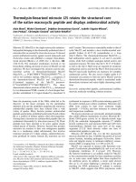
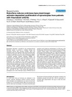
Figure 1 Outline of the miRTRAP program, Ciona abundance versus conservation, neighbor window. (a) Schematic illustration of the miRTRAP
program. The algorithm first identifies read regions that do not overlap repeats or tRNAs. The genomic region up to 150 nucleotides around the indi-
vidual read is folded using RNAfold. Then, all read products within the hairpin window are identified as 5p-miR/3p-miR, 5p-moR/3p-moR or loop based
on their positions relative to the hairpin and loop. Each read region is then evaluated by a set of filters to remove those incompatible with the bio-
chemical rules of miR biogenesis. All the rejected read regions are used to filter the initial set of candidate loci to produce a list of positive predictions.
(b) Average antisense product displacement (AAPD) score distribution from the Ciona dataset shows that the majority of known miRs have an AAPD
score of zero, while non-miR loci have a broad distribution and peaks at 8 and 10. (c) The difference between the non-miR neighbor counts within
windows centered at known miRs and non-miR loci in Ciona. Whereas non-miR neighbor counts centered around non-miR loci increases sharply as
window sizes expand, all known miR loci have non-miR neighbor counts equal or fewer than 10.
(a) (b)
(c)
Hendrix et al. Genome Biology 2010, 11:R39
/>Page 4 of 12
miR-like hairpins, and it is possible, therefore, that miR-
TRAP does not perform as well with less deeply
sequenced libraries. To address this, we performed miR-
TRAP predictions using a reduced dataset containing
1,015,781 randomly sampled reads from the original
Ciona small RNA library set. Among the 380 candidate
Ciona miRs from the original prediction, only 245 exceed
the minimal threshold of 5 sequenced products per locus.
Of these 245 miRs, 226 were identified with the reduced
dataset. In addition, miRTRAP also predicted 44 false
positive loci. These rates are comparable to the results
obtained with the original dataset containing eight-fold
more information.
In addition, we compared the performance of miR-
TRAP and miRDeep on a published set of Drosophila
melanogaster small RNA libraries [17], consisting of
871,776 aligned reads from over 20 different develop-
mental stages and tissues. There are 152 annotated D.
melanogaster miRs in miRBase and 148 of these have
sequencing reads in the library datasets. miRDeep pre-
dicted 109 of the annotated miRs, representing a discov-
ery rate of 72%. By comparison, miRTRAP predicted 134
of the 148 annotated miRs (90% discovery rate), and after
removing exonic loci another 38 novel predictions were
identified (Figure 2b). Manual examination of these new
candidates identified 19 plausible miRs, including at least
one mirtron (Figure 2c, d). None belong to known miR
families but two tandem miR loci were identified within a
previously identified Drosophila miR cluster (see supple-
mental text in Additional file 1). Thus, miRTRAP effec-
tively identifies not only known Drosophila miRs (90%
recovery rate) but also novel candidates.
An overview of predicted Ciona miRNAs
We have identified as many as 380 putative miRNA genes
in the C. intestinalis genome through a combination of
computaional prediction and manual curation (Addi-
tional files 2 and 3) [18]. This is roughly five times more
than previously predicted. More than 72% of the
sequenced library reads are derived from predicted
miRNA loci. The ratio of miR versus miR* products is
highly skewed toward the mature miR, with the less
abundant product constituting less than 2%. However, for
some loci, the relative abundance of miR to miR* switches
between developmental stages, for example, mir-92-4,
mir-132, mir-2248, and mir-2286, supporting the possi-
bility that the biogenesis of miR and miR* products might
be subject to developmental regulation.
Loop sequences from miR hairpins were rarely cloned
(30 out of 380). These sequences sometimes represent
precise Dicer processing products from pre-miRNAs in
the case of short loops (for example, mir-1497), or result
from random degradation of longer loops (for example,
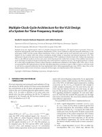
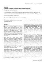
Figure 2 Comparison of miRTRAP with miRDeep. (a) miRTRAP out-
performed miRDeep for the Ciona library data set, identifying approxi-
mately five times more miRs. In addition, it identified 11 mirtron/half-
mirtrons, while miRDeep found only 1. (b) For the Drosophila small
RNA data set, miRTRAP identified 25 more known fly miRs than miRD-
eep. In particular, miRTRAP found 12 out of 14 mirtrons, while miRDeep
identified only 3. (c) Example of a novel Drosophila mirtron predicted
by miRTRAP. (d) A novel Drosophila miR/miR* containing locus predict-
ed by miRTRAP.
(a)
(b)
(c)
(d)
Hendrix et al. Genome Biology 2010, 11:R39
/>Page 5 of 12
mir-1). Nevertheless, they are extremely rare compared
to other miR associated products, constituting less than
0.0076% of the total miR-derived sequencing reads.
As described previously, there are abundant moRs in
Ciona [4]. Roughly half of the 70 miR genes detected ear-
lier were shown to produce moR and/or moR* products.
Nearly half of the expanded collection of miR genes (172
out of 380) identified in this study produce moRs from at
least one side of the hairpin. Indeed, the presence of
moRs lends support for a putative prediction. This obser-
vation confirms that moR production is a general feature
of the Ciona miR biogenesis pathway. However, moRs are
still rare compared to miR and miR* products, compris-
ing less than 1% of the total miR-associated reads.
Nearly one-third of the 380 predicted miRs (119 out of
380) appear to arise from introns, whereas 246 miR loci
are located in intergenic regions. We also observed four
cases where the predicted miR sequences overlap exonic
sequences (see below).
miRNAs play important regulatory roles during animal
development and their expression levels are expected to
change over time [19]. To evaluate the dynamics of Ciona
miR expression, we mapped changes in the levels of indi-
vidual miRs in unfertilized eggs, early embryos, late
embryos and adults. The relative expression levels of
individual miRs are normalized to the total reads from
each library. Of 380 predicted miRNAs, 316 are
expressed in the unfertilized egg, suggesting a strong
maternal miRNA contribution in Ciona embryogenesis.
In the early embryo, 342 out of 380 miRs are expressed,
and the ratio drops as embryogenesis proceeds (305 out
of 380 in late embryo library). Only 249 out of 380 of the
miRs exhibit expression in the adult. The low adult
expression rate likely results from the unequal contribu-
tion of different tissue types in the adult body; neverthe-
less, some miRs are most highly expressed at the adult
stage, for example, the let-7 family members, which regu-
late developmental timing in a variety of animals [20].
Phylogenetic conservation of urochordate miRNAs within
the deuterostome lineage
The evolution of miRNA families has been suggested to
correlate with increases in morphological complexity of
animal groups [21]. Cladograms of conserved Ciona miRs
[22,23] are consistent with urochordates, not cephalo-
chordates, as the closet living relatives of the vertebrates
[24]. With the identification of hundreds of new Ciona
miRs, we sought to investigate whether there are more
conserved miR families in the chordate lineage. We com-
pared predicted Ciona miR sequences to known miRs in
amphioxus, zebrafish, Xenopus, chicken, mouse, and
human (miRBase release 13). Saccoglossus (hemichor-
date) and S. purpuratus (echinoderm) were used as out-
groups. To define family membership, we required an
exact seed match (nucleotides 2 to 7 of the mature
sequence), and no more than four mismatches in the
mature miR sequence of a known member of this family
in the other species considered. This definition correctly
assigns all known Ciona miRs to their families, indicating
the method is both accurate and sensitive to detect family
information from mature miRNA sequences.
Altogether, 25 new Ciona miRs in 19 families were
identified that are conserved in other deuterostomes (Fig-
ure 3; Additional file 4). These include several well-con-
served miRs that were thought to be missing in Ciona,
including mir-7, mir-8 and mir-9. Thus, it would appear
that Ciona has retained most of the deuterostome miRs.
This supports the general observation that miRs are
rarely lost during evolution [25,26].
We also identified nine miR families that were previ-
ously thought to be vertebrate specific, including mir-15,
27, 96, 126, 132, 183, 196, 367, and 454. It is currently
unclear whether these miRs arose at the base of the chor-
dates or are specific to vertebrates and urochordates. A
recent study of amphioxus small RNAs [27] identified
mir-96 and mir-183, suggesting at least some of these
miRs might be present throughout the chordate lineage.
Finally, four conserved miR families, mir-10, 99, 190 and
216 were not identified, suggesting that they are either
expressed at levels below the detection limits or were lost
in the Ciona lineage.
Besides conserved miRs, we identified 20 Ciona-spe-
cific miR families (mir-2200 through mir-2219; Addi-
tional file 5). Most contain fewer than four members and
are usually organized as tandem duplications, such as Ci-
mir-2205 to mir-2219. However, in a few cases, closely
related miRs are organized within large genomic clusters.
For example, there is an approximately 4-kb miR cluster
containing 25 linked miRs that are grouped into three
closely related families differing by just a single nucle-
otide in the seed sequence (9 Ci-mir-2200, 7 Ci-mir-2201
and 9 Ci-mir2203). A second large cluster contains 11
miRs that group into 4 paralogous families (3 Ci-mir-
2200, 3 Ci-mir-2201, 4 Ci-mir-2204 and 2 Ci-2217). Inter-
estingly, some of the miRs located in these two clusters
belong to the same family, suggesting a common origin
for many of the novel Ciona miRs.
Phylogenetic signature of Ciona and urochordate miRNAs
The phylogenetic analysis of predicted Ciona miRs iden-
tified 19 new evolutionarily conserved family members.
Given the unique phylogenetic position and life history of
urochordates, we asked whether these newly predicted
miRs are also conserved in a divergent ascidian species,
Ciona savignyi, whose genome has been sequenced and is
often used for phylogenetic footprinting comparisons
[28,29]. We used the full genome alignment between the
two Ciona species [30] to determine the degree of conser-
Hendrix et al. Genome Biology 2010, 11:R39
/>Page 6 of 12
vation of both the 5p and 3p products of predicted C.
intestinalis miRs. To evaluate conservation, we use the
same criteria for miR family associations discussed above
(Additional file 6).
Of the 41 C. intestinalis miRs that have at least one
homolog in other deuterostomes, 35 are also conserved
in C. savignyi. mir-8, 9, 27, 29, 132 and 153 were not iden-
tified by sequence alignment, possibly due to gaps in the
C. savignyi genome assembly or loss of synteny over the
course of divergence between the two species (over 100
million years).
Thirty-five C. intestinalis miRs have full hairpin
sequences conserved in C. savignyi so that both miR and
miR* products are conserved. Interestingly, only 11 of
these correspond to the 41 known C. intestinalis family
members. The remaining 24 appear to be specific to
ascidians. Besides these 35 highly conserved full miR
hairpins, an additional 44 5p-miR and 31 3p-miR
sequences are also conserved, bringing the total con-
served ascidian miRs to 110. Interestingly, the 25-miR
cluster on scaffold 70 and 11-miR cluster on scaffold 20 in
C. intestinalis are not conserved in C. savignyi, suggesting
these clusters may have arisen in C. intestinalis through
recent tandem duplications.
Prevalence of antisense miRs in Ciona
Antisense miRs were originally observed for miR iab-4 in
the Drosophila Hox complex [11,12,31]. Several addi-
tional examples were subsequently identified [32]. In
these examples, a miR locus is transcribed bidirectionally
and each transcript contains a stable hairpin structure
that is processed to produce distinct miR products. Due
to the highly specific secondary structures associated
with transcripts from each strand, the two hairpin arms
almost always overlap, thus producing small RNA prod-
ucts that complement one another. The biological signifi-
cance of a single locus producing miRs from both
directions is unclear. In the case of Drosophila iab-4/iab-
8, iab-8 is produced from the opposite side of iab-4*; thus,
its sequence matches iab-4 and presumably targets the
same mRNAs. The two iab-miRs are expressed in mutu-
ally exclusive cells during Drosophila development [11].
Here, we undertook the comprehensive, genome-wide
identification of all antisense miRs in Ciona. Numerous
Ciona miR loci produce antisense products. For example,
three of the miR loci within the scaffold 20 gene cluster
have antisense products (Figure 4a). There are examples
of antisense miR, miR* and even antisense moR products
(for example, miR-2246 in Figure 4b). Altogether, 44 of
the 380 predicted miR loci appear to express antisense
products. In general, products from one strand are much
more abundant than the antisense products. Thus, exten-
sive sequence coverage is required to identify such prod-
ucts. Occasionally, the antisense product is nearly as
abundant as the sense miR product.
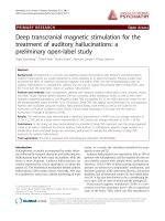
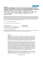
Figure 3 Phylogeny of Ciona miRNA families in the deuterostome
lineage. Newly identified conserved Ciona miR families (shaded cir-
cles) and previously known Ciona miRs (dark circles) are grouped with
homologous miR families from representative deuterostome species
(echinoderm, Strongylocentrotus purpuratus; hemichordate, Saccoglos-
sus kowalevskii; Amphioxus, Branchiostoma floridae). Missing miRs are
shown as empty circles. It is evident from the phylogenetic tree that
the miR repertoire from Ciona is closely related to the vertebrate miRs.
Echinoderm
Hemichordate
Amphioxus
Zebrafish
X.tropicalis
Chicken
Mouse
Human
Ciona
mir-1
mir-184
mir-219
mir-375
let-7
mir-182
mir-153
mir-135
mir-133
mir-125
mir-124
mir-10
mir-34
mir-33
mir-31
mir-29
mir-92
mir-9
mir-8
mir-7
mir-216
mir-190
mir-137
mir-99
mir-96
mir-454
mir-367
mir-196
mir-183
mir-132
mir-126
mir-27
mir-15
mir-281
mir-242
Hendrix et al. Genome Biology 2010, 11:R39
/>Page 7 of 12
If the major antisense miR product overlaps with miR*
from the sense strand, then it might contain the same
seed sequence as the sense miR and target the same
mRNAs, as seen for the Drosophila iab-4/iab-8 miRs.
However, if antisense miRs overlap with the sense miR
product, then the seed sequences are likely to be comple-
mentary and therefore possess distinct target specifici-
ties. Thus, bidirectional production of miR products may
expand the regulatory potential of a given miR locus by
targeting different sets of genes. Recent studies have
shown that large regions of the vertebrate genome are bi-
directionally transcribed [33], thereby raising the possi-
bility that many miR loci could produce antisense prod-
ucts.
Mitrons and exonic miRs in Ciona
Mirtrons arise from small hairpin-folding introns (58 to
70 nucleotides) processed from the nascent transcript by
the splicing machinery [34,35], thereby bypassing the
Drosha/DGCR8 microprocessor complex. Once in the
cytoplasm, mirtron hairpins and canonical miR hairpins
are both processed by Dicer to produce mature miRs. In a
Drosha knockdown cell line, production of canonical
miRs is diminished, but mirtrons are unaffected [36]. We
observed a total of four mirtrons in Ciona (Figure 5a;
Table S3 in Additional file 1). Recent studies identified
another class of mirtrons whereby only one end of the
hairpin is located at the intron-exon boundary, while the
other end is within the intron sequence [37]. These so-
called half mirtrons may be processed by a combination
of the splicing machinery and the microprocessor com-
plex. There are seven such examples in Ciona (Figure 5b;
Table S3 in Additional file 1).
In addition to intronic miRs, we also observed a class of
miRNAs deriving from mature mRNAs (Figure 5c).
These miRs are produced from local hairpin folding
within exons or UTR sequences and are supported by
EST reads spanning the hairpin. We refer to these as
exonic miRs. There are four examples in Ciona, and some
produce both miR and miR* sequences (Table S3 in Addi-
tional file 1). Presumably, the processing of exonic miRs
disrupts the stability or function of the resident mRNA,
raising the possibility that they are used as part of a
homeostasis mechanism to ensure a fixed stoichiometry
of miR and mRNA products. Recent studies have shown
that Drosha can cleave the DGCR8 mRNA, which con-
tains long hairpins [38], although it is unclear whether
these hairpins produce miRNAs. Alternatively, these loci
could arise from intronic regions of unannotated alterna-
tive splicing variants.
Discussion
We have presented a new computational method for the
systematic identification of miRs using high-throughput
sequence information. The method identified approxi-
mately 400 miRs in the Ciona genome, nearly a five-fold
increase compared with previous studies relying on tradi-
tional methods [4,22,39]. A number of conserved miR
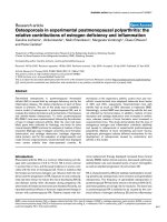
Figure 4 Prevalence of antisense miRs in Ciona. (a) In the scaffold 20 11-miR cluster, three miR loci have antisense reads that exactly match the
sense miR/miR* products. (b) Secondary structures of Ci-mir-2217-1 and its associated antisense locus, miR-2217-1-as, both form highly symmetric
hairpins, on which the miR and miR* products are indicated as lines. (c) In one case, we observed the antisense locus of Ci-mir-2246 produces not only
miR/miR*, but also a 5p-moR product. (d) Secondary structures and product distribution of Ci-mir-2246 and Ci-mir-2246-as.
(a) (b)
(c) (d)
Hendrix et al. Genome Biology 2010, 11:R39
/>Page 8 of 12
Figure 5 Non-canonical miR examples from Ciona. (a) A classic example of mirtron Ci-mir-2219-2 shows the miR and miR* products are produced
from the precisely spliced intron from gene ci100134440. (b) In some cases, only one of the miR/miR* products abuts the splice junction, while the
other product is fully inside the intron. A so-called half-mirtron example, Ci-mir-2227, is represented. (c) Ci-mir-2233 produces a miR/miR* pair from a
perfectly structured hairpin, which overlaps with a protein coding exon in the gene ci0100152310.
(a)
(b)
(c)
Hendrix et al. Genome Biology 2010, 11:R39
/>Page 9 of 12
genes were identified, such as miR-8, which were missed
in previous assays. In addition, two large clusters were
identified that encode novel miRs found only in C. intesti-
nalis. Finally, we identified a number of novel intronic
miRs, antisense miRs (and moRs), and even a few exam-
ples of putative miRs arising from exonic regions of pro-
tein coding genes, as discussed below.
Computational prediction of miRs
The miRTRAP program includes several critical criteria
that encompass the basic mechanisms of miR biogenesis
[40]. Basically, the biochemical machinery that processes
pre-miRNA hairpins produces short RNA products in
stereotypic spatial patterns. The more extensive the
sequence information, the more likely these miRNA pro-
cessing products will be identified. Thus, by defining a
minimal set of criteria for the distribution of sequences
from a given locus, it is possible to determine whether
these products conform to the known mechanisms of
miR biogenesis. This approach requires accurate assign-
ment of small RNA sequences on their relative positions
along the hairpin, that is, miR/miR*, moR/moR* and
loop. This poses a challenge because products can be het-
erogeneous due to imprecise biochemical processing,
errors in library preparation or sequencing (for example,
Ci-let-7s; details in Additional file 3). Non-canonical
hairpin structures create additional challenges to the sys-
tematic and accurate identification of miRs on a whole-
genome scale. For example, some long hairpins have
extremely extended loops that produce smaller degrada-
tion products, which complicate the identification of
authentic miR/miR* products (for example, Ci-miR-1
produces two non-overlapping loop products; Additional
file 3). Moreover, some hairpins possess not one loop, but
two closely adjacent minor hairpins that together form a
so-called double loop structure (for example, Ci-mir-375,
Ci-mir-2304). To overcome these problems, we devel-
oped a detailed identification scheme for all possible
miR-derived products from a hairpin fold region that can
accommodate the aforementioned atypical structures
(Figure S1 in Additional file 1). This allows miRTRAP to
evaluate whether any given products can possibly arise
from miR biogenesis pathways.
Numerous genomic regions produce short RNA reads
that do not derive from the miR biogenesis pathway, but
they nonetheless can resemble a miR-producing hairpin.
These might arise from random RNA degradation of long
transcripts [41], RNA interference-mediated processing
of endo siRNA products [42], piwi-RNA processing [14],
and so on. To eliminate these false miR hairpins, we took
advantage of the genomic contexts of authentic miR loci.
miRs are only rarely associated with offset antisense
products and authentic antisense miRs almost always
fully overlap sense miRs (Figure 1b). Thus, by calculating
the average shift of products from the opposite strand,
miRTRAP is able to eliminate many miR-like hairpins.
Another critical filter employed by miRTRAP is based on
our observation that miR hairpins are usually located in
genomic regions devoid of non-miR small RNAs. Statisti-
cal analysis revealed a significant difference in the num-
ber of these non-miR neighbors between known
annotated miRs and non-miRs (Figure 1c). This might be
due to the highly ordered secondary structures of long
pri-miRNA precursor RNAs [43].
Together, these two features, overlap of sense and anti-
sense products and diminished non-miR small RNA
sequences in neighboring regions, significantly reduced
the number of false positive predictions. It is worth not-
ing that the miRTRAP analysis of Drosophila small RNA
library sequences has a higher false detection ratio than
that obtained in Ciona (24% versus 19%), probably due to
the low coverage of the Drosophila libraries [17]. miR-
TRAP performs better when more sequencing data are
available. However, this is not a concern given the rapid
advances of high-throughput sequencing technology.
Future small RNA sequencing studies will have far more
extensive coverage than what is currently available. miR-
TRAP should be a useful tool for such studies.
Unique features of Ciona microRNA biogenesis pathways
The basic mechanisms of miRNA biogenesis are con-
served across animals and plants [10,44], and the applica-
tion of these rules is critical for the accurate prediction of
novel miRs. However, Ciona possesses several unique
features of miR production that are only rarely observed
in other species. Of particular note is the prevalence of
moRs, which arise from the regions immediately flanking
the locations of the mature miR and miR* products. Alto-
gether, 40 of 80 previously identified miR loci produce
moRs from at least one arm of the hairpin [4]. Here, we
have obtained evidence that 172 of 380 miR loci can pro-
duce moR sequences. Another interesting feature con-
cerning moRs is their production from tightly linked miR
clusters in Ciona. The 23-miR cluster on scaffold 71
spans an approximately 4-kb genomic region. There is
often little or no intervening sequence between the 3p-
moR product of one miR and the 5p-moR of the down-
stream miR. It is unclear how these densely packed miRs
are processed from large poly-cistronic precursor RNAs.
A surprising finding of this study is the prevalence of
antisense miR products in the Ciona genome. Such prod-
ucts have been only rarely seen in other species. In con-
trast, approximately 12% of the predicted miR loci in
Ciona appear to produce at least one antisense miR or
moR product. It is unclear whether such loci are bi-direc-
tionally transcribed in the same tissue or are expressed in
a mutually exclusive manner as seen for the iab-4/8 locus
in Drosophila [11]. In principle, co-expression could lead
Hendrix et al. Genome Biology 2010, 11:R39
/>Page 10 of 12
to the production of endo siRNA products [13,45] rather
than distinct pri-miRNA hairpins unique to each strand.
But it is possible that the stable stem-loop hairpin struc-
tures can inhibit the formation of double-stranded RNAs
and suppress endo siRNA production from these bi-
directionally transcribed miR loci.
Finally, we observed four cases where a miR/miR* pair
is produced from exonic regions. A few such examples
have been reported before [46-48]. However, these prod-
ucts are quite rare, so it is currently unclear whether they
represent bona fide miRs. The mirDeep program failed to
identify most of the mirtrons in either the Ciona or fly
dataset, while miRTRAP systematically identified most of
the known cases in Drosophila.
Phylogeny of chordate microRNAs
It has been documented that miR phylogenies accurately
reflect animal evolutionary trees, leading to speculation
that gains of miRs correlate with increases in morpholog-
ical complexity [26]. Despite the retrograde development
of adult ascidians during metamorphosis, Ciona none-
theless retains all the major chordate miR families. The
miR phylogenies (Figure 3) are entirely consistent with
the recent proposal that Ciona is more closely related to
vertebrates than amphioxus [24]. Specifically, we identi-
fied nine miR families that are unique to chordates. Con-
versely, mir-281 is specifically lost in the vertebrate
lineage after the divergence of urochordates, but is pres-
ent in all other deuterostomes as well as protostomes.
Within the urochordate lineage, most of the C. intesti-
nalis miR families are also conserved in a distantly related
ascidian species, C. savignyi, supporting the notion that
these miRs are present in the urochordate subphylum
instead of arising through convergent evolution in C.
intestinalis. In addition, we identified 69 miRs that are
only found in ascidians. They probably represent uro-
chordate-specific innovations after the last shared ances-
tor of vertebrates and urochordates.
In summary, the miRTRAP method permits the sys-
tematic identification of miRs from deep sequence infor-
mation. This method increased the number of identified
miR loci in Ciona from 80 to nearly 400 genes. Approxi-
mately half of these genes produce non-conventional miR
products, including moRs or antisense miRNAs. Phyloge-
netic analysis of this comprehensive set of miR loci sug-
gests that Ciona is more closely related to vertebrates
than amphioxus, a conclusion previously suggested by the
systematic comparison of protein coding genes [24]. In
addition to most of the conserved chordate-specific miR
loci, Ciona contains many ascidian-specific miRs and a
number of novel miRs that probably arose from tandem
duplication events at two major clusters only in the C.
intestinalis lineage. The miRTRAP method also success-
fully identified novel miRs in the well-studied Drosophila
genome, and we expect that its application to other
genomes will reveal additional novel miRs.
Materials and methods
Library preparation, sequencing and Northern analysis
Ciona stage-specific small RNA library preparation and
Illumina sequencing were performed as previously
described [4]. Sequence data were submitted to the NCBI
GEO database (GSE21078). Northern hybridization anal-
ysis was performed using DNA oligo probes at 37°C in
Ambion Oligo-UltraHyb buffer [4].
Read processing, alignment and the miRTRAP algorithm
Reads from each library were trimmed using a procedure
described in Shi et al. [4] to globally optimize read quality
over all start and stop positions using quality parameters
computed with ELAND. The reads were then aligned to
the Ciona genome (JGI version 1.0) using BLAST with an
E-value of 10, a word size of 7, and a gap penalty of
10,000. Hits to the genome were then filtered to only
include those with an E-value ≤ 0.01.
After the reads have been aligned to the genome, read
regions are defined. A read region is defined as a contigu-
ous span of overlapping reads. Only reads with fewer
than five hits to the genome are considered for the pur-
poses of defining the read regions. Read regions shorter
than 160 nucleotides and that do not overlap a repeat
region or a tRNA are then used as candidate loci to be
tested as a possible miR.
Our approach for the identification of microRNAs
using high-throughput sequencing reads is to compute a
set of quantities for each candidate locus, and by using
thresholds for each quantity we define a space of values
that contain the microRNA loci.
A key challenge to the program is to designate all read
products on a potential hairpin as corresponding to miR/
miR*, moR/moR* and/or loops because our program
relies on this information to test whether the products are
consistent with miRNA biogenesis. Once candidate loci
are folded, all reads that overlap the locus are grouped to
define 'products', and these products are then identified
as miR, moR, or loop products according to Figure S1 in
Additional file 1.
Many quantities we consider pertain to the structure of
the hairpin and positions of reads. The distance between
a miR and moR on the same arm of the hairpin, the offset
of the 5' positions of products that overlap at least 2
nucleotides on the same arm of the hairpin, and the offset
of overlapping products on opposite arms of the hairpin
are used to evaluate the spacing and distribution of prod-
ucts. The 5' heterogeneity, defined as the fraction of reads
within the miR product with the same 5' position as the
predominant splice variant of this product, is evaluated
for the most abundant miR product. Furthermore, we
Hendrix et al. Genome Biology 2010, 11:R39
/>Page 11 of 12
define the AAPD as the average distance between sense
and antisense products that overlap, and apply this mea-
sure across all sense products that overlap antisense
products. Additionally, the minimum number of base
pairs per nucleotide for either a miR or miR* product is
used to evaluate the locus.
Two additional quantities take into account informa-
tion from the sequencing data outside the candidate
locus under consideration. The average number of hits to
the genome for reads within the most abundant miR
product is evaluated as an additional level of repeat filter-
ing. Finally, after producing a list of predicted positive
loci using the above measures, we define the non-miR-
neighbor-count as the number of read regions that do not
overlap a predicted positive locus within a ± 1-kb window
surrounding the locus in question. All read regions,
including those overlapping repeat regions, tRNAs, and
those longer than 160 nucleotides, are considered for this
calculation.
Each of these quantities has user-defined thresholds
that can be adjusted to meet the desired level of strin-
gency of the predictions. The default values used in this
analysis are summarized in Table S1 in Additional file 1.
The software for miRTRAP and other resources are avail-
able on our website [49].
Additional material
Abbreviations
AAPD: average antisense product displacement; miRNA/miR: microRNA; miR-
TRAP: miRNA Tests for Read Analysis and Prediction; moR: microRNA-offset-
RNA; piRNA: Piwi-interacting RNA; siRNA: small interfering RNA; UTR: untrans-
lated region.
Authors' contributions
WS and DH designed the study. WS performed the molecular genetic experi-
ments and DH performed the computational analysis. WS and DH analyzed the
data. WS, DH and ML wrote the manuscript. All authors read and approved the
final manuscript.
Acknowledgements
We thank Benjamin Haley for sharing his extensive knowledge of microRNAs
and help with interpretations. Additionally, we thank Daniel S Rokhsar and Shu
Shenqiang for thoughtful discussions and help performing the alignment of
reads. This work was supported by NIH grant 5R24GM075049-04 to ML.
Author Details
Department of Molecular and Cell Biology, Division of Genetics, Genomics and
Development, Center for Integrative Genomics, University of California,
Berkeley, 142 LSA#3200, Berkeley, CA 94720-3200, USA
References
1. He L, Hannon GJ: MicroRNAs: small RNAs with a big role in gene
regulation. Nat Rev Genet 2004, 5:522-531.
2. Liu J: Control of protein synthesis and mRNA degradation by
microRNAs. Curr Opin Cell Biol 2008, 20:214-221.
3. Stefani G, Slack FJ: Small non-coding RNAs in animal development. Nat
Rev Mol Cell Biol 2008, 9:219-230.
4. Shi W, Hendrix D, Levine M, Haley B: A distinct class of small RNAs arises
from pre-miRNA-proximal regions in a simple chordate. Nat Struct Mol
Biol 2009, 16:183-189.
5. Langenberger D, Bermudez-Santana C, Hertel J, Hoffmann S, Khaitovich P,
Stadler PF: Evidence for human microRNA-offset RNAs in small RNA
sequencing data. Bioinformatics 2009, 25:2298-2301.
6. Yousef M, Showe L, Showe M: A study of microRNAs in silico and in vivo:
bioinformatics approaches to microRNA discovery and target
identification. Febs J 2009, 276:2150-2156.
7. Grad Y, Aach J, Hayes GD, Reinhart BJ, Church GM, Ruvkun G, Kim J:
Computational and experimental identification of C. elegans
microRNAs. Mol Cell 2003, 11:1253-1263.
8. Lai EC, Tomancak P, Williams RW, Rubin GM: Computational
identification of Drosophila microRNA genes. Genome Biol 2003, 4:R42.
9. Burgt A van der, Fiers MW, Nap JP, van Ham RC: In silico miRNA
prediction in metazoan genomes: balancing between sensitivity and
specificity. BMC Genomics 2009, 10:204.
10. Kim VN, Han J, Siomi MC: Biogenesis of small RNAs in animals. Nat Rev
Mol Cell Biol 2009, 10:126-139.
11. Tyler DM, Okamura K, Chung WJ, Hagen JW, Berezikov E, Hannon GJ, Lai
EC: Functionally distinct regulatory RNAs generated by bidirectional
transcription and processing of microRNA loci. Genes Dev 2008,
22:26-36.
12. Stark A, Bushati N, Jan CH, Kheradpour P, Hodges E, Brennecke J, Bartel DP,
Cohen SM, Kellis M: A single Hox locus in Drosophila produces
functional microRNAs from opposite DNA strands. Genes Dev 2008,
22:8-13.
13. Okamura K, Lai EC: Endogenous small interfering RNAs in animals. Nat
Rev Mol Cell Biol 2008, 9:673-678.
14. Gunawardane LS, Saito K, Nishida KM, Miyoshi K, Kawamura Y, Nagami T,
Siomi H, Siomi MC: A slicer-mediated mechanism for repeat-associated
siRNA 5' end formation in Drosophila. Science 2007, 315:1587-1590.
15. Friedlander MR, Chen W, Adamidi C, Maaskola J, Einspanier R, Knespel S,
Rajewsky N: Discovering microRNAs from deep sequencing data using
miRDeep. Nat Biotechnol 2008, 26:407-415.
16. Ciona intestinalis Genome Browser [ />gbrowse/cionaV1/]
17. Ruby JG, Stark A, Johnston WK, Kellis M, Bartel DP, Lai EC: Evolution,
biogenesis, expression, and target predictions of a substantially
expanded set of Drosophila microRNAs. Genome Res 2007,
17:1850-1864.
18. Predicted Ciona miRNA details [ />getMicroRNASearch.cgi]
19. Neilson JR, Zheng GX, Burge CB, Sharp PA: Dynamic regulation of miRNA
expression in ordered stages of cellular development. Genes Dev 2007,
21:578-589.
20. Tennessen JM, Thummel CS: Developmental timing: let-7 function
conserved through evolution. Curr Biol 2008, 18:R707-708.
21. Sempere LF, Cole CN, McPeek MA, Peterson KJ: The phylogenetic
distribution of metazoan microRNAs: insights into evolutionary
complexity and constraint. J Exp Zool B Mol Dev Evol 2006, 306:575-588.
22. Prochnik SE, Rokhsar DS, Aboobaker AA: Evidence for a microRNA
expansion in the bilaterian ancestor. Dev Genes Evol 2007, 217:73-77.
Additional file 1 Supplemental methods, Supplemental Figures 1 to 3
and Supplemental Tables 1 to 3.
Additional file 2 Supplemental Table 4. List of all predicted Ciona miRs
with genomic location, mature sequences (5p-, 3p-) and folds.
Additional file 3 Supplemental Table 5. Details of Ciona miR products.
Additional file 4 Supplemental Table 6. Details of conserved Ciona miR
family members.
Additional file 5 Supplemental Table 7. Details of Ciona specific family
members.
Additional file 6 Supplemental Table 8. List of conservation between C.
intestinalis and C. savignyi miRs.
Received: 12 October 2009 Revised: 19 January 2010
Accepted: 6 April 2010 Published: 6 April 2010
This article is available from: 2010 Hendrix et al.; licensee BioMed Central Ltd. This is an open access article distributed under the terms of the Creative Commons A ttribution License ( which permits unrestricted use, distribution, and reproduction in any medium, provided the original work is properly cited.Genome Biology 2010, 11:R39
Hendrix et al. Genome Biology 2010, 11:R39
/>Page 12 of 12
23. Heimberg AM, Sempere LF, Moy VN, Donoghue PC, Peterson KJ:
MicroRNAs and the advent of vertebrate morphological complexity.
Proc Natl Acad Sci USA 2008, 105:2946-2950.
24. Delsuc F, Brinkmann H, Chourrout D, Philippe H: Tunicates and not
cephalochordates are the closest living relatives of vertebrates. Nature
2006, 439:965-968.
25. Wheeler BM, Heimberg AM, Moy VN, Sperling EA, Holstein TW, Heber S,
Peterson KJ: The deep evolution of metazoan microRNAs. Evol Dev
2009, 11:50-68.
26. Peterson KJ, Dietrich MR, McPeek MA: MicroRNAs and metazoan
macroevolution: insights into canalization, complexity, and the
Cambrian explosion. Bioessays 2009, 31:736-747.
27. Chen X, Li Q, Wang J, Guo X, Jiang X, Ren Z, Weng C, Sun G, Wang X, Liu Y,
Ma L, Chen JY, Zen K, Zhang J, Zhang CY: Identification and
characterization of novel amphioxus microRNAs by Solexa sequencing.
Genome Biol 2009, 10:R78.
28. Johnson DS, Davidson B, Brown CD, Smith WC, Sidow A: Noncoding
regulatory sequences of Ciona exhibit strong correspondence
between evolutionary constraint and functional importance. Genome
Res 2004, 14:2448-2456.
29. Hill MM, Broman KW, Stupka E, Smith WC, Jiang D, Sidow A: The C.
savignyi genetic map and its integration with the reference sequence
facilitates insights into chordate genome evolution. Genome Res 2008,
18:1369-1379.
30. C. intestinalis and C. savignyi Alignment [ />ciona]
31. Bender W: MicroRNAs in the Drosophila bithorax complex. Genes Dev
2008, 22:14-19.
32. Azuma-Mukai A, Oguri H, Mituyama T, Qian ZR, Asai K, Siomi H, Siomi MC:
Characterization of endogenous human Argonautes and their miRNA
partners in RNA silencing. Proc Natl Acad Sci USA 2008, 105:7964-7969.
33. Katayama S, Tomaru Y, Kasukawa T, Waki K, Nakanishi M, Nakamura M,
Nishida H, Yap CC, Suzuki M, Kawai J, Suzuki H, Carninci P, Hayashizaki Y,
Wells C, Frith M, Ravasi T, Pang KC, Hallinan J, Mattick J, Hume DA, Lipovich
L, Batalov S, Engstrom PG, Mizuno Y, Faghihi MA, Sandelin A, Chalk AM,
Mottagui-Tabar S, Liang Z, Lenhard B, Wahlestedt C: Antisense
transcription in the mammalian transcriptome. Science 2005,
309:1564-1566.
34. Okamura K, Hagen JW, Duan H, Tyler DM, Lai EC: The mirtron pathway
generates microRNA-class regulatory RNAs in Drosophila. Cell 2007,
130:89-100.
35. Ruby JG, Jan CH, Bartel DP: Intronic microRNA precursors that bypass
Drosha processing. Nature 2007, 448:83-86.
36. Babiarz JE, Ruby JG, Wang Y, Bartel DP, Blelloch R: Mouse ES cells express
endogenous shRNAs, siRNAs, and other Microprocessor-independent,
Dicer-dependent small RNAs. Genes Dev 2008, 22:2773-2785.
37. Glazov EA, Cottee PA, Barris WC, Moore RJ, Dalrymple BP, Tizard ML: A
microRNA catalog of the developing chicken embryo identified by a
deep sequencing approach. Genome Res 2008, 18:957-964.
38. Han J, Pedersen JS, Kwon SC, Belair CD, Kim YK, Yeom KH, Yang WY,
Haussler D, Blelloch R, Kim VN: Posttranscriptional crossregulation
between Drosha and DGCR8. Cell 2009, 136:75-84.
39. Norden-Krichmar TM, Holtz J, Pasquinelli AE, Gaasterland T:
Computational prediction and experimental validation of Ciona
intestinalis microRNA genes. BMC Genomics 2007, 8:445.
40. Bartel DP: MicroRNAs: genomics, biogenesis, mechanism, and function.
Cell 2004, 116:281-297.
41. Ruby JG, Jan C, Player C, Axtell MJ, Lee W, Nusbaum C, Ge H, Bartel DP:
Large-scale sequencing reveals 21U-RNAs and additional microRNAs
and endogenous siRNAs in C. elegans. Cell 2006, 127:1193-1207.
42. Nilsen TW: Endo-siRNAs: yet another layer of complexity in RNA
silencing. Nat Struct Mol Biol 2008, 15:546-548.
43. Baskerville S, Bartel DP: Microarray profiling of microRNAs reveals
frequent coexpression with neighboring miRNAs and host genes. Rna
2005, 11:241-247.
44. Chen X: MicroRNA biogenesis and function in plants. FEBS Lett 2005,
579:5923-5931.
45. Werner A, Carlile M, Swan D: What do natural antisense transcripts
regulate? RNA Biol 2009, 6:43-48.
46. Das S: Evolutionary origin and genomic organization of micro-RNA
genes in immunoglobulin lambda variable region gene family. Mol Biol
Evol 2009, 26:1179-1189.
47. Kim VN, Nam JW: Genomics of microRNA. Trends Genet 2006, 22:165-173.
48. Rodriguez A, Griffiths-Jones S, Ashurst JL, Bradley A: Identification of
mammalian microRNA host genes and transcription units. Genome Res
2004, 14:1902-1910.
49. Ciona microRNA Prediction [ />CionaMicroRNAData.html]
doi: 10.1186/gb-2010-11-4-r39
Cite this article as: Hendrix et al., miRTRAP, a computational method for the
systematic identification of miRNAs from high throughput sequencing data
Genome Biology 2010, 11:R39