Differential Diagnosis in Neurology and Neurosurgery - part 5 ppsx
Bạn đang xem bản rút gọn của tài liệu. Xem và tải ngay bản đầy đủ của tài liệu tại đây (793.54 KB, 35 trang )
127
Primary malignant neo-
plasms
– Nasopharyngeal carci-
noma
– Rhabdomyosarcoma
– Multiple myeloma The most common primary bone tumor originating in
the central skull base
– Solitary plasmacy-
toma
– Osteosarcoma The second most common primary bone tumor after
multiple myeloma
– Chondrosarcomas
Posterior skull base,
clivus
Includes the clivus below the spheno-occipital syn-
chondrosis, the petrous temporal bone, the pars lat-
eralis and squamae of the occipital bones, and sur-
rounds the foramen magnum
Lesions in the temporal
bone
Lesions in the foramen
magnum
Clival and paraclival le-
sions
– Chordoma Chordomas or chondrosarcomas usually originate
from the sacrococcygeal region, the spheno-occipital
region (40%), or the vertebrae. Both these tumors
represent 6 –7 % of primitive skull base lesions, and
they are very rare, representing only 0.2% of intra-
cranial tumors. Differential diagnosis of intracranial
chordomas vs. invasive and calcified tumors includes:
ț Chromophobe adenoma
ț Mucinous adenocarcinoma
ț Meningioma
ț Craniopharyngioma
ț Schwannoma
ț Nasopharyngeal carcinoma
ț Salivary gland tumors
– Chondrosarcomas
– Metastasis
ț Regional exten-
sion
E.g., nasopharyngeal squamous-cell carcinoma
ț Hematogenous
extracranial sites
E.g., lung, prostate, breast
– Meningioma
– Osteomyelitis Including Gradenigo’s syndrome
– Multiple myeloma
– Plasmacytoma
– Histiocytosis
Skull Base
Tsementzis, Differential Diagnosis in Neurology and Neurosurgery © 2000 Thieme
All rights reserved. Usage subject to terms and conditions of license.
128
caudate nucleus
head
thalamus
claustrum
diaphragm
of sella
anterior
clinoid
process
CN III
CN IV
CN V1
CN V2
venous spaces
of cavernous sinus
corpus callosum
putamen
internal capsule
3rd
ventricle
globus
pallidus
mamillary
body
optic
chiasm
infundi-
bular
stalk
pituitary gland
internal
carotid artery
CN VI
temporal
lobe
sphenoid
sinus
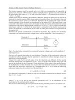
Fig. 13 Suprasellar and parasellar lesions. Diagram of the cavernous sinus and
its contents; the sellar, suprasellar, and parasellar structures
Jugular foramen lesions
– Neoplastic masses
ț Paragangliomas Chemodectomas or glomus tumors; parasympathetic
paraganglia located in the jugular bulb adventitia and
in various sites of the head and neck, especially the
carotid body, glomus jugulare, and glomus tympani-
cum
ț Metastases – Regional extension (e.g., nasopharyngeal carci-
noma, lymph node metastatic disease)
– Hematogenous extracranial sites (e.g., lung, pros-
tate, breast)
ț Nerve sheath
tumors
Uncommon location
– Schwannomas of cranial nerves IX and XI
– Neurofibromas
– Epidermoid tumor
Chondroid, chordo-
ma lesions
ț Meningioma
Intracranial Tumors
Tsementzis, Differential Diagnosis in Neurology and Neurosurgery © 2000 Thieme
All rights reserved. Usage subject to terms and conditions of license.
129
Nonneoplastic masses
– Prominent jugular
bulb
“Pseudomass”—normal variant
– Jugular vein thrombo-
sis
– Osteomyelitis
Diffuse skull base le-
sions
Neoplastic masses
– Metastases
– Multiple myeloma,
plasmacytoma
– Meningioma
– Lymphoma Primary or secondary; uncommon, but increasing in
incidence, causing leptomeningeal disease and multi-
ple cranial nerve palsies
Nonneoplastic masses
– Fibrous dysplasia The most common benign skeletal disorder in adoles-
cents and young adults. In the most common monos-
totic type, 25% of skull and facial bones are involved,
compared with 40– 60% in the polyostotic type, caus-
ing facial deformities and cranial nerve palsies
– Paget’s disease
– Eosinophilic granulo-
ma
Cavernous sinus lesions
(Fig. 13)
Unilateral
– Schwannoma Cranial nerves III, IV, V, and VI
– Meningioma These tend to follow the lateral margin of the
cavernous sinus, and may extend posteriorly along the
tentorial margin, with a dovetail appearance on MRI.
May encase or distort the cavernous portion of the
ICA
– Metastasis E.g., adenoid cystic carcinoma, basal-cell carcinoma,
lymphoma, mucoepidermoid carcinoma, melanoma,
and schwannoma, showing perineural spread through
the basal skull foramen and into the brain
– Vascular lesions E.g., ectatic carotids, caroticocavernous fistula,
cavernous carotid aneurysm, cavernous hemangioma,
and cavernous sinus thrombosis
– Chordoma
– Lymphoma
– Chondrosarcoma
– Lipoma
– Infection E.g., actinomycosis, Lyme disease, and herpes zoster
can also demonstrate perineural involvement
Skull Base
Tsementzis, Differential Diagnosis in Neurology and Neurosurgery © 2000 Thieme
All rights reserved. Usage subject to terms and conditions of license.
130
– Idiopathic inflam-
matory disease
Tolosa–Hunt syndrome: characterized by recurrent at-
tacks of retro-orbital pain, defects in cranial nerves III,
IV, Va, and VI, with spontaneous remission and
prompt response to steroid therapy
Bilateral
– Extensive and ag-
gressive pituitary
adenoma
– Meningioma
– Metastases
– Thrombosis of the
cavernous sinus
May occur as part of a septic process associated with
spontaneous dural malformations, or may result from
an interventional or surgical procedure
ICA: internal carotid artery; MRI: magnetic resonance imaging.
Choroid Plexus Disease
Differential diagnosis:
Tumors
Choroid plexus papil-
loma
Choroid plexus carci-
noma
Meningioma
Ependymoma, sub-
ependymoma
Neurofibroma
Glioblastoma, astrocy-
toma
Oligodendroglioma
Tuberous sclerosis, sub-
ependymal giant-cell
astrocytoma
CNS lymphoma
PNET E.g., medulloblastomas, ependymoblastomas, pineo-
blastomas, cerebral neuroblastomas, medullo-
epitheliomas, melanotic vermian PNET of infancy
Metastases
Nonneoplastic tumor-
like lesions
Epidermoid tumor
Dermoid tumor
Intracranial Tumors
Tsementzis, Differential Diagnosis in Neurology and Neurosurgery © 2000 Thieme
All rights reserved. Usage subject to terms and conditions of license.
131
Gliomatosis Cerebri
This is a diffusely infiltrative neoplasm, with variably undifferentiated
astrocytes and without a necrotic center. Gliomatosis cerebri presents as
a diffuse involvement of the cerebral hemispheres, leading to progres-
sive changes in personality, headaches, and impaired mental status.
Positron-emission tomography (PET) scanning with methionine shows
isotope accumulation in the diffusely infiltrative tumorous area, with
greater accuracy than computed tomography or magnetic resonance
imaging. The definitive diagnosis is at autopsy. The prognosis is variable,
with survival measured in months to years.
Differential diagnosis:
Low-grade glioma
Oligodendroglioma
Gliomatosis cerebri
Leptomeningeal gliomatosis
Encephalitis
Diffuse and demyelinating disease
Pseudotumor cerebri
Nonneoplastic cysts
Colloid cyst
Rathke’s cleft cyst
Neuroglial (neuroepi-
thelial) cyst
Vascular malforma-
tions
Choroid plexus angio-
mas
Phakomatosis E.g., Sturge-Weber syndrome
Infection
Choroid plexitis Pathogens include Cryptococcus and Nocardia
Other
Inflammation
Sarcoidosis
Xanthogranuloma
CNS: central nervous system; PNET: primitive neuroectodermal tumor.
Gliomatosis Cerebri
Tsementzis, Differential Diagnosis in Neurology and Neurosurgery © 2000 Thieme
All rights reserved. Usage subject to terms and conditions of license.
132
Tolosa–Hunt Syndrome
Idiopathic inflammatory disease of the cavernous sinus.
Sarcoidosis
Meningioma
Lymphoma
Metastatic and neurotropic spread of tumor into the cavernous sinus
Infections (e.g., actinomycosis, mucormycosis, aspergillosis)
Recurrence of Malignant Gliomas
An enlarging lesion at the site of a previously treated glioma most prob-
ably represents a regrowth of an incompletely treated initial tumor, and
is less likely to be the development of a new pathological entity. In the
differential diagnosis of an enlarging lesion at the site of a previously
eradicated malignant glioma, the clinician should consider the follow-
ing possibilities.
Development of a dis-
tinct new tumor
In cases of genetic predisposition to tumor develop-
ment shared by cells in the area:
– Multiple gliomas in patients with tuberous sclerosis
– Multiple neurofibromas developing along the same
nerve root in patients with neurofibromatosis
Growth of a tumor
with related pathology
A tumor with related histopathology may supplant the
original tumor.
– The astrocytic component of a mixed glioma re-
placing its previously treated oligodendrocytic
component
– A gliosarcoma can arise from a previously treated
glioblastoma
Growth of a secondary
tumor
The initial treatment may induce a secondary tumor
of a different type:
– A parasellar sarcoma after irradiation for a pituitary
adenoma
– A glioblastoma in the radiation field of a menin-
gioma
Metastatic tumor at
the original tumor site
E.g., a breast metastasis within a pituitary adenoma
Intracranial Tumors
Tsementzis, Differential Diagnosis in Neurology and Neurosurgery © 2000 Thieme
All rights reserved. Usage subject to terms and conditions of license.
133
Nonneoplastic lesions Nonneoplastic lesions can mimic tumor growth:
– Radiation necrosis after focal high-dose irradiation
–
Abscess formation at the site of the tumor resection
Congenital Posterior Fossa Cysts and Anomalies
Dandy–Walker com-
plex
In 70% of cases, the syndrome has a number of as-
sociated anomalies, such as hydrocephalus, agenesis
of the corpus callosum, nuclear dysplasia of the brain
stem, and other cerebrocerebellar heterotopias
Dandy–Walker malfor-
mation
Large posterior fossa and CSF cyst, high transverse
sinuses and tentorial insertion, vermian, cerebellar
hemispheric and brain stem hypoplasia in 25% of
cases
Dandy–Walker variant Mild vermian hypoplasia, moderately enlarged fourth
ventricle although the posterior fossa is typically of
normal size, the brain stem is normal, and there is a
variable degree of vermian hypoplasia
Other posterior fossa
cysts
Arachnoid and neuro-
epithelial cysts
Arachnoid cysts are formed by a splitting of the
arachnoid membrane with layers of thickened fibrous
connective tissue, whereas neuroepithelial or glio-
ependymal cysts are lined with a low cuboidal-colum-
nar epithelium
Megacisterna magna The fourth ventricle appears normal and the vermis
and cerebellar hemispheres are normal, but occa-
sionally the posterior fossa can be enlarged, with
prominent scalloping of the occipital bones
Isolated fourth ventricle After ventriculoperitoneal shunt, leading to secondary
aqueductal stenosis, but in addition the CSF outflow
from the fourth ventricle is prevented, or its absorp-
tion is prevented, e.g., in patients in whom the hydro-
cephalus is due to or associated with an inflammatory
meningeal process, such as infection or hemorrhage
Pulsion diverticulum In advanced hydrocephalus, the thin ventricular wall
may dehisce into the adjacent subarachnoid space,
forming diverticula commonly in the inferomedial wall
of the atria, the suprapineal recess, and through the
incisure, causing downward displacement of the cere-
bellum
Congenital Posterior Fossa Cysts and Anomalies
Tsementzis, Differential Diagnosis in Neurology and Neurosurgery © 2000 Thieme
All rights reserved. Usage subject to terms and conditions of license.
134
Intracranial Tumors
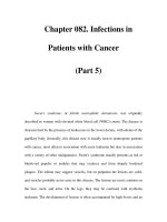
3. Arachnoid cyst of the 4th ventricle. Sagittal T1 WI showing dilatation of the 4th
ventricle and isodense signal with the cerebrospinal fluid.
4. Hemangioblastoma. Coronal T1 WI demonstrates a cystic space-occuping le-
sion with a small postcontrast enhancing mural nodule.
5. Epidermoid cyst. Axial T1WI with a solid extrinsic space-occupying mass with
smooth margins and a relative heterogeneity, which causes smooth erosion of
the occipital bone and exerts mild compression on the left cerebellar hemi-
sphere.
6. Epidermoid cyst. Coronal T1 WI shows a solid extrinsic space-occupying mass
with well-defined margins, it is non-contrast enhancing and causes erosion of
the occipital bone.
Miscellaneous cerebel-
lar hypoplasias
Chiari type IV malfor-
mation
Absent or severely hypoplastic cerebellum and small
brain stem
Jouber t’s syndrome Split or segmented vermis, transmitted by autosomal
recessive genes
Rhombencephalo-
synapsis
Agenesis of the vermis and midline fusion of the cere-
bellar hemispheres and peduncles
Tectocerebellar dys-
raphia
Vermian hypoplasia, occipito-encephalocele, and dor-
sal brain stem traction
Lhermitte–Duclos dis-
ease or dysplastic cere-
bellar gangliocytoma
Gross thickening of the cerebellar folia, hypertrophy of
the granular cell layer, and axonal hypermyelination of
the molecular cell layer
CSF: cerebrospinal fluid.
Posterior Fossa Cysts
(Fig. 14)
Dandy–Walker complex
Megacisterna magna
Arachnoid cyst
Nonneoplastic cysts
Inflammatory
Enterogenous
Neoplastic cysts
– Hemangioblastoma
– Pilocytic astrocytoma
Cyst-like tumors
– Dermoid
– Epidermoid
Tsementzis, Differential Diagnosis in Neurology and Neurosurgery © 2000 Thieme
All rights reserved. Usage subject to terms and conditions of license.
135
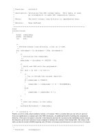
Fig. 14 Posterior fossa cysts
1. Dandy-Walker cyst. Proton density axial MRI T2 WI presenting a cystic dilata-
tion of the cisterna magna that communicates with the 4th ventricle. There is
an associated atrophy of the cerebellar vermis and a smooth erosion of the
occipital bone.
2. Dandy-Walker cyst. Proton density sagittal T2 WI (same case). The com-
munication of the cyst with the 4th ventricle and the significant vermian atro-
phy are noted. There is also elevation of the confluence of sinuses and of the
tentorium cerebelli.
Posterior Fossa Cysts
Tsementzis, Differential Diagnosis in Neurology and Neurosurgery © 2000 Thieme
All rights reserved. Usage subject to terms and conditions of license.
136
Enhancing Lesions in Children and Young Adults
Imaging differential diagnoses for a peripheral enhancing lesion in a
child or young adult include the following.
Glioblastoma
Ganglioglioma
Gangliosarcoma
Malignant astrocytoma
Meningioma
Meningiosarcoma
Oligodendroglioma
Juvenile pilocytic astrocytoma
Solitary metastasis
Pleomorphic xanthoastrocytoma
Fibrous histiocytoma
Fibrous xanthomas
Tumoral Hemorrhage
Intratumoral hemorrhage may be suspected in the appropriate clinical
circumstances, for example in patients with known malignancy, in
elderly nonhypertensive persons, and in patients who had progressive
symptoms before the hemorrhage ictus. Hemorrhage has been noted in
about 1 % of brain tumors, whereas underlying tumors have been re-
ported in up to 10% of cases with intracranial hemorrhage.
Metastatic lesions are usually seen as well-defined, round masses lo-
cated around the gray-white junction, and they show contrast enhance-
ment and moderate edema. Hemorrhagic metastases are usually seen as
areas of high signal intensity on T1-weighted images and T2-weighted
images, with a relative absence of hemosiderin deposition.
Brain tumors associated with hemorrhage include the following.
Primary brain tumors
Malignant astrocytoma
– Anaplastic astrocy-
toma
– Glioblastoma multi-
forme
Of the adult gliomas, glioblastoma multiforme (GBM)
is the one most often associated with intratumoral
hemorrhage and subarachnoid seeding
Intracranial Tumors
Tsementzis, Differential Diagnosis in Neurology and Neurosurgery © 2000 Thieme
All rights reserved. Usage subject to terms and conditions of license.
137
Oligodendroglioma
(neurocytoma)
Although intraventricular neurocytomas have a more
benign course, they are more often subject to hemor-
rhage than oligodendrogliomas, which may suggest
the diagnosis
Meningioma
Pituitary adenoma
Hemangioblastoma
Acoustic neurinoma
Lymphomas Hemorrhage is rare in lymphomas
Metastatic brain
tumors
Lung cancer Bronchial carcinomas spread to the CNS in 30% of
cases; oat-cell carcinoma is the most frequent,
whereas squamous-cell carcinoma is the least fre-
quent subtype to metastasize to the brain
Breast cancer It is estimated that 18–30 % of patients with breast
cancer will develop brain metastases
Malignant melanoma Third most common neoplasm, with a propensity for
metastatic spread to the brain, after the lung and
breast
Renal-cell carcinoma
Thyroid cancer
Gastrointestinal primary
tumors
Choriocarcinoma
Retinoblastoma
CNS: central nervous system.
Brain Metastases
A known history of systemic cancer and the presence of multiple lesions
on magnetic resonance imaging (MRI) make the diagnosis of metastatic
brain tumor probable. Even a typical scan only suggests, but does not
prove, that the lesion is a brain metastasis and not another lesion, such
as a primary brain tumor or a cerebral abscess. Stereotactic needle bi-
opsy is required for definitive diagnosis.
Brain Metastases
Tsementzis, Differential Diagnosis in Neurology and Neurosurgery © 2000 Thieme
All rights reserved. Usage subject to terms and conditions of license.
138
Differential diagnosis:
Primary brain tumors
Meningioma – Meningiomas show homogeneous contrast en-
hancement, a relative lack of peritumoral edema,
and attachment to the dura. Metastatic cancers
may also arise from the dura, and can even be sup-
plied by the external carotid artery, making the dis-
tinction between metastasis and meningioma im-
possible except by biopsy.
– If the neurological symptoms have developed very
slowly, or if the MRI suggests a lesion neighboring
the falx or the inner skull table, the diagnosis is in
favor of a meningioma
– It should also be borne in mind that breast cancer
may metastasize to a meningioma
Astrocytoma Brain metastasis presents as a spherical mass, whereas
primary gliomas are usually irregular, and present fin-
ger-like extensions of contrast enhancing tumor run-
ning along the white matter tracts and bundles
Primary brain lym-
phoma
These lesions of ten present as uniform, multiple, peri-
ventricular lesions on MRI, with irregular margins that
are not discrete
Acoustic neurinoma
and pituitary adenoma
Almost impossible to distinguish from metastatic
brain tumors in the same areas
Vascular disorders
Cerebral infarction – Acute infarctions do not enhance, and the MRI find-
ings may be entirely normal for 24– 48 hours after
the event
– Contrast enhancement of the pial surface of the
overlying cortical gyri develops 1– 3 weeks after
the ictus, unlike the ring-like enhancing lesion of a
brain metastasis
– Several weeks postictally, the contrast enhance-
ment in an infarct diminishes and gradually disap-
pears, and the ischemic area becomes hypointense
Cerebral hemorrhage – Acute hemorrhage is hyperdense on a noncontrast
CT scan, but may have a normal appearance on
MRI
– Contrast enhancement 3 – 6 weeks postictally dem-
onstrates an isodense clot with a ring enhance-
ment, resembling a metastasis or an abscess. Early
enhancement suggests tumoral hemorrhage
Intracranial Tumors
Tsementzis, Differential Diagnosis in Neurology and Neurosurgery © 2000 Thieme
All rights reserved. Usage subject to terms and conditions of license.
139
Infections Cerebral abscess usually occurs in patients with re-
duced immunity, and particularly in those suffering
from Hodgkin’s disease and other lymphomas, condi-
tions in which brain abscesses are more common than
metastatic brain tumors
Toxoplasma abscess This is the most common parasitic CNS infection, and
has a predilection to lodge in the basal ganglia as a
single mass
Multiple nocardia ab-
scesses
These develop in 50% of immunosuppressed patients
with Nocardia pulmonary infection.
Progressive multifocal
leukoencephalopathy
(PML)
An infection of the oligodendrocytes caused by the JC
polyomavirus, affecting patients with depressed cellu-
lar immunity due to lymphoma or chronic lympho-
cytic leukemia, or after prolonged chemotherapy
Differential features
– CT and MRI help identify brain abscesses. The en-
hancing ring of an abscess is generally thinner and
more uniform than the ring of a tumor. The capsule
of an abscess is characteristically thicker near the
corte x, where oxygenation is better, and somewhat
thinner near the ventricular surface.
– With suspected Toxoplasma abscesses, a therapeu-
tic trial with sulfadiazine and pyrimethamine has a
rapid response, and this can establish the diagnosis
without the need for a biopsy. With other sus-
pected abscesses, stereotactically directed needle
biopsy performed early in the diagnostic work-up
both establishes the diagnosis and reveals the in-
volved organism for the appropriate antibiotic ther-
apy
– CT and MRI in PML reveal multifocal, punched-out
lesions of the white matter, with no mass effect
and usually no contrast enhancement. Nonenhanc-
ing lymphomas may be similar. A definitive diagno-
sis is secured only by biopsy
Radiation necrosis CT and MRI reveal a hypodense or isodense ring-en-
hancing brain lesion, surrounded by edema. Differen-
tiating between radiation necrosis and recurrent brain
metastases in a patient previously irradiated for a
brain metastasis may be impossible without needle bi-
opsy
Methotrexate
leukoencephalopathy
Causes bilateral white matter lesions and ventricular
enlargement. The lesions show a reduced density on
CT scanning and appear hyperintense on T2-weighted
MRI without enhancement, a feature that distinguish-
es the condition from a brain metastasis
Brain Metastases
Tsementzis, Differential Diagnosis in Neurology and Neurosurgery © 2000 Thieme
All rights reserved. Usage subject to terms and conditions of license.
140
Multiple sclerosis MS lesions may be single or multiple, and contrast-
enhancing, which makes them indistinguishable from
brain tumors. However, MS lesions do not enhance
after 6 – 8 weeks, and other new nonenhancing lesions
may be present, which is unlikely with brain
metastases
Miscellaneous
Transient changes in CT or MRI sometimes follow focal
or generalized epilepsy in the absence of underlying
primary or metastatic brain tumor. These lesions dis-
appear within a few weeks after control of the seizures
CNS: central nervous system; CT: computed tomography; MRI: magnetic resonance imaging;
MS: multiple sclerosis; PML: progressive multifocal leukoencephalopathy.
Subarachnoid Space Metastases
Between 6% and 18% of central nervous system (CNS) metastases involve
the arachnoid and subarachnoid space, or the pia, or both. The sub-
arachnoid space can be diffusely or focally involved by spread from a pri-
mary CNS tumor, or by an extraneural malignancy. The typical locations
for metastatic seeding are at the basal cisterns, the cerebellopontine
angle cistern, the suprasellar cisterns, along the course of the cranial
nerves, and over the convexities. Subtle leptomeningeal and sub-
arachnoid space metastatic disease is identified in up to 45% of cases
using contrast-enhanced magnetic resonance imaging (MRI) scans.
Cerebrospinal fluid (CSF) cytology provides definitive diagnosis of lepto-
meningeal carcinomatosis, with abnormal CSF noted in up to 55% of
cases after the first spinal tap and in up to 90% after the third. If lumbar
puncture is contraindicated or the CSF cytology is equivocal,
gadolinium-enhanced MRI is a useful diagnostic tool.
Sources of sub-
arachnoid metastases
Children
Primary brain tumors
– Primary neuroec-
todermal tumors
(PNETs)
ț Medulloblastoma
ț Ependymoblastoma
– Pineal tumors Germinoma, pineoblastoma
– Choroid plexus car-
cinoma
Primary extracranial
tumors
– Neuroblastoma
Intracranial Tumors
Tsementzis, Differential Diagnosis in Neurology and Neurosurgery © 2000 Thieme
All rights reserved. Usage subject to terms and conditions of license.
141
– Lymphoma
– Leukemia
Adults
Primary brain tumors
– Glioblastoma multi-
forme, anaplastic
astrocytoma
– Oligodendroglioma
– Primary lymphoma
Primary extracranial tu-
mors
– Lung cancer
– Breast cancer
– Malignant melanoma
– Gastrointestinal carci-
noma
– Ovary
– Lymphoma
– Leukemia
Differential diagnosis:
Cranial meningeal carci-
nomatosis
Meningitis
– Acute bacterial menin-
gitis
– Chronic meningitis Fungal and granulomatous meningitis. Chronic menin-
gitides have a predilection to invade the basal cisterns
ț Tuberculous meningitis
ț Coccidioidomycosis imitans meningitis
ț Cryptococcus neoformans meningitis
ț Neurocysticercosis
Noninfectious inflam-
matory diseases
– Sarcoidosis
Lymphoma
Leukemia
Posttraumatic basal cran-
ial adhesions
Intrathecal chemother-
apy, radiation
Idiopathic pachymenin-
gitis
Subarachnoid Space Metastases
Tsementzis, Differential Diagnosis in Neurology and Neurosurgery © 2000 Thieme
All rights reserved. Usage subject to terms and conditions of license.
142
Hyperprolactinemia
Hyperprolactinemia in women leads to amenorrhea, galactorrhea, and
osteoporosis, while in men it may result in diminished sexual drive and
impotence, or may be asymptomatic. The degree of hyperprolactinemia
is directly related to the functionality of the prolactin-secreting tumor.
Serum prolactin levels over 200 ng/mL correlate well with the presence
of a prolactinoma. Normal prolactin levels are in the ranges of 1– 20 ng/
mL in men, and 1 –25 ng/mL in women.
Differential diagnosis:
Nonpathological causes
Pregnancy
Early nursing periods
Nipple stimulation
Coitus
Sleep
Stress
Exercise
Diseases
True prolactinomas
Pituitary traumatic stalk section
Pituitary stalk compression from chromophobe macroadenomas
Empty sella syndrome
Hypothalamic disorders
– Tumors (e.g., craniopharyngiomas)
– Histiocytosis X
– Sarcoidosis
Primary hypothyroidism
Chiari–Frommel syndrome
Renal failure
Liver cirrhosis
Drugs
Dopamine antagonists (e.g., phenothiazine-like drugs)
Reserpine
–
α-methyl
– Dopa
Opiate derivatives (e.g., morphine)
Prostaglandin F
2α
Thyrotropin-releasing hormone
Estrogens
Intracranial Tumors
Tsementzis, Differential Diagnosis in Neurology and Neurosurgery © 2000 Thieme
All rights reserved. Usage subject to terms and conditions of license.
143
Demyelinating Disease and Brain Atrophy
Multifocal White Matter Lesions
Multiple sclerosis
Hypertension and
ischemic white matter
lesions (leukokraurosis)
Increases with age, and has also been seen with
chronic hypertension. There are two types of ischemic
white matter lesions:
– Lesions involving the watershed distribution of the
major brain arteries
– Lesions caused by intrinsic disease of the small
penetrating medullary arteries (arteriolar sclerosis)
Perivascular (Virchow–
Robin) spaces
Enlargement of these perivascular spaces with age
and hypertension, associated with thinning, pallor and
atrophy of the adjacent myelin, is called état criblé
Metastases
Trauma, nonvascular
white matter injury
Diffuse axonal shearing caused by acceleration, decel-
eration, and rotation forces on the brain
Inflammatory E.g., Lyme disease, cysticercosis
Vasculitides
– Systemic lupus ery-
thematosus
– Sjögren’s syndrome
– Behçet’s disease
– Moyamoya disease
– Amyloid angiopathy
– Polyarteritis nodosa
Primary CNS lymphoma
Migraine Mysterious lesions of the frontal lobe, centrum semi-
ovale, and basal ganglia, possibly due to microemboli
from increased platelet aggregation during migraine
attacks
Inherited leukoencepha-
lopathy
Secondary leukoence-
phalopathy
– Acute disseminated
encephalomyelitis
(ADEM)
Tsementzis, Differential Diagnosis in Neurology and Neurosurgery © 2000 Thieme
All rights reserved. Usage subject to terms and conditions of license.
144
– Progressive multifocal
encephalopathy
(PML)
– Binswanger’s disease Subcortical arteriosclerotic encephalopathy
– Postanoxic encepha-
lopathy
– Osmotic demyelin-
ation, or central
pontine myelolysis
– Alcoholism (Marchia-
fava–Bignami syn-
drome)
– Drugs Methamphetamine, cocaine, heroin
– Toxins Hexachlorophene, lead, isoniazid, chemotherapeutic
agents, eclampsia
– Radiation changes
Dysmyelinating diseases
– Metachromatic
leukodystrophy
(MLD)
The most common type, resulting from a deficiency of
the enzyme arylsulfatase A
– Adrenoleukodystro-
phy
E.g., associated with adrenal cortical insufficiency and
the accumulation of very long chain fatty acids in the
white matter, adrenal cortex, and plasma due to im-
pairment in peroxisomes of
β-oxidation
– Alexander’s disease
– Canavan’s disease Deficiency of the enzyme aspartoacyclase
– Krabbe’s disease Deficiency of
β-galactosidase
CNS: central nervous system.
Multiple Sclerosis–Like Lesions
Multiple sclerosis (MS) is a clinical diagnosis that should never be made
using neuroimaging alone. In 78–95% of clinically diagnosed MS
patients, gadolinium-enhanced magnetic resonance imaging (MRI) fea-
tures include ovoid periventricular, infratentorial, temporal lobe, and
corpus callosum white matter lesions that are isointense to hypointense
on T1-weighted images, and show high intensity on proton density and
T2-weighted images. Many conditions have to be taken into account in
the differential diagnosis of multiple white matter high-signal abnor-
malities on proton density and T2-weighted images. Other conditions
may produce lesions with or without enhancement, and can occur in a
patient population similar to that with MS. The list of diseases with clini-
cal and neuroimaging features similar to those of multiple sclerosis in-
cludes the following.
Demyelinating Disease and Brain Atrophy
Tsementzis, Differential Diagnosis in Neurology and Neurosurgery © 2000 Thieme
All rights reserved. Usage subject to terms and conditions of license.
145
Neurosarcoidosis The granulomatous process invades and thromboses
affected blood vessels, and produces a granulomatous
angiitis similar to primary angiitis of the CNS. High-
intensity white matter in sarcoid may be indistinguish-
able from MS
Lyme disease Neuroborreliosis. Approximately 10– 15% of patients
with Lyme disease have CNS involvement. High-signal
contrast-enhancing subcortical abnormalities on pro-
ton density and T2-weighted images on MRI in the
frontal and parietal lobes, the basal ganglia and pons,
cranial nerves (facial nerve)
Vasculitides Multisystem immune-related vasculitis, with CNS in-
volvement in 10–49% of cases, e.g. systemic lupus
erythematosus, Behçet’s disease. May resemble MS
clinically and due to a white matter lesion pattern in
the brain and spinal cord
Neurosyphilis Contrast-enhanced MRI shows patchy enhancement
involving the basal ganglia or the middle cerebral
ar tery territories
Tuberculosis Single or multiple lesions located in the cerebral hemi-
sphere and basal ganglia in adults, and in the cerebel-
lum in children. On MRI with gadolinium injection, a
hypodense rim may separate the hyperintense center
from the peripheral hyperintense edema on T2-
weighted images, and T1-weighted images often
show nodular enhancement
Viral infection
Devic’s disease, or neuro-
myelitis optica
Diffuse sclerosis
(Schilder’s disease)
An acute, rapidly progressing form of MS with bi-
lateral, relatively symmetric and large areas of demy-
elination, often involving the centrum semiovale and
the occipital lobes; seen usually in childhood, and
rarely in those over 40
Myelopathy
Acute disseminated
encephalomyelitis
Acute monophasic inflammatory demyelination, dis-
tinguished from MS by its clinical course—a single
acute episode including fever and headache. The loca-
tions and characteristics of the lesions on the MRI may
be indistinguishable from MS
Multiple Sclerosis–Like Lesions
Tsementzis, Differential Diagnosis in Neurology and Neurosurgery © 2000 Thieme
All rights reserved. Usage subject to terms and conditions of license.
146
Baló’s disease (concen-
tric sclerosis)
Represents a histological MS lesion with alternating
concentric regions of demyelination and normal brain
Hypertension and
ischemic white matter
lesions
In elderly patients with malignant hypertension, high-
signal patchy or diffuse bilateral periventricular white
matter abnormalities, most likely representing small-
vessel disease manifesting as lacunar, deep white mat-
ter infarctions
Virchow–Robin spaces Dilated perivascular spaces enlarge with age and hy-
pertension and occur in characteristic locations, typi-
cally in the basal ganglia, around the ventricular atria,
centrum semiovale, brain stem. The perivascular
spaces remain isodense to CSF, whereas lesions are
hypodense on the proton density – weighted MRI
sequence
Lesions associated with
migraine
High-intensity abnormalities in the centrum semiovale
and frontal white matter in young patients under
40. The lesions appear to be a diffuse process, possibly
resulting from platelet microemboli or primary neu-
ronal damage related to the pathophysiology of mi-
graine
Multi-infarct dementia,
leukoareosis, and
Binswanger’s disease
Affects the elderly population, and the predominant
clinical manifestations are cognitive and behavioral
disorders. The MRI shows periventricular white matter
and centrum ovale watershed infarcts, similar in ap-
pearance to the demyelinating lesions of MS; however,
in contrast to the MS lesions, there are no associated
lesions in the basal ganglia, brain stem, or occipital
horns, and there is sparing of the subcortical U fibers
Normal aging In healthy individuals of 52 – 72 years of age, atrophic
periventricular demyelination has been found in 53.4%
and white matter infarcts are seen in 13.4%. Incident al
white matter T2 hyperintensities occur frequently in
elderly people
Metastases and brain
abscesses
Rarely produce lesional patterns quite similar to MS.
The presence of a mass effect and a clinical history
suggesting a remote source for the lesions is impor-
tant
Motor neuron disease
Intracranial tumor Especially brain stem, cerebellum
Vitamin B
12
deficiency Gastrectomy, gastric carcinoma, malabsorption syn-
dromes
CSF: cerebrospinal fluid; CNS: central nervous system; MRI: magnetic resonance imaging; MS:
multiple sclerosis.
Demyelinating Disease and Brain Atrophy
Tsementzis, Differential Diagnosis in Neurology and Neurosurgery © 2000 Thieme
All rights reserved. Usage subject to terms and conditions of license.
147
Cerebellar Atrophy
Toxic
– Alcohol abuse The most common cause, with the vermis more ex-
tensively involved
– Long-term drug use ț Phenytoin (dilantin)
ț Phenobarbital
– Mercury poisoning
Hereditary, degenerative
– Olivopontocerebellar
degeneration
– Shy–Drager disease
– Friedreich’s ataxia
– Hereditary cerebellar
atrophy
– Louis–Bar syndrome,
or ataxia teleangi-
ectasia
Ischemia E.g., chronic vertebrobasilar atherosclerotic disease
Paraneoplastic syn-
dromes
– Neuroblastoma
– Hodgkin’s disease
– Cancer Ovarian, gastrointestinal, lung, breast
Cerebral Atrophy
Alzheimer-type demen-
tia
Diffuse cortical atrophy, especially in the temporal
lobes and hippocampal- parahippocampal area, and
dilation of more than 3 mm in diameter of the
choroidal-hippocampal fissure complex and dilation of
the temporal horns
Pick’s disease Severe atrophy of the anterior frontal and temporal
lobes, with swollen nerve cells and intracytoplasmic
inclusions (Pick’s bodies)
Parkinson’s disease Altered intensity of small and basal ganglia in the sub-
stantia nigra
Progressive supranu-
clear palsy (Steele–
Richardson–Olszewski
syndrome)
Third ventricular dilation, midbrain atrophy, and en-
largement of the interpeduncular cistern
Cerebral Atrophy
Tsementzis, Differential Diagnosis in Neurology and Neurosurgery © 2000 Thieme
All rights reserved. Usage subject to terms and conditions of license.
148
Creutzfeldt–Jakob dis-
ease
Frontal predominance atrophy, abnormal intensity of
the basal ganglia
Multi-infarct dementia White matter and deep gray lacunae, central pontine
infarcts and strokes of different ages
Dyke–Davidoff–Masson
syndrome
E.g., hemiatrophy of one hemisphere
Porencephaly E.g., from trauma, infection, and perinatal ischemia
Miscellaneous causes
– Previous infections
– Long-standing multi-
ple sclerosis
– Extensive traumatic
brain injury
– Chronic use of ste-
roids
– Radiation injury
– Intrathecal chemo-
therapy
– Starvation, anorexia
– Dehydration
Dementia
Dementia is very common, and is the most disabling psychiatric dis-
order in the adult population. The incidence increases exponentially
with age, from 0.5% at age 40 years up to 20% of the population aged 80
years and over. Over 80% of patients with dementia suffer from a small
number of conditions, associated with characteristic types of pathology
and different etiologies.
Etiology % of dementia cases
Alzheimer’s disease 45
Cerebrovascular disease 15
Cortical Lewy body disease 10
Head trauma 3
Parkinson’s disease 3
Motor neuron disease 2
Other 5
AIDS dementia (prion disease) > 1
Unknown 15
AIDS: acquired immune deficiency syndrome.
Demyelinating Disease and Brain Atrophy
Tsementzis, Differential Diagnosis in Neurology and Neurosurgery © 2000 Thieme
All rights reserved. Usage subject to terms and conditions of license.
149
Differential diagnosis
Degenerative disorders
Presenile dementia
– Alzheimer’s disease
– Pick’s disease
– Cortical Lewy body
disease
– Prion disease
– Huntington’s chorea
Senile dementia
Cerebrovascular dis-
ease
Multi-infarct dementia A series of relatively large infarcts damaging a suffi-
cient volume of brain results in dementia. Neuro-
pathological calculations indicate that infarct volumes
that total over 50 mL are of ten associated with de-
mentia, and that a total infarct volume over 100 mL is
always associated with dementia. Vascular dementia
may coexist with Alzheimer’s disease in 20% of cases,
and smaller volumes of infarct could therefore con-
tribute significantly to the dementia symptoms
Cerebral embolism
Cerebral hemorrhage
Subarachnoid hemor-
rhage
Disseminated lupus ery-
thematosus
Transient ischemic at-
tacks
Head injury
Acute head injury
Subdural hematoma
Posttraumatic dementia
Hypoxia
Post– cardiac arrest
– Heart failure
– Myocardial infarction
Respiratory disorders
Carbon monoxide poi-
soning
Dementia
Tsementzis, Differential Diagnosis in Neurology and Neurosurgery © 2000 Thieme
All rights reserved. Usage subject to terms and conditions of license.
150
Intracranial tumors
Infections
Intracranial
– Encephalitis
– Meningitis
– Meningoencephalitis E.g., general paresis
– AIDS dementia
General E.g., Urinary tract, bronchopneumonia, topical infection
Epilepsy
Toxic disorders
Drugs E.g., Alcohol, barbiturates, opiates, amphetamines,
LSD, cocaine, tricyclic antidepressants, steroids,
lithium, l-dopa, cycloserine, digoxin, MAOIs, cy-
closerine, isoniazid
Heavy metals E.g., Lead, mercury, manganese
Metabolic disorders
Acute
– Electrolyte distur-
bance
– Uremia
– Hepatic encephalo-
pathy
– Hypoglycemia
– Porphyria
– Endocrine diseases E.g., thyrotoxicosis, diabetes mellitus, Addison’s dis-
ease, parathyroid disorder, hypopituitarism
– Vitamin deficiencies E.g., thiamine, B
12
, nicotinic acid
Chronic
– Chronic alcoholic de-
mentia
– Heavy metals
– Myxedema, hypogly-
cemia, hypopituitar-
ism
– Vitamin deficiency E.g., thiamine—Korsakof f’s psychosis; nicotinic acid—
pellagra; vitamin B
12
and folic acid
Other disorders affect-
ing the CNS
Multiple sclerosis
Parkinson’s disease
Normal pressure hydro-
cephalus
AIDS: acquired immune deficiency syndrome; CNS: central nervous system; LSD: lysergic acid
diethylamide; MAOI: monoamine oxidase inhibitor.
Demyelinating Disease and Brain Atrophy
Tsementzis, Differential Diagnosis in Neurology and Neurosurgery © 2000 Thieme
All rights reserved. Usage subject to terms and conditions of license.
151
Cerebrovascular Disease
Cerebral Infarction in Young Adults
Cerebrovascular
atherosclerosis
Thrombotic or embolic
Embolism
Cardiac source
– Valvular Mitral stenosis, prosthetic valve, infective endocardi-
tis, marantic endocarditis, Libman–Sacks endocarditis,
mitral annulus calcification, mitral valve prolapse, cal-
cific aor tic stenosis
– Atrial fibrillation and
sick sinus syndrome
– Acute myocardial in-
farction and/or left
ventricular aneurysm
– Left atrial myxoma
– Cardiomyopathy
Paradoxical embolism or
pulmonary source
– Pulmonary AVM Including Osler–Weber–Rendu disease
– Atrial and ventricular
septal defects with
right-to-left shunt
– Patent foramen ovale
with shunt
– Pulmonary vein
thrombosis
– Pulmonary and medi-
astinal tumors
Other
– Aortic cholesterol
embolism
– Transient embolic
aortitis
– Emboli distal to un-
ruptured aneurysm
– Fat embolism syn-
drome
Tsementzis, Differential Diagnosis in Neurology and Neurosurgery © 2000 Thieme
All rights reserved. Usage subject to terms and conditions of license.