NEUROLOGICAL FOUNDATIONS OF COGNITIVE NEUROSCIENCE - PART 4 potx
Bạn đang xem bản rút gọn của tài liệu. Xem và tải ngay bản đầy đủ của tài liệu tại đây (405.21 KB, 30 trang )
the disease, they produce prototypical (e.g., “horse”
for “hippopotamus” and for any other large animal)
or superordinate responses (“animal”), but only
in very advanced cases are cross-category errors
produced.
This characteristic progression appears most
readily interpretable in terms of a hierarchically
structured semantic system, in which specific
information is represented at the extremities of a
branching “tree of knowledge.” More fundamental
distinctions, such as the division of animate beings
into land animals, water creatures, and birds, are
thought to be represented closer to the origin of
the putative hierarchy, with living versus nonliving
things at the very top. The defining characteristics
of higher levels are inherited by all lower points
(Collins & Quillian, 1969). Such a model has intu-
itive appeal and the deficits of semantic dementia
can be seen as a progressive pruning back of the
semantic tree (Warrington, 1975).
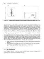
An alternative account, which we favor, is based
on the concept of microfeatures in a distributed
connectionist network (McClelland et al., 1995;
McClelland & Rumelhart, 1985). The basic idea is
illustrated in figure 4.4. An advantage of such a
model is that the low-level “features” of individual
concepts need only be represented once, while a
hierarchical model requires distinctive features to
be represented separately for every concept for
which they are true (e.g., “has a mane” for both lion
and horse). Category membership is then under-
stood as an emergent property of the sharing of ele-
ments of these patterns between concepts and thus
becomes a matter of degree—another intuitively
appealing property. A distributed feature network
could predict preservation of superordinate at the
expense of finer-grained knowledge, as seen in
semantic dementia, because even in a network that
had lost the representations of many individual
attributes, category coordinates would continue to
possess common elements, allowing judgments
about category membership to be supported long
after more fine-grained distinctions had become
impossible.
John R. Hodges 78
upright
eats fish webbed feet
large likes cold
has feathers
has legs
has a beak
lays eggs
hoots
predator
nocturnal
flies
small
eats insects
red breast
Figure 4.4
Distributed representation (microfeature) model illustrating penguin (thin line), owl (dashed line), and robin (thick line).
Do Patients with Semantic Dementia Show
Category-Specific Loss of Knowledge?
Living versus Nonliving Things
Semantic memory impairment that selectively
affects some categories of knowledge and spares
others has been most extensively documented in
patients with herpes simplex virus encephalitis, who
typically demonstrate a memory advantage for non-
living over living and natural things (animals, fruit,
etc.) (Pietrini et al., 1988; Warrington & Shallice,
1984). The complementary dissociation, which
effectively rules out any explanation based exclu-
sively on either lower familiarity or a greater degree
of visual similarity among the exemplars of living
categories, has also been described, typically in
patients who have suffered ischemic strokes in the
territory of the left middle cerebral artery (for a
review see Caramazza, 1998; Gainotti, Silveri,
Daniele, & Giustolisi, 1995).
The simplest interpretation of this phenomenon
would be that the neural representations of different
categories are located in separate cortical regions
(Caramazza, 1998; Caramazza & Shelton, 1998).
An alternative hypothesis is, however, that the
attributes critical to the identification of items
within these two broad domains differ in kind.
According to this view, one group of items, domi-
nated by living things, depends more strongly
on perceptual attributes, while another, mostly
artifacts, depends on their functional properties
(Warrington & Shallice, 1984). Support for the
sensory–functional dichotomy as a basis for cate-
gory specificity came initially from a group study
of patients showing this phenomenon. In these
patients the impaired categories did not always
respect the living versus manmade distinction
(Warrington & McCarthy, 1987). In particular, body
parts were found to segregate with nonliving things
while fabrics, precious stones, and musical instru-
ments behaved more like living things. The division
of knowledge into these fundamental subtypes has
been supported by positron emission tomography
activation studies of normal volunteers (Martin et
al., 1996; Mummery et al., 1999), but studies exam-
ining the status of perceptual and functional knowl-
edge in patients with category-specific impairments
have provided only limited endorsement of the
hypothesis (DeRenzi & Lucchelli, 1994; Silveri &
Gainotti, 1988).
The picture in semantic dementia presents a
similar inconsistency. When asked to provide defi-
nitions of common concepts, these patients volun-
teer very little visuoperceptual information. For
instance, when asked to describe a horse, they
typically produce phrases such as “you ride them,”
“they race them,” and “you see them in fields,” but
only rarely comment on their size, shape, color, or
constituent parts (Lambon Ralph, Graham,
Patterson, & Hodges, 1999). In view of the striking
temporal lobe involvement, the sensory–functional
theory might be confidently expected to predict a
significant advantage for artifact categories on tests
of naming or comprehension. When considered as
a group, the expected pattern does emerge in these
patients (albeit to a rather modest degree), but a
striking category effect is only rarely seen in indi-
vidual cases (Garrard, Lambon Ralph, & Hodges,
2002).
It seems, therefore, that lesion location and type
of information are not the sole determinants of
category specificity. Whether the additional factors
relate mainly to brain region (it has been hypo-
thesized, for instance, that involvement of medial
temporal structures may be important) (Barbarotto,
Capitani, Spinnler, & Trivelli, 1995; Pietrini et al.,
1988) or to some unidentified aspect of cognitive
organization, is as yet unclear.
Knowledge of People versus Objects: The Role of
Right and Left Temporal Lobes
A number of earlier authors had suggested an
association between right temporal atrophy and the
selective loss of knowledge of persons (DeRenzi,
1986; Tyrrell, Warrington, Frackowiak, & Rossor,
1990), but the first fully documented case, V.H., was
reported by our group in 1995 (Evans, Heggs,
Antoun, & Hodges, 1995). Initially, V.H. appeared
Semantic Dementia 79
to have the classic features of modality-specific
prosopagnosia, i.e., a severe inability to identify
familiar people from their faces, but much better
performance on names and voices. With time,
however, it became clear that the deficit was one
of a loss of knowledge about people affecting all
modalities of access to knowledge. V.H. was unable
to identify a photograph of Margaret Thatcher (the
patient was English) or to provide any information
when presented with the name, yet general seman-
tic and autobiographical memory remained intact
(Kitchener & Hodges, 1999). We hypothesized a
special role for the right temporal lobe in the repre-
sentation of knowledge about people (Evans et al.,
1995). As with most clear predictions, subsequent
studies have produced rather conflicting data. While
further patients with predominantly right-sided
atrophy have all shown a severe loss of knowledge
of persons, we have also observed significant
(though not selective) impairments of such knowl-
edge in patients with a predominantly left-sided
abnormality, suggesting that knowledge of people is
especially vulnerable to temporal atrophy on either
side (Hodges & Graham, 1998).
With regard to familiar objects rather than people,
our working hypothesis is that conceptual knowl-
edge is represented as a distributed network across
both the left and right temporal neocortex. This con-
clusion is supported by some, but not all, sources of
relevant evidence. For example, PET results with
normal participants would lead one to believe that
essentially all of the semantic action occurs in the
left hemisphere (Mummery et al., 1999; Vanden-
berghe et al., 1996). Our tentative claim for
bilateral representation of general conceptual know-
ledge is based on evidence from semantic
dementia. Deficits in semantic tests (such as naming
objects, matching words and pictures, sorting, or
making associative semantic judgments) are seen
not only in patients with predominantly left
temporal atrophy (e.g., Breedin, Saffran, & Coslett,
1994; Hodges et al., 1994; Lauro-Grotto et al.,
1997; Mummery et al., 1999; Snowden, Griffiths,
& Neary, 1994; Tyler & Moss, 1998; Vandenberghe
et al., 1996) but also in those with mainly right-
sided damage (e.g., Barbarotto et al., 1995; Hodges
et al., 1995; Knott et al., 1997).
V.H., the patient just described whose unilateral
anterior right temporal atrophy produced a selective
deficit for recognition and knowledge of people
(Evans et al., 1995), went on to develop a more
generalized semantic deficit in conjunction with the
spread of atrophy to the left temporal region
(Kitchener & Hodges, 1999). The opposite scenario
has occurred in two patients whose semantic
dementia began with a phase of unilateral left
anterior temporal changes in association with only
minimal semantic abnormality. Both cases were
shown to have a progressive anomia and developed
more pervasive semantic breakdown only when
the pathology spread to involve both temporal
lobes.
The most dramatic cognitive difference that has
emerged from our analyses of patients with greater
left than right atrophy (L > R), in contrast to those
with greater abnormality on the right (R > L), is not
in the extent or pattern of the semantic impairment
per se, but rather in its relationship to anomia. This
relationship was explored in a combined cross-
sectional and longitudinal analysis in which we
plotted the patient’s picture-naming score for the
forty-eight concrete concepts in our semantic
battery as a function of the corresponding level of
semantic deficit—defined for this purpose as the
patient’s score on a word-picture matching test for
the same forty-eight items. This analysis reveals
that for a given level of semantic impairment, the L
> R patients are substantially more anomic on
average than the R > L cases. The nature of the
naming errors is also different in the two subgroups;
although all patients make some of each of the three
main naming-error types seen in semantic dementia
(which, as noted earlier, are single-word semantic
errors, circumlocutions, and omissions), there are
relatively more semantic errors in the R > L patients
and relatively more failures to respond at all in the
L > R group.
Our account of this pattern is that semantic
representations of concrete concepts are distributed
across left and right temporal regions, but because
John R. Hodges 80
speech production is so strongly lateralized to the
left hemisphere, the semantic elements on the
left side are much more strongly connected to
the phonological representations required to name
the concepts. This explains how a patient in the
early stages of semantic dementia with atrophy
exclusively on the left side can be significantly
anomic, with only minor deficits on semantic tasks
that do not require naming (Lambon Ralph et al.,
1999).
Modalities of Input and Output
One of the continuing debates in the field has related
to the issue of whether knowledge is divided
according to the modality of input or output. Put
simply, when you hear or see the word “asparagus,”
is the semantic representation activated by this input
the same as or different from the conceptual knowl-
edge tapped by seeing or tasting it? Likewise, when
you speak about or name a hammer, is the concep-
tual representation that drives speech production the
same as or different from the semantic knowledge
that guides your behavior when pick up and use
a hammer? The latter kind of knowledge is often
referred to, by theorists who hold that it is a
separate system, as action semantics (Buxbaum,
Schwartz, & Carew, 1997; Lauro-Grotto et al.,
1997; Rothi, Ochipa, & Heilman, 1991).
Our hypothesis, based upon work in semantic
dementia, is that central semantic representations
are modality free. We tend to side with the theorists
arguing for one central semantic system (e.g.,
Caramazza, Hillis, Rapp, & Romani, 1990; Howard
& Patterson, 1992), rather than those proposing
separate modality-specific semantic systems (e.g.,
Lauro-Grotto et al., 1997; McCarthy & Warrington,
1988; Rothi et al., 1991; Shallice & Kartsounis,
1993). This view has been formed mainly by the
fact that none of the cases of semantic dementia
that we have studied have demonstrated a striking
dissociation between different modalities of input
or output and the following studies.
Are There Two Separate Systems for Words and
Objects?
To address this question, we recently (Lambon
Ralph et al., 1999) evaluated definitions of concrete
concepts provided by nine patients with semantic
dementia (including A.M.) (table 4.1). The stimulus
materials consisted of the forty-eight items from
the semantic battery described earlier (Hodges &
Patterson, 1995). Each patient was asked, on dif-
ferent occasions, to define each concept both in
response to a picture of it and in response to its
spoken name. The definitions were scored in a
variety of ways, including an assessment of whether
the patient’s definition achieved the status of “core
concept”: that is, the responses provided sufficient
information for another person to identify the
concept from the definition.
The view that there are separate verbal and visual
semantic systems predicts no striking item-specific
similarities across the two conditions. In keeping
with our alternative expectation, however, there was
a highly significant concordance between definition
success (core concept) and words and pictures refer-
ring to or depicting the same item. The number
of definitions containing no appropriate semantic
information was significantly larger for words than
for the corresponding pictures. This difference
might be taken by theorists preferring a multiple-
systems view as indicating the relative preservation
of visual semantics, but we argue that it is open to
the following alternative account: The mapping
between an object (or a picture of it) and its con-
ceptual representation is inherently different from
the mapping between a word and its central concept.
Although not everything about objects can be
inferred from their physical characteristics, there
is a systematic relationship between many of the
sensory features of an object or picture and its
meaning. This relationship is totally lacking for
words; phonological forms bear a purely arbitrary
relationship to meaning. Expressed another way,
real objects or pictures afford certain properties
(Gibson, 1977); words have no affordances. Unless
one is familiar with Turkish, there is no way of
Semantic Dementia 81
knowing whether piliç describes a chicken, an
aubergine, or a fish (actually it is a chicken). When
conceptual knowledge is degraded, it therefore
seems understandable that there should be a number
of instances where a patient would be able to
provide some information, even though it is
impoverished, in response to a picture, but would
draw a complete blank in response to the object’s
name.
When the nine patients were analyzed as indi-
vidual cases and definitions were scored for the
number of appropriate features that they contained,
seven patients achieved either equivalent scores for
the two stimulus conditions or better performance
for pictures than words, but the remaining two
patients in fact scored more highly in response to
words than to pictures. Furthermore, these latter two
were the only two cases whose bilateral atrophy on
MRI was clearly more severe in the right temporal
lobe than on the left.
This outcome might be thought to provide even
stronger support for separable verbal and visual
semantic systems, with verbal representations
more reliant on left hemisphere structures, and
visual representations based more on a right hemi-
sphere semantic system. Once again, this was not
our interpretation. In any picture–word dissociation,
one must consider the possibility that the patient
has a presemantic deficit in processing the stimulus
type, yielding poorer performance. For the two
patients who provided more concept attributes for
words than pictures, their clear central semantic
impairment (indicated by severely subnormal defi-
nitions for words as well as pictures) was combined
with abnormal presemantic visuoperceptual pro-
cessing. For example, both had low scores on
matching the same object across different views;
and one of the cases (also reported in Knott et al.,
1997) was considerably more successful in nam-
ing real objects (21/30) than line drawings of the
same items (2/30), reflecting difficulty in extracting
the necessary information for naming from the
somewhat sparse visual representation of a line
drawing. We have concluded that none of our
results require an interpretation in terms of separate
semantic representations activated by words and
objects.
Is There a Separate Action Semantic System?
Our recent investigations addressing this general
issue were motivated by the claim (e.g., Buxbaum
et al., 1997; Lauro-Grotto et al., 1997; Rothi et al.,
1991) that there is a separate “action semantic”
system that can be spared when there is insufficient
knowledge to drive other forms of response—not
only naming, but even nonverbal kinds of respond-
ing such as sorting, word–picture matching, or
associative matching of pictures or words. This
view is promoted by frequent anecdotal reports
that patients with semantic dementia, who fail a
whole range of laboratory-based tasks of the latter
kind, function normally in everyday life (e.g.,
Snowden, Griffiths, & Neary, 1995). We too have
observed many instances of such correct object use
in patients, although there are also a number of
counterexamples (see A.M. above). Nevertheless,
the documented successes in object use by patients
with severe semantic degradation require explana-
tion. We have recently tried to acquire some
evidence on this issue (Hodges, Bozeat, Lambon
Ralph, Patterson, & Spatt, 2000; Hodges et al.,
1999b).
The ability of six patients with semantic
dementia to demonstrate the use of twenty everyday
objects such as a bottle opener, a potato peeler, or a
box of matches was assessed. The patients also per-
formed a series of other semantic tasks involving
these same objects, including naming them, match-
ing a picture of the object with a picture of the loca-
tion in which it is typically found (a potato peeler
with a picture of a kitchen rather than a garden)
or to the normal recipient of the object’s action (a
potato peeler with a potato rather than an egg). In
addition, the patients performed the novel tool test
designed by Goldenberg and Hagmann (1998) in
which successful performance must rely on problem
solving and general visual affordances of the tools
and their recipients, since none of these correspond
to real, familiar objects.
John R. Hodges 82
The results of these experiments can be summa-
rized in terms of the questions that we framed. (1)
Are patients with semantic dementia generally
much more successful in using real objects than
would be expected from their general semantic per-
formance? No. (2) If a patient’s success in object
use varies across different items, can this usually be
predicted on the basis of his or her success in other,
nonusage semantic tasks for the same objects? On
the whole, yes. (3) Where there is evidence for
correct use of objects for which a patient’s knowl-
edge is clearly impaired, can this dissociation be
explained by preservation of general mechanical
problem-solving skills combined with real-object
affordances, rather than requiring an interpretation
of retained object-specific action semantics? Yes.
In other words, we have obtained no convincing
evidence for a separate action semantic system
that is preserved in semantic dementia.
The patient successes appear to be explicable in
terms of two main factors. The first is that the
patients have good problem-solving skills and that
many objects give good clues to their function. The
second is that success with objects is significantly
modulated by factors of exemplar-specific familiar-
ity and context. As demonstrated by the ingenious
experiments of Snowden et al. (1994), a patient who
knows how to use her own familiar teakettle in the
kitchen may fail to recognize and use both the
experimenter’s (equally kettlelike but unfamiliar)
teakettle in the kitchen and her own teakettle when
it is encountered out of a familiar context (e.g., in
the bedroom). Our experimental assessments of
object use involved standard examples of everyday
objects, but these were not exemplars previously
used by and known to the patients, and moreover
they were presented in a laboratory setting, not in
their normal contexts.
Conclusions and Future Directions
Clearly, a great deal has been learned about
the neural basis of semantic memory, and the
relationship between semantic and other cognitive
processes, from the study of patients with semantic
dementia. Despite this, much remains to be done. In
particular, there is a dearth of clinicopathological
studies that combine good in vivo neuropsycholog-
ical and imaging data with postmortem brain
analysis. The role of left and right temporal lobe
structures in specific aspects of semantic memory
remains controversial, but can be addressed by the
longitudinal analysis of rare cases who present with
predominant left over right temporal lobe atrophy.
The recent finding of asymmetrical medial tempo-
ral (hippocampal and/or entorhinal) atrophy despite
good episodic memory processing in early seman-
tic dementia also raises a number of important
issues for future study.
Until very recently, the study of memory in non-
human primates has focused almost exclusively on
working memory and paradigms thought to mirror
human episodic memory. It is now believed that
some object-based tasks (e.g., delayed matching
and nonmatching-to-sample) more closely resemble
human semantic memory tests, and that animals
failing such tasks after perirhinal ablation have
deficits in object recognition and/or high-level
perceptual function (see Murray & Bussey, 1999;
Simons et al., 1999). This radical departure has
stimulated interest in the role of the human perirhi-
nal cortex in semantic memory and the relationship
between perception and knowledge in humans. A
number of projects exploring parallels between
monkey and human semantic memory are already
under way and promise to provide further exciting
insights over the next few years.
Acknowledgment
This chapter is dedicated to my neuropsychology col-
league and friend, Karalyn Patterson, who has inspired
much of the work described in this chapter; and to the
research assistants, graduate students, and postdoctoral
researchers who have made the work possible. We have
been supported by the Medical Research Council, the
Wellcome Trust, and the Medlock Trust.
Semantic Dementia 83
References
Baddeley, A. D. (1976). The Psychology of memory. New
York: Basic Books.
Barbarotto, R., Capitani, E., Spinnler, H., & Trivelli, C.
(1995). Slowly progressive semantic impairment with
category specificity. Neurocase, 1, 107–119.
Bishop, D. V. M. (1989). Test for the reception of grammar
(2nd ed.), London: Medical Research Council.
Breedin, S. D., Saffran, E. M., & Coslett, H. B. (1994).
Reversal of the concreteness effect in a patient with
semantic dementia. Cognitive Neuropsychology, 11,
617–660.
Buxbaum, L. J., Schwartz, M. F., & Carew, T. G. (1997).
The role of semantic memory in object use. Cognitive
Neuropsychology, 14, 219–254.
Caramazza, A. (1998). The interpretation of semantic
category-specific deficits: What do they reveal about
the organization of conceptual knowledge in the brain?
Introduction. Neurocase, 4, 265–272.
Caramazza, A., Hillis, A. E., Rapp, B. C., & Romani, C.
(1990). The multiple semantics hypothesis: Multiple
confusions. Cognitive Neuropsychology, 7, 161–189.
Caramazza, A., & Shelton, J. R. (1998). Domain-specific
knowledge systems in the brain: The animate-inanimate
distinction. Journal of Cognitive Neuroscience, 10, 1–34.
Cermak, L. S. (1984). The episodic/semantic distinction in
amnesia. In L. R. Squire & N. Butters (Eds.), The neu-
ropsychology of memory (pp. 52–62). New York: Guilford
Press.
Collins, A. M., & Quillian, M. R. (1969). Retrieval time
from semantic memory. Journal of Verbal Learning and
Verbal Behaviour, 8, 240–247.
Corkin, S., Amaral, D. G., Gonzalez, R. G., Johnson, K.
A., & Hyman, B. T. (1997). H.M.’s medial temporal lobe
lesion: Findings from magnetic resonance imaging.
Journal of Neuroscience, 17, 3964–3979.
Croot, K., Patterson, K., & Hodges, J. R. (1998). Single
word production in non-fluent progressive aphasia. Brain
and Language, 61, 226–273.
Crovitz, H. F., & Schiffman, H. (1974). Frequency of
episodic memories as a function of their age. Bulletin of
the Psychonomic Society, 4, 517–518.
Cummings, J. L., & Duchen, L. W. (1981). Kluver-Bucy
syndrome in Pick’s disease: Clinical and pathological
correlations. Neurology, 31, 1415–1422.
DeRenzi, E. (1986). Prosopagnosia in two patients with
CT scan evidence of damage confined to the right hemi-
sphere. Neuropsychologia, 24, 385–389.
DeRenzi, E., & Lucchelli, F. (1994). Are semantic systems
separately represented in the brain? The case of living
category impairment. Cortex, 30, 3–25.
Diesfeldt, H. F. A. (1992). Impaired and preserved seman-
tic memory functions in dementia. In L. Backman (Ed.),
Memory functioning in dementia (pp. 227–263). Amster-
dam: Elsevier.
Edwards-Lee, T., Miller, B., Benson, F., Cummings, J. L.,
Russell, G. L., Boone, K., & Mena, I. (1997). The
temporal variant of frontotemporal dementia. Brain, 120,
1027–1040.
Eslinger, P. J. (1998). Autobiographical memory after
temporal and frontal lobe lesions. Neurocase, 4, 481–
495.
Evans, J. J., Breen, E. K., Antoun, N., & Hodges, J. R.
(1996). Focal retrograde amnesia for autobiographical
events following cerebral vasculitis: A connectionist
account. Neurocase, 2(1), 1–12.
Evans, J. J., Heggs, A. J., Antoun, N., & Hodges, J. R.
(1995). Progressive prosopagnosia associated with selec-
tive right temporal lobe atrophy: A new syndrome? Brain,
118, 1–13.
Foundas, A. L., Daniels, S. K., & Vasterling, J. J. (1998).
Anomia: Case studies with lesion localization. Neurocase,
4, 35–43.
Gainotti, G., Silveri, M. C., Daniele, A., & Giustolisi, L.
(1995). Neuroanatomical correlates of category-specific
semantic disorders: A critical survey. Memory, 3, 247–264.
Galton, C. J., Patterson, K., Graham, K., Lambon Ralph,
M. A., Williams, G., Antoun, N., Sahakian, B. J., &
Hodges, J. R. (2001). Differing patterns of temporal
atrophy in Alzheimer’s disease and semantic dementia.
Neurology, 57, 216–225.
Garrard, P., Lambon Ralph, M. A., & Hodges, J. R. (2002).
Semantic dementia: A category-specific paradox. In
E. M. E. Forde & G. W. Humphreys (Eds.), Category
specificity in brain and mind. East Sussex: Psychology
Press.
Garrard, P., Perry, R., & Hodges, J. R. (1997). Disorders
of semantic memory. Journal of Neurology, Neurosurgery
and Psychiatry, 62(5), 431–435.
Gibson, J. J. (1977). The theory of affordances. In R.
Shaw, J. Bransford, (Eds.), Perceiving, acting and
knowing: Toward an ecological psychology. Hillsdale,
NY: Lawrence Erlbaum Associates.
John R. Hodges 84
Girling, D. M., & Berrios, G. E. (1994). On the relation-
ship between senile cerebral atrophy and aphasia. (Trans-
lation of A. Pick, Über die beziehungen der senilen
hirnatrophie zur aphasie. Prager Medicinische Wochen-
schrift 1892, 17, 165–167.) History of Psychiatry, 8,
542–547.
Girling, D. M., & Berrios, G. E. (1997). On the sym-
ptomatology of left-sided temporal lobe atrophy.
(Translation of Zur symptomatologie der linksseitigen
schäfenlappenatrophie. Monatschrift fÜr Psychiatrie und
Neurologie, 1904, 16, 378–388.) History of Psychiatry, 8,
149–159.
Girling, D. M., & Markova, I. S. (1995). Senile atrophy as
the basis for focal symptoms. (Translation of A. Pick,
Senile hirnatrophie als grundlage von herderescheinungen.
Wiener klinische Wochenschrift 1901, 14, 403–404.)
History of Psychiatry, 6, 533–537.
Goldenberg, G., & Hagmann, S. (1998). Tool use and
mechanical problem solving in patients with apraxia.
Neuropsychologia, 36, 581–589.
Graham, K. S., & Hodges, J. R. (1997). Differentiating the
roles of the hippocampal complex and the neocortex in
long-term memory storage; evidence from the study of
semantic dementia and Alzheimer’s disease. Neuropsy-
chology, 11, 77–89.
Graham, K. S., Hodges, J. R., & Patterson, K. (1994). The
relationship between comprehension and oral reading in
progressive fluent aphasia. Neuropsychologia, 32, 299–
316.
Graham, K. S., Simons, J. S., Pratt, K. H., Patterson, K.,
& Hodges, J. R. (2000a). Insights from semantic demen-
tia on the relationship between episodic and semantic
memory. Neuropsychologia, 38, 313–324.
Graham, N. L., Patterson, K., & Hodges, J. R. (2000b).
The impact of semantic memory impairment on spelling:
Evidence from semantic dementia. Neuropsychologia, 38,
143–163.
Greene, J. D. W., Hodges, J. R., & Baddeley, A. D. (1995).
Autobiographical memory and executive function in early
dementia of Alzheimer type. Neuropsychologia, 33,
1647–1670.
Hodges, J. R. (1995). Retrograde amnesia. In A. D.
Baddeley, B. A. Wilson, & F. N. Watts (Eds.), Handbook
of memory disorders (pp. 81–109). Chichester, UK: Wiley.
Hodges, J. R. (2000). Pick’s disease: Its relationship to
semantic dementia, progressive aphasia and frontotempo-
ral dementia. In J. O’Brien, D. Ames, & A. Burns (Eds.),
Dementia, 2nd Edition (pp. 747–758). London: Hodder
Headline.
Hodges, J. R., Bozeat, S., Lambon Ralph, M. A.,
Patterson, K., & Spatt, J. (2000). The role of conceptual
knowledge in object use: Evidence from semantic
dementia. Brain, 123, 1913–1925.
Hodges, J. R., Garrard, P., & Patterson, K. (1998). Seman-
tic dementia. In A. Kertesz & D. G. Munoz (Eds.),
Pick’s disease and Pick complex (pp. 83–104). New York:
Wiley-Liss.
Hodges, J. R., & Graham, K. S. (1998). A reversal of
the temporal gradient for famous person knowledge in
semantic dementia: Implications for the neural organisa-
tion of long-term memory. Neuropsychologia, 36(8),
803–825.
Hodges, J. R., Graham, N., & Patterson, K. (1995). Chart-
ing the progression in semantic dementia: Implications
for the organisation of semantic memory. Memory, 3,
463–495.
Hodges, J. R., & Patterson, K. (1995). Is semantic memory
consistently impaired early in the course of Alzheimer’s
disease? Neuroanatomical and diagnostic implications.
Neuropsychologia, 33(4), 441–459.
Hodges, J. R., & Patterson, K. (1996). Nonfluent pro-
gressive aphasia and semantic dementia: A comparative
neuropsychological study. Journal of the International
Neuropsychological Society, 2, 511–524.
Hodges, J. R., & Patterson, K. E. (1997). Semantic
memory disorders. Trends in Cognitive Science, 1, 67–
72.
Hodges, J. R., Patterson, K., Oxbury, S., & Funnell, E.
(1992a). Semantic dementia: Progressive fluent aphasia
with temporal lobe atrophy. Brain, 115, 1783–1806.
Hodges, J. R., Patterson, K., & Tyler, L. K. (1994).
Loss of semantic memory: Implications for the modu-
larity of mind. Cognitive Neuropsychology, 11, 505–
542.
Hodges, J. R., Patterson, K., Ward, R., Garrard, P., Bak,
T., Perry, R., & Gregory, C. (1999a). The differentiation
of semantic dementia and frontal lobe dementia (temporal
and frontal variants of frontotemporal dementia) from
early Alzheimer’s disease: A comparative neuropsycho-
logical study. Neuropsychology, 13, 31–40.
Hodges, J. R., Salmon, D. P., & Butters, N. (1992b).
Semantic memory impairment in Alzheimer’s disease:
Failure of access or degraded knowledge? Neuropsy-
chologia, 30, 301–314.
Semantic Dementia 85
Hodges, J. R., Spatt, J., & Patterson, K. (1999b). What and
How: Evidence for the dissociation of object knowledge
and mechanical problem-solving skills in the human brain.
Proceedings of the National Academy of Sciences U.S.A.,
96, 9444–9448.
Howard, D., & Patterson, K. (1992). Pyramids and
palm trees: A test of semantic access from pictures
and words. Bury St. Edmunds, UK: Thames Valley Test
Company.
Kitchener, E., & Hodges, J. R. (1999). Impaired knowl-
edge of famous people and events and intact auto-
biographical knowledge in a case of progressive right
temporal lobe degeneration: Implications for the organi-
zation of remote memory. Cognitive Neuropsychology, 16,
589–607.
Knott, R., Patterson, K., & Hodges, J. R. (1997). Lexical
and semantic binding effects in short-term memory:
Evidence from semantic memory. Cognitive Neuropsy-
chology, 14, 1165–1218.
Lambon Ralph, M., Graham, K. S., Patterson, K., &
Hodges, J. R. (1999). Is a picture worth a thousand
words? Evidence from concept definitions by patients
with semantic dementia. Brain and Language, 70, 309–
335.
Lauro-Grotto, R., Piccini, C., & Shallice, T. (1997).
Modality-specific operations in semantic dementia.
Cortex, 33, 593–622.
Martin, A., Wiggs, C. L., Ungerleider, L. G., & Haxby, J.
V. (1996). Neural correlates of category-specific knowl-
edge. Nature, 379, 649–652.
McCarthy, R. A., & Warrington, E. K. (1988). Evidence
for modality-specific meaning systems in the brain.
Nature, 334, 428–430.
McClelland, J. L., McNaughton, B. L., & O’Reilly, R. C.
(1995). Why there are complementary learning systems in
the hippocampus and neocortex: Insights from the suc-
cesses and failures of connectionist models of learning and
memory. Psychological Review, 102, 419–457.
McClelland, J. L., & Rumelhart, D. E. (1985). Distributed
memory and the representation of general and specific
information. Journal of Experimental Psychology, 114(2),
159–188.
Mesulam, M. M., & Weintraub, S. (1992). Primary
progressive aphasia. In F. Boller (Ed.), Heterogeneity of
Alzheimer’s disease (pp. 43–66). Berlin: Springer-Verlag.
Mummery, C. J., Patterson, K. E., Hodges, J. R., & Wise,
R. J. S. (1996). Generating “Tiger” as an animal name and
a word beginning with T: Differential brain activation.
Proceeedings of the Royal Society of London, Ser. B, 263,
989–995.
Mummery, C. J., Patterson, K., Price, C. J., Ashburner, J.,
Frackowiak, R. S. J., & Hodges, J. R. (2000). A
voxel-based morphometry study of semantic dementia:
The relationship between temporal lobe atrophy and
semantic dementia. Annals of Neurology, 47, 36–45.
Mummery, C. J., Patterson, K., Wise, R. J. S., Vanden-
bergh, R., Price, C. J., & Hodges, J. R. (1999). Disrupted
temporal lobe connections in semantic dementia. Brain,
122, 61–73.
Murray, E. A., & Bussey, T. J. (1999). Perceptual-
mnemonic functions of the perirhinal cortex. Trends in
Cognitive Science, 3, 142–151.
Patterson, K., Graham, N., & Hodges, J. R. (1994).
The impact of semantic memory loss on phonological
representations. Journal of Cognitive Neuroscience, 6,
57–69.
Patterson, K., & Hodges, J. R. (1992). Deterioration of
word meaning: Implications for reading. Neuropsy-
chologia, 30, 1025–1040.
Patterson, K. E., & Hodges, J. R. (1994). Disorders of
semantic memory. In A. D. Baddeley, B. A. Wilson, &
F. N. Watts (Eds.), Handbook of memory disorders
(pp. 167–187). Chichester, UK: Wiley.
Patterson, K., & Lambon Ralph, M. A. (1999). Selective
disorders of reading? Current Opinion in Neurobiology, 9,
235–239.
Patterson, K., Suzuki, T., Wydell, T., & Sasanuma, S.
(1995). Progressive aphasia and surface alexia in Japan-
ese. Neurocase, 1(2), 155–166.
Pietrini, V., Nertempi, P., Vaglia, A., Revello, M. G., Pinna,
V., & Ferro-Milone, F. (1988). Recovery from herpes
simplex encephalitis: Selective impairment of specific
semantic categories with neuroradiological correlation.
Journal of Neurology, Neurosurgery and Psychiatry, 51,
1284–1293.
Price, C. J. (1998). The functional anatomy of word com-
prehension and production. Trends in Cognitive Science,
2, 281–288.
Rothi, L. J. G., Ochipa, C., & Heilman, K. M. (1991).
A cognitive neuropsychological model of limb praxis.
Cognitive Neuropsychology, 8, 443–458.
Schwartz, M. F., Marin, O. S. M., & Saffran, E. M. (1979).
Dissociations of language function in dementia: A case
study. Brain and Language, 7, 277–306.
John R. Hodges 86
Shallice, T., & Kartsounis, L. D. (1993). Selective
impairment of retrieving people’s names: A category-
specific disorder? Cortex, 29, 281–291.
Silveri, M. C., & Gainotti, G. (1988). Interaction between
vision and language in category-specific semantic impair-
ment. Cognitive Neuropsychology, 3, 677–709.
Simons, J. S., Graham, K. S., & Hodges, J. R. (1999).
What does semantic dementia reveal about the functional
role of the perirhinal cortex? Trends in Cognitive Sciences,
3, 248–249.
Snowden, J. S., Goulding, P. J., & Neary, D. (1989).
Semantic dementia: A form of circumscribed cerebral
atrophy. Behavioural Neurology, 2, 167–182.
Snowden, J. S., Griffiths, H. L., & Neary, D. (1994).
Semantic dementia: Autobiographical contribution to
preservation of meaning. Cognitive Neuropsychology, 11,
265–288.
Snowden, J. S., Griffiths, H. L., & Neary, D. (1995). Auto-
biographical experience and word meaning. Memory, 3,
225–246.
Snowden, J. S., Griffiths, H. L., & Neary, D. (1996a). Pro-
gressive language disorder associated with frontal lobe
degeneration. Neurocase, 2, 429–440.
Snowden, J. S., Griffiths, H. L., & Neary, D. (1996b).
Semantic-episodic memory interactions in semantic
dementia: Implications for retrograde memory function.
Cognitive Neuropsychology, 13, 1101–1137.
Tulving, E. (1972). Episodic and semantic memory. In E.
Tulving & W. Donaldson (Eds.), Organisation of memory
(pp. 381–403). New York: Academic Press.
Tulving, E. (1983). Elements of episodic memory. Oxford:
Clarendon Press.
Tulving, E. (1995). Organization of memory: Quo vadis.
In M. S. Gazzaniga (Ed.), The cognitive neurosciences (pp.
839–847). Cambridge, MA: MIT Press.
Tyler, L. K., & Moss, H. E. (1998). Going, going, gone
? Implicit and explicit tests of conceptual knowledge
in a longitudinal study of semantic dementia. Neuropsy-
chologia, 36, 1313–1323.
Tyrrell, P. J., Warrington, E. K., Frackowiak, R. S. J., &
Rossor, M. N. (1990). Progressive degeneration of
the right temporal lobe studied with positron emission
tomography. Journal of Neurology, Neurosurgery and
Psychiatry, 53, 1046–1050.
Vandenberghe, R., Price, C., Wise, R., Josephs, O., &
Frackowiak, R. S. J. (1996). Functional anatomy of a
common semantic system for words and pictures. Nature,
383, 254–256.
Waltz, J. A., Knowlton, B. J., Holyoak, K. J., Boone,
K. B., Mishkin, F. S., de Menezes Santos, M., Thomas,
C. R., & Miller, B. L. (1999). A system for relational
reasoning in human prefrontal cortex. Psychological
Science, 10, 119–125.
Warrington, E. K. (1975). Selective impairment of seman-
tic memory. Quarterly Journal of Experimental Psychol-
ogy, 27, 635–657.
Warrington, E. K., & McCarthy, R. A. (1987). Categories
of knowledge: Further fractionation and an attempted inte-
gration. Brain, 110, 1273–1296.
Warrington, E. K., & Shallice, T. (1984). Category-
specific semantic impairments. Brain, 107, 829–854.
Semantic Dementia 87
This page intentionally left blank
Geoffrey K. Aguirre
Topographical disorientation (hereafter, TD) refers
to the selective loss of way-finding ability within
the locomotor environment. Despite sharing this
general impairment and a diagnostic label, patients
with TD present in a rather heterogeneous manner,
with considerable variability in the precise nature
of their cognitive deficit and lesion site. This vari-
ability in clinical presentation might be expected,
given the tremendous complexity of way-finding
and the multifaceted solutions that are brought to
bear on the challenge. It should further be clear that
many general impairments, which have little to do
with representation of environmental information
per se (e.g., blindness, global amnesia, paralysis)
might prevent a person from successfully traveling
from their home to a well-known destination.
Historically, the treatment of TD as a neurological
disorder has been a bit of a muddle, with con-
siderable debate regarding the singular, “essential
nature” of the disorder and confusion regarding
the terminology used to describe the cases. (For a
historical review see Barrash, 1998, or Aguirre and
D’Esposito, 1999.)
Despite these challenges, the complexities of TD
yield to an understanding of the behavioral elements
of way-finding and an appreciation of the parcella-
tion of cognitive function within the cortex. I
consider here a framework that can be used to cat-
egorize cases of TD based upon the behavioral
impairment and the location of the responsible
lesion. I begin with four cases of TD, which provide
a sense of the range of disabilities seen. Next,
I consider the cognitive processes involved in
way-finding and the interpretation of clinical tests
of disoriented patients. The cases presented initially
are then revisited in greater detail, and a four-
part “taxonomy” of TD explored. Finally, I dis-
cuss the results of recent neuropsychological and
functional neuroimaging studies of environmental
representation.
5
Topographical Disorientation: A Disorder of Way-Finding Ability
Case Reports
Case 1: A patient reported by Levine and colleagues
(Levine, Warach, & Farah, 1985) presented with severe
spatial disorientation following development of intracere-
bral hemorrhages. He would become lost in his own house
and was unable to travel outside without a companion
because he was completely unable to judge which direc-
tion he needed to travel. The patient demonstrated a right
homonymous hemianopia, but had intact visual acuity and
no evidence of prosopagnosia, object agnosia, or achro-
matopsia. His disabilities were most strikingly spatial. He
had difficulty fixating on individual items within an array,
demonstrated right-left confusion for both external space
and his own limbs, and could not judge relative distance.
He became grossly disoriented in previously familiar
places; was unable to learn his way around even simple
environments; and provided bizarre descriptions of routes.
A computed tomography (CT) scan revealed bilateral
posterior parietal lesions extending into the posterior
occipital lobe on the left.
Case 2: Patient T.Y. (Suzuki, Yamadori, Hayakawa, &
Fujii, 1998) presented with severe difficulties in finding
her way to her doctor’s office, a route which she had rou-
tinely walked over the previous 10 years. Although T.Y.
initially demonstrated unilateral spatial neglect and con-
structional apraxia, these resolved over the following
weeks. She did have a stable, incomplete, left lower quad-
rantanopsia. She was without object agnosia or prosopag-
nosia, and had intact visual and spatial memory as
measured by standard table-top tests. Despite an intact
ability to recognize her house and famous buildings, T.Y.
was unable to state the position from which the photo-
graphs of these structures were taken. She was also utterly
unable to judge her direction of heading on a map while
performing a way-finding task through a college campus.
In contrast to these deficits, T.Y. was able to draw accu-
rate maps and provide verbal directions to places familiar
to her prior to her disability. A magnetic resonance
imaging (MRI) scan revealed a subcortical hemorrhage
involving primarily the right posterior cingulate.
Case 3: Patient A.H. (Pallis, 1955) woke one morning to
find that he could not recognize his bedroom and became
lost trying to return from the toilet to his room. In
addition to a central scotoma, he developed achromatop-
sia and marked prosopagnosia. He was without neglect,
left-right confusion, or apraxia. His primary and most dis-
tressing complaint was his inability to recognize places.
While he could intuit his location within his hometown
from the turns he had taken and the small details he might
notice (i.e., the color of a particular park bench), he was
unable to distinguish one building from another, for
example, mistaking the post office for his pub. His trouble
extended to new places as well as previously familiar
locales. Vertebral angiography revealed defective filling of
the right posterior cerebral artery.
Case 4: Patient G.R. (Epstein, DeYoe, Press, Rosen, &
Kanwisher, 2001) developed profound difficulties learning
his way around new places following cardiac surgery. In
addition to his way-finding complaints, G.R. demonstrated
a left hemianopsia, right upper quadrantanopsia, and
dyschromatopsia. He had no evidence of neglect, left-right
confusion, or apraxia, and no prosopagnosia or object
agnosia. G.R. did have subtle memory impairments on
formal testing, with greater disability for visual than verbal
material. Despite being able to follow routes marked on
maps, G.R. was totally unable to learn new topographical
information, including the appearance of environmental
features and exocentric spatial relationships. He was
unimpaired in navigating through environments familiar
to him prior to the onset of his symptoms. An MRI scan
revealed bilateral damage to the parahippocampal gyri,
with extension of the right lesion posteriorly to involve
the inferior lingual gyrus, medial fusiform gyrus, and
occipital lobe.
Normative Way-Finding and Clinical Tests
People employ a variety of strategies and repre-
sentations when solving way-finding tasks. These
variations have been attributed to subject variables
(e.g., gender, age, length of residence), differences
in environmental characteristics (e.g., density of
landmarks, regularity of street arrangements), and
differences in knowledge acquisition (e.g., naviga-
tion versus map learning). One basic tenet of
environmental psychology studies is that these dif-
ferences are largely the result of differences in
representation; a subject not only improves his or
her knowledge of the environment with increasing
familiarity, for example, but comes to represent
that knowledge in qualitatively different ways with
experience (Appleyard, 1969; Piaget, Inhelder, &
Szeminska, 1960; Siegel, Kirasic, & Kail, 1978;
Siegel & White, 1975). This shift in representation
in turn supports the ability to produce more accu-
rate, flexible, and abstract spatial judgments. Speci-
fically, a distinction has frequently been drawn
between representations of the environment that are
route based and those that are more “maplike.” This
gross division has appeared under many labels
(i.e., taxon versus locale, O’Keefe & Nadel, 1978;
procedural versus survey, Thorndyke & Hayes,
1982; route versus configural, Siegel & White,
1975; network versus vector map, Byrne, 1982), but
they generally possess the same basic structure.
Most environmental representation is predicated
on the ability to recognize specific locations where
navigational decisions are executed. This perceptual
ability is called “landmark (or place) recognition”
and is thought to be the first “topographic” ability
acquired in developing infants (Piaget et al., 1960).
Subjects improve in their ability to successfully
identify environmental features with developmen-
tal age and there is considerable between-subject
agreement as to what constitutes a useful landmark
(Allen, Kirasic, Siegel, & Norman, 1979). For
example, buildings located at street intersections
seem to provide primary anchor points for real-
world navigational learning (Presson, 1987).
Route knowledge describes the information that
encodes a sequential record of steps that lead from
a starting point, through landmarks, and finally to a
destination. This representation is essentially linear,
in that each landmark is coupled to a given instruc-
tion (i.e., go right at the old church), which leads to
another landmark and another instruction, repeated
until the goal is reached. Indeed, the learning of
landmark-instruction paths has been likened to
the learning of stimulus-response pairs (Thorndyke,
1981). While more information can be stored
along with a learned route—for example, distances,
the angles of turns and features along the route
Geoffrey K. Aguirre 90
(Thorndyke & Hayes, 1982)—there is evidence that
subjects often encode only the minimal necessary
representation (Byrne, 1982).
Descriptions of route learning also emphasize its
grounding in an egocentric coordinate frame. It is
assumed that a set of transformations take place by
which the retinal position of an image is combined
with information regarding the position of the eyes
in the orbits and the position of the head upon the
neck in order to represent the location of an object
with reference to the body. This is called an “ego-
centric (or body-centered) space” and is the domain
of spatial concepts such as left and right. Orienta-
tion is maintained within a learned route by repre-
senting an egocentric position with respect to a
landmark (i.e., pass to the left of the grocery store,
then turn right). A final, and crucial, aspect of route
knowledge is its presumed inflexibility. Because a
route encodes only a series of linear instructions, the
representation is fragile in that changes in crucial
landmarks or detours render the learned path
useless.
Whereas route learning is conducted within ego-
centric space, maplike representations are located
within the domain of exocentric space, in which
spatial relations between objects within the envi-
ronment, including the observer, are emphasized
(Taylor & Tversky, 1992). A developmental disso-
ciation between egocentric and exocentric spatial
representation has been demonstrated in a series
of experiments by Acredolo (1977), indicating that
these two coordinate frames are represented by
adult subjects. In order to generate a representation
of exocentric space, egocentric spatial decisions
must be combined with an integrated measure of
one’s motion in the environment. While a tree may
be to my right now, if I walk forward ten paces
and turn around, the tree will now be to my left.
Though the egocentric position of the landmark has
changed, I am aware that the tree has not moved;
the exocentric position has remained invariant. A
representation of this invariance is made available
by combining the egocentric spatial judgments with
a measure of the vector motion that was undertaken.
An important lesson from this cursory review is
that the particular type of representation that a
subject generates of his or her environment can be
dependent upon (1) the subject’s developmental
age, (2) the duration of a subject’s experience with
a particular environment, (3) the manner in which
the subject was introduced to the environment
(i.e., self-guided exploration, map reading), (4) the
level of differentiation (detail) of the environment,
and (5) the tasks that the subject is called upon to
perform within the space. The multiplicity and
redundancy of strategies that may be brought to bear
upon way-finding challenges make the interpreta-
tion of standard clinical tests of topographical ori-
entation problematic. For example, asking a patient
to describe a route in his or her town is not guaran-
teed to evoke the same cognitive processes for
different routes, let alone different subjects. Since
these commonly employed tests of topographical
orientation (i.e., describing a route, drawing a map)
are poorly defined with regard to the cognitive
processes they require, it is always possible to
provide a post hoc explanation for any particular
deficit observed.
This inferential complication is further con-
founded by the ability of patients to store a partic-
ular representation in any one of several forms.
Consider, for example, the frequently employed
bedside test of producing a sketch map. Patients are
asked to draw a simple map of a place (e.g., their
home, their town, the hospital) with the intention
of revealing intact or impaired exocentric (i.e.,
maplike) representations of space. It is possible
however, to produce a sketch map of a place without
possessing an exocentric representation (Pick,
1993). For example, complete route knowledge of
a place, combined with some notion of the relative
path lengths composing the route segments, is suf-
ficient to allow the construction of an accurate
sketch map. Thus, while a subject may be able to
produce a sketch map of a place, this does not nec-
essarily indicate that the subject ever possessed
or considered an exocentric representation of that
place prior to the administration of the test (Byrne,
Topographical Disorientation 91
1982). Alternatively, it is possible that considerable
experience with map representations of a place
would lead a subject to develop a “picturelike” rep-
resentation. If, for example, a subject has had the
opportunity to consult or draw maps of his home or
hometown several times previously, then he might
be able to draw a map of that place in the same
manner that he might draw a picture of an object.
In a similar manner, impairments in one area of
topographical representation might lead to poor per-
formance on tests that ostensibly probe a different
area of competence. For example, if a patient is
asked to describe a route through a well-known
place, it is frequently assumed that the patient is
relying only upon intact egocentric spatial knowl-
edge. However, it is entirely possible that if pro-
ducing a verbal description of a route is not a
well-practiced behavior, the subjects engage in
an imaginal walk along the route to produce the
description (Farrell, 1996). In this case, deficits in
the ability to represent and manipulate information
about the appearance of landmarks would also
impair performance. Thus, given that subjects might
have to generate maplike representations only at the
time of testing, and given that this process can be
dependent upon route representations which them-
selves may require intact representations of envi-
ronmental landmarks, it is conceivable that tertiary
impairments in producing a sketch map might
be produced by primary impairments in landmark
recognition!
How then are we to proceed in interpreting the
clinical tests given to patients with TD? The only
possible means of gaining inferential knowledge of
these disorders is to obtain additional information
regarding the nature of the impairment. One simple
approach is to attach credence to the patient’s
description of their disability. As will be examined
later, some categories of TD give rise to rather con-
sistent primary complaints across patients. When
these reports are sufficiently clear and consonant,
they provide a reasonable basis for theorizing. Nat-
urally, there are limitations to this approach as well.
Patient reports might simply be wrong (Farrell,
1996); the case reported by DeRenzi and Faglioni
(1962) offers an example in which the patient’s
claim of intact recognition for buildings and envi-
ronmental features was at odds with his actual
performance.
Additional clinical tests, with more transparent
interpretations, may also be used to help inter-
pret topographical impairments. Demonstrations of
stimulus-specific deficits in visual memory and im-
pairments of egocentric spatial representation have
been particularly helpful. For example, Whiteley
and Warrington (1978) introduced tests of visual
recognition and matching of landmarks, which have
led to a deeper understanding of one type of TD. Of
course, such tests themselves require careful inter-
pretation and monitoring. As has been demonstrated
for general object agnosia, patients can maintain
intact performance on such tasks by using markedly
altered strategies (Farah, 1990).
While more complex clinical tests have been
employed, these frequently are as subject to various
interpretations as the original patient deficit. For
example, the stylus-maze task (Milner, 1965), in
which the subject must learn an invisible path
through an array of identical bolt heads, has been
widely applied. Despite the vague similarity of
maze learning and real-world navigation, it is con-
ceivable that failure to successfully complete the
task might be due to a number of cognitive impair-
ments that are unrelated to way-finding; indeed,
neuropsychological studies that have employed this
test have noted that many patients who are impaired
on the stylus maze task have no real-world orienta-
tion difficulties whatsoever (Newcombe & Ritchie,
1969) and vice versa (Habib & Sirigu, 1987). Other
tests that have been applied with varying degrees
of success include the Semmes Extrapersonal
Orientation Test, which requires retention and
updating of right-left orientation, and tests of geo-
graphical knowledge (i.e., is Cincinnati east or west
of Chicago?), which seem to bear no relationship to
TD per se.
The ability of patients to compensate for their
deficits and the techniques that they use are also
informative. For example, it has long been noted
that some patients navigate by reference to an exten-
Geoffrey K. Aguirre 92
sive body of minute environmental features, such as
distinctive doorknobs, mailboxes, and park benches
(Meyer, 1900). As discussed later, this compensa-
tory strategy speaks both to the nature of the impair-
ment and to the intact cognitive abilities of the
patient.
Finally, the traditional sketch map production
and route description tests can provide useful infor-
mation in some situations. Consider the case of a
patient who is able to generate accurate sketch maps
of places that were unfamiliar prior to sustaining the
lesion and that the patient has only experienced
through direct exploratory contact. In this situation,
the patient must have an intact ability to represent
spatial relationships (either egocentric or exocen-
tric) to have been able to generate this representa-
tion. In a similar vein, the demonstration of intact
representational skills using these “anecdotal” clin-
ical measures may be interpreted with slightly more
confidence than impairments.
Neuropsychological Studies of Way-Finding
While the early neurological literature regarding TD
contains almost exclusively case studies, the 1950s
and 1960s witnessed the publication of a number of
group and neuropsychological studies. The research
from this era has been ably reviewed and evaluated
by Barrash (1998). Essentially, these studies empha-
sized that lesions of the “minor hemisphere” (right)
were most frequently associated with topographical
difficulties and the studies initiated the process of
distinguishing types of disorientation. The modern
era of neuropsychological investigation of TD
began with Maguire and colleagues’ (Maguire,
Burke, Phillips, & Staunton, 1996a) study of the
performance of patients with medial temporal
lesions on a standardized test of real-world way-
finding. One valuable contribution of this study
was to emphasize the importance of evaluating
TD within the actual, locomotor environment, as
opposed to the use of table-top tests.
Twenty patients who had undergone medial tem-
poral lobectomy (half on either side) were tested
on a videotaped route-learning task. While these
patients denied frank TD and did not have any
measurable general memory impairments, they
were impaired relative to controls on tests of route-
learning and judgment of exocentric position. It is
interesting that patients with left or right excisions
had roughly equivalent impairments.
Another report (Bohbot et al., 1998) also exam-
ined the involvement of the hippocampal formation
in topographical learning. Fourteen patients with
well-defined thermocoagulation lesions of the me-
dial temporal lobes were tested on a human analog
of the Morris (Morris, Garrud, Rawlins, & O’Keefe,
1982) water maze task. Patients with lesions con-
fined to the right parahippocampal cortex were
impaired more than those with lesions of the left
parahippocampal cortex, right or left hippocampus,
and epileptic controls.
The focus on the medial temporal lobes in general
(and the hippocampus in particular) in these studies
derives from the compelling finding in rodents of
“place cells” within the hippocampus. Considered
in more detail later, these neurons are “tuned” to fire
maximally when the rodent is within a particular
position within an exocentric space. The existence
of these neurons led to the proposal that the hip-
pocampus is the anatomical site of the “cognitive
map” of exocentric space emphasized by O’Keefe
and Nadel (1978). As we will see, the role of the
hippocampus and its adjacent structures in human
navigation is still rather uncertain, but the studies of
Maguire (1996a) and Bohbot (1998) demonstrated
that lesions within the medial temporal lobes could
impair real-world navigation.
The neuropsychological study by Barrash and
colleagues (Barrash, Damasio, Adolphs, & Tranel,
2000) is notable for its comprehensive examination
of patients with lesions distributed throughout
the cortex on a real-world route-finding test. One
hundred and twenty-seven patients with stable,
focal lesions were asked to learn a complex, one-
third-mile route through a hospital. The primary
finding was that lesions to several discrete areas
of the right hemisphere were frequently associated
(>75% of the time) with impaired performance on
Topographical Disorientation 93
the route-learning test. The identified area extended
from the inferior medial occipital lobe (lingual and
fusiform gyri) to the parahippocampal and hip-
pocampal cortices, and also included the intrapari-
etal sulcus and white matter of the superior parietal
lobule. A much smaller region of the medial occip-
ital lobe and parahippocampus on the left was also
identified. This study is valuable in that it identifies
the full extent of cortical areas that are necessary in
some sense for the acquisition of new topographi-
cal knowledge.
There are two important caveats, however, which
were well recognized and discussed by the authors
of the study. First, the patients were studied using
a comprehensive navigation task. As has been dis-
cussed, there are many different underlying cogni-
tive impairments that might lead to the final
common pathway of route-learning deficits. There-
fore, the various regions identified as being neces-
sary for intact route learning might each be involved
in the task in a very different way. Second, because
the patients have “natural” as opposed to experi-
mentally induced lesions, the identification of the
necessary cortical regions cannot be accepted un-
critically. For example, while lesions of the right
hippocampus were associated with impaired per-
formance, a high proportion of patients with hip-
pocampal damage also have parahippocampal
damage because of the distribution of the vascular
territories. If so, it is possible that damage to the
parahippocampus alone is sufficient, and that the
finding of an association between hippocampal
lesions and impaired performance is the erroneous
result of an anatomical confound.
Both of these objections can be addressed by
using alternative approaches. By studying the
precise cognitive deficits present in patients with
localized lesions, the cognitive, way-finding respon-
sibility of each identified region can be more pre-
cisely defined. In addition, functional neuroimaging
studies in humans (although strictly providing for
different kinds of inference) can be used to refine
anatomical identifications without reliance upon
the capricious distributions of stroke lesions. We
discuss this in greater detail later.
A Taxonomy of Topographical Disorientation
Now armed with the distribution of cortical lesion
sites known to be associated with route-learning
impairments and with an understanding of the
behavioral basis of way-finding, we can return to
the cases presented originally. As we will see, these
four cases each serve as an archetype for a particu-
lar variety of TD. These four varieties of TD are
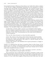
summarized in table 5.1, and the lesion site prima-
rily responsible for each disorder is illustrated in
figure 5.1.
Egocentric Disorientation (Case 1)
The patient described by Levine, Warach, and Farah
(1985) demonstrated profound way-finding difficul-
ties within his own home and new places following
bilateral damage to the posterior parietal cortex.
While he (and a number of similar patients: M.N.N.,
Kase, Troncoso, Court, & Tapia, 1977; Mr. Smith,
Hanley, & Davies, 1995; G.W., Stark, Coslett, &
Saffran, 1996; and the cases of Holmes & Horax,
1919) has been described as topographically disori-
ented, it is clear that his impairments extended far
beyond the sphere of extended, locomotor space. To
quote Levine and Farah:
[His] most striking abnormalities were visual and spatial.
. . . He could not reach accurately for visual objects,
even those he had identified, whether they were presented
in central or peripheral visual fields. When shown two
objects, he made frequent errors in stating which
was nearer or farther, above or below, or to the right or
left
He could not find his way about. At 4 months after the
hemorrhages, he frequently got lost in his own house and
never went out without a companion Spatial imagery
was severely impaired. He could not say how to get from
his house to the corner grocery store, a trip he had made
several times a week for more than 5 years. In contrast, he
could describe the store and its proprietor. His descriptions
of the route were frequently bizarre: “I live a block away.
I walk direct to the front door.” When asked which direc-
tion he would turn on walking out of his front door, he
said, “It’s on the right or left, either way.” When,
Geoffrey K. Aguirre 94
seated in his room, he was blindfolded and asked to point
to various objects named by the examiner, he responded
[very poorly]. (Levine, Warach, & Farah, 1985, p. 1013)
These patients, as a group, had severe deficits in
representing the relative location of objects with
respect to the self. While they were able to gesture
toward objects they could see, for example, this
ability was completely lost when their eyes were
closed. Performance was impaired on a wide range
of visual-spatial tasks, including mental rotation
and spatial span tasks. It thus seems appropriate to
locate the disorder within the egocentric spatial
frame. Indeed, Stark and colleagues (1996) have
suggested that one of these patients (G.W.) had sus-
tained damage to a spatial map that represents in-
formation within an egocentric coordinate system.
It is interesting that these cases suggest that neural
systems capable of providing immediate informa-
tion on egocentric position can operate independ-
ently of systems that store this information (Stark
et al., 1996).
These patients were uniformly impaired in way-
finding tasks in both familiar and novel environ-
ments. Most remained confined to the hospital or
home, willing to venture out only with a compan-
ion (Kase et al., 1977; Levine et al., 1985). Route
descriptions were impoverished and inaccurate
(Levine et al., 1985; Stark et al., 1996) and sketch
map production disordered (Hanley & Davies,
1995). In contrast to these impairments, visual-
object recognition was informally noted to be intact.
Patient M.N.N. was able to name objects correctly
without hesitation, showing an absence of agnosic
features in the visual sphere. Patient G.W. had no
difficulty in recognizing people or objects and case
2 of Levine et al. (1985) was able to identify
common objects, pictures of objects or animals,
familiar faces, or photographs of the faces of family
members and celebrities.
Unfortunately, these patients were not specifi-
cally tested on visual recognition tasks employing
landmark stimuli. As noted earlier, Levine and
colleagues reported that their case 2 was able to
describe a grocery store and its proprietor, but this
Topographical Disorientation 95
Figure 5.1
Locations of lesions responsible for varieties of topo-
graphical disorientation: (1) the posterior parietal cortex,
associated with egocentric disorientation; (2) the posterior
cingulate gyrus, associated with heading disorientation;
(3) the lingual gyrus, associated with landmark agnosia;
and (4) the parahippocampus, associated with anterograde
disorientation. These sites are illustrated in the right hemi-
sphere since the great majority of cases of topograph-
ical disorientation follow damage to right-sided cortical
structures.
does not constitute a rigorous test. It is possible that
despite demonstrating intact object and face recog-
nition abilities, patients with egocentric disorienta-
tion will be impaired on recognition tasks that
employ topographically relevant stimuli. Thus, until
these tests are conducted, we can offer only the pos-
sibility that these patients are selectively impaired
within the spatial sphere.
It seems plausible that the way-finding deficits
that these patients display are a result of their pro-
found disorientation in egocentric space. As noted
earlier, route-based representations of large-scale
space are formed within the egocentric spatial
domain. This property of spatial representation was
well illustrated by Bisiach, Brouchon, Poncet, &
Rusconi’s 1993 study of route descriptions in a
patient with unilateral neglect. Regardless of the
direction that the subject was instructed to imagine
traveling, turns on the left-hand side tended to be
ignored. Thus, the egocentric disorientation that
these patients display seems sufficient to account
for their topographical disorders. In this sense, it is
perhaps inappropriate to refer to these patients as
selectively topographically disoriented—their dis-
ability includes forms of spatial representation that
are clearly not unique to the representation of large-
scale, environmental space.
Barrash (1998) has emphasized the variable dura-
tion of the symptoms of TD. In particular, many
patients who demonstrate egocentric disorientation
in the days and weeks following their lesion gradu-
ally recover near-normal function. Following this
initial period, patients can demonstrate a pattern
of deficits described by Passini, Rainville, & Habib
(2000) as being confined to “micro” as opposed to
“macroscopic” space. Their distinction is perhaps
more subtle than the egocentric versus exocentric
classification made here, because the recovered
patients may demonstrate impairments in the mani-
pulation of technically nonegocentric spatial infor-
mation (e.g., mental rotation), but do not show gross
way-finding difficulties.
Those egocentrically disoriented patients for
whom lesion data are available all have either bilat-
eral or unilateral right lesions of the posterior pari-
etal lobe, commonly involving the superior parietal
lobule. Studies in animals (both lesion and electro-
physiologically based) support the notion that
neurons in these areas are responsible for the repre-
sentation of spatial information in a primarily ego-
centric spatial frame. Homologous cortical areas in
monkeys contain cells with firing properties that
represent the position of stimuli in both retinotopic
and head-centered coordinate spaces simultane-
Geoffrey K. Aguirre 96
Table 5.1
A four-part taxonomy of topographical disorientation
Lesion site Disorder label Proposed impairment Model case
Posterior parietal Egocentric Unable to represent the location G.W. (Stark, Coslett, Saff,
disorientation of objects with respect to self 1996)
Posterior cingulate Heading Unable to represent direction of T.Y. (Suzuki, Yamador,
gyrus disorientation orientation with respect to external Hayakawa, Fujii, 1998)
environment
Lingual gyrus Landmark Unable to represent the appearance A.H. (Pallis, 1955)
agnosia of salient environmental stimuli
(landmarks)
Parahippocampus Anterograde Unable to create new representations G.R. (Epstein, Deyoe, Press,
disorientation of environmental information Rosen, Kanwisher 2001)
ously (i.e., planar gain fields; Anderson, Snyder, Li,
& Stricanne, 1993). Notably, cells with exocentric
firing properties have not been identified in the
rodent parietal cortex, although cells responsive
to complex conjunctions of stimulus egocentric
position and egomotion have been reported
(McNaughton et al., 1994).
Heading Disorientation (Case 2)
While the previous group of patients evidenced a
global spatial disorientation, rooted in a fundamen-
tal disturbance of egocentric space, a second group
of patients raises the intriguing possibility that
exocentric spatial representations can be selectively
damaged. These are patients who are both able to
recognize salient landmarks and who do not have
the dramatic egocentric disorientation described
earlier. Instead, they seem unable to derive direc-
tional information from landmarks that they do
recognize. They have lost a sense of exocentric
direction, or “heading” within their environment.
Patient T.Y. (Suzuki et al., 1998) presented with
great difficulty in way-finding following a lesion
of the posterior cingulate gyrus. She showed no
evidence of aphasia, acalculia, or right-left disori-
entation, object agnosia, prosopagnosia, or achro-
matopsia. She also had intact verbal and visual
memory as assessed by the Wechsler Memory
Scale, intact digit span, and normal performance on
Raven’s Progressive Matrices. Her spatial learning
was intact, as demonstrated by good performance
on a supraspan block test and the Porteus Maze test.
In contrast to these intact abilities, T.Y. was unable
to state the position from which photographs of
familiar buildings were taken. Or judge her direc-
tion of heading on a map while performing a way-
finding task through a college campus.
Three similar patients have been reported by
Takahashi and colleagues (Takahashi, Kawamura,
Shiota, Kasahata, & Hirayama, 1997). Like patient
T.Y., they were unable to derive directional infor-
mation from the prominent landmarks that they
recognized. The patients were able to discriminate
among buildings when several photographs were
displayed and were able to recognize photographs
of familiar buildings and landscapes near their
homes. The basic representation of egocentric
space, both at immediate testing and after a 5-
minute delay, was also demonstrated to be pre-
served. In contrast to these preserved abilities,
Takahashi et al.’s patients were unable to describe
routes between familiar locations and could not
describe the positional (directional) relationship
between one well-known place and another. In addi-
tion, the three patients were unable to draw a sketch
map of their hospital floor. A patient (M.B.) reported
by Cammalleri et al. (1996) had similar deficits.
Takahashi and colleagues suggested that their
patients had lost the sense of direction that allows
one to recall the positional relationships between
one’s current location and a destination within a
space that cannot be fully surveyed in one glance.
This can also be described as a sense of heading, in
which the orientation of the body with respect to
external landmarks is represented. Such a represen-
tation would be essential for both route-following
and the manipulation of maplike representations of
place. The possibility of isolated deficits in the
representation of spatial heading is an intriguing
one. These patients have a different constellation of
deficits from those classified as egocentrically dis-
oriented, and the existence of these cases suggests
that separate cortical areas mediate different frames
of spatial representation.
Patient T.Y., Takahashi’s three patients, and
patient M.B. had lesions located within the right
retrosplenial (posterior cingulate) gyrus. Figure
5.2 shows the lesion site in patient T.Y. It is inter-
esting that this area of the cortex in the rodent has
been implicated in way-finding ability. Studies in
rodents (Chen, Lin, Green, Barnes, & McNaughton,
1994) have identified a small population of cells
within this area that fire only when the rat is main-
taining a certain heading, or orientation within
the environment. These cells have been dubbed
“head-direction” cells (Taube, Goodridge, Golob,
Dudchenko, & Stackman, 1996), and most likely
Topographical Disorientation 97
generate their signals based upon a combination of
landmark, vestibular, and idiothetic (self-motion)
cues. Representation of the orientation of the body
within a larger spatial scheme is a form of spatial
representation that might be expected to be drawn
upon for both route-based and map-based naviga-
tion. Neuroimaging studies in humans (considered
later) have also added to this account.
Landmark Agnosia (Case 3)
The third class of topographically disoriented
patient can be described as landmark agnosic, in
that the primary component of their impairment is
an inability to use prominent, salient environmental
features for orientation. The patients in this category
of disorientation are the most numerous and best
studied.
Patient A.H. described by C. A. Pallis in 1955,
woke to find that he could not recognize his
bedroom and became lost trying to return from the
toilet to his room. He also noted a central “blind
spot,” an inability to see color, and that all faces
seemed alike. He quickly became lost upon leaving
his house, and was totally unable to recognize what
had previously been very familiar surroundings.
Upon admission, the patient was found to have
visual field deficits consistent with two adjacent,
upper quadrantic scotomata, each with its apex at
the fixation point. A.H. had no evidence of neglect,
was able to localize objects accurately in both the
left and right hemifields and had intact stereognos-
tic perception, proprioception, and graphaesthetic
sense. There was no left-right confusion, acalculia,
or apraxia. General memory was reported as com-
pletely intact. A.H.’s digit span was eight forward
and six backward, and he repeated the Babcock
sentence correctly on his first try.
The patient had evident difficulty recognizing
faces. He was unable to recognize his medical atten-
dants, wife, or daughter, and failed to identify pic-
tures of famous, contemporary faces. He had similar
difficulty identifying pictures of animals, although
a strategy of scrutinizing the photos for a critical
detail that would allow him to intuit the identity of
the image was more successful here than for the pic-
tures of human faces. For example, he was able to
identify a picture of a cat by the whiskers.
His primary and most distressing complaint was
his inability to recognize places:
In my mind’s eye I know exactly where places are, what
they look like. I can visualize T . . . square without diffi-
culty, and the streets that come into it I can draw you
Geoffrey K. Aguirre 98
Figure 5.2
MRI scan of patient T.Y., revealing a right-sided, posterior cingulate gyrus lesion. (Images courtesy of Dr. K. Suzuki.)
a plan of the roads from Cardiff to the Rhondda Valley.
. . . It’s when I’m out that the trouble starts. My reason
tells me I must be in a certain place and yet I don’t rec-
ognize it. It all has to be worked out each time. For
instance, one night, having taken the wrong turning, I was
going to call for my drink at the Post Office I have to
keep the idea of the route in my head the whole time, and
count the turnings, as if I were following instructions that
had been memorized. (Pallis, 1955, p. 219)
His difficulty extended to new places as well as pre-
viously familiar locales: “It’s not only the places I
knew before all this happened that I can’t remem-
ber. Take me to a new place now and tomorrow
I couldn’t get there myself” (Pallis, 1955, p. 219).
Despite these evident problems with way-finding,
the patient was still able to describe and draw maps
of the places that were familiar to him prior to his
illness, including the layout of the mineshafts in
which he worked as an engineer.
Patient A.H. is joined in the literature by a
number of well-studied cases, including patients
J.C. (Whiteley & Warrington, 1978), A.R. (Hécaen,
Tzortzis, & Rondot, 1980), S.E. (McCarthy, Evans,
& Hodges, 1996), and M.S. (Rocchetta, Cipolotti,
& Warrington, 1996); several of the cases reported
by Landis, Cummings, Benson, & Palmer (1986);
and the cases reported by Takahashi, Kawamura,
Hirayama, & Tagawa (1989); Funakawa, Mukai,
Terao, Kawashima, & Mori (1994), and Suzuki,
Yamadori, Takase, Nagamine, & Itoyama (1996).
These patients have several features in common: (1)
disorientation in previously familiar and novel
places, (2) intact manipulation of spatial informa-
tion, and (3) an inability to identify specific build-
ings. In other words, despite a preserved ability to
provide spatial information about a familiar envi-
ronment, the patient is unable to find his or her way
because of the inability to recognize prominent
landmarks.
This loss of landmark recognition, and its relative
specificity, has been formally tested by several
authors, usually by asking the subject to iden-
tify pictures of famous buildings. Patient S.E.
(McCarthy et al., 1996) was found to be markedly
impaired at recalling the name or information about
pictures of famous landmarks and buildings com-
pared with the performance of control subjects and
his own performance recalling information about
famous people.
Patient M.S. (Rocchetta et al., 1996) performed
at chance level on three different delayed-
recognition memory tests that used pictures of (1)
complex city scenes, (2) previously unfamiliar
buildings, and (3) country scenes. M.S. was also
found to be impaired at recognizing London
landmarks that were familiar before his illness.
Takahashi and colleagues (1989) obtained seven-
teen pictures of the patient’s home and neighbor-
hood. The patient was unable to recognize any of
these, but he could describe from memory the trees
planted in the garden, the pattern printed on his
fence, the shape of his mailbox and windows, and
was able to produce an accurate map of his house
and hometown.
In contrast, tests of spatial representation have
generally shown intact abilities in these patients.
Patients S.E., M.S., and J.C. were all found to have
normal performance on a battery of spatial learning
and perceptual tasks that included Corsi span,
Corsi supraspan, and “stepping-stone” mazes
(Milner, 1965). (Patient A.R., however, was found
to be impaired on the last of these tests.) In general,
the ability to describe routes and produce sketch
maps of familiar places is intact in these patients.
As discussed previously, these more anecdotal
measures of intact spatial representation should be
treated with some caution because there is con-
siderable ambiguity as to the specific nature of the
cognitive requirements of these tasks. Nonetheless,
the perfectly preserved ability of patients A.R. and
A.H. to provide detailed route descriptions, and the
detailed and accurate maps produced by S.E., A.H.,
and Takahashi’s patient (Takahashi et al., 1989),
are suggestive of intact spatial representations of
some kind. (Patient M.S., however, was noted to
have poor route description abilities.) Particularly
compelling, moreover, are reports of patients pro-
ducing accurate maps of places that were not famil-
iar prior to the lesion event (Cole & Perez-Cruet,
1964; Whiteley & Warrington, 1978). In this case,
Topographical Disorientation 99
the patient can only be drawing upon preserved
spatial representational abilities to successfully
transform navigational experiences into an exocen-
tric representation.
Several neuropsychological deficits have been
noted to co-occur with landmark agnosia, specifi-
cally, prosopagnosia (Cole & Perez-Cruet, 1964;
Landis et al., 1986; McCarthy et al., 1996; Pallis,
1955; Takahashi et al., 1989) and achromatopsia
(Landis et al., 1986; Pallis, 1955), along with some
degree of visual field deficit. These impairments do
not invariably accompany landmark agnosia (e.g.,
Hécaen et al., 1980), however, and are known to
occur without accompanying TD (e.g., Tohgi et al.,
1994). Thus it is unlikely that these ancillary im-
pairments are actually the causative factor of TD.
More likely, the lesion site that produces landmark
agnosia is close to, but distinct from, the lesion sites
responsible for prosopagnosia and achromatopsia.
There is also evidence that landmark agnosics
have altered perception of environmental features,
in addition to the loss of familiarity (as is the case
with general-object agnosics and prosopagnosics;
Farah, 1990). For example, Hécaen’s patient A.R.
was able to perform a “cathedral matching” task
accurately, but “he [AR] spontaneously indicated
that he was looking only for specific details ‘a
window, a doorway but not the whole.’
Places were identified by a laborious process of
elimination based on small details” (Hécaen et al.,
1980, p. 531).
An additional hallmark feature of landmark
agnosia is the compensatory strategy employed by
these patients. The description of patient J.C. is
typical:
He relies heavily on street names, station names, and
house numbers. For example, he knows that to get to the
shops he has to turn right at the traffic lights and then left
at the Cinema When he changes his place of work he
draws a plan of the route to work and a plan of the inte-
rior of the “new” building. He relies on these maps and
plans He recognizes his own house by the number
or by his car when parked at the door. (Whiteley &
Warrington, 1978, pp. 575–576)
This reliance upon small environmental details,
called variously “signs,” “symbols,” and “land-
marks” by the different authors, is common to all of
the landmark agnosia cases described here and pro-
vides some insight into the cognitive nature of the
impairment. First, it is clear that these patients are
capable of representing the strictly spatial aspect of
their position in the environment. In order to make
use of these minute environmental details for way-
finding, the patient must be able to associate spatial
information (if only left or right turns) with partic-
ular waypoints. This is again suggestive evidence
of intact spatial abilities. Second, although these
patients are termed “landmark agnosics,” it is not
the case that they are unable to make use of any
environmental object with orienting value. Instead,
they seem specifically impaired in the use of high-
salience environmental features, such as buildings,
and the arrangement of natural and artificial stimuli
into scenes. Indeed, these patients become disori-
ented within buildings, suggesting that they are no
longer able to represent a configuration of stimuli
that allows them to easily differentiate one place
from another. It thus seems that careful study
of landmark agnosics may provide considerable
insight into the normative process of selection and
utilization of landmarks.
The parallels between prosopagnosia and land-
mark agnosia (which we might refer to as synorag-
nosia, from the Greek for landmark) are striking.
Prosopagnosic patients are aware that they are
viewing a face, but do not have access to the effort-
less perception of facial identity that characterizes
normal performance. They also develop compensa-
tory strategies that focus on the individual parts
of the face, often distinguishing one person from
another by careful study of the particular shape of
the hairline, for example.
The lesion sites reported to produce landmark
agnosia are fairly well clustered. Except for patient
J.C. (who suffered a closed head injury and for
whom no imaging data are available) and patient
M.S. (who suffered diffuse small-vessel ischemic
disease), the cases of landmark agnosia reviewed
here all had lesions either bilaterally or on the right
Geoffrey K. Aguirre 100
side of the medial aspect of the occipital lobe,
involving the lingual gyrus and sometimes the
parahippocampal gyrus. The most common mecha-
nism of injury is an infarction of the right posterior
cerebral artery.
The type of visual information that is represented
in this critical area of the lingual gyrus is an open
question. Is this a region involved in the represen-
tation of all landmark information, or simply certain
object classes that happen to be used as landmarks?
How would such a region come to exist? One
account of the “lingual landmark area” (Aguirre,
Zarahn, & D’Esposito, 1998a) posits the existence
of a cortical region predisposed to the representa-
tion of the visual information employed in way-
finding. Through experience, this area comes to
represent environmental features and visual config-
urations that have landmark value (i.e., that tend to
aid navigation). We might imagine that such a spa-
tially segregated, specialized area would develop
because of the natural correlation of some landmark
features with other landmark features (Polk &
Farah, 1995). Furthermore, such a region might
occupy a consistent area of cortex from person to
person as a result of the connection of the area with
other visual areas (e.g., connections to areas with
large receptive fields or to areas that process “optic
flow”).
We have a sense from environmental psychology
studies of the types of visual features that would
come to be represented in such an area: large,
immobile things located at critical, navigational
choice points in the environment. Certainly build-
ings fit the bill for western, urban dwellers. We
might suspect that in other human populations that
navigate through entirely different environments,
different kinds of visual information would be rep-
resented. In either case, lesions to this area would
produce the pattern of deficits seen in the reported
cases of landmark agnosia. Evidence for such an
account has been provided by neuroimaging studies
in intact human subjects, which are considered
below.
Anterograde Disorientation and the Medial
Temporal Lobes (Case 4)
Our discussion so far has focused on varieties of
TD that follow damage to neocortical structures.
The posterior parietal cortex, the posterior cingulate
gyrus, and areas of the medial fusiform gyrus have
all been associated with distinct forms of naviga-
tional impairments. Despite this, much of the extant
TD literature has been concerned with an area of
the paleocortex: the medial temporal lobes. As
mentioned previously, this focus on the medial tem-
poral lobes derives from the compelling neuro-
physiological finding that hippocampal cells in the
rodent fire selectively when the freely moving
animal is in certain locations within the environ-
ment. The existence of these place cells is the
basis for theories that offer the hippocampus as a
repository of information about exocentric spatial
relationships (O’Keefe & Nadel, 1978). Additional
evidence regarding the importance of the hip-
pocampus in animal spatial learning was provided
by Morris and colleagues (1982), who reported that
rats with hippocampal lesions were impaired on a
test of place learning, the water maze task. The
specificity of the role played by the hippocampus
(i.e., Ammon’s horn, the dentate gyrus, and the
subiculum) in spatial representation has subse-
quently been debated at length (e.g., Cohen &
Eichenbaum, 1993).
At the very least, it is clear that selective (neuro-
toxic), bilateral lesions of this structure in the rodent
greatly impair performance in place learning tasks
such as the water maze (Jarrard, 1993; Morris,
Schenk, Tweedie, & Jarrard, 1990). The central role
of the hippocampus in theories of spatial learning in
animals has influenced the neurological literature on
TD to some extent. For example, many case reports
of topographically disoriented patients with neocor-
tical damage are at pains to relate the lesion loca-
tion to some kind of disruption of hippocampal
function (e.g., through disconnection or loss of
input).
In recent decades, the “cognitive map” theory
has come to be contrasted with models of medial
Topographical Disorientation 101
temporal lobe function in the realm of long-term
memory. In this account, which is supported prima-
rily through lesion studies in human patients, the
hippocampus is responsible for the initial formation
and maintenance of “declarative” memories, which
over a period of months are subsequently consoli-
dated within the neocortex and become independ-
ent of hippocampal function.
What of the impact of medial temporal lesions
upon navigational ability? It is clear that unilateral
lesions of the hippocampus do not produce any
appreciable real-world way-finding impairments in
humans (DeRenzi, 1982). While one study (Vargha-
Khadem et al., 1997) has reported anterograde way-
finding deficits in the setting of general anterograde
amnesia following bilateral damage restricted to
the hippocampus, this obviously cannot be consid-
ered a selective loss. Other studies of patients with
bilateral hippocampal damage have not commented
upon anterograde way-finding ability (Rempel-
Clower, Zola, Squire, & Amaral, 1996; Scoville
& Milner, 1957; Zola-Morgan, Squire, & Amaral,
1986). Retrograde loss of way-finding knowledge in
these patients is not apparently disproportionate to
losses in other areas (Rempel-Clower et al., 1996)
and this knowledge can be preserved (Milner,
Corkin, & Teuber, 1968; Teng & Squire, 1999).
Cases have been reported, however, of topo-
graphical impairment that is primarily confined to
novel environments, although it is not associated
with lesions to the hippocampus per se. It is inter-
esting that this anterograde TD, described in two
patients by Ross (1980), one patient by Pai (1997),
the patient of Luzzi, Pucci, Di Bella, & Piccirilli
(2000), and the first two cases of Habib and Sirigu
(1987), appears to affect both landmark and spatial
spheres. By far the most comprehensive study has
been provided by Epstein and colleagues (2001) in
their examination of patients G.R. and C.G.
At the time of testing, G.R. was a well-educated,
60-year-old man who had suffered right and left
occipital-temporal strokes 2 years previously.
Figure 5.3 shows the lesion site in this patient.
These strokes had left him with a left hemianopsia
and right upper quadrantanopsia and dyschro-
matopsia. He had no evidence of neglect, left-right
Geoffrey K. Aguirre 102
Figure 5.3
MRI scan of patient G.R., revealing bilateral damage to the parahippocampal gyri, with extension of the right lesion
posteriorly to involve the inferior lingual gyrus, medial fusiform gyrus, and occipital lobe. (Images courtesy of Dr. R.
Epstein.)